Embed Size (px)
Citation preview

www.elsevier.com/locate/pneurobio
Progress in Neurobiology 77 (2005) 215–251
From brainstem to cortex: Computational models of
saccade generation circuitry
B. Girard *, A. Berthoz
Laboratoire de Physiologie de la Perception et de l’Action, UMR 7152, CNRS-College de France, 11 place Marcelin Berthelot,
75231 Paris Cedex 05, France
Received 1 October 2004; received in revised form 27 October 2005; accepted 1 November 2005
Abstract
The brain circuitry of saccadic eye movements, from brainstem to cortex, has been extensively studied during the last 30 years. The wealth of
data gathered allowed the conception of numerous computational models. These models proposed descriptions of the putative mechanisms
generating this data, and, in turn, made predictions and helped to plan new experiments.
In this article, we review the computational models of the five main brain regions involved in saccade generation: reticular formation saccadic
burst generators, superior colliculus, cerebellum, basal ganglia and premotor cortical areas. We present the various topics these models are
concerned with: location of the feedback loop, multimodal saccades, long-term adaptation, on the fly trajectory correction, strategy and metrics
selection, short-term spatial memory, transformations between retinocentric and craniocentric reference frames, sequence learning, to name the
principle ones.
Our objective is to provide a global view of the whole system. Indeed, narrowing too much the modelled areas while trying to explain too much
data is a recurrent problem that should be avoided. Moreover, beyond the multiple research topics remaining to be solved locally, questions
regarding the operation of the whole structure can now be addressed by building on the existing models.
# 2005 Elsevier Ltd. All rights reserved.
Keywords: Saccade generation circuitry; Computational models; Brainstem; Superior colliculus; Cerebellum; Basal ganglia; Cortex
Contents
1. Introduction . . . . . . . . . . . . . . . . . . . . . . . . . . . . . . . . . . . . . . . . . . . . . . . . . . . . . . . . . . . . . . . . . . . . . . . . . . . . . . . . . 216
2. Reticular formation saccadic burst generators . . . . . . . . . . . . . . . . . . . . . . . . . . . . . . . . . . . . . . . . . . . . . . . . . . . . . . . . . . 217
2.1. Robinson (1975). . . . . . . . . . . . . . . . . . . . . . . . . . . . . . . . . . . . . . . . . . . . . . . . . . . . . . . . . . . . . . . . . . . . . . . . . . 218
2.2. Jurgens et al. (1981) . . . . . . . . . . . . . . . . . . . . . . . . . . . . . . . . . . . . . . . . . . . . . . . . . . . . . . . . . . . . . . . . . . . . . . . 218
2.3. van Gisbergen et al. (1985) . . . . . . . . . . . . . . . . . . . . . . . . . . . . . . . . . . . . . . . . . . . . . . . . . . . . . . . . . . . . . . . . . . 219
2.4. Tweed and Vilis (1985) . . . . . . . . . . . . . . . . . . . . . . . . . . . . . . . . . . . . . . . . . . . . . . . . . . . . . . . . . . . . . . . . . . . . . 219
2.5. Grossberg and Kuperstein (1986) . . . . . . . . . . . . . . . . . . . . . . . . . . . . . . . . . . . . . . . . . . . . . . . . . . . . . . . . . . . . . . 220
Abbreviations: aCG, anterior cingulate cortex; BG, basal ganglia; BN, superior colliculus burst neurons; BUN, superior colliculus build-up neurons; CBLM,
cerebellum; CMAC, cerebellar model arithmetic computer; DLPFC, dorsolateral prefrontal cortex; EBN, reticular formation excitatory burst neurons; EP,
entopeduncular nucleus; FEF, frontal eye fields; FOR, fastigial oculomotor area; GABA, g-aminobutyric acid; GCZ, gateable cortical zone; GP, globus pallidus;
GPe, external part of globus pallidus; GPi, internal part of globus pallidus; IBN, reticular formation inhibitory burst neurons; IFN, inhibitory feedback neurons; IO,
inferior olive; IT, inferotemporal cortex; LIP, lateral intraparietal cortex; LLB, reticular formation long-lead burst neurons; MLB, reticular formation medium-lead
burst neurons; MN, ocular motoneurons; NRTP, nucleus reticularis tegmenti pontis; OPN, reticular formation omnipause neurons; PFC, prefrontal cortex; PPC,
posterior parietal cortex; pre-SEF, presupplementary eye fields; QV, quasi-visual neurons; RI, resettable integrator; SBG, reticular formation saccadic burst
generators; SC, superior colliculus; SEF, supplementary eye fields; SNc, substantia nigra pars compacta; SNr, substantia nigra pars reticulata; SRT, saccade reaction
time; STN, subthalamic nucleus; STT, spatio-temporal transform; TLLB, tectal long-lead burst neurons; TN, reticular formation tonic neurons; V4, extrastriate visual
cortex area 4; VTA, ventral tegmental area; WTA, winner-takes-all mechanism
* Corresponding author. Tel.: +33 1 44 27 13 91; fax: +33 1 44 27 13 82.
E-mail address: [email protected] (B. Girard).
0301-0082/$ – see front matter # 2005 Elsevier Ltd. All rights reserved.
doi:10.1016/j.pneurobio.2005.11.001

B. Girard, A. Berthoz / Progress in Neurobiology 77 (2005) 215–251216
2.6. Scudder (1988). . . . . . . . . . . . . . . . . . . . . . . . . . . . . . . . . . . . . . . . . . . . . . . . . . . . . . . . . . . . . . . . . . . . . . . . . . . 220
2.7. Moschovakis (1994) . . . . . . . . . . . . . . . . . . . . . . . . . . . . . . . . . . . . . . . . . . . . . . . . . . . . . . . . . . . . . . . . . . . . . . . 221
2.8. Nichols and Sparks (1995). . . . . . . . . . . . . . . . . . . . . . . . . . . . . . . . . . . . . . . . . . . . . . . . . . . . . . . . . . . . . . . . . . . 222
2.9. Quaia and Optican (1997) . . . . . . . . . . . . . . . . . . . . . . . . . . . . . . . . . . . . . . . . . . . . . . . . . . . . . . . . . . . . . . . . . . . 222
2.10. Breznen and Gnadt (1997). . . . . . . . . . . . . . . . . . . . . . . . . . . . . . . . . . . . . . . . . . . . . . . . . . . . . . . . . . . . . . . . . . . 222
2.11. Gancarz and Grossberg (1998) . . . . . . . . . . . . . . . . . . . . . . . . . . . . . . . . . . . . . . . . . . . . . . . . . . . . . . . . . . . . . . . . 223
2.12. Discussion . . . . . . . . . . . . . . . . . . . . . . . . . . . . . . . . . . . . . . . . . . . . . . . . . . . . . . . . . . . . . . . . . . . . . . . . . . . . . . 223
3. The superior colliculus . . . . . . . . . . . . . . . . . . . . . . . . . . . . . . . . . . . . . . . . . . . . . . . . . . . . . . . . . . . . . . . . . . . . . . . . . . 224
3.1. Ottes et al. (1986) . . . . . . . . . . . . . . . . . . . . . . . . . . . . . . . . . . . . . . . . . . . . . . . . . . . . . . . . . . . . . . . . . . . . . . . . 225
3.2. van Gisbergen et al. (1987) . . . . . . . . . . . . . . . . . . . . . . . . . . . . . . . . . . . . . . . . . . . . . . . . . . . . . . . . . . . . . . . . . . 225
3.3. van Opstal and van Gisbergen (1989) . . . . . . . . . . . . . . . . . . . . . . . . . . . . . . . . . . . . . . . . . . . . . . . . . . . . . . . . . . . 226
3.4. Tweed and Vilis (1990) . . . . . . . . . . . . . . . . . . . . . . . . . . . . . . . . . . . . . . . . . . . . . . . . . . . . . . . . . . . . . . . . . . . . . 226
3.5. Droulez and Berthoz (1991, 1992) . . . . . . . . . . . . . . . . . . . . . . . . . . . . . . . . . . . . . . . . . . . . . . . . . . . . . . . . . . . . . 226
3.6. Waitzman et al. (1991) . . . . . . . . . . . . . . . . . . . . . . . . . . . . . . . . . . . . . . . . . . . . . . . . . . . . . . . . . . . . . . . . . . . . . 227
3.7. Dominey and Arbib (1992) . . . . . . . . . . . . . . . . . . . . . . . . . . . . . . . . . . . . . . . . . . . . . . . . . . . . . . . . . . . . . . . . . . 228
3.8. Lefevre and Galiana (1992) . . . . . . . . . . . . . . . . . . . . . . . . . . . . . . . . . . . . . . . . . . . . . . . . . . . . . . . . . . . . . . . . . . 229
3.9. Krommenhoek et al. (1993, 1996) and Krommenhoek and Wiegerinck (1998) . . . . . . . . . . . . . . . . . . . . . . . . . . . . . . . 229
3.10. Arai et al. (1994, 1999) and Das et al. (1995) . . . . . . . . . . . . . . . . . . . . . . . . . . . . . . . . . . . . . . . . . . . . . . . . . . . . . 229
3.11. Optican (1994) . . . . . . . . . . . . . . . . . . . . . . . . . . . . . . . . . . . . . . . . . . . . . . . . . . . . . . . . . . . . . . . . . . . . . . . . . . . 230
3.12. van Optsal and Hepp (1995) . . . . . . . . . . . . . . . . . . . . . . . . . . . . . . . . . . . . . . . . . . . . . . . . . . . . . . . . . . . . . . . . . 230
3.13. Grossberg et al. (1997) . . . . . . . . . . . . . . . . . . . . . . . . . . . . . . . . . . . . . . . . . . . . . . . . . . . . . . . . . . . . . . . . . . . . . 230
3.14. Bozis and Moschovakis (1998). . . . . . . . . . . . . . . . . . . . . . . . . . . . . . . . . . . . . . . . . . . . . . . . . . . . . . . . . . . . . . . . 231
3.15. Anastasio et al. (2000) and Patton et al. (2002) . . . . . . . . . . . . . . . . . . . . . . . . . . . . . . . . . . . . . . . . . . . . . . . . . . . . 232
3.16. Trappenberg et al. (2001) . . . . . . . . . . . . . . . . . . . . . . . . . . . . . . . . . . . . . . . . . . . . . . . . . . . . . . . . . . . . . . . . . . . 232
3.17. Discussion . . . . . . . . . . . . . . . . . . . . . . . . . . . . . . . . . . . . . . . . . . . . . . . . . . . . . . . . . . . . . . . . . . . . . . . . . . . . . . 233
4. Cerebellum . . . . . . . . . . . . . . . . . . . . . . . . . . . . . . . . . . . . . . . . . . . . . . . . . . . . . . . . . . . . . . . . . . . . . . . . . . . . . . . . . . 234
4.1. Dean et al. (1994) . . . . . . . . . . . . . . . . . . . . . . . . . . . . . . . . . . . . . . . . . . . . . . . . . . . . . . . . . . . . . . . . . . . . . . . . 235
4.2. Dean (1995). . . . . . . . . . . . . . . . . . . . . . . . . . . . . . . . . . . . . . . . . . . . . . . . . . . . . . . . . . . . . . . . . . . . . . . . . . . . . 236
4.3. Schweighofer et al. (1996a, b) . . . . . . . . . . . . . . . . . . . . . . . . . . . . . . . . . . . . . . . . . . . . . . . . . . . . . . . . . . . . . . . . 236
4.4. Lefevre et al. (1998); Quaia et al. (1999) and Optican and Quaia (2002) . . . . . . . . . . . . . . . . . . . . . . . . . . . . . . . . . . 237
4.5. Gancarz and Grossberg (1999) . . . . . . . . . . . . . . . . . . . . . . . . . . . . . . . . . . . . . . . . . . . . . . . . . . . . . . . . . . . . . . . . 237
4.6. Ebadzadeh and Darlot (2003). . . . . . . . . . . . . . . . . . . . . . . . . . . . . . . . . . . . . . . . . . . . . . . . . . . . . . . . . . . . . . . . . 239
4.7. Discussion . . . . . . . . . . . . . . . . . . . . . . . . . . . . . . . . . . . . . . . . . . . . . . . . . . . . . . . . . . . . . . . . . . . . . . . . . . . . . . 239
5. Basal ganglia. . . . . . . . . . . . . . . . . . . . . . . . . . . . . . . . . . . . . . . . . . . . . . . . . . . . . . . . . . . . . . . . . . . . . . . . . . . . . . . . . 239
5.1. Dominey and Arbib (1992) and Dominey et al. (1995) . . . . . . . . . . . . . . . . . . . . . . . . . . . . . . . . . . . . . . . . . . . . . . . 240
5.2. Brown et al. (2004) . . . . . . . . . . . . . . . . . . . . . . . . . . . . . . . . . . . . . . . . . . . . . . . . . . . . . . . . . . . . . . . . . . . . . . . 241
5.3. Discussion . . . . . . . . . . . . . . . . . . . . . . . . . . . . . . . . . . . . . . . . . . . . . . . . . . . . . . . . . . . . . . . . . . . . . . . . . . . . . . 243
6. Cortex . . . . . . . . . . . . . . . . . . . . . . . . . . . . . . . . . . . . . . . . . . . . . . . . . . . . . . . . . . . . . . . . . . . . . . . . . . . . . . . . . . . . . 243
6.1. Zipser and Andersen (1988). . . . . . . . . . . . . . . . . . . . . . . . . . . . . . . . . . . . . . . . . . . . . . . . . . . . . . . . . . . . . . . . . . 243
6.2. Dominey and Arbib (1992) and Dominey et al. (1995) . . . . . . . . . . . . . . . . . . . . . . . . . . . . . . . . . . . . . . . . . . . . . . . 243
6.3. Gancarz and Grossberg (1999) . . . . . . . . . . . . . . . . . . . . . . . . . . . . . . . . . . . . . . . . . . . . . . . . . . . . . . . . . . . . . . . . 244
6.4. Brown et al. (2004) . . . . . . . . . . . . . . . . . . . . . . . . . . . . . . . . . . . . . . . . . . . . . . . . . . . . . . . . . . . . . . . . . . . . . . . 245
6.5. Deneve et al. (1999, 2001) . . . . . . . . . . . . . . . . . . . . . . . . . . . . . . . . . . . . . . . . . . . . . . . . . . . . . . . . . . . . . . . . . . 245
6.6. Mitchell and Zipser (2001) . . . . . . . . . . . . . . . . . . . . . . . . . . . . . . . . . . . . . . . . . . . . . . . . . . . . . . . . . . . . . . . . . . 246
6.7. Mitchell and Zipser (2003) . . . . . . . . . . . . . . . . . . . . . . . . . . . . . . . . . . . . . . . . . . . . . . . . . . . . . . . . . . . . . . . . . . 247
6.8. Discussion . . . . . . . . . . . . . . . . . . . . . . . . . . . . . . . . . . . . . . . . . . . . . . . . . . . . . . . . . . . . . . . . . . . . . . . . . . . . . . 247
7. Conclusion . . . . . . . . . . . . . . . . . . . . . . . . . . . . . . . . . . . . . . . . . . . . . . . . . . . . . . . . . . . . . . . . . . . . . . . . . . . . . . . . . . 248
7.1. Beyond the parts: modelling the whole . . . . . . . . . . . . . . . . . . . . . . . . . . . . . . . . . . . . . . . . . . . . . . . . . . . . . . . . . . 248
7.2. Dynamics considerations . . . . . . . . . . . . . . . . . . . . . . . . . . . . . . . . . . . . . . . . . . . . . . . . . . . . . . . . . . . . . . . . . . . . 248
Acknowledgements . . . . . . . . . . . . . . . . . . . . . . . . . . . . . . . . . . . . . . . . . . . . . . . . . . . . . . . . . . . . . . . . . . . . . . . . . . . . 249
References . . . . . . . . . . . . . . . . . . . . . . . . . . . . . . . . . . . . . . . . . . . . . . . . . . . . . . . . . . . . . . . . . . . . . . . . . . . . . . . . . . 249
1. Introduction
There are three main different types of primate eye
movements (slow, fast and vergence movements), that are
controlled by partially separate brain structures (Henn, 1993).
The slow movements include the vestibulo-ocular reflex, the
slow-phase of the optokinetic reflex and the smooth pursuit.
The fast movements are the fast-phase of the optokinetic reflex
and the saccades. Saccades are used by species (like humans
and primates) whose retinas have a central high-resolution
region (the fovea) to explore visual scenes by redirecting gaze
from one important visual stimulus requiring precise analysis to
another. Their speed may reach 1000 � s�1 in some primate
species.

B. Girard, A. Berthoz / Progress in Neurobiology 77 (2005) 215–251 217
Fig. 1. Saccade-related brain areas (macaque monkey). Cblm: cerebellum; CD: caudate nucleus, input of the basal ganglia; FEF: frontal eye fields; LIP: lateral
intraparietal area; SC: superior colliculus; SEF: supplementary eye fields; SG: saccadic burst generator; SNr: substantia nigra pars reticulata, output of the basal
ganglia. From Hikosaka et al. (2000), Physiol. Rev., used with permission.
The mechanics of saccadic eye movements are relatively
simple when compared to limb movements, which use multiple
joints and operate with varying loads. Saccadic eye movements
have therefore been studied for the intrinsic interest of
understanding how they are generated, but also as a simple
way to more generally study motor and premotor mechanisms
in the brain.
Numerous brain regions are involved in the generation of
saccades (Berthoz, 1996; Moschovakis et al., 1996), from the
cortex down to the brainstem (Fig. 1). The closest to the
movement execution are the vertical and horizontal saccadic
burst generators (SBG), two sets of nuclei of the reticular
formation which directly drive the ocular motoneurons (Scudder
et al., 2002). Their function is to produce, from eye displacement
instructions issued from higher level structures, the commands
appropriate to generate saccades with the desired metrics. They
are supposed to ensure accuracy by monitoring their own
commands through an efferent copy-based feedback.
The superior colliculus (SC) is, with the frontal eye fields
(FEF), the main structure sending saccade orders to the SBG
(Moschovakis, 1996). The SC is a place of convergence and
integration, often designed as the final common path of saccades.
It receives projections carrying simple visual, auditive and
somatosensory information along with more cognitive signals,
where the sensory inputs are affected by attention, motivation
and context. The SC drives the orientation of the whole body: it
does not control the eye directionwith regards to the head, but the
gaze direction. Therefore, it activates not only the SBG but also,
for example, the neck muscles.
The commands directed from the SC to the SBG are under
the influence of adaptive modulations issued from the
cerebellum (CBLM). It provides the SBG with additional
input during saccades which are interpreted as: (1) a calibration
of the system induced by long-term adaptation of the saccadic
gain and (2) an on the fly correction of every single saccade,
made necessary by the apparent variability of the rest of the
saccade generating circuitry (Optican and Robinson, 1980).
The activity of the SC is gated by inhibitory inputs issued
from a set of subcortical nuclei called the basal ganglia (BG)
(Hikosaka and Wurtz, 1983a, b, c, d; Chevalier and Deniau,
1993; Hikosaka et al., 2000). Whereas the cortical areas
generate numerous motor orders which are directly sent to the
SC, they also project to the BG which is implicated in choosing
which orders to execute, by disinhibiting the corresponding
subregion of the SC. The role of the BG might however not be
restricted to that metric selection (Handel and Glimcher, 1999).
Atop all these structures, many cortical areas are involved in
saccade generation: the posterior parietal cortex (PPC), the
dorsolateral prefrontal cortex (DLPFC), the anterior cingulate
cortex (aCG), the presupplementary, supplementary and frontal
eye fields (pre-SEF, SEF and FEF, respectively) (Platt et al.,
2004). They provide rich inputs for the SC which allow the
selection of targets by cognitive processes influenced by
motivational and attentional states, along with the possible use
of working memory or sequence learning capabilities (Pierrot-
Deseilligny et al., 2003).
Numerous computational models of all these saccade-
related brain regions have been proposed in the last 30 years.
They helped to understand their operation, functionality and
interconnections by proposing computational mechanisms and
predictions that could be tested experimentally. However, most
of them were restricted to one or a few subparts of the whole
circuitry. This may sometimes cause problems when one
attempts to replicate with such restricted models experimental
results that are indeed generated by another brain structure or
by interactions with other structures. The objective of this paper
is to review the computational models of saccade-related brain
circuits, from brainstem to cortex, in order to propose an
ensemble view of the system, of the numerous problems
remaining to be solved at each level and of the relationships
between each levels.
The computational models of the five categories of brain
areas involved in saccade generation are reviewed in the next
sections in the following order: reticular formation saccadic
burst generators, superior colliculus, cerebellum, basal ganglia
and cortex. Each of these sections has its own specific
discussion, while global considerations are provided in a final
conclusion.
2. Reticular formation saccadic burst generators
The reticular formation saccadic burst generators generate
activations transmitted to vertical and horizontal ocular
motoneurons. The ocular motoneurons have a ‘‘burst-tonic’’
discharge pattern: the tonic activity is proportional to the eye
position along the vertical or horizontal axis and the super-
B. Girard, A. Berthoz / Progress in Neurobiology 77 (2005) 215–251218
imposed bursts, corresponding to saccades, are proportional to
the amplitude of the saccade. The tonic activity is provided by
the tonic neurons (TN) of two neural integrators (Moschovakis,
1997) located in the interstitial nucleus of Cajal (vertical
integrator) and nucleus prepositus hypoglossi (horizontal
integrator), in interaction with the vestibular nuclei. The bursts
of activity are provided by two distinct burst generators
(horizontal and vertical) composed of a set of neuron classes
having specific patterns of activity (Scudder et al., 2002):
� Medium-lead burst neurons (MLB): these neurons emit
bursts of discharge beginning before saccade onset, they are
composed of excitatory and inhibitory burst neurons.
� E
xcitatory burst neurons (EBN): the EBNs are active duringipsilaterally directed saccades, their afferents are the superior
colliculus tectal long-lead burst neurons (TLLB), the
brainstem long-lead burst neurons (LLB) and the IBNs.
They project to ipsilateral motoneurons and to the tonic
neurons of the neural integrators.
� I
nhibitory burst neurons (IBN): the IBNs are active duringipsilaterally directed saccades, their afferents are the TLLBs,
the LLBs and the EBNs. They project to contralateral
motoneurons and to the tonic neurons of the neural
integrators.
� O
mnipause neurons (OPN): these neurons discharge toni-cally during fixation and stop during saccades. They project
to and inhibit the MLBs and therefore function as a saccade
temporal switch.
� L
ong-lead burst neurons: the LLBs emit bursts well beforethe saccade, often reaching their maximum firing rate at
saccade onset. LLBs with various properties (direction
selective, direction and amplitude selective, etc.) and putative
roles (relay between the SC and the MLBs, latch controlling
OPN activity, relay between the SC and the cerebellum) were
found in various brainstem nuclei.
Numerous computational models of these neurons interac-
tions have been proposed, most of them exploring the putative
feedback mechanisms ensuring saccade accuracy.
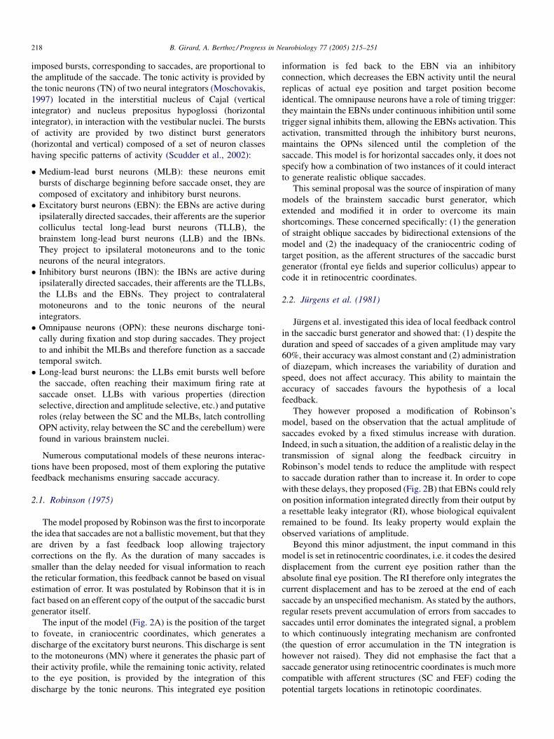
2.1. Robinson (1975)
Themodel proposed by Robinson was the first to incorporate
the idea that saccades are not a ballistic movement, but that they
are driven by a fast feedback loop allowing trajectory
corrections on the fly. As the duration of many saccades is
smaller than the delay needed for visual information to reach
the reticular formation, this feedback cannot be based on visual
estimation of error. It was postulated by Robinson that it is in
fact based on an efferent copy of the output of the saccadic burst
generator itself.
The input of the model (Fig. 2A) is the position of the target
to foveate, in craniocentric coordinates, which generates a
discharge of the excitatory burst neurons. This discharge is sent
to the motoneurons (MN) where it generates the phasic part of
their activity profile, while the remaining tonic activity, related
to the eye position, is provided by the integration of this
discharge by the tonic neurons. This integrated eye position
information is fed back to the EBN via an inhibitory
connection, which decreases the EBN activity until the neural
replicas of actual eye position and target position become
identical. The omnipause neurons have a role of timing trigger:
they maintain the EBNs under continuous inhibition until some
trigger signal inhibits them, allowing the EBNs activation. This
activation, transmitted through the inhibitory burst neurons,
maintains the OPNs silenced until the completion of the
saccade. This model is for horizontal saccades only, it does not
specify how a combination of two instances of it could interact
to generate realistic oblique saccades.
This seminal proposal was the source of inspiration of many
models of the brainstem saccadic burst generator, which
extended and modified it in order to overcome its main
shortcomings. These concerned specifically: (1) the generation
of straight oblique saccades by bidirectional extensions of the
model and (2) the inadequacy of the craniocentric coding of
target position, as the afferent structures of the saccadic burst
generator (frontal eye fields and superior colliculus) appear to
code it in retinocentric coordinates.
2.2. Jurgens et al. (1981)
Jurgens et al. investigated this idea of local feedback control
in the saccadic burst generator and showed that: (1) despite the
duration and speed of saccades of a given amplitude may vary
60%, their accuracy was almost constant and (2) administration
of diazepam, which increases the variability of duration and
speed, does not affect accuracy. This ability to maintain the
accuracy of saccades favours the hypothesis of a local
feedback.
They however proposed a modification of Robinson’s
model, based on the observation that the actual amplitude of
saccades evoked by a fixed stimulus increase with duration.
Indeed, in such a situation, the addition of a realistic delay in the
transmission of signal along the feedback circuitry in
Robinson’s model tends to reduce the amplitude with respect
to saccade duration rather than to increase it. In order to cope
with these delays, they proposed (Fig. 2B) that EBNs could rely
on position information integrated directly from their output by
a resettable leaky integrator (RI), whose biological equivalent
remained to be found. Its leaky property would explain the
observed variations of amplitude.
Beyond this minor adjustment, the input command in this
model is set in retinocentric coordinates, i.e. it codes the desired
displacement from the current eye position rather than the
absolute final eye position. The RI therefore only integrates the
current displacement and has to be zeroed at the end of each
saccade by an unspecified mechanism. As stated by the authors,
regular resets prevent accumulation of errors from saccades to
saccades until error dominates the integrated signal, a problem
to which continuously integrating mechanism are confronted
(the question of error accumulation in the TN integration is
however not raised). They did not emphasise the fact that a
saccade generator using retinocentric coordinates is much more
compatible with afferent structures (SC and FEF) coding the
potential targets locations in retinotopic coordinates.

B. Girard, A. Berthoz / Progress in Neurobiology 77 (2005) 215–251 219
Fig. 2. Saccadic burst generator models (1). (A) Robinson’s model; (B) Jurgens et al. model; (C) Grossberg and Kuperstein model; (D) Scudder’s model. EBN:
excitatory burst neurons; IBN: inhibitory burst neurons; MLB: medium-lead burst neurons; LLB: long-lead burst neurons; OPN: omnipause neurons; TN: tonic
neurons (integrators); MN: motoneurons; RI: hypothetic resettable integrator neurons; A: hypothetic arousal neurons; IFN: hypothetic inhibitory feedback neurons.
Filled arrows: inhibitory connections; empty arrows: excitatory connections. Plain and dashed lines distinguish agonist/antagonist circuits, ‘r’ and ‘l’ subscripts,
respectively, stand for the right and left circuits.
This model was neither subject to an implementation nor to
simulations in the work described by this original paper, but
was in following papers (Arai et al., 1994, 1999; Dean et al.,
1994; Nichols and Sparks, 1995; Das et al., 1995; Dean, 1995;
Breznen and Gnadt, 1997).
2.3. van Gisbergen et al. (1985)
van Gisbergen and colleagues studied the dynamic proper-
ties of oblique saccades, showing that: (1) the onset of
movement was simultaneous for horizontal and vertical
components of the movement and (2) the movement along
the component needing the shortest displacement is stretched
(increased duration and reduced peak velocity) to last as long as
the other one, consequently generating straight trajectories.
This added new constraints on the coupling of unidirectional
saccade generators models to get realistic full-range 2D
saccadic burst generator and led them to propose that a vectorial
pulse could be generated before being decomposed in vertical
and horizontal components.
Their computational model is not as precise as the preceding
ones in terms of neuronal circuitry, but assumes that the
retinotopically coded signals of the SC and the FEF are
transformed in a vectorial eye velocity command. This
command is coded in an array of direction-selective long-lead
burst cells, where each neuron is associated with a direction and
its firing rate represents the amplitude of the desired saccade.
This intermediate stage of computations drives the decomposed
coding in the vertical and horizontal saccadic burst generators,
therefore ensuring their simultaneous onset and the temporal
stretching of the shortest component. Such an architecture
necessitates the synaptic weights from the LLBs to the vertical
and horizontal EBNs to follow sine and cosine functions,
respectively.
This model was proved to better fit the obtained data on
oblique saccades than a model lacking the intermediate coding
stage and therefore generating synchronous but independent
movements along the two axis.
2.4. Tweed and Vilis (1985)
Tweed and Vilis were also interested in modelling a 2D
saccadic burst generator and developed the van Gisbergen et al.
model. Their main proposals were that: (1) keeping the
B. Girard, A. Berthoz / Progress in Neurobiology 77 (2005) 215–251220
direction-sensitive LLB array of van Gisbergen et al. (1985),
the local feedback should not occur in the EBNs but in the
LLBs, before the component decomposition, and (2) because
the third-order dynamic of the eye muscles is probably different
on the vertical and horizontal axes, three parameters linked with
position, velocity and acceleration are needed for calibration of
control on each axis.
The inclusion of the local feedback loop at the level of the
LLBs has an interesting side-effect: it generates the experi-
mentally observed saccade averaging, i.e. when two different
saccade commands from the superior colliculus are simulta-
neously fed in the model, the resulting saccade is directed to the
vector average of the two positions rather than to their sum.
However, a consensus has now emerged to consider that this
phenomenon is the result of lateral interactions in the collicular
maps (see Section 3).
Because of their second point, they considered that the eye
plant was controlled by a composite signal mixing the output of
the EBNs (coding velocity and acceleration), the TNs (coding
position) and the bursts-tonic neurons – usually considered as
the motoneurons – (coding velocity and position) using three
gains (g1, g2 and g3; Fig. 3). As the information concerning
position, velocity and acceleration is encoded in these three
populations of neurons, a linear combination of their output
allows the calibration of the controller of the third-order
dynamic eye plant.
Like the van Gisbergen et al. model, this model is not as
close as Robinson’s or Jurgens et al. models to the anatomy and
physiology of the corresponding neural circuitry (OPNs and
IBNs not included, integration mechanisms not detailed, etc.).
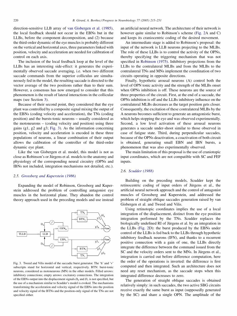
2.5. Grossberg and Kuperstein (1986)
Expanding the model of Robinson, Grossberg and Kuper-
stein addressed the problem of controlling antagonist eye
muscles in the horizontal plane. They abandon the control
theory approach used in the preceding models and use instead
Fig. 3. Tweed and Vilis model of the saccadic burst generator. The ‘h’ and ‘v’
subscripts stand for horizontal and vertical, respectively. BTN: burst-tonic
neurons, considered as motoneurons (MN) in the other models. Filled arrows:
inhibitory connections; empty arrows: excitatory connections. The integration
of the EBNs output into the displacement signalsDh andDv is not specified, but
the use of a mechanism similar to Scudder’s model is evoked. The mechanisms
transforming the acceleration and velocity signal of the EBNs into the position
and velocity signal of the BTNs and the position-only signal of the TNs are not
specified either.
an artificial neural network. The architecture of their network is
however quite similar to Robinson’s scheme (Fig. 2A and C)
and keeps its craniocentric coding of the desired movement.
An intermediate stage is added to Robinson’s proposal: the
input of the network is LLB neurons projecting to the MLBs.
The role of these LLBs is to control the activity of the OPNs,
thereby specifying the triggering mechanism that was not
specified in Robinson (1975). Inhibitory projections from the
LLBs to the contralateral MLBs and from the MLBs to the
contralateral TNs and MNs implement the coordination of two
circuits operating in opposite directions.
Finally, hypothetic arousal neurons (A) control both the
level of OPN tonic activity and the strength of the MLBs onset
when OPNs inhibition is off. These neurons are the source of
three properties of the circuit. First, at the end of a saccade, the
OPNs inhibition is off and the LLBs inhibitory influence on the
contralateral MLBs decreases as the target position gets closer.
Consequently, the excitation of these contralateral MLBs by the
A neurons becomes sufficient to generate an antagonistic burst,
which helps stopping the eye and was observed experimentally.
Second, a low level activation of these arousal neurons
generates a saccade under-shoot similar to those observed in
case of fatigue state. Third, during perpendicular saccades,
because of the OPNs deactivation, a coactivation of both circuit
is obtained, generating small EBN and IBN bursts, a
phenomenon that was also experimentally observed.
The main limitation of this proposal is the use of craniotopic
input coordinates, which are not compatible with SC and FEF
inputs.
2.6. Scudder (1988)
Building on the preceding models, Scudder kept the
retinocentric coding of input orders of Jurgens et al., the
artificial neural network approach and the control of antagonist
muscles of Grossberg and Kuperstein, and addressed the
problem of straight oblique saccades generation raised by van
Gisbergen et al. and Tweed and Vilis.
Using retinotopic coordinates implies the use of a local
integration of the displacement, distinct from the eye position
integration performed by the TNs. Scudder replaces the
biologically undefined RI of Jurgens et al. by an integration in
the LLBs (Fig. 2D): the burst produced by the EBNs under
control of the LLBs is fed back to the LLBs through hypothetic
inhibitory feedback neurons (IFN), and thanks to a recurrent
positive connection with a gain of one, the LLBs directly
integrate the difference between the command issued from the
SC and the velocity orders sent to the MNs. In Jurgens et al.,
integration is carried out before difference computation, here
the order of the operations is inverted: the difference is first
computed and then integrated. Such an architecture does not
need any reset mechanism, as the saccade stops when this
integrated difference decreases to zero.
The generation of straight oblique saccades is obtained
relatively simply: in such saccades, the two active SBG circuits
receive exactly the same burst as input (supposedly generated
by the SC) and share a single OPN. The amplitude of the

B. Girard, A. Berthoz / Progress in Neurobiology 77 (2005) 215–251 221
saccade component they, respectively, generate depend on a
weight applied to the SC burst, defined by the locus of activity
on the SC map (see Section 3). The duration of the saccade,
defined by the burst duration and the shared OPN gating, is the
same for both components. As the SBG circuits operate
linearly, their activities are similar but scaled by the weights
respectively, applied to their common input. The oblique
saccade trajectories are thus straight.
The coordination with the contralateral circuit is assumed by
the IBNs, which is biologically more realistic than the mixed
excitatory–inhibitory neurons used by Grossberg and Kuper-
stein. The biases imposed on EBNs, IBNs and OPNs firing rate
have a similar role as the A neurons excitations in the Grossberg
and Kuperstein model, as they are also the source of an
antagonistic burst at the end of the saccade and as varying their
values accounts for the slowed responses observed in case of
fatigue.
Finally, this model reproduces the results obtained by
microstimulation of the OPNs at the beginning or in the middle
of a saccade: the saccade stops during stimulation and then
resumes.
Scudder states a few limits of his model: (1) the EBNs firing
rate is too high in saccades larger than 15 �, compared to
experimental data; (2) the saccade averaging is not supported,
but he assumes that this phenomenon is a property of the
afferent structures (SC and FEF); (3) the model can produce the
staircase saccades observed during prolonged stimulation of a
Fig. 4. Saccadic burst generator models (2). (A) Moschovakis’ model; (B) Nichols
neurons; IBN: inhibitory burst neurons; LLB: long-lead burst neurons; OPN: omnipa
resettable integrator neurons; LI: hypothetic leaky integrator; A: hypothetic arous
arrows: excitatory connections. Plain and dashed lines distinguish agonist/antagonis
and down circuits.
SC site, but these do not match correctly with the observations
(amplitude and range of variation are too small).
2.7. Moschovakis (1994)
Moschovakis concentrates on the vertical saccadic burst
generator. He observes that neurons having properties similar to
the hypothetic IFNs of Scudder exist for the downward part of
the system, they are located in the interstitial nucleus of Cajal.
However, such neurons were not found in the upward part of the
system, while neurons resembling Jurgens et al. RI exist in the
nucleus of posterior commissure. He therefore proposes a
neural network model of the upward saccadic burst generator
inspired by the proposal of Jurgens et al., using a RI which is
simply reset by the tonic inhibition of the OPNs between
saccades (Fig. 4A).
The results obtained in the simulations are consistent with
the known sensibility of the EBNs (1.5 spike per degree) and
with the main sequence data in monkeys (saccade duration
versus amplitude and maximal velocity versus amplitude
relationships). Moreover, using a architecture similar to
Scudder’s proposal, the model generates straight oblique
saccades, microstimulation of the OPNs interrupts or truncates
saccades, whether they happen near the end of the saccade or
not, and staircase saccades are evoked by continuous superior
colliculus stimulation over a wide range of amplitudes. Finally,
in Scudder’s model, as well as in this one, a monotonic relation
and Sparks model; (C) Gancarz and Grossberg model. EBN: excitatory burst
use neurons; TN: tonic neurons (integrators); MN: motoneurons; RI: hypothetic
al neurons; COMP: comparator. Filled arrows: inhibitory connections; empty
t circuits, ‘r’, ‘l’, ‘u’ and ‘d’ subscripts, respectively, stand for the right, left, up

B. Girard, A. Berthoz / Progress in Neurobiology 77 (2005) 215–251222
exists between the curves of the instantaneous motor error
plotted against TLLBs instantaneous firing rate. The inter-
pretation that this experimentally observed relation is indicative
of a causal relationship between TLLBs discharge and motor
error, which gave rise to models including the superior
colliculus in the local feedback loop (see Section 3.6), is
therefore invalid.
A first limitation of the model concerns the fact that the RI
is a perfect integrator; however, a version of the model using a
leaky integrator RI can be built and was used in recent works of
the team (see Bozis and Moschovakis, 1998 and Section 3.14).
Another one is the use of linear non-saturating EBNs: a non-
linear transfer function can be used instead, the consequences
being that the TLLB-to-EBN weight then has a non-linear
relation with desired displacement and that the shape of the
saccade duration versus amplitude function gets steeper.
Finally, experimentally observed staircase saccades have their
amplitude reduced step after step, which is not the case here.
However, this variation might be caused by the effects of direct
superior colliculus connections to the motoneurons, bypassing
the saccadic burst generator (Moschovakis et al., 1998); thus,
models of the saccade generator do not have to reproduce
them.
2.8. Nichols and Sparks (1995)
The amplitude of a saccade evoked by stimulation of the
superior colliculus usually depends on its location on the
retinotopic collicular map. However, when evoked shortly after
a visual saccade, this amplitude varies and this variation decays
exponentially with the intersaccadic interval. Nichols and
Sparks investigated that point and proposed a modified version
of the Jurgens et al. model (Fig. 4B). Being interested in this
temporal phenomenon, they only kept the essentials of the
model: the decoupled integration of relative displacement (in a
leaky integrator) for the local feedback on the one hand and of
the absolute position (in the TNs) on the other hand. They
simply extended this basic scheme for the control of upward
and downward movements.
Instead of using an unspecified mechanism to reset the local
integrator, they relied on its leak only. This precise choice
explains the phenomenon of amplitude variation: if a saccade is
evoked just after another one, the leaky integrator is not
completely discharged and the remaining charge affects the
saccade as if a displacement had already been performed. The
exponential decay of the leaky integrator charge after a saccade
explains the exponential decay of this amplitude effect.
This Jurgens et al. extension was voluntarily kept very
simple (OPNs and IBNs removed, use of single integrators for
both the upward and downward movements) as its main point
was to study the effect of a purely leaky integrator in the local
feedback. However, the results concerning saccade amplitude
variation are not very well understood, as the decay process
seems to start from the beginning of the saccade (Schlag et al.,
1998) rather than at its end and as it was also proven that
normometric saccades can be produced shortly after a previous
saccade (Goossens and Van Opstal, 1997).
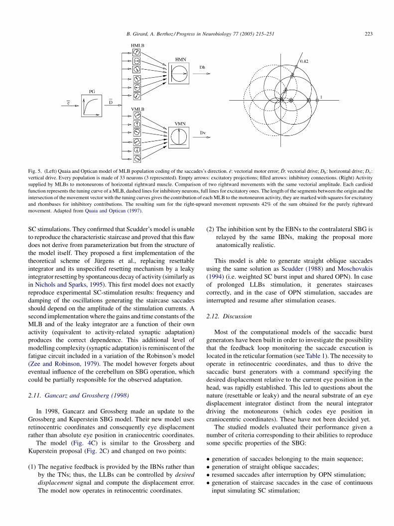
2.9. Quaia and Optican (1997)
Physiological evidence that the MLBs do not necessarily
reach their maximal firing rate for movements aligned with the
direction of the motoneurons they project to incited Quaia and
Optican to propose a model of population-coding of saccade
direction in the MLBs. The on-direction of the MLBs (the
direction for which they reach their maximal firing rate) may in
fact be titled away from the direction of the target motoneurons
by as much as 70�, and their tuning curve can be fit by a
Gaussian function centered around this on-direction.
This model uses four populations of MLBs (up, down, left
and right) having varied on-directions and realistic patterns of
activity (Fig. 5, left) to code the vectorial pulse D corresponding
to the order issued from the superior colliculus. Each pair of
populations associated to one axis project to the corresponding
motoneurons, they exert an opposite effect (excitation or
inhibition) depending on their associated direction. For
example, the up MLBs excite the up motoneurons and inhibit
the down motoneurons. Unlike van Gisbergen et al. or Tweed
and Vilis, such a model does not rely on an explicit
decomposition of the superior colliculus signal into its
horizontal and vertical components and thus avoids the use
of sinusoidal connection weights.
The behaviour of this model during oblique saccades is quite
interesting, as component stretching occurs even if the superior
colliculus signal is not decomposed. This property emerges from
the specificfiringpatterns of theMLBs and the repartitionof their
on-directions which makes the sum of agonist and antagonist
MLBs activities smaller as the direction of movement diverges
from the component axis (Fig. 5, right). The generated saccades
are almost perfectly straight, because the neural and mechanical
components of the model are perfectly symmetric. However, the
introduction of asymmetries in the MLBs on-directions
repartition or in the plant gains generates slightly curved
saccades which are in accordance with experimental results.
This model is not a complete model of the saccadic burst
generators: first, it does not explain how the varied on-directions
ofMLBs are generated, as its unphysiological to assume that it is
a characteristic of each neuron. Second, it does not propose a
feedbackmechanism in accordancewith its distributed coding of
the direction of movement. In fact, the estimation of the eye
displacement signal is not explicitly available, as it is distributed
among MLBs; therefore, a local feedback mechanism would
have to recompose it for integration, and then to decompose the
integrated signal to feed each MLB. Including the SC in the
feedback loop could simplify the problem, the authors however
signal many flaws in the two main theories (see Section 3) and
consequently suggest to switch to new models of feedback, and
especially to pay more attention to the role of cerebellum in
ensuring saccade accuracy (see the cerebellum models of the
same team, Section 4.4).
2.10. Breznen and Gnadt (1997)
Breznen and Gnadt studied the response of two models
(Jurgens et al., 1981; Scudder, 1988) to simulated continuous
B. Girard, A. Berthoz / Progress in Neurobiology 77 (2005) 215–251 223
Fig. 5. (Left) Quaia and Optican model of MLB population coding of the saccades’s direction. e: vectorial motor error; D: vectorial drive; Dh: horizontal drive; Dv:
vertical drive. Every population is made of 33 neurons (3 represented). Empty arrows: excitatory projections; filled arrows: inhibitory connections. (Right) Activity
supplied by MLBs to motoneurons of horizontal rightward muscle. Comparison of two rightward movements with the same vectorial amplitude. Each cardioid
function represents the tuning curve of aMLB, dashed lines for inhibitory neurons, full lines for excitatory ones. The length of the segments between the origin and the
intersection of the movement vector with the tuning curves gives the contribution of eachMLB to the motoneuron activity, they are marked with squares for excitatory
and rhombuses for inhibitory contributions. The resulting sum for the right-upward movement represents 42% of the sum obtained for the purely rightward
movement. Adapted from Quaia and Optican (1997).
SC stimulations. They confirmed that Scudder’s model is unable
to reproduce the characteristic staircase and proved that this flaw
does not derive from parameterization but from the structure of
the model itself. They proposed a first implementation of the
theoretical scheme of Jurgens et al., replacing resettable
integrator and its unspecified resetting mechanism by a leaky
integrator resetting by spontaneous decay of activity (similarly as
in Nichols and Sparks, 1995). This first model does not exactly
reproduce experimental SC-stimulation results: frequency and
damping of the oscillations generating the staircase saccades
should depend on the amplitude of the stimulation currents. A
second implementationwhere the gains and time constants of the
MLB and of the leaky integrator are a function of their own
activity (equivalent to activity-related synaptic adaptation)
produces the correct dependence. This additional level of
modelling complexity (synaptic adaptation) is reminiscent of the
fatigue circuit included in a variation of the Robinson’s model
(Zee and Robinson, 1979). The model however forgets about
eventual influence of the cerebellum on SBG operation, which
could be partially responsible for the observed adaptation.
2.11. Gancarz and Grossberg (1998)
In 1998, Gancarz and Grossberg made an update to the
Grossberg and Kuperstein SBG model. Their new model uses
retinocentric coordinates and consequently eye displacement
rather than absolute eye position in craniocentric coordinates.
The model (Fig. 4C) is similar to the Grossberg and
Kuperstein proposal (Fig. 2C) and changed on two points:
(1) T
he negative feedback is provided by the IBNs rather thanby the TNs; thus, the LLBs can be controlled by desired
displacement signal and compute the displacement error.
The model now operates in retinocentric coordinates.
(2) T
he inhibition sent by the EBNs to the contralateral SBG isrelayed by the same IBNs, making the proposal more
anatomically realistic.
This model is able to generate straight oblique saccades
using the same solution as Scudder (1988) and Moschovakis
(1994) (i.e. weighted SC burst input and shared OPN). In case
of prolonged LLBs stimulation, it generates staircases
correctly, and in the case of OPN stimulation, saccades are
interrupted and resume after stimulation ceases.
2.12. Discussion
Most of the computational models of the saccadic burst
generators have been built in order to investigate the possibility
that the feedback loop monitoring the saccade execution is
located in the reticular formation (see Table 1). The necessity to
operate in retinocentric coordinates, and thus to drive the
saccadic burst generators with a command specifying the
desired displacement relative to the current eye position in the
head, was rapidly established. This led to questions about the
nature (resettable or leaky) and the neural substrate of an eye
displacement integrator distinct from the neural integrator
driving the motoneurons (which codes eye position in
craniocentric coordinates). These have not been decided yet.
The studied models evaluated their performance given a
number of criteria corresponding to their abilities to reproduce
some specific properties of the SBG:
� g
eneration of saccades belonging to the main sequence;� g
eneration of straight oblique saccades;� r
esumed saccades after interruption by OPN stimulation;� g
eneration of staircase saccades in the case of continuousinput simulating SC stimulation;

B. Girard, A. Berthoz / Progress in Neurobiology 77 (2005) 215–251224
Table 1
Topics addressed by the reviewed saccadic burst generator models
Model LFM SOS AMC SI SCS SA AB CoA
Robinson (1975) �Jurgens et al. (1981) �van Gisbergen et al. (1985) �Tweed and Vilis (1985) � � �Grossberg and Kuperstein (1986) � � � �Scudder (1988) � � � � � �Moschovakis (1994) � � � � � � �Nichols and Sparks (1995) � �Quaia and Optican (1997) � �Breznen and Gnadt (1997) � �Gancarz and Grossberg (1998) � � � � � � �
LFM: local feedback mechanism; SOS: straight oblique saccades; AMC: coordination of antagonist muscles control; SI: saccade interruption by OPN stimulation;
SCS: staircase saccades; SA: saccades averaging (a property usually associated to superior colliculus processing); AB: antagonistic burst at the end of a saccade; CoA:
coactivation of perpendicular SBG in purely horizontal or vertical saccades. (�) The model addressed the specific topic and (�) the experimental result was not
explicitly simulated but should be explained by the model.
� g
eneration of a small burst by the antagonist SBG at the endof a saccade;
� c
oactivation of the vertical (respectively, horizontal) SBGsduring a purely horizontal (respectively, vertical) saccade.
However, the choice of evaluating isolated SBG models by
their ability to produce main sequence saccades might not be a
good one: when the cerebellum is deactivated or lesioned,
saccades do not belong to the main sequence anymore
(Robinson et al., 1993). If the main sequence is the result of
SBG–cerebellum interactions, then SBG model parameters
should be tuned using data gathered in animals with lesioned or
deactivated cerebellum. Concerning the generation of straight
oblique saccades, it seems that the mechanism proposed by
Scudder (1988) (weighted SC burst input and shared OPN) and
used since in many models including the SBGs (Moschovakis,
1994; Gancarz and Grossberg, 1998; Arai et al., 1994; Lefevre
et al., 1998, to name a few) is the simplest one and provides the
best explanation of the phenomenon. Thus, we suggest that
future models of the SBG should meet the last five criteria
before exploring any new exotic property of the SBG.
Table 1 first summarizes which models explored the
mechanisms of a feedback loop local to the SBG. It then
distinguishes which models were dedicated to the study of a
very specific point and the broader ones, which tried to explain
as much experimental data as possible. Among these, it appears
that Moschovakis (1994) and Gancarz and Grossberg (1998)
are the most complete.
The preceding remark concerning the role of the cerebellum
in the generation of main sequence saccades and the simulation
of saccade averaging with the sole SBG proposed by Tweed and
Vilis (1985) advocates for a careful approach of modelling,
where one should avoid narrowing to much on a single brain
area and consider the role of its interactions with its
neighboring circuits, before trying to explain with that single
area more experimental data than what it can afford.
The Quaia and Optican (1997) model is the only one which
uses realistic population coding of the desired movement in the
MLBs, the other models lump neurons in single computational
units. This specific approach led them to questioning the
existence of a feedback loop local to the SBG (see Section 2.9).
They also rejected the numerous proposals to include the
superior colliculus in a larger feedback loop (see Section 3),
mainly because the SC does not appear to be crucial to ensure
saccade accuracy. This resulted in models (Lefevre et al., 1998;
Quaia et al., 1999; Optican and Quaia, 2002) where the control
of accuracy is controlled by the cerebellum only. Their point of
view is a bit extreme, as, in their models, the superior colliculus
and SBGs have great difficulties generating saccades – be them
inaccurate – without the help of the cerebellum. However, they
opened a path that should followed, where the SBG operation is
explored in interaction with the SC and the cerebellum and
where a feedback loop local to the SBGs might not be
necessary.
3. The superior colliculus
The superior colliculus is a multilayered structure. Its
superficial layers are visual, they receive direct retinal inputs
and are topographically organized: their rostral parts respond to
visual stimuli close to the fovea while peripheral ones activate
more caudal sites (see Fig. 6 for a precise diagram of the
mapping in monkeys). Neurons of this layer consequently
discharge for targets situated in a limited area of the visual field
– the visual field of the neuron – and their activity follows a
bell-shape tuning function centered around a preferred position.
The deeper layers of the SC are presaccadic, they are also called
visuomotor as they exhibit small visual bursts before the motor
bursts, which are similar to those of the visual cells and usually
not sufficient to generate a saccade. They are also topogra-
phically organized: the neurons discharge before saccades
directed to a specific region of the visual field – the movement
field of the neuron – which is in register with the visual fields of
the visual neurons situated above them.
The neurons of the intermediate and deep layers are
classified in four main categories (Mays and Sparks, 1980;
Wurtz and Optican, 1994; Moschovakis, 1996; Moschovakis
et al., 1996):

B. Girard, A. Berthoz / Progress in Neurobiology 77 (2005) 215–251 225
Fig. 6. Mapping from polar coordinates in visual space (right) to Cartesian coordinates in the primate SC (left). Adapted from Optican, 95.
� T
he fixation cells are located in the rostral SC (in themonkey), they exhibit increased activity during fixation (even
in the absence of a fixation visual stimulus), stop firing during
the saccades and also display continuous activity during
pursuit. They are considered as inhibitors of saccadic eye
movements.
� T
he burst neurons (BN or tectal long-lead burst neurons) havea very low firing rate during fixation and generate bursts of
spikes immediately before saccade onset, if the saccade target
is located in their movement field.
� T
he build-up neurons (BUN) are located deeper in the SCthan the burst cells, they also have very low firing rate during
fixation, but begin to discharge at the target onset and the
firing rate slowly increases until saccade onset. The
movement fields of the build-up cells are different from
those of the burst cells: they are not centered around a
preferred position, but begin at a given position and extend to
any position with a larger amplitude.
� T
he quasi-visual cells (QV) are neither visual nor motor cells.They respond to visual stimulus in their visual field, like visual
cells, even if a saccade towards that stimulus will not be
performed.Theymay, unlikevisual cells, continue to discharge
for awhile after stimulus removal, with longer discharges if the
stimulus is a saccade target. They can also discharge without
any visual stimulus at all if a saccade to the position
corresponding to the receptive field of the cell is about to be
elicited. However, the activity pattern in that case is not tightly
linked to the saccade onset, as in motor (BN and BUN) cells.
The precise role of these cells is not clearly defined, an
interpretation of their activity as a dynamic spatial memory
(Droulez and Berthoz, 1991) of stimuli locations is proposed
by Bozis and Moschovakis (1998) (see Section 3.14).
Models of the SC were at first interested in the description of
the mappings of the visual fields on the superficial SC and of the
motor fields on the deeper SC. Neural network models then
allowed the study of the internal dynamics of the SC maps
(implying lateral excitations and inhibition) and the possible
roles of velocity and/or position feedback (simple remapping of
targets after saccades or involvement in the SBG control loop
by spatial integration of displacement).
3.1. Ottes et al. (1986)
In 1986, Ottes et al. proposed a model of the structure of
collicular neural maps and of the shape of collicular visuomotor
fields, based on the initial mapping experimentally obtained by
Robinson (1972) with monkeys. The mapping between the
position of a stimulus in the retina and the location of activated
cells in the SC is based on a logarithmic function (Fig. 6) and
the mapping between this location and the corresponding
saccade vector is defined as the inverse function. The activity
profile generated in the SC map by a stimulus is defined as a 2D
Gaussian function of constant size, mapped in physical
coordinates on the collicular surface. The combination of
these mappings and activity profiles accurately simulates the
increase of the visual and motor fields size with the distance to
retina center and the skewness of their sensitivity profile.
This quantification of mapping of the visual space onto the
physical surface of the colliculus was essential to further
proceed with quantitative modelling of the working superior
colliculus. As a consequence, it was extensively used in
subsequent studies.
3.2. van Gisbergen et al. (1987)
van Gisbergen et al. modelled the ensemble coding of
movement in the collicular map. They used the SCmapping and
population activity proposed by Ottes et al. (1986) and tested
the hypothesis that the resulting saccade vector is the sum of the
saccade vectors associated to each SC visuomotor cell,
weighted by the cell activity.

B. Girard, A. Berthoz / Progress in Neurobiology 77 (2005) 215–251226
1 Quaternions are a four-component extension of complex numbers and are of
common use to represent rotations and rotations composition.2 The eyes have three degrees of rotational freedom. The Listing’s Law
however states that, to avoid eye torsion, the static positions that the eye can
assume are described by Euler axes that lie in a plane perpendicular to the
primary position, called Listing’s plane.
The choice of using a vector sum to define the resulting
saccade has many implications. The first is that limited lesions
(1 mm in diameter) of the SC do not eliminate the
corresponding saccades from the saccades repertoire, as the
stimulation is larger than the lesion and therefore generates
some activity in the SC cells. However, saccades whose
population activity partially overlaps the lesioned region are
systematically hypometric, as the vector sum lacks the
contribution of the lesioned cells. It was later observed (Lee
et al., 1988) that in such a situation, saccades ‘‘diverge’’ from
the lesion site, which does not automatically result in
hypometric saccades.
Another effect of summation is that it causes edge effects
that limit the maximum amplitude of saccades as stimulation
Gaussians close to the limits of the SC are truncated.
Finally, in case of double stimulation, the resulting saccade
is the sum of the two vectors associated to the sites of
stimulation instead of the average. This last point was solved in
the 1989 model (van Opstal and van Gisbergen, 1989).
3.3. van Opstal and van Gisbergen (1989)
Following the 1987 model, van Gisbergen’s team proposed
an improved version of it, integrating a non-linearity in the
collicular cells firing rate and some lateral inhibitions within the
SC.
The collicular cells firing rate is simply set to 0 for negative
activations and depends linearly on positive activations until it
reaches its maximum (500 spikes/s). This allowed them to
reproduce qualitatively the fact that the amplitude of
electrically evoked saccades depends on the strength of the
current and does not increase further beyond a certain strength.
As various results suggested the existence of lateral long
range inhibitory interactions within the SC, these were simply
modelled by the addition of global inhibition constants
generated by each active cell on every other cells. This
modifies the shape of the activity profile in the SC and reduces
its global level of activation in the case of double stimulation.
The amplitude of a saccade in the case of double stimulation is
therefore reduced, so that it corresponds to an average saccade
weighted by the relative current strength rather than a vector
sum.
These modifications produce a more realistic model;
however, the effect of lateral short-range excitations and
long-range inhibitions was modelled as purely static. Conse-
quently, the resulting SC activity profile remained bimodal.
Following models (Arai et al., 1994, for example) using a
neural network approach showed that the simulation of the
dynamic of such a connection pattern can results in unimodal
activity, depending on the strength of the connections.
3.4. Tweed and Vilis (1990)
In an early work, Tweed and Vilis (1987) studied the effects
the non-commutativity of the rotations has on 3D eye
movements, and especially the fact that the derivative of
orientation is not angular velocity. To solve the problem of
computing head orientation from vestibular velocity signals,
they proposed a transformation based on the use of
quaternions.1 They also showed that the simple integrators
used in 1D feedback models of control of saccades should be
replaced by such systems in 3D models.
This led them to propose a model specifically dedicated to
quaternion-coded saccades. This has the advantage of solving
the problem of rotation non-commutativity, of simply explain-
ing why the amplitude of a saccade is affected by stimulation
position in the SC map only and of replicating weighted
averaging resulting from double stimulations.
The model SC is composed of map of neurons without
lateral connections. As each SC cell codes for a particular eye
rotation, it sends four projections to the SBG, whose relative
strengths code the four components of the corresponding
quaternion. The angle coded by a quaternion is not affected by
multiplication by a non-zero scalar; therefore, variations in the
frequency of activation of the SC cells do not change the
metrics of the generated saccade. The generation of weighted
averaging saccades in the case of double stimulations with
different frequencies directly derives from the same property.
Finally, Tweed and Vilis proposed that extending their model to
take into account the Listing Law2 would result in the addition
of a dedicated module situated upstream from the SC.
Solving the problem of the non-commutativity of rotations
with quaternions is quite elegant. However, the discovery of
orbital pulleys and of their role in changing the axes of action
of the extraocular muscles depending on eye position may
simplify the problem. Indeed, Quaia and Optican (1998)
showed that if the pulleys are correctly placed, the saccadic
circuitry can be commutative, because the eye plant itself
converts the neural signal into the appropriate rotation of the
eye. Moreover, in such a configuration, the implementation
of the Listing’s Law is simplified. Nevertheless, Tweed and
Vilis (1990) rose the problem of integrating rotations in 3D,
often neglected in earlier – and also most of the following –
studies.
3.5. Droulez and Berthoz (1991, 1992)
Droulez and Berthoz proposed a neural network model of a
dynamic spatial memory. It is not explicitly modelling the SC,
but is a theoretical proposal of generic premotor mechanisms
used to store the position of targets and to update this position
according to movements performed. These might occur in the
SC but also in cortical areas.
Considering the case of saccade-related brain structures, the
dynamic spatial memory is a 2D retinotopic map of
interconnected neurons, each of them coding for a specific
eye displacement. Lateral short-range bell-shaped excitatory

B. Girard, A. Berthoz / Progress in Neurobiology 77 (2005) 215–251 227
Fig. 7. Principle of moving hill models of the SC. (A) The position of potential targets is stored as hills of activations in the retinotopic map. (B) The positions are
continuously updated during the execution of a saccade towards target 1, thanks to velocity feedback, until the corresponding hill of activation reaches the center of the
map.
connections ensure that any input, representing the position of a
target, is stored as a hill of activation centered around the
neuron representing this position (Fig. 7A). The position of
these hills is affected by velocity feedback (efferent copy of
motor commands), allowing the remapping of target positions
during the execution of saccades (Fig. 7B).
Such a mechanism is not limited to target remapping: the
continuous update of the position of the potential targets allows
to specifically track the current target position during the
execution of the saccade. Therefore, a SC model based on that
principle could be integrated in the feedback loop of the
saccadic burst generator, and thus provide an implementation of
a principle first proposed by Keller (1980), but computationally
unspecified at that time. This idea of moving hills was included
in many following SC (Lefevre and Galiana, 1992; Optican,
1994; Grossberg et al., 1997) and cortical map (Dominey and
Arbib, 1992; Mitchell and Zipser, 2003) models.
Fig. 8. Waitzman et al. model of the SC. The TLLB are placed inside the feedback lo
Parameter a is specific to each TLLB, as it varies with the amplitude of the sacca
3.6. Waitzman et al. (1991)
Waitzman et al. studied the variation of the firing rate of
TLLBs during the execution of saccades directed to their
movement fields and discovered that there is a quasi-linear
relationship between this rate and the radial error. This result
led them to propose a model including the SC inside the
feedback loop of the saccadic burst generator.
Their model is a modification of Jurgens et al. model of the
saccadic burst generator, where the resettable integrator which
provides position error feedback performs an inverse temporal
to spatial transform before projecting to the SC (Fig. 8). The
dynamic of this model is different from the Droulez and
Berthoz moving hills, and could instead be described as a
stationary decreasing hill.
Including the SC in the feedback loop necessitates to
proceed to both spatial to temporal (SC output) and temporal to
op. Filled arrows: inhibitory connections; empty arrows: excitatory connections.
de.
B. Girard, A. Berthoz / Progress in Neurobiology 77 (2005) 215–251228
spatial (RI output) transformations. The potential functional
advantages derived from such a structure were not assessed.
Moreover, Moschovakis showed in his 1994 study of the
saccadic burst generator (see Section 2.7) that it is possible to
reproduce the relationship between TLLB firing rate and radial
error with both Scudder’s and his own model, where the SC is
kept outside the feedback loop. The inclusion of the SC into the
feedback loop is thus not necessary to explain the Waitzman
et al. experimental results.
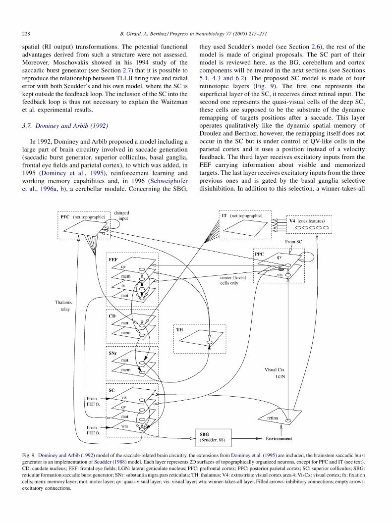
3.7. Dominey and Arbib (1992)
In 1992, Dominey and Arbib proposed a model including a
large part of brain circuitry involved in saccade generation
(saccadic burst generator, superior colliculus, basal ganglia,
frontal eye fields and parietal cortex), to which was added, in
1995 (Dominey et al., 1995), reinforcement learning and
working memory capabilities and, in 1996 (Schweighofer
et al., 1996a, b), a cerebellar module. Concerning the SBG,
Fig. 9. Dominey and Arbib (1992) model of the saccade-related brain circuitry, the e
generator is an implementation of Scudder (1988) model. Each layer represents 2D s
CD: caudate nucleus; FEF: frontal eye fields; LGN: lateral geniculate nucleus; PFC:
reticular formation saccadic burst generator; SNr: substantia nigra pars reticulata; TH
cells; mem:memory layer; mot: motor layer; qv: quasi-visual layer; vis: visual layer;
excitatory connections.
they used Scudder’s model (see Section 2.6), the rest of the
model is made of original proposals. The SC part of their
model is reviewed here, as the BG, cerebellum and cortex
components will be treated in the next sections (see Sections
5.1, 4.3 and 6.2). The proposed SC model is made of four
retinotopic layers (Fig. 9). The first one represents the
superficial layer of the SC, it receives direct retinal input. The
second one represents the quasi-visual cells of the deep SC,
these cells are supposed to be the substrate of the dynamic
remapping of targets positions after a saccade. This layer
operates qualitatively like the dynamic spatial memory of
Droulez and Berthoz; however, the remapping itself does not
occur in the SC but is under control of QV-like cells in the
parietal cortex and it uses a position instead of a velocity
feedback. The third layer receives excitatory inputs from the
FEF carrying information about visible and memorized
targets. The last layer receives excitatory inputs from the three
previous ones and is gated by the basal ganglia selective
disinhibition. In addition to this selection, a winner-takes-all
xtensions from Dominey et al. (1995) are included, the brainstem saccadic burst
urfaces of topographically organized neurons, except for PFC and IT (see text).
prefrontal cortex; PPC: posterior parietal cortex; SC: superior colliculus; SBG:
: thalamus; V4: extrastriate visual cortex area 4; VisCx: visual cortex; fx: fixation
wta: winner-takes-all layer. Filled arrows: inhibitory connections; empty arrows:
B. Girard, A. Berthoz / Progress in Neurobiology 77 (2005) 215–251 229
(WTA) mechanism, whose neural substrate is not specified,
selects the actual target of the forthcoming saccade.
This SC model is the first one to include to include the QV
cells and the influence of the basal ganglia over the SC (tonic
inhibition with selective disinhibition of restricted areas of the
SC map). However, as if this BG selection was not sufficient, a
final WTA is added. More recent models showed that the BG
selection is sufficient (Bozis and Moschovakis, 1998; Brown
et al., 2004) and the WTA therefore unnecessary.
3.8. Lefevre and Galiana (1992)
Lefevre and Galiana explored further the idea that the SC
could be included in the SBG feedback loop and proposed a SC-
centered model belonging to the moving hill family.
The model SC is one-dimensional map of laterally
interconnected neurons (short-range excitations) receiving a
gaze velocity feedback directed to its most caudal sites only.
This map drives a model including eye and head control
(modified version of Galiana et al., 1992 model of eye–head
coordination) through linearly weighted projections (increasing
with the distance to the fovea). However, the amplitude of
produced saccades does not depend linearly on the stimulation
position on the SC map. This emerging property is somewhat
reminiscent of the non-linear mapping modelled by Ottes et al.
(1986); however, a bidimensional extension of the model would
probably be necessary to compare this non-linearity with the
logarithmic mapping of the SC.
In this model, the gaze-velocity feedback is supposed to be
provided to the SC by projections from the prepositus
hypoglossi. The authors however mention that: (1) the
projections from the prepositus hypoglossi reach the whole
SC rather than its caudal part only, distributed feedback over the
SC should therefore be modelled, and (2) prepositus hypoglossi
cells carry a mixture of eye position, eye velocity and gaze
velocity, a property that will be fully exploited in the Arai et al.
(1999) model. Finally, even if it is beyond the scope of this
report, note that this model is the first SC model dealing with
gaze control during saccades, thus including both eye and head
dynamics.
3.9. Krommenhoek et al. (1993, 1996) and Krommenhoek
and Wiegerinck (1998)
Krommenhoek et al., also considering that the SC is in the
saccadic burst generator feedback loop, studied in 1993 the
question of the necessity of a step of craniocentric coding (a
proposition from Robinson, 1975) in order to solve the
remapping of activity in the SC. To do so, they used a
methodology very similar to the one used by Zipser and
Andersen (1988) in their model of area 7a of the PPC (see
Section 6.1). They trained (back-propagation algorithm) two
non-linear feedforward neural network architectures to produce
the actual motor error coded in retinocentric coordinates as a
hill of activation on a 2D map, using the initial retinal error and
the initial and actual positions of the eye in craniocentric
coordinates.
They showed that the simplest model (all inputs connected
to a single hidden layer) does not have to explicitly compute to
the position of the target in craniocentric coordinates in order to
achieve the task. Indeed, the eye displacement, which does not
depend on the reference frame, can be obtained by subtracting
the two eye positions in craniocentric coordinates. Conse-
quently, the activity in the resulting hidden layer is a distributed
coding of the motor error in retinotopic coordinates. The second
network first combines the retinal error and the initial eye
position in a hidden layer before a second hidden layer
combines the output of hidden layer 1 with the actual eye
position. In that configuration, the first hidden layer codes the
target position in craniocentric coordinates and the second
codes the motor error in retinocentric coordinates. The role of
these hidden layers is proposed to be similar to the one of,
respectively, area 7a and LIP in the posterior parietal cortex.
In these experiments, the hidden layers were not 2D maps
but simple populations of neurons without any topological
significance. Consequently, this study proposes an interesting
framework for analysing the gain field coding of coordinates by
populations of neurons, which was further developed in the
1996 paper (Krommenhoek et al., 1996). However, it might
seem a bit unrealistic to consider that parietal cortex is
incorporated in the saccadic burst generator feedback during
the execution of saccades, because of the long delays implied
by such a large loop.
The 1998 paper (Krommenhoek and Wiegerinck, 1998)
studies the behaviour of the model in a situation of double
stimulation. It is capable of generating an averaging saccade,
this averaging takes place in the first hidden layer (representing
area 7a of posterior parietal cortex) and not in the SC. It is
influenced by the total stimulation intensity and the initial eye
position. The model is however unable to account for the
bistability (the generated saccade is directed to either one or the
other target) obtained in double stimulations when the locations
of stimulation are more than 30 � away or when the latency is
longer than 300 ms.
3.10. Arai et al. (1994, 1999) and Das et al. (1995)
Arai et al. proposed in 1994 a model where the SC is
incorporated to the feedback control loop, belonging to the
decreasing hill of activity family. Its architecture is quite similar
to other proposals, its main originality is that the weights of the
lateral connections in the motor layer of the SC were obtained
by a supervised learning algorithm using real neurons data as
training set.
The network is made of two layers: the visual-only layer
relays the visual input to the motor layer. The cells of the motor
layer are laterally interconnected and project to a SBG model
belonging to the Jurgens et al. family of models (driven by eye
displacement and using a resettable integrator). The weights of
the lateral interconnections, which where generated by
supervised learning based on electrophysiological recordings,
consisted of short-range excitation and long-range inhibitions.
Feedback from the SBG was provided as additional input to the
motor layer, the learning algorithm converged (i.e. satisfactory

B. Girard, A. Berthoz / Progress in Neurobiology 77 (2005) 215–251230
Fig. 10. Optican 1994 model of the superior colliculus. BN: burst neurons;
BUN: build-up neurons; FX: fixation cells; SBG: brainstem saccadic burst
generator. Filled arrows: inhibitions; empty arrows: excitations. Adapted from
Optican (1994).
duplication of real cells activity profiles) for both velocity and
position feedback. The excitatory weights of the projections to
the SBG were also obtained by supervised learning, but using a
criterion of resulting saccade accuracy. A global shunting
inhibition over the SC is used to control the onset of the
saccade. This inhibition is supposed to be generated by either
the FEF or the basal ganglia, and canceled during the generation
of the saccade. It however lacked the spatial selectivity one may
expect from basal ganglia output.
This model generates saccades in accordance with the main
sequence, it can generate express saccades (saccades with a
very short latency) if the inhibition is absent and accurately
accomplishes saccade averaging. As a result of the learning
algorithm used, the activity profile of the motor layer neurons is
qualitatively similar to recorded cells.
In the case of saccade interruption by OPN stimulation, the
model was not able to resume saccades correctly. However,
refinements of this initial model (Das et al., 1995; Arai et al.,
1999) resulted in better results, albeit a slight hypermetria
tendency remained and the SC neurons did not completely turn
off during the interruption, in contradiction to experimental
data. The last version of the model used both velocity and eye
displacement feedback. The weights of these feedback
connections over the SC are learnt using a back-propagation
algorithm which gives better results using both velocity and
displacement. This led the authors to suggest that neurons
carrying feedback to the SC might not carry pure velocity or
displacement signals, but a mixture of both.
3.11. Optican (1994)
In 1994, Optican proposed a model preserving the idea that
the SC might be in the saccade trajectory control feedback
loop. However, where models with the SC outside the loop
produce a desired displacement signal and models with the SC
inside the loop produce the displacement error, Optican
suggests that the SC produces both a desired displacement
signal and an actual displacement signal. The first one is
produced by the BNs, which are unaffected by velocity
feedback, and the second one by the BUNs, which thus
constitute a spatial equivalent of the RI in the Jurgens et al.
family of SBG models.
The model is composed of two layers (Fig. 10). The first one
contains the BUNs and the fixation cells (fixation cells are
located centrally and inhibit the second layer), it receives
sensory inputs signalling the presence of potential targets and
velocity feedback provided by the vertical and horizontal
saccadic burst generators. The second one is the BNs layer,
these have lateral inhibitory connections, implementing a
spatial winner-takes-all. The two layers are interconnected by
bidirectional excitations which connect BN and BUN neurons
having the same movement fields. The difference between the
BUN layer signal (actual displacement) and the BN layer signal
(desired displacement) is computed in the horizontal and
vertical saccadic burst generators.
This model is the only one which integrates actual
displacement rather than displacement error in the SC, so that
the error is computed in the SBG. This specificity, which was
never reused in following models, has the advantage of
proposing different functional roles for the BNs and BUNs.
3.12. van Optsal and Hepp (1995)
Building on experimental results showing that the activity of
the neurons in the deep layers of the SC is modulated by initial
eye position, van Optsal and Hepp (1995) proposed a very
simple model of the SC motor map which could produce the
three-dimensional rotation axis for saccades in Listing’s plane.
The model SC is a retinotopic map where every movement
field considered is covered by nine cells. Each of these nine
cells has the same sensibility to eye displacement in its
movement field as its eight neighbors, but this basic pattern is
modulated by eye position (what Zipser and Andersen, 1988
defined as gain fields; see Section 6.1) in a specific way, as each
cell has a specific preferred eye position. The gain field
modulation is 1% of the cell’s peak firing rate per degree change
in eye position. This map simultaneously encodes the desired
eye displacement (spatially) and the eye position (by gain
fields). van Opstal and Hepp showed that projections to a simple
perceptron trained using the Widrow–Hoff learning rule
allowed the decoding of both variables and also the
computation a good approximation of the ocular rotation axis
in Listing’s plane.
Despite its extreme simplicity with regards to known
anatomy and physiology of the SC, this model suggests an
interesting role for gain fields: allowing the computation of
rotations compliant with Listing’s Law at the level of the SC.
3.13. Grossberg et al. (1997)
As the superior colliculus is an area of multisensory
integration, Grossberg et al. proposed the SACCART model,
specifically dedicated to that aspect of the SC operation. As
depicted in Fig. 11, their model contains both a burst neurons
map (stationary decreasing hill of activity) and a build-up
neurons map (moving hill of activity).

B. Girard, A. Berthoz / Progress in Neurobiology 77 (2005) 215–251 231
Fig. 11. SACCART model (Grossberg et al., 1997) of multisensory integration in the SC. Filled arrows: inhibitory connections; empty arrows: excitatory
connections. Adapted from Grossberg et al. (1997).
The BN map receives an input from the retina via the
superficial layers of the SC and is therefore capable of initiating
visual saccades on its own. This visual saccade part of the SC is
used for the calibration of the connections between the SC and
the SBG, as a visual feedback is available at the end of the
saccade to evaluate its accuracy, which is not the case for
memory saccades.
The BUN map is in charge of integrating the information for
the non-visual saccades, from the PPC (attentional system able
to store one target location), the PFC (working memory storing
sequences of saccades) and the auditory cortex (auditory
saccades). The target locations in these areas are supposed to be
coded in craniocentric coordinates and the transformation into
retinocentric coordinates subtracting eye position is made by
specific weighting of their projections to BUN map, obtained
by learning. These non-visual saccades do not necessarily have
a visual feedback available at the end of the saccade to be
calibrated, they therefore take advantage of the accuracy
developed by the visual pathway.
Three types of inhibitions affect the activity on both SC
maps and limit the spatial extend of the reverberation generated
by their excitatory interconnections: a global shunting
inhibition from the mesencephalic reticular formation, a
time-selective inhibition from the SC fixation cells and a
spatial-selective inhibition from the basal ganglia.
This model was tested in simple visual saccades and also in
memory, overlap, gap and multimodal tasks. It is the first one to
study the multimodality of the SC and the associated problem
of calibration, it also provides an interesting interpretation of
the respective roles of BNs and BUNs, different from the
Optican (1994) proposal.
3.14. Bozis and Moschovakis (1998)
In his 1994 model of the SBG – called MSH – Moschovakis
showed that the correlation between the variation of the firing
rate of the TLLB and the displacement error does not
necessarily imply that the SC is in the control feedback loop.
The Bozis and Moschovakis model of the SC is therefore built
on the MSH and is not included in this feedback loop.
The model is composed of two main layers. The first one is a
motor TLLB layer very similar to earlier models (van Opstal
and van Gisbergen, 1989; Arai et al., 1994; Das et al., 1995;
Grossberg et al., 1997), where localised hills of activity are
generated by local lateral excitations and global or long-range
inhibition. The weight distribution of lateral excitations is
derived from synaptic button density measures. The global
inhibition depends on the total TLLB activity and is provided
by speculative inhibitory neurons. The dynamics of this layer is
however different from previous proposals: the TLLB-
inhibitory neurons loop is supposed to give rise to an oscillating
activity, where the hill of activity on the map repetitively grows
and vanishes as long as there is an excitatory input on a single
TLLB. An additional inhibition provided by basal ganglia gates
the saccade onset and offset, allowing a single oscillation to
occur. The detailed mechanisms of this oscillating system are
however unclear, and thus difficult to replicate.
The second layer of the model is composed of the visual cells
of the superficial SC, which contact a complex network of
predictive visual and quasi-visual cells in the deep SC. This
network works as a spatial memory which projects to the TLLB
layer and where the position of disappearing visual target is
stored. An eye displacement feedback from the SBG is capable
B. Girard, A. Berthoz / Progress in Neurobiology 77 (2005) 215–251232
of shifting the hills of activity of this memory in order to update
the position of targets after a saccade occurred. This circuit
operates in a qualitatively similar manner as the dynamic
spatial memory proposed by Droulez and Berthoz (1991);
however, this feedback is not used for the control of the
occurring saccade metrics as in Lefevre and Galiana (1992), but
only for the memory update as in Dominey and Arbib (1992).
The choice of the targets to be stored in this memory is
supposed to be the result of the gating of a second group of basal
ganglia inhibitory cells which were shown to decrease their
activity at the presentation of a target and to recover their tonic
rate only after a saccade towards that specific target. This
selective inhibitory mechanism is however unspecified by the
model. Bozis and Moschovakis acknowledge the fact that such
a dynamic memory also probably exists in cortical areas, but
they propose that they are parallel and complementary rather
than hierarchically organized (Dominey and Arbib, 1992).
This model exhibits saccade averaging in the case of double
stimulation, amplitude modifications in the case of local lesions
which are compatible with the results from Lee et al. (1988),
and finally, correct vector subtraction in double saccade task. It
does not include BUN, however the authors note that slightly
modifying the parameters of the model allows the generation of
activities in the TLLB layer similar to BUN activity.
3.15. Anastasio et al. (2000) and Patton et al. (2002)
Anastasio and colleagues are interested in multisensory
integration in the SC, and more specifically in the so-called
multisensory enhancement. A subset of neurons from the deep
layers (about one-quarter of them in primates) is multisensory,
as they are affected by inputs from various sensory modalities
(visual and auditory, for example). Stimuli occurring at the
same time and place can increase the response of a multisensory
neuron with regards to its response to only one of these stimuli
(multisensory enhancement). This enhancement, defined as the
ratio of the discharge increase in a combination of multisensory
inputs over the maximal discharge obtained with only one of
the sensory component, is larger for small inputs than for large
ones (inverse effectiveness). The model they propose focuses
on these multisensory neurons only, trying to grasp these two
properties.
The model is built in a Bayesian framework, it is
hypothesised that the deep SC neurons compute the probability
that a target is present in their receptive fields given the sensory
inputs and prior knowledge about environment (target
appearance frequency) and sensor (reliability) properties.
Three likelihood functions of the input firing rates (no target
present, a target in one modality or in another one) are modelled
by three Poisson distributions with different means. The larger
the difference between the mean of the ‘‘no target’’ distribution
and the two others, the easier the discrimination between the
two possibilities (target present or not). When this difference is
small, the distributions overlap on a wide firing rate range, and
on that range, discrimination is poor. However, when two input
modalities are available, even if the overlap is large for both of
them, an enhancement of the discrimination is obtained, which
is subject to inverse effectiveness. Finally, an information
theoretic analysis (Patton et al., 2002) suggests that the
numerous unimodal neurons might be those for which the
means of the distributions are separated enough, so that a good
discrimination between target absence or presence can be
obtained, and for which the addition of a second modality does
therefore not add much information.
This work is an interesting first step in modelling the SC
using a Bayesian framework. It does however not currently
explore the mechanism that would allow neurons to carry out
the Bayesian computation, it is restricted to the computation of
the deep SC neurons (BN or BUN) and should be extended to
other neurons and structures. Finally, the hypothesis that the
deep SC neurons compute target presence probability is quite
simplifying, knowing that the activity of these neurons is rather
sensorimotor and reflects a target selection process, probably
mediated by the projections from the basal ganglia.
3.16. Trappenberg et al. (2001)
Trappenberg et al. studied in 2001 the influence of the lateral
connections in the SC (short-range excitation and long-range
inhibitions) on the saccade response time (SRT) in various
experimental settings.
Their model is made of two layers: the first one simulates the
laterally interconnected SC fixation and build-up cells, it
integrates both exogenous and endogenous stimulations; the
second one simulates the burst cells and is under control of the
previous layer (BN discharge when the BUN with the same
movement field reaches 80% of its maximum firing rate).
Exogenous stimulations are purely visual stimuli, while
endogenous stimulations are the results of cortical processes
taking into account visual stimuli, task context and a priori
knowledge of target position. The lateral connections in the first
layer have a short-range excitation/long-range inhibition
pattern similar to those of earlier models (van Opstal and
van Gisbergen, 1989; Arai et al., 1994; Das et al., 1995; Bozis
and Moschovakis, 1998). The connection weights are here
tuned using indirect experimental data: a distractor is presented
after the fixation point offset and before the target onset, the
activity profile of the SC cell corresponding to the target is then
amplified or inhibited, depending on distance between target
and distractor; if one supposes that this modification is caused
by the activity of the cells corresponding to the distractor
through lateral interconnections in the SC, an approximation of
their weighting can be deduced.
The dynamic generated by these lateral interconnections can
explain various effects. Concerning the gap effect, the reduced
reaction time is caused by the removal of the fixation point
which implies a cancelation of the fixation cells long-range
inhibition over the build-up cells, which therefore reach the
threshold of BN activation faster. The model also explains the
fact that the inhibitory or excitatory effects of a distractor are
stronger when it is displayed in close temporal proximity with
the target. Antisaccades were simulated by first sending the
exogenous visual stimuli to the SC, followed by an endogenous
signal exciting the cells corresponding to the opposite saccade:

B. Girard, A. Berthoz / Progress in Neurobiology 77 (2005) 215–251 233
Table 2
Topics addressed by the reviewed superior colliculus models
Model Model features Evaluation criteria
MAP LAT FBL LES DSA MTR MMS
Ottes et al. (1986) �van Gisbergen et al. (1987) � �van Opstal and van Gisbergen (1989) � � � �Tweed and Vilis (1990) �Droulez and Berthoz (1991) � � �Waitzman et al. (1991) �Dominey and Arbib (1992) �Lefevre and Galiana (1992) � � �Krommenhoek et al. (1993) � �Arai et al. (1994) � � � �Optican (1994) �Grossberg et al. (1997) � � �Bozis and Moschovakis (1998) � � � �Anastasio et al. (2000) �Trappenberg et al. (2001) �
MAP:modelling of the topological mapping of the SC; LAT: simulated lateral interconnections in collicular maps; FBL: SC included in the SBG feedback loop; LES:
effects of localised SC lesions; DSA: double stimulation averaging; MTR: multiple targets remapping; MMS: multimodal saccades.
the build-up activity generated by the visual signal is not strong
enough to generate a saccade on its own, it however inhibits the
endogenous signal and thus justifies the increased SRT. A priori
knowledge of the target position was simulated by an
endogenous input preceding the visual input and exciting the
cells of the area with the highest probability of target onset. It
increased or decreased the SRT depending on the actual
position of the target.
This model, which is structurally close to earlier ones,
proposes a detailed study of the effects of the lateral interactions
in the SC on SRT, providing a link between saccade behavioural
studies and the corresponding neural substrate.
3.17. Discussion
The modelling of logarithmic visuomotor mapping of the
monkey SC did not generates much controversy, but was not
much used in models (see Table 2, MAP column). Concerning
the transformation of this SC spatial code into the SBG
temporal one, the so-called spatio-temporal transformation or
STT,most of themodels built on the idea of vanGisbergen et al.
(1987). It is based on a spatially (and often also temporally)
stereotyped hill of activity on the SC, which can easily be
decoded using an appropriate weighted sum of this activity.
However, assuming such a perfect stereotypy is a bit
unrealistic, given the experimental results concerning SC
population activity (Anderson et al., 1998). Without stereo-
typy, an additional normalization step is needed. While
Optican and Quaia (2002) (see Section 4.4) thus prefer to
negate the existence of a STTand to delegate the whole task of
controlling the amplitude and direction of saccades to the
cerebellum,Groh (2001) proposes an architecture that explains
microstimulation results and should be further explored in
future SC models. The question of how precisely vertical
saccades and saccades close to the vertical, probably involving
activity on both colliculi, are coded in the SC and decoded in
the STT was rapidly addressed in van Gisbergen et al. (1987)
and is not solved yet.
Non-commutativity of 3D rotations and implementation of
the Listing’s Law have been important preoccupations of
early modellers. However, the use of quaternion coding,
proposed by Tweed and Vilis (1990) in order to cope with the
non-commutativity, is probably not necessary. Quaia and
Optican (1998) showed that orbital pulleys may convert the
command of a commutative saccadic circuitry into the correct
eye rotation. Moreover, the pulleys also simplify the
implementation of the Listing’s Law: as long as the pulse
vector, defined by the horizontal and vertical EBN activity,
lies in Listing’s plane, the Listing’s Law is respected. This
adjustment of the pulse might be generated by a simple gain-
field coding in the SC, similar to the proposal of van Optsal
and Hepp (1995).An important progress in the understanding of the intrinsic
computations of the SC came from the evolution of the
modelling methodology. In the first relatively abstract models
(van Gisbergen et al., 1987; van Opstal and van Gisbergen,
1989), the temporal dimension was neglected, while the explicit
simulation of the neural activity opened a new pathway and
allowed the study of the evolution of SC activity during the
execution of a saccade. This was of primary importance as
evidence of lateral short-range excitatory and long-range
inhibitory interconnections in the collicular maps was
accumulating. Numerous models explicitly include such
connections (see Table 2, LAT column), and attribute to them
various roles: spatial memory (Droulez and Berthoz, 1991; Arai
et al., 1994), shift of activity locus during saccades (Droulez
and Berthoz, 1991; Lefevre and Galiana, 1992), generation of
oscillatory activity (Bozis and Moschovakis, 1998) or influence
over the selection of saccade target (Trappenberg et al., 2001).
However, beyond these various – and sometimes contradictory
– simulation results, a theoretical study of the intra-map
dynamics is now needed.
B. Girard, A. Berthoz / Progress in Neurobiology 77 (2005) 215–251234
After the relative failure of a purely SBG-based feedback
loop, the possible inclusion of the SC in this loop was studied
and is still debated. Models addressing it also question the
relative roles of the two populations of motor cells (BN and
BUN).
The correlation between the BNs firing rate and the
remaining displacement error inspired the stationary decreas-
ing hill family of models including the SC in the SBG control
loop (Waitzman et al., 1991; Arai et al., 1994, 1999; Das et al.,
1995). These models were however contradicted by the results
obtained in paradigms, where saccades are interrupted by either
OPN stimulations or eye blinks (Keller and Edelman, 1994;
Goossens and van Opstal, 2000), as the monotonic relations
between instantaneous firing rate and dynamic motor error are
lost in the resumed activity. It was moreover shown
(Moschovakis, 1994) that this correlation can be obtained in
models lacking causal relationship, i.e. where the SC is outside
the feedback loop.
The ‘‘open’’ motor fields of BUNs (with a lower but not an
upper bound in amplitude) along with their slowly increasing
firing rate led to the moving hill family of models, where the
remaining displacement error is monitored by the continuously
updated position of the hill of activity with regards to the foveal
area of the map (Droulez and Berthoz, 1991; Lefevre and
Galiana, 1992). These models also suffer from flaws: the center
of gravity of BUN activity does not significantly move on the
SC map and this spreading activity starts before saccade onset
(Anderson et al., 1998), and in interrupted saccades, the locus
of the resumed activity does not move rostrally as predicted, but
stays at the initial position (Keller and Edelman, 1994;
Goossens and van Opstal, 2000).
Finally, the latest developments of these models led to
hybrid solution assigning complementary roles to BNs and
BUNs in order to ensure saccade accuracy (Optican, 1994;
Grossberg et al., 1997). Electrophysiological studies (van
Opstal and Frens, 1996) however revealed that collicular
activity undergoes large changes depending on the modality of
the saccade (activity decreased down to 70% or increased up to
350%) which do not change the saccade properties. The
existence of a feedback from the SBG does not necessarily
imply that it is used to control saccade dynamics: it can also be
useful for simply remapping the position of multiple potential
targets on collicular maps. This was included in some models
(see Table 2), and in particular in the Bozis and Moschovakis
(1998) model, which was the only one to include the QV cells
as the substrate of this dynamic spatial memory.
The different models reflect different views of the respective
functions of the BNs and BUNs. In Optican (1994), they
contribute to two different SC outputs (BNs produce a desired
displacement and BUNs actual displacement). In Grossberg
et al. (1997), the BNs are used by visual saccades and subserve
the calibration of the saccadic system, while the BUNs are the
interface with other modalities. Finally, Quaia et al. (1999), in
their cerebellum-centered model (reviewed in Section 4.4),
suggested that they are in fact the two extremes of a continuum
of neurons targeted by different cortical areas, some providing
saccadic command (burst activity) and the others anticipatory
saccadic plans (build-up activity). In that case, they would be
functionally indistinguishable as long as saccade execution is
concerned.
In addition to the various ways they implement the STT,
exploit the lateral interconnections and consider the respective
roles of BNs and BUNs, the reviewed models can be
distinguished by the various SC properties they simulate (see
Table 2):
� The effect of local lesions: the metrics of saccades evoked by
stimulation of cells close to the lesion are shifted away with
regards to the metrics of the saccades associated to the
lesioned area.
� T
he double target averaging: short-latency saccades evokedby two targets close from one another tend to end in a position
between the two targets, in the case of double stimulation of
the SC, the ending position is the barycentre of the two
positions, weighted by stimulation current strength.
� M
ultisensory enhancement and inverse efficiency: multi-sensory neurons’ activity is increased by inputs from various
sensory modalities compared to the single modality activity;
this enhancement is relatively larger for weaker inputs.
� G
ap effect and other SRT effects: saccade reaction time isaffected by the suppression of the fixation point before the
presentation of the target (gap effect), the presence of
distractors and preliminary knowledge of the task.
Future models should be able to simulate these properties
properly, and could also explore a bit more the role of the QV
cells. Finally, the influence of the basal ganglia over the SC is
often considered as a purely external inhibitory input (Dominey
and Arbib, 1992; Arai et al., 1994; Grossberg et al., 1997; Bozis
and Moschovakis, 1998). However, the SC and the BG form
various loops involving either the superficial or the intermediate
layers of the SC (McHaffie et al., 2005). The hierarchical point
of view, where the BG control the SC, is probably a too simple
story; modelling effort should therefore study the interconnec-
tion of SC and BG models to fully explain the corresponding
selection processes.
4. Cerebellum
The models of the saccadic burst generators (see Section 2)
were often evaluated by their ability to accurately reproduce
motoneuron firing patterns and, using a model of the eye plant,
to produce saccades in accordance with the main sequence data
recorded in primates. These models however neglected the fact
that the motoneurons activity during saccades is not solely
driven by the SBG, but is also significantly influenced by
projections from the cerebellum. Indeed, damage or inactiva-
tion of the cerebellar afferent areas induces hypermetria,
alteration of the main sequence and an increased variability of
the amplitude (Optican and Robinson, 1980).
The architecture of the cerebellum is extremely regular and
very well known (Fig. 12). Inputs to the cerebellum are carried
by mossy fibers which project to a huge population of granule
cells, which is thought to recode these inputs; the granule cells
send T-shaped parallel fibers to the superficial layer of the

B. Girard, A. Berthoz / Progress in Neurobiology 77 (2005) 215–251 235
Fig. 12. Schematic diagram of the structural organization of the cerebellum.
Filled arrows: inhibitory connections; empty arrows: excitatory connections.
cerebellum; these parallel fibers have synaptic contacts with
Purkinje cells dendrites; Purkinje cells project to the cerebellar
central nuclei which are the output of the cerebellum;
additional intermediate inhibitory cells play a role of activity
regulation. A classical point of view considers that this
structure performs supervised learning. Purkinje cells would
behave like perceptrons: they have access to a large input
space, and the plasticity mechanisms (long-term depression—
LTD) taking place at the granulo-Purkinje synpases would be
driven by teaching or error signals emitted by the climbing
fibers coming from the inferior olive. This interpretation of the
cerebellum’s operation is however challenged by recent results
concerning the plasticity of other cerebellar synapses and by
the existence of long-term potentiation (LTP) processes
(Hansel et al., 2001), and alternate models are accordingly
Fig. 13. Dean et al. (1994) model of long-term adaptation of saccadic gain by cer
reticularis tegmenti pontis. Empty arrows: excitatory projections; filled arrows: inh
being proposed (see Medina and Mauk, 2000 and Section 4.4,
for example).
Numerous experimental results agree on the fact that the
lobules VIc and VII of the vermis are the areas of the cerebellar
cortex involved in saccade generation. These lobules draw
ipsilateral and topographically organized projections towards a
subpart of the caudal fastigial nuclei (a cerebellar central
nuclei) called fastigial oculomotor area (FOR). FOR affects the
saccade generation circuitry at the level of the saccade
generator, by projections to MLBs.
Models of the saccade-related circuitry including the
cerebellum are broadly subdivided in two classes: those
derived from supervised learning modelling of the cerebellum,
which are concerned with long-term calibration and adaptation
of saccade gain, and those interested in the ability of the
cerebellum to affect individual saccades on the fly in order to
compensate for the variability of the rest of the saccadic system.
4.1. Dean et al. (1994)
The model proposed by Dean et al. studies the influence of
the cerebellum on long-term adaptation in the brainstem
circuitry. It is inspired by Kawato’s (1990) ideas on supervised
learning in biological systems, concerning the use of the
commands generated for corrective movements (here, correc-
tive saccades) as an error signal for the supervised learning
system.
Its architecture is relatively simple (Fig. 13): the saccade
order, issued from the superior colliculus, is sent to a SBG
model of the Jurgens et al. family (see Section 2.2) with a high
feedback gain and also to a cerebellum module which can
modify this gain. The cerebellum is an implementation of the
cerebellar model arithmetic computer (CMAC) of Albus (1971,
1981), which has to learn to produce the correct gain, given the
collicular command, the initial eye position and an estimation
of the error. The principle of the CMAC is to learn to produce
the correct output given a specific input by simply building a
ebellar inputs. CMAC: cerebellar model arithmetic computer; NRTP: nucleus
ibitory connections.

B. Girard, A. Berthoz / Progress in Neurobiology 77 (2005) 215–251236
lookup table. However, as a pure lookup table approach would
not be tractable because of huge memory requirements when
coding numerous dimensions with a high resolution, inputs are
sent to parallel tables with low input resolution, and the CMAC
output is the average of the output of the tables.
The amplitude of a saccade generated by a given
motoneuron output varies depending on the initial stretching
state of the muscle, i.e. the initial eye position. As the saccadic
burst generator model does not adapt its command depending
on eye position, the cerebellum module is successfully trained
to learn to correct this effect. Satisfactory training could be
achieved with a perfect error signal providing the exact gain the
cerebellum should have chosen (a signal that can be generated
in simulation but unavailable to animals), but it was also
achieved with simpler and more biomimetic signals: the
distance between actual and desired gaze position and even a
+1/ �1 signalling under- or over-shoot without quantitative
evaluation of error.
The adaptations to the weakening of one eye muscle and to
target displacement during saccade was also successfully
tested. However, the experimental finding that adaptation speed
is faster in case of over-shoot is not reproduced by the model,
suggesting that error signals concerning under- and over-shoot
might be handled differently.
This model first proposed a long-term adaptation mechan-
ism for the saccade generation circuitry involving a model of
the cerebellum. The CMAC architecture is however quite
abstract and does not produce artificial neuron activations that
could be compared to electrophysiological results. Finally, the
mechanism that could allow the cerebellum to modify the gain
of the feedback loop is not specified; experimental results tend
to show that the cerebellum affects the SBG by adding
excitatory or inhibitory influences on the burst rather than by
gain modification (see next section).
4.2. Dean (1995)
In his 1995 work, Dean studies how the electrophysiological
findings concerning FOR neurons firing patterns during
saccades could be integrated in a saccadic burst generator
model including the cerebellar influence.
For that purpose, he uses a bidirectional SBG model
derived from the Jurgens et al. proposal (see Section 2.2) to
which a realistic 5 ms delay is integrated in the feedback loop,
as initially suggested by Jurgens et al. A first observation
deriving from this SBG model, without cerebellum influence,
is that the inclusion of this delay generates a systematic
hypermetria, which was overlooked in earlier SBG modelling
studies and which cannot be corrected by a simple
modification of the feedback gain. This SBG model is then
modified in order to produce the degradations concerning
hypermetria and main sequence alteration observed in
cerebellum inactivation studies. This is done by decreasing
the feedback gain and increasing the slope parameter of the
EBN transfer function.
To correct the hypermetria, the simulated activity of the FOR
neurons during a saccade is projected on the EBNs of the SBG
model. Actual FOR neurons have a tonic baseline activity,
produce bursts during contra- and ipsilateral saccades and
sometimes pause either before or after the burst elicited for a
large saccade. The variability of firing pattern between cells and
for the same cell during different saccades is considerable. As
only the bursts of activity seem to be related to saccades
parameters, the tonic and pause activities were ignored.
Contralateral bursts seem to begin approximately 12 ms before
saccades, mainly affect the beginning of the saccade and have
an excitatory effect. Their amplitude and duration depend on
the saccade amplitude. Ipsilateral burst onset varies with the
amplitude of saccades, affects the end of saccades, has an
inhibitory effect and its amplitude and duration also depend on
the saccade amplitudes. The circuitry generating these bursts is
not explicitly modelled, rather, bursts mimicking the observed
ones are fed into the SBGmodel as additional inputs. The actual
target for the FOR neurons is not clearly established but
probably lies in the SBG, Dean chose to affect the input of the
EBN, where the comparison between SC order and feedback is
made.
The resulting model corrects the systematic errors induced
by the delay in the feedback loop (hypermetria and main
sequence alteration). Qualitatively, the command issued from
the cerebellum is giving an additional boost in acceleration in
the beginning of the saccade, followed by a stronger brake,
allowing faster saccades without loss of accuracy. Finally,
noting the variability of the FOR neurons activity and the loss of
accuracy induced by cerebellar inactivation, Dean suggests that
this activity could also reflect the on the fly correction of
saccade trajectory induced by the variability of the brainstem
saccadic circuitry, but without specifying the mechanism.
4.3. Schweighofer et al. (1996a, b)
The cerebellar module added by Schweighofer et al. to the
Dominey and Arbib model (1992) also deals with the long-term
adaptation of saccadic gain. It proposes a neural network model
comprising granule, Purkinje and nuclear cells of the
cerebellum and also of inferior olive neurons. It therefore
allows comparison with electrophysiological results.
The granule cells are the input of the cerebellar network,
they receive visual input, proprioceptive input (providing eye
position) and feedback from the EBNs. The visual input is
supposed to come from the cortex rather than from the SC via
the nucleus reticularis tegmenti pontis (NRTP), because
experimental results show that saccades generated by SC
stimulation cannot be subject to adaptation. The granule cells
recode their inputs and project to two Purkinje cells (right and
left) with a set of weights, which are randomly initialized and
subject to adaptation. Each Purkinje cell inhibits one FOR
neuron. The FOR neurons receive excitation from the EBNs,
this activity is shaped by the inhibition of the Purkinje cells and
then transmitted back to EBNs and IBNs. However, modifying
the EBN activity does not change the metrics of the saccade, as
they are involved in a control feedback loop. Thus, to allow
cerebellar adaptation of saccade metrics, Schweighofer et al.
hypothesise the EBNs are divided in two populations, a first one

B. Girard, A. Berthoz / Progress in Neurobiology 77 (2005) 215–251 237
embedded in the SBG feedback loop and projecting to the FOR
and a second one, outside the loop, under control of the FOR
and projecting to the motoneurons.
The adaptation of the granule–Purkinje synapses is
controlled by an error signal generated in the inferior olive
(IO) using proprioceptive rather than visual error estimation.
The model IO neuron may fire a spike when it simultaneously
receives memory input, signalling the occurrence of a saccade
in the past, and the signal that the target was not foveated,
provided by frontal eye fields fixation neurons. In that case, the
probability that a spike is emitted increases with the strength of
the proprioceptive input signalling the occurrence of a
corrective saccade, i.e. with the amplitude of this corrective
saccade. As the IO error input at the level of the granule–
Purkinje synapses is delayed until the occurrence of the
corrective saccade, the synapse has a time window of eligibility
during which IO input induces long-term depression. In order to
avoid all weights to tend to zero, a biologically unspecified
weight normalization mechanism is also added.
The mossy fiber inputs and the FOR neurons activity of the
model are similar to electrophysiological results. Specifically,
the occasional pauses in FOR activity are reproduced, they are
caused by too large Purkinje cells inhibitions and tend to
disappear with learning. However, the simplification in the
Purkinje cells modelling (only two such cells modelled) does
not allow comparison with experimental results. The model is
able to learn the non-linearities of the saccadic burst generator
and eye plant system, despite the coarse (binary) error signal. In
target displacement adaptation, the finding that re-learning is
faster is reproduced by the model, and is caused by the weight
normalization mechanism. A major drawback of this model is
the lack of input from the SC via NRTP, as this pathway is
generally regarded as essential to the relations of cerebellum
with the saccadic circuitry.
This study adds to the previous proposals of Dean a
quantitative modelling of the cerebellar neural circuitry
involved in the shaping of FOR neurons activity. This makes
comparisons with electrophysiological results possible, and
thus helps evaluate its verisimilitude.
4.4. Lefevre et al. (1998); Quaia et al. (1999) and Optican
and Quaia (2002)
SC models do not explain why lesions of the SC do not
generate permanent accuracy deficits, while it is well
established that cerebellum lesions result in such deficits
(increased variability of both amplitude and direction of
saccades). Quaia, Lefevre and Optican therefore investigated
the hypothesis that the cerebellar contribution to saccades is
adapted on the fly to compensate for the variability of the rest of
the saccadic system.
They propose that two parallel circuits control the saccade:
(1) The SC provides a ‘‘go’’ signal through the deactivation of
the OPN by the fixation cells and a ‘‘directional drive’’
depending on the location of a hill of activity on the SC
map.
(2) T
he cerebellum provides some additional drive, controlsthe progress of the saccade (via a spatial integration of
eye velocity) allowing on the fly adjustments of trajectory
and finally stops the saccade using contralateral IBN
activation.
The main hypothesis of the model, derived from this
proposal, is that the cerebellum has a topographical organiza-
tion similar to FEF or SC maps. The spatial integration is
implemented by projections from the FEF and the SC (via the
NRTP) specifying the position of the target and velocity
feedback projections from the MLBNs (Fig. 14), interacting
in a manner reminiscent of the SC moving hill models. As a
consequence of this cerebellar spatial integration, the SBG
part of the model does not include any resettable or leaky
integrator.
Another interesting stipulation of the model is that the BN
and BUN neurons do not constitute two separate populations,
but instead represent the two extremes of a continuum. The
burst part of their activity is supposed to be produced by the
FEF and specifies the target position, while the build-up
comes from the lateral intraparietal cortex (LIP) and could
represent an anticipatory remapping of the position of the
target to be foveated. SC neurons receive weighted connec-
tions from both areas, BN putting more weight on FEF input
and BUN on LIP input. They are, as a consequence,
functionally identical, at least as far as saccade control is
concerned. The potential role of BUN in target selection,
calibration of maps in other modalities (as in Grossberg et al.,
1997), or determination of reaction time is evoked. The
resulting SC model does neither belong to the decreasing hill
of activity family nor to the moving hill of activity family as
both of them were judged inadequate, its role is only to
designate the goal of the ongoing saccade.
The model is not only capable of correcting saccade
trajectories but also reproduces some lesion effects: FOR
lesions induce longer and slower saccades while SC lesions
induce curvature and speed changes. However, experimentally
observed latency increase is not obtained.
The topological organization of the FOR which is the
basis of cerebellar spatial integration in this model has not
been much studied and quite unclear given available data
(Ron and Robinson, 1973), an alternative is therefore to be
proposed.
Some aspects of this model are similar to the Dean (1995)
model (additional initial acceleration and final deceleration);
however, the main difference between the two is that here the
integration is completely removed from the SBG and assumed
by the cerebellum only. As a conclusion, this model does not
only study the role of the cerebellum in saccade generation, but
proposes a completely new view of the respective roles of the
SBG, the SC and cerebellum.
4.5. Gancarz and Grossberg (1999)
Learning in the saccadic system comprises at least two types
of learning: gain learning which allows to calibrate the
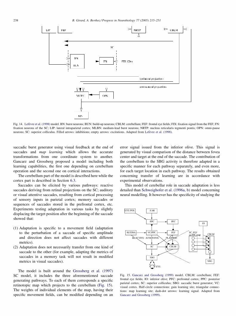
B. Girard, A. Berthoz / Progress in Neurobiology 77 (2005) 215–251238
Fig. 14. Lefevre et al. (1998) model. BN: burst neurons; BUN: build-up neurons; CBLM: cerebellum; FEF: frontal eye fields; FIX: fixation signal from the FEF; FN:
fixation neurons of the SC; LIP: lateral intraparietal cortex; MLBN: medium-lead burst neurons; NRTP: nucleus reticularis tegmenti pontis; OPN: omni-pause
neurons; SC: superior colliculus. Filled arrows: inhibitions; empty arrows: excitations. Adapted from Lefevre et al. (1998).
saccadic burst generator using visual feedback at the end of
saccades and map learning which allows the accurate
transformations from one coordinate system to another.
Gancarz and Grossberg proposed a model including both
learning capabilities, the first one depending on cerebellum
operation and the second one on cortical interactions.
The cerebellum part of the model is described here while the
cortex part is described in Section 6.3.
Saccades can be elicited by various pathways: reactive
saccades deriving from retinal projections on the SC; auditory
or visual attentive saccades, resulting from cortical processing
of sensory inputs in parietal cortex; memory saccades or
sequences of saccades stored in the prefrontal cortex, etc.
Experiments testing adaptation in various tasks by slightly
displacing the target position after the beginning of the saccade
showed that:
(1) A
daptation is specific to a movement field (adaptationto the perturbation of a saccade of specific amplitude
and direction does not affect saccades with different
metrics).
(2) A
daptation does not necessarily transfer from one kind ofsaccade to the other (for example, adapting the metrics of
saccades in a memory task will not result in modified
metrics in visual saccades).
Fig. 15. Gancarz and Grossberg (1999) model. CBLM: cerebellum; FEF:
frontal eye fields; IO: inferior olive; PFC: prefrontal cortex; PPC: posterior
parietal cortex; SC: superior colliculus; SBG: saccadic burst generator; VC:
visual cortex. Half-circle connections: gain learning site; triangular connec-
tions: map learning site; dash-dot arrows: learning signal. Adapted from
Gancarz and Grossberg (1999).
The model is built around the Grossberg et al. (1997)
SC model, it includes the three aforementioned saccade
generating pathways. To each of them corresponds a specific
retinotopic map which projects to the cerebellum (Fig. 15).
The weights of individual elements of the map, having their
specific movement fields, can be modified depending on an
error signal issued from the inferior olive. This signal is
generated by visual comparison of the distance between fovea
center and target at the end of the saccade. The contribution of
the cerebellum to the SBG activity is therefore adapted in a
specific manner for each pathway separately, and even more,
for each target location in each pathway. The results obtained
concerning transfer of learning are in accordance with
experimental observations.
This model of cerebellar role in saccade adaptation is less
detailed than Schweighofer et al. (1996a, b) model concerning
neural modelling. It however has the specificity of studying the
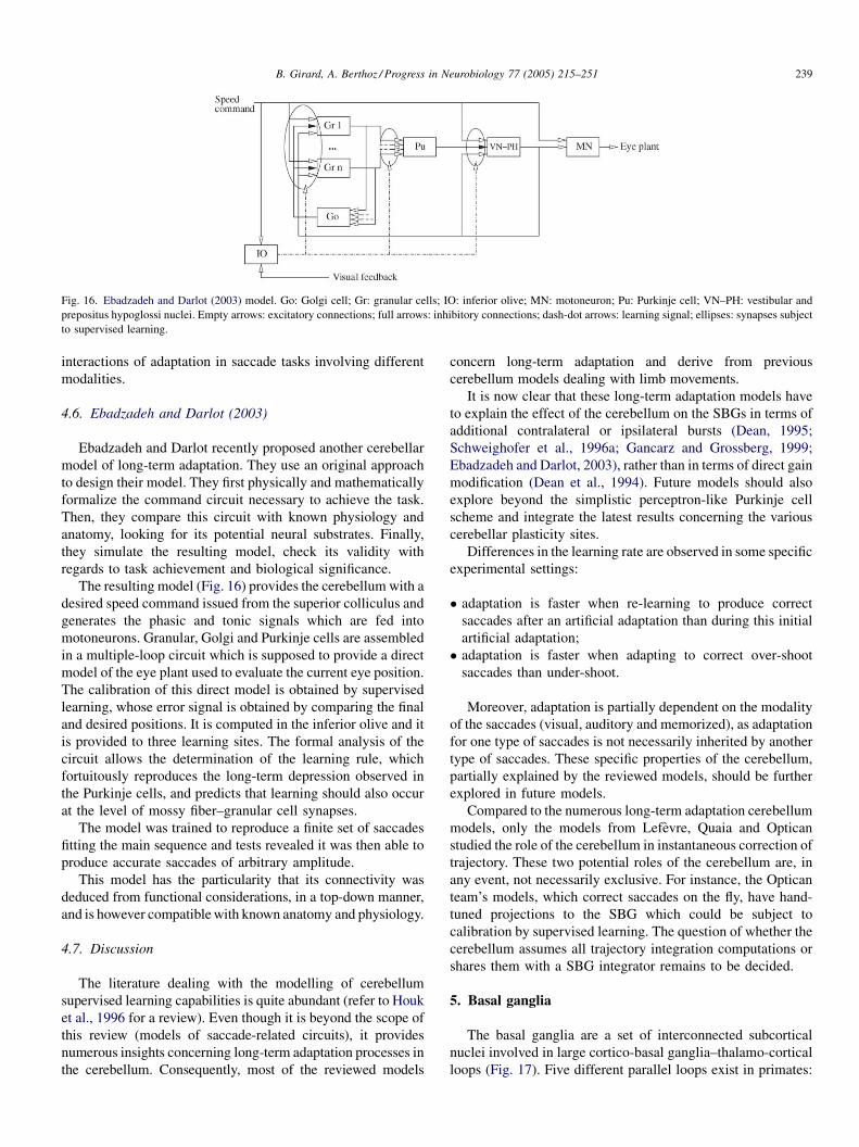
B. Girard, A. Berthoz / Progress in Neurobiology 77 (2005) 215–251 239
Fig. 16. Ebadzadeh and Darlot (2003) model. Go: Golgi cell; Gr: granular cells; IO: inferior olive; MN: motoneuron; Pu: Purkinje cell; VN–PH: vestibular and
prepositus hypoglossi nuclei. Empty arrows: excitatory connections; full arrows: inhibitory connections; dash-dot arrows: learning signal; ellipses: synapses subject
to supervised learning.
interactions of adaptation in saccade tasks involving different
modalities.
4.6. Ebadzadeh and Darlot (2003)
Ebadzadeh and Darlot recently proposed another cerebellar
model of long-term adaptation. They use an original approach
to design their model. They first physically and mathematically
formalize the command circuit necessary to achieve the task.
Then, they compare this circuit with known physiology and
anatomy, looking for its potential neural substrates. Finally,
they simulate the resulting model, check its validity with
regards to task achievement and biological significance.
The resulting model (Fig. 16) provides the cerebellum with a
desired speed command issued from the superior colliculus and
generates the phasic and tonic signals which are fed into
motoneurons. Granular, Golgi and Purkinje cells are assembled
in a multiple-loop circuit which is supposed to provide a direct
model of the eye plant used to evaluate the current eye position.
The calibration of this direct model is obtained by supervised
learning, whose error signal is obtained by comparing the final
and desired positions. It is computed in the inferior olive and it
is provided to three learning sites. The formal analysis of the
circuit allows the determination of the learning rule, which
fortuitously reproduces the long-term depression observed in
the Purkinje cells, and predicts that learning should also occur
at the level of mossy fiber–granular cell synapses.
The model was trained to reproduce a finite set of saccades
fitting the main sequence and tests revealed it was then able to
produce accurate saccades of arbitrary amplitude.
This model has the particularity that its connectivity was
deduced from functional considerations, in a top-down manner,
and is however compatiblewith known anatomy and physiology.
4.7. Discussion
The literature dealing with the modelling of cerebellum
supervised learning capabilities is quite abundant (refer to Houk
et al., 1996 for a review). Even though it is beyond the scope of
this review (models of saccade-related circuits), it provides
numerous insights concerning long-term adaptation processes in
the cerebellum. Consequently, most of the reviewed models
concern long-term adaptation and derive from previous
cerebellum models dealing with limb movements.
It is now clear that these long-term adaptation models have
to explain the effect of the cerebellum on the SBGs in terms of
additional contralateral or ipsilateral bursts (Dean, 1995;
Schweighofer et al., 1996a; Gancarz and Grossberg, 1999;
Ebadzadeh and Darlot, 2003), rather than in terms of direct gain
modification (Dean et al., 1994). Future models should also
explore beyond the simplistic perceptron-like Purkinje cell
scheme and integrate the latest results concerning the various
cerebellar plasticity sites.
Differences in the learning rate are observed in some specific
experimental settings:
� a
daptation is faster when re-learning to produce correctsaccades after an artificial adaptation than during this initial
artificial adaptation;
� a
daptation is faster when adapting to correct over-shootsaccades than under-shoot.
Moreover, adaptation is partially dependent on the modality
of the saccades (visual, auditory and memorized), as adaptation
for one type of saccades is not necessarily inherited by another
type of saccades. These specific properties of the cerebellum,
partially explained by the reviewed models, should be further
explored in future models.
Compared to the numerous long-term adaptation cerebellum
models, only the models from Lefevre, Quaia and Optican
studied the role of the cerebellum in instantaneous correction of
trajectory. These two potential roles of the cerebellum are, in
any event, not necessarily exclusive. For instance, the Optican
team’s models, which correct saccades on the fly, have hand-
tuned projections to the SBG which could be subject to
calibration by supervised learning. The question of whether the
cerebellum assumes all trajectory integration computations or
shares them with a SBG integrator remains to be decided.
5. Basal ganglia
The basal ganglia are a set of interconnected subcortical
nuclei involved in large cortico-basal ganglia–thalamo-cortical
loops (Fig. 17). Five different parallel loops exist in primates:

B. Girard, A. Berthoz / Progress in Neurobiology 77 (2005) 215–251240
Fig. 17. Overview of basal ganglia nuclei and connectivity in a rat brain. EP: entopeduncular nucleus; GP: globus pallidus; SNc: substantia nigra pars compacta; SNr:
substantia nigra pars reticulata; STN: subthalamic nucleus. Striatum’s patch and matrix: see text. Empty arrows: excitatory projections; full arrows: inhibitory
projections; dash-dot arrows: dopaminergic projections.
motor, oculomotor, prefrontal (dorsolateral prefrontal and
lateral orbitofrontal) and limbic loops. They have similar
internal connectivity but interact with different cortical areas
and brainstem nuclei. The oculomotor loop is of primary
interest concerning saccade generation, as it interacts with the
frontal eye fields and the parietal posterior cortex and projects
to the SC, gating the loci of activation on the collicular map.
The prefrontal loop involving the dorsolateral prefrontal cortex
is incidentally involved in saccade generation as it is involved in
working memory processes enabling the learning and restitu-
tion of sequences of saccades.
The input nuclei of the basal ganglia circuit are the striatum
(including the putamen, caudate nucleus and nucleus
accumbens) and the subthalamic nucleus (STN). The striatum
can be divided in cytoarchitectonically and neurochemically
distinct subpopulations of neurons, a set of isolated areas
called patches, surrounded by the so-called matrix. The matrix
of the striatum has inhibitory projections to the globus pallidus
(GP, or external part of globus pallidus, GPe, in primates),
entopeduncular nucleus (EP, or internal part of globus pallidus,
GPi, in primates) and substantia nigra pars reticulata (SNr). EP
and SNr constitute the output (inhibitory) of the BG, while the
GP is part of an internal circuit involving the STN: the GP
inhibits the EP and the STN, while the STN excites the GP, the
EP and the SNr.
The patches of the striatum project to the dopaminergic
system (substantia nigra pars compacta, SNc, and ventral
tegmental area, VTA) which projects back to thewhole striatum,
to the STN and to the frontal cortex. This circuitry seems to be
central in the neural implementation of reinforcement learning
capabilities.
The basal ganglia circuitry of a given loop is constituted of
parallel channels: each nucleus can be subdivided in distinct
groups of neurons, each group of a given nucleus projecting to a
specific group in the target nucleus. The STN is an exception as
its excitation seems to be diffuse.
The role of the BG is interpreted as a generic role of
selection: it selects which channel inside a loop has to be
activated at a given moment. This selection occurs via
disinhibition: at rest, the output nuclei of the BG are tonically
active and keep their thalamic and brainstem targets under
constant inhibition, when multiple channels are activated, the
BG operate a winner-takes-all computation and, as a result, the
output inhibition corresponding to the winning channel is
interrupted, allowing the activation of the targeted circuit.
The complex internal circuitry of the basal ganglia has long
been interpreted as constituted of so-called direct and indirect
pathways (Fig. 18, left). The direct is inhibitory to the EP/SNr,
the indirect is excitatory (as a result of a cascade of two
inhibitions and one excitation), and the balance between the
two opposite effects is a key feature of basal ganglia operation.
This scheme is however outdated as new experimental data
showed that it neglects important connections, it has now been
replaced by a direct, indirect and hyperdirect pathways
interpretation (Fig. 18, right).
The two models of saccade generation including the basal
ganglia explored their role in selecting the saccade target on the
SC and in storing in working memory the location of potential
targets.
5.1. Dominey and Arbib (1992) and Dominey et al. (1995)
The BG subpart of the Dominey and Arbib model is based on
the old direct/indirect pathways interpretation and moreover
only includes the direct pathway. Two parallel subloops are
distinguished in the BG: a first one projecting to the SC,
enabling the selection of the target, and a second one projecting
back to the FEF via the thalamus, in charge of storing potential
targets in working memory.
The striatum and the SNr only are modelled (Fig. 9). The
channels of both circuits are elements of a retinotopic map
which allows the specification of the saccade metrics. The

B. Girard, A. Berthoz / Progress in Neurobiology 77 (2005) 215–251 241
Fig. 18. Interpretations of the BG connectivity. (Left) Direct/indirect pathways and (right) direct, indirect and hyperdirect pathways. D1: striatal neurons with D1-
type dopamine receptors; D2: striatal neurons with D2-type dopamine receptors; EP: entopeduncular nucleus; GP: globus pallidus; SNr: substantia nigra pars
reticulata; STN: subthalamic nucleus. Empty arrows: excitatory projections; full arrows: inhibitory projections; dashed arrows: neglected connections in the direct/
indirect pathways interpretation; bold arrows: pathways.
striatum of the selection circuit receives inputs from both
visual and memory layers of the FEF, the learned weighting
of these inputs induces the selective inhibition of the SNr
which in turn disinhibits specific areas of the SC, enabling
the execution of the chosen saccade. The memory circuit
receives inputs from the memory layer of the FEF only, and
its SNr projects to the thalamus. The disinhibition of the
thalamus allows the onset of a reverberation between the
thalamus and the memory layer of the FEF, which is the base
of the memory.
An extension of the model (Dominey et al., 1995) includes
plasticity of the cortico-striatal synapses and dopamine
reward signals. Additional cortico-striatal projections from
the inferior temporal cortex (IT) and from the prefrontal
cortex (PFC) provide additional information concerning the
context and the execution of sequences. The IT provides
contextual information relative to the nature of cues (color,
shape, etc.) that can be presented before the selection of the
target and the execution of the saccade. The PFC provides an
input vector with unique patterns of activity for each step
during the execution of a given sequence. The model is then
able to learn to choose among two similar potential targets
depending on precueing and to reproduce sequences of
saccades.
The whole Dominey and Arbib model was tested in single,
memory and double saccades and some of its simulated cell
activity patterns were qualitatively compared with electro-
physiological recordings.
This model of the saccade generation circuitry is the first to
include the basal ganglia, and thus to propose mechanisms
generating the SNr responses measured by Hikosaka andWurtz
(1983a, b, c, d). It is also the most complete to date in terms of
included brain regions, and thus the only one to produce a
global understanding of the operation of the whole system,
proposing explanations of how individually studied areas
interact together during the execution of saccades.
5.2. Brown et al. (2004)
As new experimental data concerning the BG have been
accumulated in the last 10 years, Brown et al. proposed a new
model of the role of the BG in selecting both the relevant
strategy for saccade generation and the metrics of the saccade,
once the strategy is chosen. The cortical part of this model, and
especially the description of the laminar structure of the FEF, is
presented in Section 6.4.
The oculomotor BG loop is, in this model also, divided in
two parallel subloops (Fig. 19). The first one is an IT/FEF–BG–
thalamus–FEF loop whose role is to select which strategy will
drive the forthcoming saccade decision. These strategies can
be based on: (1) the state and position of the fixation point, (2)
the state and position of the target or (3) the nature of some
object in the visual field, independently from its position (cue).
This circuit therefore has three channels associated to these
three strategies. The second subloop is a PPC/FEF–BG–SC–
FEF loop, it has to select the metrics of the saccade by
disinhibiting a location on the SC map. The channels of this
circuit correspond to the movement fields of a retinotopic map.
The decision is taken using a saliencemap provided by the FEF
and depending on the strategy decision taken by the first
circuit.
The internal circuitry of the BG is modelled using recent
knowledge on nuclei interconnections:
� T
he direct pathway is the place where selection occurs,thanks to an on-center off-surround architecture. Each
channel in the direct part of the striatum receives specific
excitatory input related to its role only in conjunction with
diffuse inhibitory input common to all channels and
generated by a combination of all excitatory inputs in the
striatal g-aminobutyric acid (GABA) interneurons. For
example, in the first BG subloop, the channel corresponding
to the strategy based on target state and position is excited by

B. Girard, A. Berthoz / Progress in Neurobiology 77 (2005) 215–251242
Fig. 19. Brown et al. (2004) model. BG: basal ganglia; FEF: frontal eye fields; GABA: GABAergic inhibitory striatal interneurons; GPe: external segment of the
globus pallidus; IT (a/p): anterior/posterior inferotemporal cortex; PFC: prefrontal cortex; PPC: posterior parietal cortex; SC: superior colliculus; SNr: substantia
nigra pars reticulata; STN: subthalamic nucleus; V4: extrastriate visual cortex area 4. Empty arrowheads: excitatory projections; full arrowheads: inhibitory
projections; filled semi-circles: plastic synapses. Adapted from Brown et al. (2004).
subpart of the FEF corresponding to this strategy, while it is
inhibited by a signal generated by the whole FEF.
� T
he indirect pathway is used to generate a trainable stopsignal used in tasks involving a delay before the response. It
operates by increasing the STN activity: this provides
additional activation of the SNr/GPi thus increasing their
inhibitory output and blocking the selection process.
� T
he hyperdirect pathway is only present in the first BGsubloop, it is under control of the FEF output layer and is used
by the winning strategy to lock the BG in order to prevent the
selection of another strategy before the execution of orders.
The selection of a strategy by the direct pathway involves
increased activity in FEF output layer, transmitted to the STN
which blocks the BG subloop selection process in a similar
manner as the indirect pathway.
The cortico-striatal synapses of the first subloop are
trainable using dopamine input signalling the occurrence of
a reward or a punishment. A burst of dopamine above baseline
level (reward) generates a weight increase of the recently
activated channels in the direct pathway, favouring the last
decision taken in future similar contexts. Conversely, dips in the
dopamine baseline level (punishment) generate weight
increases in the indirect pathway, which will tend to suspend
the last decision taken in future similar contexts.
The model was tested in five standard saccade tasks
(fixation, single saccade, overlap, gap and memory saccade)
and was able to learn to generate adequate saccades in each
situation. The activity profile of some model cells was shown to
be qualitatively similar to their experimentally observed
counterparts.
Various aspects of the BG part of the model are subject to
discussion:
� C
onsidering that the indirect pathway is dedicated to delayedsaccade responses seems to be a bit too much task-specific
and not necessarily useful in real-life situations. The
hypothesis that such a complex cascade of inhibitions and
excitation evolved for this sole purpose is debatable.
� I
n the first BG subloop, two GABAergic striatal interneuronsfamilies are distinguished: some are projecting specifically to
the neurons belonging to the direct part of the striatum and
have an additional thalamic input, as the others are dedicated
to the indirect pathway. Knowing that these interneurons are
B. Girard, A. Berthoz / Progress in Neurobiology 77 (2005) 215–251 243
not numerous in the striatum and seem to have broad and
extensive projections over the whole striatum, such a
discriminative targeting is unprobable.
� T
he plasticity of the cortico-striatal synapses in the first BGsubloop depends on secondary messenger traces. These are
however computed in a different manner for direct and
indirect pathways (in the indirect pathway, the trace depends
on the thalamic input) even if their inputs are quite similar. No
justification for this difference is proposed.
� T
he plasticity of the cortico-striatal synapses of the secondBG subloop is not assigned any role.
� C
oncerning the connectivity of the second BG subloop, theselectivity of the SC feedback over the indirect pathway is not
justified, nor the lack of any cortical modulation of the STN
activity.
� F
inally, the experimentally observed memory-related SNrcells (Hikosaka and Wurtz, 1983c), which can be necessary
for efficient target remapping in the SC (Bozis and
Moschovakis, 1998), are not included in the SC-projecting
BG subloop.
Despite these small flaws, this BG model is much more up-
to-date than the Dominey and Arbib proposal, it is also the first
BG model (even including BG models not related to saccade
generation) taking into consideration the laminar structure of
the cortex, and specifically the fact that striatal and STN inputs
originate from different layers.
5.3. Discussion
The role of the oculomotor loop of the basal ganglia in the
saccade-related brain circuitry has not been subject to many
modelling studies. The BG clearly have a selective influence
over the SC, which was often included in SC models by SNr
external inhibitory input (Arai et al., 1994, 1999; Das et al.,
1995; Grossberg et al., 1997; Bozis and Moschovakis, 1998).
As mentioned in Section 3.17, the BG do not form loops with
cortical areas only, but also with SC superficial and
intermediate layers. The dynamics of these loops can only
be studied by modelling the BG–SC interactions with up-to-
date models of both regions.
As many models of the other basal ganglia loops performing
selection have been proposed (see Gillies and Arbruthnott,
2000; Joel et al., 2002, for recent reviews), some of them could
easily be adapted to encompass the oculomotor one (Gurney
et al., 2001, for example).
Finally, the involvement of a prefrontal BG loop in working
memory has been studied and modelled (Frank et al., 2000) and
could conveniently be included in future models, providing them
with additional memory and sequences learning capabilities.
6. Cortex
As briefly stated in Section 1, many cortex areas are more or
less implicated in the saccadic premotor activity. The posterior
parietal cortex, and more specifically lateral intraparietal area
(LIP), modulates the ‘‘Where’’ stream of the cortical visual
processing by attentional processes. The dorsolateral prefrontal
cortex stores target positions, acting as a spatial working
memory, which allows temporal organization of saccades
(predictions and delays) and even saccade inhibition. The
presupplementary eye fields is involved in learning motor
programs containing saccades and projects to the supplemen-
tary eye fields (SEF) which can execute them if the context is
appropriate by sending saccade orders to the frontal eye fields.
The FEF receives convergent inputs from PPC, SEF and
DLPFC, operates the final target selection stage by interacting
with the basal ganglia and sends the corresponding motor
command to the SC and SBG. Finally, FEF, SEF and DLPFC
receive projections from the anterior cingulate cortex which
provides them with motivational modulation.
Concerning saccadic system modelling studies, the PPC and
the FEF have been the most frequently and extensively
modelled cortical areas because of their positions in the flow
information: visual ‘‘Where’’ stream input to ocular premotor
circuitry and latest ocular premotor area before subcortical
stages, respectively.
6.1. Zipser and Andersen (1988)
Zipser and Andersen (1988) studied the transformation of
visual input coded in oculocentric coordinates into cranio-
centric coordinates using eye position. This transformation is
thought to occur in the area 7a of the PPC, which receives both
eye position and retinal signals and where neurons exhibit
interesting patterns of activity, corresponding to retinocentric
receptive fields modulated by so-called gain fields coding eye
position in the head. Their model is a perceptron trained to
solve the coordinate transformation problem and happens to
simulate the area 7a gain-field activity.
The input of the perceptron is a 8� 8 array, composed of
visual neurons with 15�-wide receptive field, and two 16 neuronvectors coding the azimuth and elevation coordinates of the eye
in the head. The hidden layer is composed of 9–36 units. The
craniocentric output is either a 8� 8 map or two 16 neuron
vectors coding the coordinates of the target. This perceptron is
trained to compute the coordinate transformation using the
classical back-propagation algorithm.
After 1000 trials, the network is satisfactorily trained. The
activity of the hidden layer neurons reflects gain fields
superimposed on large receptive fields and is qualitatively
comparable to 7a neurons. These fields are however not as
complex as some of those of real neurons, especially when the
output is coded by a vector rather than by a map. These complex
receptive fields are more comparable to the hidden layer
activity before training.
The main interest of this paper is to show that the gain-field
encoding used in area 7a can efficiently perform a coordinate
transformation.
6.2. Dominey and Arbib (1992) and Dominey et al. (1995)
In their 1992 model, Dominey and Arbib simulate the
activity of neurons in the FEF and PPC, while the 1995

B. Girard, A. Berthoz / Progress in Neurobiology 77 (2005) 215–251244
extension adds simulated extrastriate visual cortex area 4 (V4),
inferior temporal cortex (IT) and prefrontal cortex. On the one
hand, FEF and PPC are made of multiple layers, each of them
organized as retinotopic maps where potential targets are
represented by hills of activity (Fig. 9). On the second hand, V4,
IT and PFC are modelled by groups of neurons that do not have
a topological organization.
The PPC is made of two layers. The first one (visual)
receives visual input from the ‘‘Where’’ pathway of the visual
cortex, signalling the position but not the nature of potential
targets present in the visual field. The second one, a quasi-visual
layer, receives direct projections from the visual layer and is
able to memorize targets positions thanks to reverberating
excitations. Beyond this memory capability, this second layer
also receives feedback from the brainstem tonic neurons
measuring the eye position and is capable of remapping the
positions of targets during eye movements, in a moving hill of
activity manner, similar to many SC models proposals. This
layer drives the activity of the visual and quasi-visual layers in
FEF and SC, respectively.
The FEF is made of four layers. The first (visual) layer only
relays the activity of the QV PPC layer to the memory and
presaccadic layers of the FEF. The second (memory) layer of
the FEF receives the memorized targets positions from the PPC
but these are stored only if the resonant activity with the
thalamus is made possible by the disinhibition of the latter by
the memory basal ganglia loop (see Section 5.1). The PPC
memory stores all perceived targets positions, whereas the FEF
memory selects those it stores depending on reinforcement
learning occurring at the level of cortico-striatal synapses. The
third layer is a fixation layer, it is active if the foveal part of the
PPC visual layer is activated, in which case it inhibits the
memory and presaccadic layers of FEF along with the
superficial and winner-takes-all layers of the superior
colliculus. Finally, the presaccadic layer gathers the activations
of the visual and memory layers of the FEF along with the
fixation inhibition signalling the presence of a target in the
fovea and projects the resulting landscape of activity to the BG
selection circuit and to the SC saccadic layer to enable saccade
generation.
The role of the PPC is to relay present and past visual inputs,
and to produce the remapping function necessary to keep this
information up-to-date after saccades. The FEF interacts with
the BG to select: (1) the targets to be stored in working memory
and (2) the targets to foveate among suggestions issued from
both vision and memory.
V4 is modelled as a group of neurons coding the features
(shape and color) of the object lying on the fovea, these project
to 25 IT neurons with random connections, therefore encoding
feature conjunctions and disjunctions. The projections from IT
to BG allow the saccade selection to learn to take into account
the nature of cues, as evoked in Section 5.1.
The PFC is a group of 25 neurons, it receives randomly
weighted inputs from the PPC, providing target positions, and
from the SC, providing saccade execution signals. It also
receives damped self-inputs with five different time constants
which provide short-term memory. The resulting activity
encodes past sequences of targets and saccades, unique patterns
being generated for each step of the execution of a sequence.
The projections from the PFC to the BG are subject to
reinforcement learning, which enables the learning of
sequences by associating specific PFC patterns with the
corresponding saccades.
The resulting architecture accounts for the visual, quasi-
visual and memory activity patterns observed in FEF and PPC.
As previously mentioned, it succeeded in tasks of simple,
memory, double and precued saccades, and also in sequence
learning tasks. This proves the efficiency of the memory,
remapping and learning properties of the simulated cortical
areas.
This model was the first one to propose an interpretation of
the cortical patterns of activity observed in saccade experi-
ments. Concerning the remapping of targets ability, the choice
is to have it performed in the PPC QV layer, this layer then
controlling its equivalents in the FEF and the SC. This could
also be performed in a parallel and redundant manner, as
suggested in the Bozis andMoschovakis (1998) model, because
in a phylogenetical point of view, this function might have
originated in the SC of animals lacking a real cortical structure
and kept since then.
6.3. Gancarz and Grossberg (1999)
The cerebellum part of the Gancarz and Grossberg (1999)
model is described in Section 6.3. Its cortical part deals with the
problem of coordinate transformations in the cortex, what they
call map learning. It is supposed to occur at the level of parietal
cortex, where information from visual (retinocentric) and
auditory (craniocentric) sources is mixed, and between the PFC
and the FEF, as the planning of saccade sequences in PFC is
thought to be in craniocentric coordinates.
The model generates saccades in one direction only and
considers one visible target at a time. The coding of positions
and coordinate changes were therefore highly simplified. The
retina is a vector of zeros with a single one at the target
position. As only one visual target has to be coded in the PPC
and FEF, a single value representing its abscissa on the
saccade axis is coded. Transformations from craniotopic to
retinotopic coordinates simply imply the subtraction of the
eye position signal provided by the tonic neurons of the
brainstem saccade generator. An ad hoc transformation
involving fixed weights and threshold allows the transforma-
tion of this value into an activation on a 1D map in the PFC,
allowing the storage of multiple targets. Finally, the reverse
transformation used to compute a single value (in PPC and
FEF) from a 1D map (retina or PFC) involves the calibration
of weights, simply using the eye position at the end of the
saccade as a teaching signal.
The disturbance of these weights by the perturbation
protocol previously evoked (see Section 4.5) is the base of the
recalibration of coordinates changes. The specific order in
which these coordinates changes occur explains why adaptation
transfers from scanning task to memory task but not the other
way.

B. Girard, A. Berthoz / Progress in Neurobiology 77 (2005) 215–251 245
This model is the only one exploring the issue of
unidirectional adaptation transfers; however, its implementa-
tion is too coarse to allow comparison of its internal cortical
state and recorded cells.
6.4. Brown et al. (2004)
The Brown et al. model included various cortical areas (PFC,
FEF and PPC) but a special interest was given to the FEF, as its
internal laminar structure was modelled and simulated. The
model distinguishes four layers and a pool of post-saccadic
cells in the FEF (Fig. 19). These layers are a sensory input layer,
a plan layer (representing layers II, III and VIa), an output layer
(layer Vb) and an arousal layer (layer VI). Input, plan and
output layers are made of three distinct retinotopic maps,
representing the three saccade strategies implemented in the
model: strategy guided by the position of target, or the position
of the fixation object or guided by the features of the foveated
object (Fig. 20). The resulting distinct trans-layer structures
associated to the strategies are called gateable cortical zones
(GCZ). The plan layer of the FEF, at the GCZ level, interacts
with the first basal ganglia circuit (see Section 5.2) in order to
select the saccade strategy, the retinotopic maps of the output
layer then interact with the second basal ganglia circuit in order
to select the precise metrics of the saccade.
The input layer receives information about the location of
salient stimuli in the visual field, the nature of the stimulus
dictating which GCZ is concerned. Additional feedforward
inhibitions act to enhance the contrast.
The arousal layer is driven by inputs from the PFC, which
stores the nature of the presented objects, and can provide an
additional drive to one of the GCZs if the cue was associated by
learning to a specific strategy.
The plan layer receives the sorted and contrast-enhanced
visual input of the input layer, local selection of saccade metrics
is made in each GCZ by lateral inhibitions and autoexcitation,
Fig. 20. FEF structure of the Brown et al. (2004) model. (A) Retinotopic maps at the l
rows correspond to different layers, columns correspond to different strategies forming i
(IT) GCZ represents the strategy consisting in the association of a specific cue (the na
position be, the corresponding input layer is therefore not a spatial map. Reprinted from
ganglia circuits interact to control planned and reactive saccades. Neural Netw. 17, 4
GCZ selection is made by reciprocal inhibitions of neurons
belonging to the different GCZ but associated to the same
metrics. The GCZ selection is reinforced by positive feedback
provided by the basal ganglia–thalamic circuit and by the
possible object-associated drive from the arousal layer. SC
feedback reinforces the activity of the units corresponding to
the selected metrics during the execution of the saccade, while
post-saccadic inhibition resets the activity after saccade
completion.
The output layer is very similar to the plan layer, the
differences being the absence of between-GCZ inhibitions and
the projections to the SC providing the saccade ‘‘go’’ signal.
The PPC is a single-layer retinotopic map. It has a phasic
activity because of self-inhibitions, is activated by the FEF
output layer and by biased visual input (peripheral targets and
left–right rather than up–down saccades are favoured) and
projects to the SC. Its functional role in the model is to elicit
reactive visual saccades, no attentional process was modelled.
The model was tested in a number of tasks listed in Section
5.2.
The choice of building two GCZs distinguishing the fixation
and the target stimuli seems a bit ad hoc and surprising as in
monkey experiments, these are often objects of the same nature.
This does not however discard the interesting idea of
hierarchical selection (first strategy and then metrics) which
is present in some basal ganglia models (Monchi et al., 2000).
This recent saccade-circuitry model is however the first one to
go into such detailed modelling of the cortical laminar
structure.
6.5. Deneve et al. (1999, 2001)
Deneve et al. address the generic problems of population
coding with noisy inputs and of computations based on
population-coded variables. Their first model (Deneve et al.,
1999) shows how variables can be extracted from noisy sensory
evel of the cortex are 2D-grids with the central unit representing the fovea and (B)
solated gateable cortical zones (GCZ) across the layers. The inferotemporal cortex
ture of an object provided by IT) with a given saccade, whatever might its spatial
Brown, J., Bullock, D., Grossberg, S., 2004.How laminar frontal cortex and basal
71–510, Copyright (2004), with permission from Elsevier.

B. Girard, A. Berthoz / Progress in Neurobiology 77 (2005) 215–251246
input and stored in recurrent networks. Building on this, the
second one (Deneve et al., 2001) shows how such networks can
be interconnected in order to perform function approximation
(computation of a variable from inputs) and cue integration (all
variables are known and interact to remove noise). This work is
not limited to cortical areas involved in saccades, it is
representative of the current state of the art (Pouget et al., 2003)
in cortex population coding and computing. However, the
example used in the second paper is of peculiar relevance, as it
addresses the integration of variables linked with saccades
generation and coordinates transformation: the retinotopic
position of a target, the craniotopic position of the eye and the
craniotopic position of the target.
The maps encoding a single variable are recurrent networks
with excitatory lateral connections (Gaussian filters) and global
divisive shunting inhibition. The cells of these maps have bell-
shaped tuning curves with regard to sensory input with a
superimposed noise. Function approximation and cue integra-
tion is based on a basis function architecture (Fig. 21).
However, the purpose of this study is not to execute simple
function approximation (computing one variable depending on
the two inputs), but to allow any of the three variables to be
computed from the two others along with cue integration.
Consequently, the connections of the three sensory layers to the
basis function layer are not feedforward but bidirectional. The
reliability of an input is represented by its level of activity.
The population coding efficiency is evaluated in the
maximum likelihood estimation framework (ML). It can
recover the best possible measure of the encoded variable.
The operations of function approximation and cue integration
are also performed with an efficiency very close to the ML
estimates, even in the case of deteriorated inputs (reduced
reliability or increased standard deviation).
Fig. 21. Deneve et al. (2001) model. (Left) Initial state of the model. (Right) Stable
dimensional map of basis function units, the four layers interconnections are bidire
(upper left) has no initial input, while in cue integration, this layer also has a noisy in
the Nature Publishing Group.
The activity of the basis function layer mimics the gain fields
observed in many cortical areas. It is predicted that multimodal
neurons should exhibit partially shifting receptive fields in all
modalities, similar to those observed in SC and in ventral intra
parietal and premotor cortices.
These models extend function approximation with popula-
tion codes using basis functions, as it performs optimally with
both function approximation and cue integration in the case of
noisy input.
6.6. Mitchell and Zipser (2001)
The Mitchell and Zipser (2001) model deals with spatial
short-term memory in parietal areas. It is a neural network
obtained by optimization, rather than by design, in order to
memorize the position of a target in eye- and head-centered
coordinates and to update it in case of saccades.
The network is made of a set of recurrently connected
neurons, its inputs are two simulated retinas together with eye
conjugate and vergence angles and velocities, its output are two
maps, respectively, eye- and head-centered. The connections of
the network are obtained by learning (back-propagation
through time algorithm), the evaluation criterion being the
ability to store the position of a flashed target and to correctly
update its position during saccades.
The resulting network contains neurons that are comparable
to parietal neurons from area 7a or LIP: their receptive field
responses are modulated by gain fields which are planar
functions of eye position. They can be divided in three classes,
depending on the nature of their receptive fields: eye-based,
head-based or intermediate. The head-based cells obtained in
the model have not been found yet in area 7a or LIP, but could
have been overlooked or could lie in another parietal area.
state stabilization. Inputs are one-dimensional, the intermediate layer is a two-
ctional. In the case of function approximation, the head-centered position layer
put. Reprinted from Deneve et al. (2001), Nat. Neurosci., with permission from
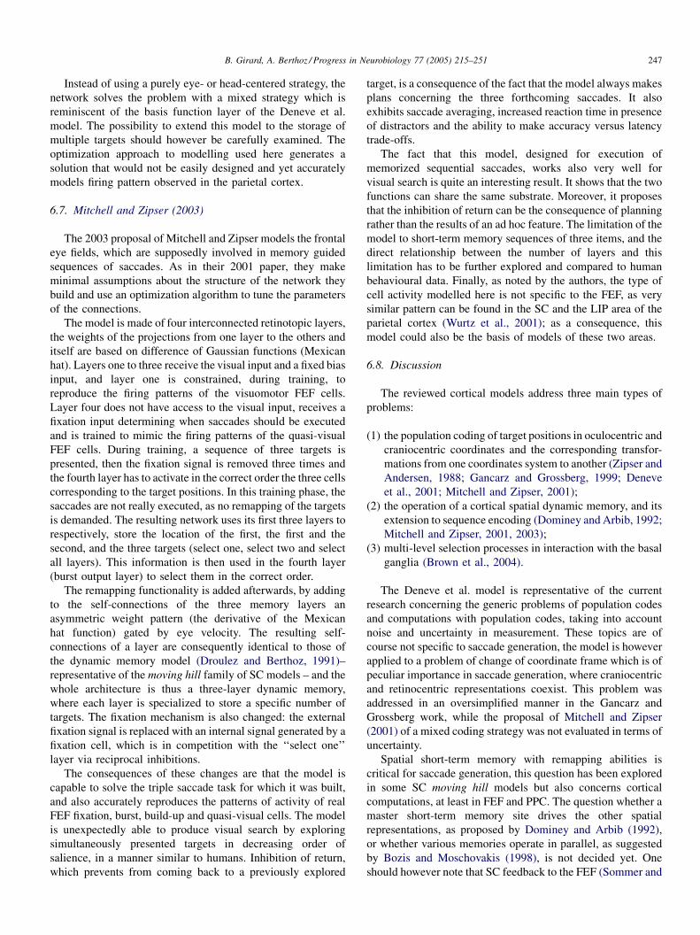
B. Girard, A. Berthoz / Progress in Neurobiology 77 (2005) 215–251 247
Instead of using a purely eye- or head-centered strategy, the
network solves the problem with a mixed strategy which is
reminiscent of the basis function layer of the Deneve et al.
model. The possibility to extend this model to the storage of
multiple targets should however be carefully examined. The
optimization approach to modelling used here generates a
solution that would not be easily designed and yet accurately
models firing pattern observed in the parietal cortex.
6.7. Mitchell and Zipser (2003)
The 2003 proposal of Mitchell and Zipser models the frontal
eye fields, which are supposedly involved in memory guided
sequences of saccades. As in their 2001 paper, they make
minimal assumptions about the structure of the network they
build and use an optimization algorithm to tune the parameters
of the connections.
The model is made of four interconnected retinotopic layers,
the weights of the projections from one layer to the others and
itself are based on difference of Gaussian functions (Mexican
hat). Layers one to three receive the visual input and a fixed bias
input, and layer one is constrained, during training, to
reproduce the firing patterns of the visuomotor FEF cells.
Layer four does not have access to the visual input, receives a
fixation input determining when saccades should be executed
and is trained to mimic the firing patterns of the quasi-visual
FEF cells. During training, a sequence of three targets is
presented, then the fixation signal is removed three times and
the fourth layer has to activate in the correct order the three cells
corresponding to the target positions. In this training phase, the
saccades are not really executed, as no remapping of the targets
is demanded. The resulting network uses its first three layers to
respectively, store the location of the first, the first and the
second, and the three targets (select one, select two and select
all layers). This information is then used in the fourth layer
(burst output layer) to select them in the correct order.
The remapping functionality is added afterwards, by adding
to the self-connections of the three memory layers an
asymmetric weight pattern (the derivative of the Mexican
hat function) gated by eye velocity. The resulting self-
connections of a layer are consequently identical to those of
the dynamic memory model (Droulez and Berthoz, 1991)–
representative of the moving hill family of SC models – and the
whole architecture is thus a three-layer dynamic memory,
where each layer is specialized to store a specific number of
targets. The fixation mechanism is also changed: the external
fixation signal is replaced with an internal signal generated by a
fixation cell, which is in competition with the ‘‘select one’’
layer via reciprocal inhibitions.
The consequences of these changes are that the model is
capable to solve the triple saccade task for which it was built,
and also accurately reproduces the patterns of activity of real
FEF fixation, burst, build-up and quasi-visual cells. The model
is unexpectedly able to produce visual search by exploring
simultaneously presented targets in decreasing order of
salience, in a manner similar to humans. Inhibition of return,
which prevents from coming back to a previously explored
target, is a consequence of the fact that the model always makes
plans concerning the three forthcoming saccades. It also
exhibits saccade averaging, increased reaction time in presence
of distractors and the ability to make accuracy versus latency
trade-offs.
The fact that this model, designed for execution of
memorized sequential saccades, works also very well for
visual search is quite an interesting result. It shows that the two
functions can share the same substrate. Moreover, it proposes
that the inhibition of return can be the consequence of planning
rather than the results of an ad hoc feature. The limitation of the
model to short-term memory sequences of three items, and the
direct relationship between the number of layers and this
limitation has to be further explored and compared to human
behavioural data. Finally, as noted by the authors, the type of
cell activity modelled here is not specific to the FEF, as very
similar pattern can be found in the SC and the LIP area of the
parietal cortex (Wurtz et al., 2001); as a consequence, this
model could also be the basis of models of these two areas.
6.8. Discussion
The reviewed cortical models address three main types of
problems:
(1) t
he population coding of target positions in oculocentric andcraniocentric coordinates and the corresponding transfor-
mations from one coordinates system to another (Zipser and
Andersen, 1988; Gancarz and Grossberg, 1999; Deneve
et al., 2001; Mitchell and Zipser, 2001);
(2) t
he operation of a cortical spatial dynamic memory, and itsextension to sequence encoding (Dominey and Arbib, 1992;
Mitchell and Zipser, 2001, 2003);
(3) m
ulti-level selection processes in interaction with the basalganglia (Brown et al., 2004).
The Deneve et al. model is representative of the current
research concerning the generic problems of population codes
and computations with population codes, taking into account
noise and uncertainty in measurement. These topics are of
course not specific to saccade generation, the model is however
applied to a problem of change of coordinate frame which is of
peculiar importance in saccade generation, where craniocentric
and retinocentric representations coexist. This problem was
addressed in an oversimplified manner in the Gancarz and
Grossberg work, while the proposal of Mitchell and Zipser
(2001) of a mixed coding strategy was not evaluated in terms of
uncertainty.
Spatial short-term memory with remapping abilities is
critical for saccade generation, this question has been explored
in some SC moving hill models but also concerns cortical
computations, at least in FEF and PPC. The question whether a
master short-term memory site drives the other spatial
representations, as proposed by Dominey and Arbib (1992),
or whether various memories operate in parallel, as suggested
by Bozis and Moschovakis (1998), is not decided yet. One
should however note that SC feedback to the FEF (Sommer and

B. Girard, A. Berthoz / Progress in Neurobiology 77 (2005) 215–251248
Wurtz, 1998), which seem to preferentially transmit burst
activity (high-pass filtering) signalling saccade occurrence,
provides the FEF with the necessary information to process
such computations by itself.
The selection aspects of the cortical part of the Brown et al.
(2004) model are intermingled with the operation of the basal
ganglia circuits and only make sense when both systems are
considered together. This once again highlights the relevance of
modelling the interactions of reciprocally connected brain
regions. The specificity of the FEF part of this model is the
interpretation of the cortical layers interactions and roles it
proposes. It is a quite promising proposal that has to be refined.
Finally, the problems of attentive modulation in the PPC, of
working memory operation in the DLPFC, of programs
learning and restitution in the pre-SEF and SEF and of
motivation modulation of all these function by aCG were
superficially or not addressed at all in saccade generation
models, and should be in future works.
7. Conclusion
This review aimed at giving a glimpse of the evolution,
during the last 30 years, of the conception of the operation of
the various brain regions implied in the saccadic system,
through the presentation of the succession of computational
models built to explore this subject. More importantly, it also
tried to help identify the current questions concerning the roles
of the various regions involved and the precise mechanisms
implementing these roles, and thus the hot topics to be
explored by future models. These were exhaustively listed in
the five previous discussions, we will thus lay stress here on
two points that we think emerge from this review: the
importance of thinking models not only in terms of local brain
regions operation, but complementarily considering interact-
ing brain regions, and, as a consequence, the necessity to
theoretically study the dynamics of the resulting recurrent
networks.
7.1. Beyond the parts: modelling the whole
We have seen in various reviewed studies examples of
models of a given brain region designed to explain experi-
mental results that happened to result from the operation of
another brain region, or from the interaction of the modelled
region with some other regions. The most representative
example of this concerns SBG-only models: they all seek to
produce accurate movements, usually fitted to the main
sequence, while it is now clear that without their associated
cerebellar circuits, the SBGs produce inaccurate movements
that do not respect the main sequence. Understanding the
accuracy of the saccadic system requires the study of, at least,
the interactions of the superior colliculus, the cerebellum and
the saccade burst generators.
Similarly, the remapping of targets in dynamic memories
implies cortical or collicular interactions with the SBGs, and
the target selection processes implicate cortico-basal–thalamo-
cortical and collicular–thalamo-basal–collicular loops.
This advocates for systemic studies of the saccadic system
(and of other brain systems also), in complement of those
focused on a specific region only. Dominey and Arbib (1992) is
an example of such an approach; however, knowledge has
accumulated since andmany of their modelling choices are now
outdated. First, the location of the control feedback loop is
subject to much debate (Scudder et al., 2002). Its presence in
the brainstem saccadic burst generators is of doubtful validity
as no entirely satisfactory neural substrate for the resettable/
leaky integrator has been found. The two theories including the
SC in the loop suffer from various flaws. The cerebellum could
be the missing piece of the feedback loop puzzle, even if current
models of integration in the cerebellum are not completely
satisfactory. Second, the old direct/indirect pathways inter-
pretation of the basal ganglia circuitry has now been replaced
by a much more complex pattern of interconnections. Modern
knowledge concerning the basal ganglia did not give rise to
numerous models of the oculomotor loop; however, interesting
proposals modelling other BG loops could be the basis of future
work. Finally, beyond PPC and FEF, the operation of the other
cortical areas involved in high level aspects of saccade
generation (short, medium and long-term memory, learning of
sequences, of response schedules, inhibition of response,
motivational modulation) could be integrated.
A revised Dominey and Arbib model, summarizing the
current knowledge of the brain circuitry involved in saccade
generation and addressing the various hot topics highlighted in
the five previous discussions, would provide a better under-
standing of the operation of the system as a whole and of the
importance of the interactions between regions which have
often be modelled independently.
7.2. Dynamics considerations
Recurrent neural network models (including internal loops)
may exhibit various dynamic behaviour, like convergence to
some equilibrium point, chaotic trajectories, oscillations, etc.
Some of the reviewed studies considered the internal dynamics
of their models, especially the SC models integrating the lateral
short-range excitation and long-range inhibitions (see Section
3.17), this was however not done in a systematic manner.
Moreover, these considerations were almost exclusively drawn
from simulations in a limited number of specific cases rather
than from generic theoretical analyses.
Nevertheless, when considering large systems composed of
multiple loops of interconnected subsystems, controlling the
dynamics is crucial to ensure correct operation of the model,
given any initial condition and sensory input. Thus, conducting
an analysis of the system’s dynamics becomes mandatory. The
models to be analysed are intrinsically non-linear, but
theoretical tools are available to study the dynamics of non-
linear systems, and some of them, like the contraction analysis
developed by Lohmiller and Slotine (1998), are relatively
simple. The contraction analysis has an additional advantage:
contracting systems connected together, using a number of
combinations (hierarchy, feedback, etc.) with minimal con-
straints, produce contracting systems (Slotine and Lohmiller,
B. Girard, A. Berthoz / Progress in Neurobiology 77 (2005) 215–251 249
2001). As an illustration of this, contraction analysis was
recently used to design a contracting basal ganglia model
composed of contracting modules (Girard et al., 2005), which
could in turn be embedded in a larger system composed of other
contracting systems.
Beyond understanding the operation of a given function or
behaviour in the brain, the modelling of the large systems, like
the saccadic circuitry, has an intrinsic scientific interest:
exploring the solutions found by evolutionary processes to
progressively interconnect new modules with older ones while
keeping the whole system operational.
Acknowledgments
This review was supported by the Bayesian Inspired Brain
and BIBA Artifacts (BIBA) project, funded by the European
Community (grant IST-2001-32115). The authors thank A.
Grantyn, A. Moschovakis and L. Rondi-Reig for valuable
discussions and for providing bibliographical material. We also
thank the three anonymous reviewers for their very constructive
comments on the manuscript.
References
Albus, J., 1971. A theory of cerebellar function. Math. Biosci. 10, 25–61.
Albus, J., 1981. Brains, behavior and robotics. In: BYTE Books, McGraw-Hill,
New Hampshire.
Anastasio, T., Patton, P., Belkacem-Boussaid, K., 2000. Using Bayes’ rule to
model multisensory enhancement in the superior colliculus. Neural Com-
put. 12 (5), 1165–1187.
Anderson, R., Keller, E., Ghandi, N., Das, S., 1998. Two dimensional saccade-
related population activity in superior colliculus in monkey. J. Neurophy-
siol. 80 (2), 798–817.
Arai, K., Das, S., Keller, E., Aiyoshi, E., 1999. A distributed model of the
saccade system: simulations of temporally perturbed saccades using posi-
tion and velocity feedback. Neural Netw. 12 (10), 1359–1375.
Arai, K., Keller, E., Edelman, J., 1994. Two-dimensional neural network model
of the primate saccadic system. Neural Netw. 7, 1115–1135.
Berthoz, A., 1996. The role of inhibition in the hierarchical gating of executed
and imagined movements. Cogn. Brain Res. 3, 101–113.
Bozis, A., Moschovakis, A., 1998. Neural network simulations of the primate
oculomotor system III. An one-dimensional, one-directional model of the
superior colliculus. Biol. Cybern. 79, 215–230.
Breznen, B., Gnadt, J., 1997. Analysis of the step response of the saccadic
feedback: computational models. Exp. Brain Res. 117 (2), 181–191.
Brown, J., Bullock, D., Grossberg, S., 2004. How laminar frontal cortex and
basal ganglia circuits interact to control planned and reactive saccades.
Neural Netw. 17 (4), 471–510.
Chevalier, G., Deniau, J.-M., 1993. Parallel architectures of the basal ganglia-
collicular relation ships. In: Berthoz, A. (Ed.), Multisensory Control of
Movement. Oxford University Press, Oxford, UK, pp. 177–184.
Das, S., Gandhi, N., Keller, E., 1995. Simulations of the primate saccadic system
using burst cell discharge from the colliculus. Biol. Cybern. 73, 509–518.
Dean, P., 1995. Modelling the role of the cerebellar fastigial nuclei in producing
accurate saccades: the importance of burst timing. Neuroscience 68, 1059–
1077.
Dean, P., Mayhem, J., Langdon, P., 1994. Learning and maintaining saccadic
accuracy: a model of brainstem-cerebellar interactions. J. Cogn. Neurosci. 6
(2), 117–138.
Deneve, S., Latham, P., Pouget, A., 1999. Reading population codes: a neural
implementation of ideal observers. Nat. Neurosci. 2 (8), 740–745.
Deneve, S., Latham, P., Pouget, A., 2001. Efficient computation and cue
integration with noisy population codes. Nat. Neurosci. 4 (8), 826–831.
Dominey, P., Arbib, M., 1992. A cortico-subcortical model for generation of
spatially accurate sequential saccades. Cereb. Cortex 2 (2), 153–175.
Dominey, P., Arbib, M., Joseph, J.-P., 1995. A model of corticostriatal plasticity
for learning oculomotor associations and sequences. J. Cogn. Neurosci. 7
(3), 311–336.
Droulez, J., Berthoz, A., 1991. A neural network model of sensoritopic maps
with predictive short-termmemory properties. Proc. Natl. Acad. Sci. U.S.A.
88, 9653–9657.
Droulez, J., Berthoz, A., 1992. The dynamic memory model and the final
oculomotor and cephalomotor integrators. In: Shimazu, H., Shinoda, Y.
(Eds.), Vestibular and Brain Stem Control of Eye, Head and Body Move-
ments. Japan Scientific Societies Press, Tokyo, pp. 221–239.
Ebadzadeh, M., Darlot, C., 2003. Cerebellar learning of bio-mechanical func-
tions of extra-ocular muscles: modeling by artificial neural networks.
Neuroscience 122 (4), 941–966.
Frank, M.J., Loughry, B., O’Reilly, R.C., 2000. Interactions between frontal
cortex and basal ganglia in working memory: a computational model. Cogn.
Affect. Behav. Neurosci. 1, 137–160.
Galiana, H., Guitton, D., Munoz, D., 1992. Modelling head-free gaze control in
the cat. In: Berthoz, A., Vidal, P., Graf, W. (Eds.), The Head–Neck Sensori-
Motor System. Oxford University Press, Oxford, UK, pp. 520–526.
Gancarz, G., Grossberg, S., 1998. A neural model of the saccade generator in the
reticular formation. Neural Netw. 11 (7–8), 1159–1174.
Gancarz, G., Grossberg, S., 1999. A neural model of saccadic eye movement
control explains task-specific adaptation. Vis. Res. 39 (18), 3123–3143.
Gillies, A., Arbruthnott, G., 2000. Computational models of the basal ganglia.
Mov. Disord. 15 (5), 762–770.
Girard, B., Tabareau, N., Slotine, J.-J., Berthoz, A., 2005. Contracting model of
the basal ganglia. In: Bryson, J., Prescott, T., Seth, A. (Eds.), Modelling
Natural Action Selection: Proceedings of an International Workshop. AISB
Press, Brighton, UK, pp. 69–76.
Goossens, H., Van Opstal, A., 1997. Local feedback signals are not distorted by
prior eye movements: evidence from visually evoked double saccades. J.
Neurophysiol. 78 (1), 533–538.
Goossens, H., van Opstal, A., 2000. Blink-perturbed saccades in monkey. II.
Superior colliculus activity. J. Neurophysiol. 83 (6), 3430–3452.
Groh, J., 2001. Converting neural signals from place codes to rate codes. Biol.
Cybern. 85, 159–165.
Grossberg, S., Kuperstein, M., 1986. Neural Dynamics of Adaptive Sensory-
Motor Control: Ballistic Eye Movements. Elsevier/North Holland, Amster-
dam.
Grossberg, S., Roberts, K., Aguilar, M., Bullock, D., 1997. A neural model of
multimodal adaptive saccadic eye movement control by superior colliculus.
J. Neurosci. 17 (24), 9706–9725.
Gurney, K., Prescott, T.J., Redgrave, P., 2001. A computational model of action
selection in the basal ganglia. I. A new functional anatomy. Biol. Cybern.
84, 401–410.
Handel, A., Glimcher, P., 1999. Quantitative analysis of substantia nigra pars
reticulata activity during a visually guided saccade task. J. Neurophysiol.
82, 3458–3475.
Hansel, C., Linden, D., DAngelo, E., 2001. Beyond parallel fiber LTD: the
diversity of synaptic and non-synaptic plasticity in the cerebellum. Nat.
Neurosci. 4 (5), 467–475.
Henn, V., 1993. Neuronal control of eye movements. In: Berthoz, A. (Ed.),
Multisensory Control of Movement. Oxford University Press, Oxford, UK,
pp. 7–26.
Hikosaka, O., Takikawa, Y., Kawagoe, R., 2000. Role of the basal ganglia in the
control of purposive saccadic eye movements. Physiol. Rev. 80 (3), 953–
978.
Hikosaka, O., Wurtz, R., 1983a. Visual and oculomotor functions of monkey
substantia nigra pars reticulata. I. Relation of visual and auditory responses
to saccades. J. Neurophysiol. 49, 1230–1253.
Hikosaka, O., Wurtz, R., 1983b. Visual and oculomotor functions of monkey
substantia nigra pars reticulata. II. Visual responses related to fixation of
gaze. J. Neurophysiol. 49, 1254–1267.
Hikosaka, O., Wurtz, R., 1983c. Visual and oculomotor functions of monkey
substantia nigra pars reticulata. III. Memory-contingent visual and saccade
responses. J. Neurophysiol. 49, 1268–1284.
B. Girard, A. Berthoz / Progress in Neurobiology 77 (2005) 215–251250
Hikosaka, O., Wurtz, R., 1983d. Visual and oculomotor functions of monkey
substantia nigra pars reticulata. IV. Relation of substantia nigra to superior
colliculus. J. Neurophysiol. 49, 1285–1301.
Houk, J., Buckingham, J., Barto, A., 1996. Models of the cerebellum and motor
learning. Behav. Brain Sci. 19 (3), 368–383.
Joel, D., Niv, Y., Ruppin, E., 2002. Actor-critic models of the basal ganglia: new
anatomical and computational perspectives. Neural Netw. 15 (4–6), 535–
547.
Jurgens, R., Becker, W., Kornhuber, H., 1981. Natural and drug-induced
variation of velocity and duration of human saccadic eye movements:
evidence for control of the neural pulse generator by local feedback. Biol.
Cybern. 39, 87–96.
Kawato, M., 1990. Feedback-error-learning neural network for supervised
motor learning. In: Eckmiller, R. (Ed.), Advanced Neural Computers.
Elsevier, Amsterdam, pp. 365–372.
Keller, E., 1980. Oculomotor specificity within subdivisions of the brain stem
reticular formation. In: Hobson, J., Brazier, M. (Eds.), The Reticular
Formation Revisited. Raven Press, New York, pp. 227–240.
Keller, E.L., Edelman, J., 1994. Use of interrupted saccade paradigm to study
spatial and temporal dynamics of saccadic burst cells in superior colliculus
in monkey. J. Neurophysiol. 72 (6), 2754–2770.
Krommenhoek, K., van Opstal, A., Gielen, C., van Gisbergen, J., 1993.
Remapping of neural activity in the motor colliculus: a neural network
study. Vis. Res. 33 (9), 1287–1298.
Krommenhoek, K., van Opstal, A., van Gisbergen, J., 1996. An analysis of
craniocentric and oculocentric coding stages in neural network model of the
saccadic system. Neural Netw. 9 (9), 1497–1511.
Krommenhoek, K., Wiegerinck, W., 1998. A neural network study of pre-
collicular saccadic averaging. Biol. Cybern. 78 (6), 465–477.
Lee, C., Rohrer, W., Sparks, D., 1988. Population coding of saccadic eye
movements by neurons in the superior colliculus. Nature 332, 357–360.
Lefevre, P., Galiana, H., 1992. Dynamic feedback to the superior colliculus in a
neural network model of the gaze control system. Neural Netw. 5, 871–890.
Lefevre, P., Quaia, C., Optican, L., 1998. Distributed model of control of
saccades by superior colliculus and cerebellum. Neural Netw. 11 (7–8),
1175–1190.
Lohmiller, W., Slotine, J., 1998. Contraction analysis for nonlinear systems.
Automatica 34 (6), 683–696.
Mays, L., Sparks, D., 1980. Dissociation of visual and saccade-related
responses in superior colliculus neurons. J. Neurophysiol. 43 (1), 207–232.
McHaffie, J., Stanford, T., Stein, B., Coizet, V., Redgrave, P., 2005. Subcortical
loops through the basal ganglia. Trends Neurosci. 28 (8), 401–407.
Medina, J., Mauk, M., 2000. Computer simulation of cerebellar information
processing. Nat. Neurosci. 3, 1205–1211.
Mitchell, J., Zipser, D., 2001. A model of visual–spatial memory across
saccades. Vis. Res. 41 (12), 1575–1592.
Mitchell, J., Zipser, D., 2003. Sequential memory-guided saccades and target
selection: a neural model of the frontal eye fields. Vis. Res. 43, 2669–2695.
Monchi, O., Taylor, J.G., Dagher, A., 2000. A neural model of working memory
processing normal subjects, Parkinson’s disease and schizophrenia for fmri
design and predictions. Neural Netw. 13, 953–973.
Moschovakis, A., 1994. Neural network simulations of the primate oculomotor
system. I. The vertical saccadic burst generator. Biol. Cybern. 70, 291–302.
Moschovakis, A., 1996. The superior colliculus and eye movement control.
Curr. Opin. Neurobiol. 6, 811–816.
Moschovakis, A., 1997. The neural integrators of the mammalian saccadic
system. Front. Biosci. 2, 552–577.
Moschovakis, A., Dalezios, Y., Petit, J., Grantyn, A., 1998. Newmechanism that
accounts for position sensitivity of saccades evoked in response to stimula-
tion of the superior colliculus. J. Neurophysiol. 80, 3373–3379.
Moschovakis, A., Scudder, C., Highstein, S., 1996. The microscopic anatomy
and physiology of the mammalian saccadic system. Prog. Neurobiol. 50,
133–254.
Nichols, M., Sparks, D., 1995. Nonstationary properties of the saccadic system:
new constraints on models of saccadic control. J. Neurophysiol. 73 (1), 431–
435.
Optican, L., 1994. Control of saccade trajectory by the superior colliculus. In:
Fuchs, A., Brandt, T., Buttner, U., Zee, D. (Eds.), Contemporary Ocular
Motor and Vestibular Research: A Tribute to David A. Robinson. Thieme,
Stuttgart, pp. 98–105.
Optican, L., Quaia, C., 2002. Distributed model of collicular and cerebellar
function during saccades. Ann. N. Y. Acad. Sci. 956, 164–177.
Optican, L., Robinson, D., 1980. Cerebellar dependent adaptive control of
primate saccadic system. J. Neurophysiol. 44, 1058–1076.
Ottes, F., van Gisbergen, J.A., Eggermont, J., 1986. Visuomotor fields of the
superior colliculus: a quantitative model. Vis. Res. 26 (6), 857–873.
Patton, P., Belkacem-Boussaid, K., Anastasio, T., 2002. Multimodality in the
superior colliculus: an information theoretic analysis. Cogn. Brain Res. 14
(1), 10–19.
Pierrot-Deseilligny, C., Muri, R., Ploner, C., Gaymard, B., Rivaud-Pechoux, S.,
2003. Cortical control of ocular saccades in humans: a model for motricity.
Prog. Brain Res. 142, 3–17.
Platt, M., Lau, B., Glimcher, P.W., 2004. Situating the superior colliculus within
the gaze control network. In: Hall, W.C., Moschovakis, A. (Eds.), The
Superior Colliculus. New Approaches for Studying Sensorimotor Integra-
tion. Methods & New Frontiers in Neuroscience. CRC Press, Boca Raton,
FL, pp. 1–34.
Pouget, A., Dayan, P., Zemel, R., 2003. Inference and computation with
population codes. Annu. Rev. Neurosci. 26, 381–410.
Quaia, C., Lefevre, P., Optican, L., 1999. Model of the control of saccades by
superior colliculus and cerebellum. J. Neurophysiol. 82 (2), 999–1018.
Quaia, C., Optican, L., 1997.Model with distributed vectorial premotor bursters
accounts for the component stretching of oblique saccades. J. Neurophysiol.
78 (2), 1120–1134.
Quaia, C., Optican, L., 1998. Commutative saccadic generator is sufficient to
control a 3-D ocular plant with pulleys. J. Neurophysiol. 79 (6), 3197–3215.
Robinson, D., 1972. Eye movements evoked by collicular stimulation in the
alert monkey. Vis. Res. 12, 1795–1808.
Robinson, D., 1975. Oculomotor control signals. In: Lennerstrand, G., Rita,
P.B. (Eds.), Basic Mechanisms of Ocular Motility and their Clinical
Implications. Pergamon, Oxford, UK.
Robinson, F., Straube, A., Fuchs, A., 1993. Role of the caudal fastigial nucleus
in saccade generation. II. Effects of muscimol inactivation. J. Neurophysiol.
70, 1741–1758.
Ron, S., Robinson, D., 1973. Eye movements evoked by cerebellar stimulation
in the alert monkey. J. Neurophysiol. 36, 1004–1022.
Schlag, J., Pouget, A., Sadeghpour, S., Schlag-Rey, M., 1998. Interactions
between natural and electrically evoked saccades. III. Is the nonstationarity
the result of an integrator not instantaneously reset? J. Neurophysiol. 79 (2),
903–910.
Schweighofer, N., Arbib, M., Dominey, P., 1996a. A model of the cerebellum in
adaptive control of saccadic gain. I. The model and its biological substrate.
Biol. Cybern. 75 (1), 19–28.
Schweighofer, N., Arbib, M., Dominey, P., 1996b. A model of the cerebellum in
adaptive control of saccadic gain. II. Simulation results. Biol. Cybern. 75
(1), 29–36.
Scudder, C., 1988. A new local feedback model of the saccadic burst generator.
Neurophysiology 59 (5), 1455–1475.
Scudder, C., Kaneko, C., Fuchs, A., 2002. The brainstem burst generator for
saccadic eye movements. A modern synthesis. Exp. Brain Res. 142, 439–
462.
Slotine, J.J.E., Lohmiller, W., 2001. Modularity, evolution, and the binding
problem: a view from stability theory. Neural Networks 14 (2), 137–
145.
Sommer, M., Wurtz, R., 1998. Frontal eye field neurons orthodromically
activated from the superior colliculus. J. Neurophysiol. 80 (6), 3331–
3335.
Trappenberg, T., Dorris, M., Munoz, D., Klein, R., 2001. A model of saccade
initiation based on the competitive integration of exogenous and endogen-
ous signals in the superior colliculus. J. Cogn. Neurosci. 13 (2), 256–271.
Tweed, D., Vilis, T., 1985. A two dimensional model for saccade generation.
Biol. Cybern. 52 (4), 219–227.
Tweed, D., Vilis, T., 1987. Implication of rotational kinematics for the oculo-
motor system in three dimensions. J. Neurophysiol. 58, 832–849.
Tweed, D., Vilis, T., 1990. The superior colliculus and spatiotemporal transla-
tion in the saccadic system. Neural Netw. 3, 75–86.

B. Girard, A. Berthoz / Progress in Neurobiology 77 (2005) 215–251 251
van Gisbergen, J., van Opstal, A., Schoenmakers, J., 1985. Experimental test of
two models for the generation of oblique saccades. Exp. Brain Res. 57 (2),
321–336.
van Gisbergen, J., van Opstal, A., Tax, A., 1987. Collicular ensemble coding of
saccades based on vector summation. Neuroscience 21 (2), 541–555.
van Opstal, A., Frens, M., 1996. Task-dependence of saccade-related activity in
monkey superior colliculus: implications for models of the saccadic system.
Prog. Brain Res. 112, 179–194.
van Opstal, A., van Gisbergen, J., 1989. A nonlinear model for collicular spatial
interactions underlying the metrical properties of electrically elicited
saccades. Biol. Cybern. 60 (3), 171–183.
van Optsal, A., Hepp, K., 1995. A novel interpretation of the collicular role in
saccade generation. Biol. Cybern. 73, 431–445.
Waitzman, D., Ma, T., Optican, L., Wurtz, R., 1991. Superior colliculus neurons
mediate the dynamic characteristics of saccades. J. Neurophysiol. 66 (5),
1716–1737.
Wurtz, R., Optican, L., 1994. Superior colliculus cell types and models of
saccade generation. Curr. Opin. Neurobiol. 4, 857–861.
Wurtz, R., Sommer, M., Pare, M., Ferraina, S., 2001. Signal transformation
from cerebral cortex to superior colliculus for the generation of saccades.
Vis. Res. 41, 3399–3412.
Zee, D., Robinson, D., 1979. A hypothetical explanation of saccadic oscilla-
tions. Ann. Neurol. 5 (5), 405–414.
Zipser, D., Andersen, R., 1988. A back-propagation programmed network that
simulates response properties of a subset of posterior parietal neurons.
Nature 331, 679–684.