Embed Size (px)
Citation preview
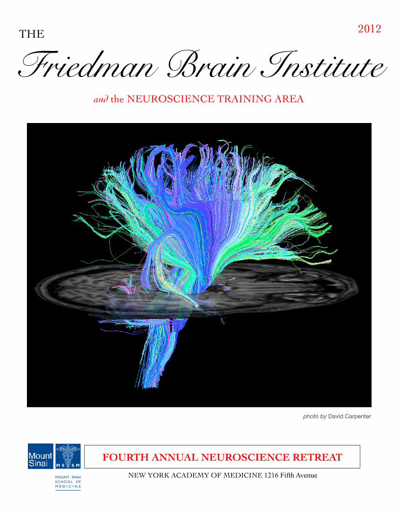
FOURTH ANNUAL NEUROSCIENCE RETREAT
Friedman Brain InstituteTHE 2012
photo by David Carpenter
and the NEUROSCIENCE TRAINING AREA
NEW YORK ACADEMY OF MEDICINE 1216 Fifth Avenue
Page 2
Director: Eric Nestler, MD, PhD, Department of Neuroscience
Friedman Brain Institute Leadership Team
ChairsJoshua Bederson, MD, Department of Neurosurgery
Wayne Goodman, MD, Department of PsychiatryJohn Morrison, PhD, Dean, Graduate School of Biological Sciences Kristjan Ragnarsson, MD, Department of Rehabilitation Medicine
Stuart Sealfon, MD, Department of Neurology
Centers of Excellence Joseph Buxbaum, MSc PhD
Chief, Center of Excellence on Neurodevelopment Disorders
Patrizia Casaccia, MD, PhDChief, Center of Excellence on Myelin Disorders: Mechanisms & Repair
Samuel Gandy, MD, PhDChief, Center of Excellence on Neurodegeneration
Patrick Hof, MDChief, Center of Excellence on Brain Aging
Yasmin Hurd, PhDChief, Center of Excellence on Mood & Motivation
Ehud Kaplan, PhDChief, Center of Excellence on Computational & Systems Neuroscience
Giulio Maria Pasinetti, MD, PhDChief, Center of Excellence on Novel Neurodiagnostics & Neurotherapeutics
Matthew Shapiro, PhDChief, Center of Excellence on Cognition & Neural Plasticity
4th Annual Neuroscience Retreat Committee
Retreat Organizers: Zhenyu Yue, PhD (Neurology) and Matthew Shapiro, PhD (Neuroscience)
Retreat Administrators: Marie Kopp, Celeste Reyes, Jenny Rivera, and Veronica Szarejko

Page 3
CONTENTSNeuroscience Retreat Schedule ...................................................................................... 4Abstracts (Presenters and Posters) .............................................................................. 5-47Ina Caesar and Daniel Christoffel........................................................................................ 5Caroline Dias and Mattea Finelli ......................................................................................... 6Jessica Walsh and Claudia Lindtner ................................................................................... 7Marianne Reddan and Jun Wang........................................................................................ 8Esther Kim and Ernesto Borrero ......................................................................................... 9Dongming Cai and Casaccia Laboratory .......................................................................... 10Rhonda Charles and Michael Chary.................................................................................. 11Dipesh Chaudhury and Kate Collins ................................................................................ 12Paula Croxson and Marshall Crumiller.............................................................................. 13Diane Damez-Werno and Elodie Drapeau......................................................................... 14Anastasiya Dzhun and Gabor Egervari.............................................................................. 15Sarah Evans and Arash Fazl ............................................................................................. 16Daniel Freire and Christopher Frenz ................................................................................ 17Allyson Friedman and Lauren Friedman .......................................................................... 18Sam Golden and Bing Gong ............................................................................................. 19Naqi Haider and Hala Harony-Nicolas .............................................................................. 20Desmond Heath and Mitra Heshmati................................................................................ 21Lap Ho and Georgia Hodes .............................................................................................. 22Terrell Holloway and Kuangfu Hsiao ................................................................................. 23Benjamin Hubert and Carlo Iomini .................................................................................... 24Michelle Jacobs and Yuji Kajiwara.................................................................................... 25Serene Keilani and Manjula Khubchandani....................................................................... 26Soong Ho Kim and Lindsay Knable .................................................................................. 27 Catharine Krebs and H. Lee Lau ....................................................................................... 28Xianting Li and Xiaochuan Liu ........................................................................................... 29Vladimir Makarov and Bridget Marcellino ......................................................................... 30Nicole McKnight and M. Mercedes Perez-Rodriguez ...................................................... 31Michael Michaelides and Effie Mitsis ................................................................................ 32Effie Mitsis and Cesar Moreno .......................................................................................... 33Jose Moreno and Hirofumi Morishita ................................................................................ 34Tiffany Morris and Daniel Ohm ......................................................................................... 35Giulio Pasinetti and Shekhar Patil ..................................................................................... 36Matthew Perkins and Rishi Puri ........................................................................................ 37Qing Xu and Justin Riceberg ............................................................................................ 38Elizabeth Schwartz and Bryan Sepulveda ........................................................................ 39Sushila Shenoy and Andrew Shin ..................................................................................... 40HaoSheng Sun and Eric Sweet ......................................................................................... 41Rita Tavares and Christopher Thompson.......................................................................... 42Nikos Tzavaras and Merina Varghese ............................................................................... 43Prashant Vempati and Rong Wang ................................................................................... 44Jonathan Wardman and Zhao Xuan.................................................................................. 45Wei Zhao and Yana Zorina ................................................................................................ 46Upcoming Events and Graduate Program information ............................................... 47
NEW YORK ACADEMY OF MEDICINE 1216 Fifth Avenue (corner of 103rd Street)
4th Annual Neuroscience Retreat
FRIEDMAN BRAIN INSTITUTE
Page 4
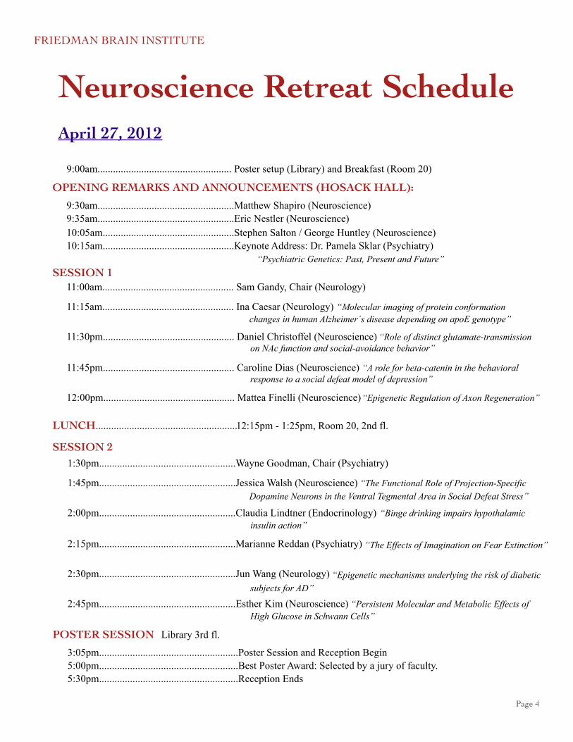
Neuroscience Retreat Schedule
9:00am.................................................... Poster setup (Library) and Breakfast (Room 20)
9:30am.....................................................Matthew Shapiro (Neuroscience)9:35am.....................................................Eric Nestler (Neuroscience)10:05am...................................................Stephen Salton / George Huntley (Neuroscience)10:15am...................................................Keynote Address: Dr. Pamela Sklar (Psychiatry)
SESSION 1 11:00am................................................... Sam Gandy, Chair (Neurology)
11:15am................................................... Ina Caesar (Neurology)
11:30pm................................................... Daniel Christoffel (Neuroscience)
11:45pm................................................... Caroline Dias (Neuroscience)
12:00pm................................................... Mattea Finelli (Neuroscience)
LUNCH.......................................................
OPENING REMARKS AND ANNOUNCEMENTS (HOSACK HALL):
12:15pm - 1:25pm, Room 20, 2nd fl.
“Molecular imaging of protein conformation
“Role of distinct glutamate-transmission
“A role for beta-catenin in the behavioral
“Epigenetic Regulation of Axon Regeneration”
“Psychiatric Genetics: Past, Present and Future”
SESSION 21:30pm.....................................................Wayne Goodman, Chair (Psychiatry)
1:45pm.....................................................Jessica Walsh (Neuroscience)
2:00pm.....................................................Claudia Lindtner (Endocrinology)
2:15pm.....................................................Marianne Reddan (Psychiatry)
2:30pm.....................................................Jun Wang (Neurology)
2:45pm.....................................................Esther Kim (Neuroscience)
“The Functional Role of Projection-Specific
insulin action”
“The Effects of Imagination on Fear Extinction”
“Epigenetic mechanisms underlying the risk of diabetic
High Glucose in Schwann Cells”
POSTER SESSION
3:05pm......................................................Poster Session and Reception Begin5:00pm......................................................Best Poster Award: Selected by a jury of faculty. 5:30pm......................................................Reception Ends
Library 3rd fl.
changes in human Alzheimer´s disease depending on apoE genotype”
on NAc function and social-avoidance behavior”
response to a social defeat model of depression”
Dopamine Neurons in the Ventral Tegmental Area in Social Defeat Stress”
“Binge drinking impairs hypothalamic
subjects for AD”
“Persistent Molecular and Metabolic Effects of
April 27, 2012
FRIEDMAN BRAIN INSTITUTE
Page 5
PresentersMolecular imaging of protein conformation changes in human Alzheimer´s disease
depending on apoE genotype
Ina Caesar1, K. Peter R. Nilsson2, Per Hammarström2, Stefan Prokop3, Frank L. Heppner3, David M. Holtzman4, Patrick R. Hof1, Sam Gandy1
1Mount Sinai School of Medicine, 2Linköping University, 3Charité–Universitätsmedizin Berlin, 4Washington University
Alzheimer’s disease (AD) is characterized by accumulation in brain of extracellular plaques of aggregated amyloid-β (Aβ) peptide and intraneuronal tangles comprised of hyperphosphorylated tau. The strongest genetic risk factor for AD is the apolipoprotein E gene (APOE) ε4 allele.
In this study, we approached the molecular mechanisms underlying how apoE isoform influences AD by imaging the structural differences of aggregated protein structures (plaques, tangles, vascular amyloid) in human postmortem AD tissue samples. The samples were matched in pairs, and differed only by being either homozygous for the APOE ε3 or ε4 allele. Luminescent conjugated oligothiophenes (LCO) were employed for the collection of conformation-dependent fluorescence spectra and were analyzed depending on apoE isoform.
We observed differences in LCO images and spectra indicating apoE isoform-dependent morphological changes and structural differences in plaques, tangles and vascular amyloid in human AD postmortem brain. ApoE isoform-dependent effects have recently been demonstrated to control Aβ clearance from brain, and we speculate that the apoE isoform-specific effects on Aβ structure that we detect with LCOs may play a role in these effects on Aβ clearance.
This work was supported by postdoctoral salary award from Swedish Research Council (Caesar), National Institute on Aging AG05138 to Mary Sano (Gandy, Hof), AG13956 (Holtzman).
Role of distinct glutamate-transmission on NAc function and social-avoidance behavior
Dan Christoffel, Russo Lab
Department of Neuroscience, Mount Sinai School of Medicine
Adaptation to stressful conditions depends on the proper functioning of synaptic plasticity mechanisms. However, repeated activation of these mechanisms by stress can lead to maladaptive behavior. The frontal cortex plays a prominent role in regulating decision-making. Dysregulation of PFC is a hallmark of many psychiatric disorders. Clinical imaging studies show depressive patients to have reduced frontal cortical activity. We previously utilized a chronic social defeat stress (CSDS) paradigm to model a subset of symptoms present in mood and anxiety disorders. Here we aim to elucidate the how release of glutamate into NAc, a brain region involved in integrating the salience of stimuli to regulate subsequent behavior, from the PFC and regulates social avoidance behavior after chronic social defeat stress. To study this circuit specific release of glutamate we will inject AAV-ChR2 into the PFC. We hypothesize that stimulation of ChR2 in PFC terminals in the NAc will have an antidepressant response in mice susceptible to social defeat. Indeed, we find that 100 Hz stimulation of PFC terminals during a social interaction test reverses social avoidance in susceptible mice. Currently, studies are underway to determine if chronic blockade of glutamate release from PFC terminals projecting to the NAc, using membrane-tethered toxins blocking Ca2+ influx of Cav2.2 and Cav2.3 channels, induces social avoidance and affects the spine density of NAc medium spiny neurons (MSNs). We hypothesize that glutamate blockade of PFC terminals will increase dendritic spine density.
page 6
A role for beta-catenin in the behavioral response to a social defeat model of depression
Dias, C.1, Mazei-Robison M.1, Feng J., Shao, N.1, Sun H.1, Damez-Werno D.1, Scobie K.1, Vialou V.1, Kennedy P.1, Neve2, R., Shen L.1, Nestler E.J.1
1Department of Neuroscience, Mount Sinai School of Medicine, 2McGovern Brain Institute, MIT
Beta-catenin is a multi-functional protein that plays an important role in the mature central nervous system, and particularly in neurological and psychiatric disease. In mature neurons beta-catenin can play a structural role at the synapse, but it is also the key effector of canonical Wnt signaling, where it acts as a transcriptional activator at LEF/TCF target genes. However, to date there has been a lack of evidence directly demonstrating the transcriptional role of beta-catenin in the context of psychiatric illness. Here we show that expressing beta-catenin in mouse nucleus accumbens (NAc), a key brain reward region, with Herpes simplex viral vectors, has anti-depressant and anxiolytic properties with regards to baseline behavior. Furthermore, when over-expressed in NAc during social defeat stress, beta-catenin mediates resilience to the development of social avoidance. We also find that social defeat stress robustly regulates both protein and mRNA levels of key components of canonical Wnt signaling, including beta-catenin and TCF 4. In order to gain an unbiased view of potential Wnt target genes, we performed massively parallel sequencing on beta-catenin ChIP (ChIP-Seq) from NAc. Preliminary data suggest that both known and novel, CNS-specific Wnt target genes associate with beta-catenin. Overall we show that canonical Wnt signaling plays an important role in mediating resilience to stress.
Epigenetic Regulations of Axon Regeneration.
Mattea Finelli1, Jamie Wong1, and Hongyan (Jenny) Zou1,2
1Department of Neuroscience and 2Department of Neurosurgery, Friedman Brain Institute, Mount Sinai School of Medicine
Adult neurons in mammalian central nervous system regenerate minimally after injury, in part, to an age-dependent decrease of neuronal axon growth capacity. To understand the molecular mechanisms underlying reactivation of axon growth capacity of adult neurons, we studied the unique capacity of adult sensory neurons in dorsal root ganglia (DRG) to regenerate under the so-called conditioning lesion paradigm. Previous work has shown that axon regeneration following a conditioning lesion is dependent on the transcription of a large number of regeneration-associated genes (RAGs). How these genes are transcriptionally regulated is still unknown.
We focused on epigenetic mechanisms, in particular, histone acetylation because we identified a marked global enrichment of histone acetylation in regenerating DRG neurons as compared to non-regenerating neurons. We showed that the levels of histone acetylation are dynamically regulated both globally and on RAG promoters. Consistently, the expression levels of histone deacetylases (HDACs) and histone acetyltransferases (HATs) are also deregulated in regenerating neurons, favouring the higher global levels of histone acetylation observed.
Treatment with a HDAC inhibitor in adult mice increases histone acetylation levels in DRG neurons both globally and on RAG promoters leading to RAGs transcription, and in turn, to the enhancement of axon growth capacity. We are now testing if modulating histone acetylation levels either pharmacologically or through genetic manipulation of HATs or HDACs in a mouse model of spinal cord injury can induce adult neurons to regenerate by way of inducing transcription of RAGs.
page 7
The Functional Role of Projection-Specific Dopamine Neurons in the Ventral Tegmental Area in Social Defeat Stress
Jessica Walsh, Allyson Friedman, Dipesh Chaudhury, Barbara Juarez, and Ming-Hu Han
Mount Sinai School of Medicine
The efficacy of novel depression treatment with deep brain stimulation implicates major depressive disorder (MDD) as a neural circuit disorder. Studies have implicated the mesolimbic dopamine (DA) system in the pathophysiology of depression, with DA neurons in the ventral tegmental area (VTA) projecting to the medial prefrontal cortex (mPFC), nucleus accumbens (NAc), and amygdala. Utilizing lumafluors, we investigated the firing rate of pathway-specific DA neurons from the VTA, and found that NAc-projection DA neurons fired higher in susceptible, but not resilient mice. In contrast, mPFC-projection DA neurons fired lower selectively in susceptible. Using optogenetics, we manipulated firing patterns of these pathways to elucidate the role in the regulation of behavioral abnormalities. We injected a transcellular Cre virus into the NAc, retrograding Cre from the NAc to the VTA, so that only NAc-projection DA neurons in the VTA express Cre. Conditional ChR2 was injected into the VTA, expressing ChR2 only in NAc-projection VTA DA neurons. We found that light activation of ChR2 in virus-infected neurons projecting to the NAc, opposed to the mPFC, induced depolarizing photocurrents and generated a susceptible phenotype when burst firing was elicited. Currently, using NpHR, we will manipulate the mPFC-projection specific pathway. These studies expand our functional understanding of the projection-specific VTA DA neurons and provide information for the target-oriented treatment of MDD.
NIMH R01MH092306.
Binge drinking impairs hypothalamic insulin action
Claudia Lindtner1, Thomas Scherer1, Elizabeth Zielinski1, Martin Fasshauer1, Michele Puchowicz2, Christoph Buettner1
1Department of Medicine and Department of Neuroscience, Mount Sinai School of Medicine, NY2Mouse Metabolic Phenotyping Center Case Western Reserve University, OH
A history of binge drinking is associated with an increased risk of the metabolic syndrome and type 2 diabetes in women. Whether binge drinking impairs glucose homeostasis and/or insulin action is unknown. Here we demonstrate that binge drinking impairs glucose tolerance in female rats. Hyperinsulinemic euglycemic clamp studies revealed that binge drinking induces hepatic insulin resistance despite intact hepatic insulin signaling as well as a marked defect in insulins ability to suppress lipolysis, a defect that persists up to 30 hrs after all ethanol has been metabolized. Since hypothalamic insulin signaling controls hepatic insulin action, we next tested if binge drinking impairs hypothalamic insulin action, defined as the ability of hypothalamic insulin to suppress hepatic glucose production and adipose tissue lipolysis. Binge drinking markedly compromised hypothalamic insulin action, i.e. hypothalamic insulin failed to suppress hepatic glucose production (hGP) and lipolysis. Signaling studies demonstrated that binge drinking decreases insulin signaling in the hypothalamus, possibly due to hypothalamic inflammation and increased expression of PTP1b. Thus, these results suggest that binge drinking induces insulin resistance in part due to a disruption of brain control of peripheral glucose and lipid partitioning that sets the stage for the metabolic syndrome and type 2 diabetes.
page 8
The Effects of Imagination on Fear Extinction
Marianne Reddan and Daniela Schiller
Department of Psychiatry, Mount Sinai School of Medicine
Imagination has a powerful effect on one’s emotional state. We hypothesize that extinction training performed in one’s imagination can effectively reduce a fear response acquired in the real world. To examine this hypothesis, we manipulate fear learning using directed imagination. All participants in this study initially underwent an imagination training session followed by auditory fear conditioning, in which one neutral tone was sometimes paired with shock and another was not. In the next phase, subjects were randomized into three groups. The first group underwent real-world extinction training (repeated tone exposures without shock). The second group was cued to imagine the tones in the same sequence presented to the first group (repeated imaginary tone exposures without shock). A third group was cued to imagine two neutral sounds from nature in order to control for the general effects of imagination on arousal. After a short break, all subjects were exposed to four unsignaled shocks in order to reinstate the fear memory. The tones were then presented again to examine fear recovery. Skin conductance response indicated fear-related arousal. Compared to the imagination control group, subjects who imagined extinction exhibited smaller conditioned fear responses after reinstatement, but did not differ from those who underwent real-world extinction training. These results suggest that directed imagination could be as effective or even more effective than real-world extinction learning. In the clinic, controlled imagination might be an effective alternative or a complementary method to exposure therapy.
Type 2 diabetes mellitus is a key risk factor for Alzheimer's disease. As diabetes and sedentary lifestyles become increasingly common, the characterization of mechanism(s) underlying diabetes-induced cognitive impairment in AD and the development of early prevention strategies are critical.
We recently discovered that there are significant changes in the expression of select chromatin modification enzymes, such as histone deacetylases (HDACs), in the brains of diabetic subjects compared to control subjects, and that these changes coincide with altered expression of proteins involved in synaptic function. Using a mouse model of diet-induced type 2 diabetes (T2DM mice), we explored the impact of type 2 diabetes on epigenetic mechanisms in the brain and found that, similar to humans, diabetic mice also showed significant up-regulation of HDACs, and these alterations coincided with increased susceptibility to oligomeric Aβ toxicity.
Our study suggest that diabetes may induce epigenetic modifications resulting in structural and/or functional changes in the brain, eventually leading to increased susceptibility to insults of AD-type neuropathology.
Acknowledgment: This work is supported by discretionary fund to G.M.P.
Epigenetic mechanisms underlying the risk of diabetic subjects for AD
Jun Wang, Bing Gong, Giulio M Pasinetti
Department of Neurology, Mount Sinai School of Medicine
page 9
Persistent Molecular and Metabolic Effects of High Glucose in Schwann Cells
Esther S. Kim, Fumiko Isoda, Charles V. Mobbs
Department of Neuroscience, Mount Sinai School of Medicine
Diabetic complications, such as diabetic neuropathy, pose a major problem to public health due to their persistent nature despite a return to normal glucose levels. This long lasting pathological effect is referred to as metabolic memory. Increased glucose metabolism during chronic high glucose can cause oxidative stress by producing NADH for use in the mitochondrial electron transport chain and produce free radical superoxide O2-. Schwann cells are the myelin forming cells in the PNS and are damaged during diabetes. Using Schwann cells as a model to study the molecular mechanisms of diabetic neuropathy, we have found that chronic (>2 months) high (25mM) glucose increases both glycolysis and oxidative stress. In addition, high glucose increased expression of glycolytic genes while decreasing the pentose phosphate pathway, a major source of NADPH for antioxidant regeneration. This gene profile is not reversed by 1-4 weeks at normal (5.6mM) glucose, thus exhibiting features of metabolic memory. Using measures of NADH and ATP we have also identified a profile that supports increased NADH production from glycolysis in chronic high glucose cells that provides further insight into the mechanism underlying metabolic memory.
AbstractsInvestigating Self-Association of Prototypic G Protein-Coupled
Receptors in Biological Membrane Models.Ernesto Borrero and Marta Filizola
Structural and Chemical Biology, Mount Sinai School of Medicine
A wealth of experimental evidence accumulated over recent years suggests that G protein-coupled receptors (GPCRs) associate with each other in the plasma membrane, forming both homo- and hetero-mers. However, the specificity and lifetime of these interactions, as well as their role in GPCR function, remain topics of intense debate. We are interested in building an understanding of the rules that govern receptor-receptor interactions and how these may lead to differences in interactions between receptor subtypes. We use molecular dynamics based calculations combined with multiscale membrane-protein representations to contribute a rigorous mechanistic insight into the organization of GPCRs in biological membrane models at a level of molecular detail that is unattainable using current experimental techniques alone. Here, we present preliminary results of a computational study aimed at simulating the self-assembly of two different, yet highly homologous prototypic receptors, beta1- and beta2-adrenergic receptors, in an explicit, closed membrane vesicle system of diameter similar to that created experimentally in collaborative studies, and consisting of the same lipid compositions and protein concentrations as those used experimentally. The main goal of these simulations is to obtain homo- and hetero-dimeric models of ligand-free receptors which can then be used to provide unique hypotheses of receptor-receptor interactions (involved in neurological, and drug abuse disorders) that can be tested experimentally through mutagenesis to help elucidate the role of receptor association in GPCR function.
1
page 10
Several lines of evidence reveal the link of endocytosis to biology of Alzheimer’s disease (AD). Dynamin 1, a small GTPase that plays a critical role in endocytosis, is recently associated with AD. We have undertaken the studies of possible roles for dynamin 1 in regulating Aβ homeostasis. Here we show that genetic perturbation of dynamin 1 (dyn1) reduces Aβ levels in cell culture. There is a dramatic reduction in beta-site APP-Cleaving Enzyme 1 (BACE-1) cleavage products of APP, and a reciprocal increase of α–secretase cleavage products. Dyn1 haploinsufficiency animals with AD transgenic background demonstrates decreased levels of intracellular amyloid at 2-3 months old. Furthermore, the amounts of cell surface BACE-1 and holoAPP are increased with dyn1 knockdown. In summary, these data suggests a novel modulatory mechanism by which an endocytic adaptor protein dyn1 affects amyloid generation through regulation of BACE-1 enzymatic activities and its subcellular localization.
Li Zhu1,2, Minghao Zhong1,3, Wenlun Tan1,2, Meng Su1,4, Pietro De Camilli5 and Dongming Cai1,2
An Endocytic Adaptor Protein, Dynamin 1 Regulates Amyloid Generation through Modulation of BACE-1
1Departments of Neurology and Alzheimer’s Disease Research Center, Mount Sinai School of Medicine, 2 James J. Peters Veterans Affairs Medical Center, 3Department of Pathology, New York Medical College,
4Department of Pathology, John Hopkins Medical Center, 5Department of Cell Biology and Program in Cellular Neuroscience, Neurodegeneration and Repair, Yale School of Medicine.
Fundings: MSSM Seed Fund; VA Career Development Award
2
Human stem cells for the repair of the damaged nervous system in multiple sclerosis
The Casaccia Laboratory Stem Cell Team*
Vera Alexeeva, Sunita D'Souza and Patrizia Casaccia
Multiple sclerosis (MS) is an autoimmune disease of the central nervous system (CNS) characterized by both white and gray matter demyelination. In recent years, stem cells have gained considerable interest in their use for repairing the damaged nervous sytem. Thus, our primary goal is to understand mechanisms for the patterning and differentiation of oligodendrocytes from both the H9 embryonic stem cell line and skin-derived fibroblasts of MS patients using the induced pluripotent stem cell (IPSC) method. In order to further characterize these cells, we are interested in the epigenetic switch that occurs during the cell fate decision of a neural precusor cell to become a neuron, astrocyte or oligodendrocyte. To achieve this goal we are immunoselecting the three cell fates and performing detailed RNASeq and characterization of the epigenetic marks. To more specifically characterize the functionality of these IPSC-derived oligodendrocytes, we plan to use these cells in in vitro myelination assays using dorsal root ganglia neuron cocultures and test their in vivo capacity to myelinate in the shiverer mutant mouse which lacks CNS myelin. A full understanding of the potential of these cells in repair will highlight their efficacy for potential repair in MS and discovery of new targets for therapeutic intervention in de/dysmyelinating disorders.
This work is supported by the NIH and the National Multiple Sclerosis Society.
3
page 11
The human AVPR1A BAC transgenic mouse: A preclinical model for elucidating the role of AVPR1A in Autism Spectrum Disorders.
*R. Charles¹, N. Takahashi¹, T. Sakurai¹, L. J. Young² and J. D. Buxbaum¹
¹Mount Sinai School of Medicine, ²Emory University
Genetic studies have demonstrated an association between arginine vasopressin receptor 1A (AVPR1A) and ASDs. Furthermore, as evidenced in rodent and primate studies, species-specific differences in the 5’ upstream region of the AVPR1A gene regulate brain AVPR1A expression pattern and thereby modulate behaviors that have relevance to ASDs. We proposed that generating a mouse expressing the human form of AVPR1A would provide a more relevant in vivo system in which we can better understand the regulation of the human AVPR1A receptor and its role in controlling behaviors associated with ASDs, while providing a potential preclinical model for the evaluation of therapeutics.
We showed that transgenic mice expressing human AVPR1A display a robust receptor protein expression pattern with some overlap with that observed in the brains of higher primates, and vastly distinct from the receptor expression profile in the wild type mouse. Additionally, given the expression changes observed in our transgenic animals in regions such as the amygdala and thalamus, we hypothesized that these mice would demonstrate predictable social and sensorimotor behavior alterations. Finally, this transgenic mouse line provides a unique opportunity to identify the cis regulatory elements of the human AVPR1A gene and possible chromatin remodeling mechanisms that permit expression variability.
Funding: Autism Science Foundation, Seaver Foundation
4
The devastating effects of cocaine addiction on behavior and the activity of single neurons are well documented. Much less is known how cocaine addiction affects the distributed activity of the brain that surely underlies much higher brain function. To better understand this, we recorded from multiple neurons in the ventral tegmental area (VTA) of urethane-anesthetized naive rats before and during intravenous administration of cocaine to assess the impact of cocaine on neural network behavior.
We found that cocaine can synchronize VTA neurons in a dose-dependent matter. Cocaine administration did not significantly alter the flow or complexity of information transmission, nor the network's computational structure. Simulations suggest that increased synchrony, which was seen experimentally, is an acute response to a rewarding stimulus, but not indicative of an addicted state.
Our results demonstrate the novel application of measures of neural population dynamics to non-sensory brain regions. They, furthermore, show how computational models are useful in relating neural population dynamics to behavior. We anticipate that these results will provide a baseline against which changes in population dynamics arising in rodent models of drug addiction could be compared. Ultimately, better understanding how drugs of abuse affect neural activity will help develop biomarkers that can both identify susceptible individuals early-on and track the efficacy of their treatment.
Supported by NIH grants EY16224, NIGMS 1P50GM071558 and R21MH093868-02.
Effect of acute cocaine administration on neural population dynamics
Michael Chary, Youping Xiao and Ehud Kaplan
Friedman Brain Institute, Mount Sinai School of Medicine
5
page 12
Dipesh Chaudhury¹, Barbara Juarez¹, Jessica Walsh², Allyson Friedman² and Ming-Hu Han¹¹Department of Pharmacology, ²Department of Neuroscience
Mount Sinai School of Medicine
The role of high frequency phasic firing of ventral tegmental area (VTA) dopamine (DA) neurons in mediating stress vulnerability is not completely understood. In a social defeat model of depression, our recent studies found that mice exhibiting a susceptible (depressive), but not resilient (non-depressive) phenotype, exhibited consistently increased phasic firing of VTA DA neurons. To investigate the casual relationship between phasic firing in these neurons and susceptibility to social defeat in freely-behaving mice, we selectively targeted DA cells by injecting a Cre-dependent viral vector AAV-ChR2 (channel rhodopsin2), into the VTA of transgenic TH-Cre mice. Through in vitro and in vivo electrophysiological recordings, we demonstrated that light activation of ChR2 reliably generated physiologically relevant low frequency tonic and high frequency phasic firing patterns in VTA DA neurons. We show that optogenetic induction of phasic, but not tonic, firing, in VTA DA neurons of mice, during a social interaction test, 24hrs after undergoing a subthreshold social defeat paradigm, induced a susceptible phenotype as measured by social avoidance and decreased sucrose preference. Furthermore, optogenetic phasic stimulation, of previously resilient, non-depressed, mice, induced the susceptible (depressed) phenotype. These studies provide direct evidence showing that the phasic firing pattern of VTA DA neurons in the brain reward circuitry encodes a signal for stress vulnerability.
Supported by R01 MH092306
Phasic firing of ventral tegmental area dopamine neurons encodes behavioral susceptibility to social defeat stress
6
Taking action in the face of fear: The neural substrates of active avoidance in humans.
Katherine A. Collins1, Michael Canete1, Christopher K. Cain2,3, Daniela Schiller1
1Departments of Psychiatry and Neuroscience, Mount Sinai School of Medicine, 2Emotional Brain Institute, Nathan Klein Institute, 3Center for Neural Science, New York University
Background: Active avoidance learning (AA) is the acquisition of behavior that minimizes exposure to danger. Most rodents exhibit AA, but some display only automatic fear reactions (freezing). AA deficits are associated with an “anxious” phenotype in animals, but the relationship between anxiety and AA in humans is unclear. To understand why some people can take action while others are “paralyzed” when faced with fear, we created a task to observe inter-individual variability in human AA.
Methods: During the experiment, participants moved a marker within a virtual game-board divided into two compartments by a narrow “bridge”. Every three seconds, a lightening bolt image appeared if the player had not crossed the “bridge” in that time period. The bolt was paired with mild electric shock in one-third of trials. To avoid all bolts and shocks, participants had to cross the “bridge” two times per trial.
Results: Our task identified “good” and “poor” performers in a sample of 28 healthy volunteers. “Good” performers made more avoidance responses than “poor” performers, who evinced slower reaction times in late versus early trials. “Poor” performers reported higher levels of state and trait anxiety.
Conclusions: We developed a probe of human AA that is the first to identify a subset of poor performers who report higher levels of anxiety. Future studies may elucidate the neural mechanisms of AA and anxiety disorders.
7
page 13
Alterations in resting-state network activity following corpus callosum section in macaque monkeys
PL Croxson, JX O’Reilly, J Sallet, MP Noonan, RB Mars, PG Browning, KL Miller, MF Rushworth, MG Baxter
Mount Sinai School of Medicine and University of Oxford
The basis for slow (<1Hz) “resting-state” fluctuations in the functional MRI blood oxygen level-dependent (BOLD) signal is still uncertain. The strength of resting-state functional connectivity often has a strong correlation with structural connectivity. However, in some cases functional connectivity is seen when there is weak or no direct structural connectivity. For example, in some human case studies patients without an intact corpus callosum still have interhemispheric connectivity. However, these patients often have agenesis of the corpus callosum, and may have formed different connections during development. We investigated the causal effect of a change in white matter connectivity on interhemispheric functional connectivity. We collected resting-state functional MRI from three rhesus macaque monkeys (2 female) under isoflurane anesthesia, before and after complete section of the corpus callosum. Contrary to the findings in human acallosal patients with preserved correlations between the hemispheres, we found that surgical section of the corpus callosum led to an almost complete abolition of correlations between the hemispheres. Our results provide evidence that cortico-cortical white matter connections are necessary for the propagation of resting-state correlations, and thus structural connectivity is necessary for functional connectivity. In addition, within-hemisphere connectivity was increased, suggesting one mechanism by which the brain may reorganize itself after injury.
Funded by the Wellcome Trust and MRC (UK)
8
Functional Processing of Edge and Color Detection in V1
Marshall Crumiller, Youping Xiao, and Ehud Kaplan
Friedman Brain Institute, Mt. Sinai School of Medicine
The primary visual cortex (V1) has long been implicated in the extraction of simple features from the visual field. Orientation pinwheels in V1 provide strong evidence for the functional organization of an edge detector, and hue maps for the early processing of color. Cytochrome oxidase blobs, found in the upper cortical layers, have eluded functional classification, but imply a segregation in the processing of low frequency (surface) versus high frequency (edge) features. The extent to which V1 provides separate functional architecture for the extraction of these features is a growing area of research.
We use natural movies, processed in different spatial frequency and color domains, to drive neurons in monkey V1. We have recently developed a novel method for the estimation of the amount of information conveyed by a large population of neurons. This method, combined with natural stimuli, is well-suited for the purpose of identifying network-level functional architecture concerned with the extraction of color and frequency information. We apply these measures of information to populations of cells in order to functionally segregate neurons based on their stimulus-specific responses. We demonstrate a tentative functional overlap between neuronal groups in the frequency and color domain, furthering the notion that the visual system utilizes a functional architecture to process separate but overlapping streams of information.
Supported by NIH grants EY16224, NIGMS 1P50GM071558 and R21MH093868-02.
9

page 14
Histone arginine methylation in the nucleus accumbens in response to chronic cocaine
D.Damez Werno1, K.N.Scobie1, H.Sun1, D.M.Dietz1, C.M. Dias1, F.Casadio2, R.L.Neve3, E.J.Nestler1
1Friedman Brain Institute, Department of Neuroscience, Mount Sinai School of Medicine, NY2Lab. of Chromatin Biology and Epigenetics, Rockefeller Univ., NY
3Dept. of Brain and Cognitive Sciences, Massachusetts Institute of Technology, Cambridge, MA
Histone methylation on Lys (K) residues has been linked to a number of neurological and psychiatric disorders, including drug addiction. In particular, we have shown that drug-induced long-term changes in gene transcription involve reductions in global levels of dimethylation of Lys9 on histone H3 (H3K9me2) in nucleus accumbens (NAc). In contrast to Lys methylation, the functional role of histone Arg (R) methylation in chromatin structure and gene transcription in brain remains underexplored. Histone Arg methylation is catalyzed by a family of enzymes called protein Arg methyltransferases (PRMTs). First, we investigated the effects of a chronic cocaine regimen (20 mg/kg, i.p., daily for 7 days) on PRMT expression in the NAc of mice. We found that mRNA levels of PRMTs 1, 5, and 6 exhibit a significant decrease 24 hours after the last cocaine injection and that mRNA level of PRMT7 trends toward a decrease. Consistent with the mRNA findings, we observed that levels of PRMT6 andPRMT7 proteins, which are respectively responsible for repressive H3R2 asymmetric dimethylation and repressive H4R3 symmetric dimethylation are downregulated. These novel findings suggest that Arg methylation of core histone tails plays an important role in addiction related changes in gene transcription in brain.
Supported by NIDA and NIMH.
10
Induced Pluripotent Stem Cells In Schizophrenia
Elodie Drapeau1, Andy Brown1, Sarah Levinson2, Joseph Friedman1, Joseph Buxbaum1
1Department of Psychiatry and 2Department of Developmental and Regenerative Biology, Mount Sinai School of Medicine
The study of schizophrenia is limited by the difficulty in accessing brain material from living subjects. The use of Inducible pluripotent stem cells (IPSCs) that can subsequently be differentiated into neurons provides a way to study schizophrenia at the cellular level. Copy number variations (CNVs) have been identified in schizophrenia. We propose to generate and characterize IPSCs and neurons from schizophrenic patients with rare penetrant CNVs.
DNA samples were obtained from subjects recruited by the Conte Center for Neuroscience of Mental Disorders and genotyped on Illumina arrays. Subjects underwent dermal biopsy to isolate fibroblasts for the generation of IPSCs. Reprogramming of fibroblasts into iPSCs was done using virus particles containing reprogramming transcription factor genes. Generated iPSC will be used to generate neuronal precursor cells (NPCs) and mature neurons.
To date, 253 subjects (153 schizophrenic, 100 control) have been phenotyped and have had blood samples collected; 143 patients and 86 controls were genotyped and several well replicated CNVs identified. Fibroblasts were obtained for 57 patients and 55 controls. A subject with a 1q21.1 deletion, a schizophrenia case without known CNV and six healthy controls were selected for first experiments. IPSCs were generated and are currently used to produce NPCs, and mature neurons. Future plans are to characterize those cells on the basis of gene expression, morphology, synapse formation and electrophysiological changes while assessing methods for high throughput reprogramming and differentiation.
11

page 15
Caprylic triglyceride as a dietary intervention in a mouse model of ALS
Anastasiya Dzhun, Giulio Maria Pasinetti
Department of Neurology, Mount Sinai School of Medicine
Energy metabolism deficiencies play a role in the death of spinal cord motor neurons in Amyotrophic Lateral Sclerosis (ALS). Previously we reported the beneficial effect of a ketogenic diet in a mouse model of ALS, possibly via improvement of energy metabolism. Caprylic triglyceride (CT) is emerging as a medical food for mild to moderate Alzheimer’s disease (AD). Impaired glucose metabolism has been observed in the brain of AD patients and the putative benefit of CT is to provide ketone bodies as alternative energy. Using G93A SOD mice, we tested whether CT can improve motor function/energy metabolism in ALS. We administered 10% (w/w) CT in the diet of ALS mice, while control mice received an isocaloric diet. The mice on CT showed an increase in plasma ketone levels, but the treatment had no effect on motor function or survival rate. Examination of spinal cord mitochondrial metabolism using Seahorse XF24 technology revealed that ALS mice had an increase in basal and maximal oxygen consumption rates in complex I compared to control mice; a similar increase in mitochondrial respiration was also observed in the motor-neuron like NSC-34 cells following treatment. Our findings show that while CT improves energy metabolism, it does not beneficially modify disease progression in ALS mice. CT may be considered as a combination treatment for improving metabolic efficiency in ALS, together with drugs that target motor neuron function and survival.
12
Germline exposure to delta 9-tetrahydrocannabinol leads to distinct behavioral and neurobiological abnormalities
Gabor Egervari, Henrietta Szutorisz, Jennifer A DiNieri, Yanhua Ren, XiaoChuan Liu, Li Shen and Yasmin L Hurd
Departments of Psychiatry, Pharmacology and Systems Therapeutics, and Neuroscience, Mount Sinai School of Medicine
Despite marijuana (Cannabis sativa) being the most frequently abused illicit substance by adolescents, the potential germline impact of cannabis on future offspring has not been studied.
We used a rat model to examine cross-generational consequences of exposure to the main psychoactive component of cannabis, delta 9-tetrahydrocannabinol (THC). Male and female rats received THC injections during adolescence, the animals were mated as adults, and offspring were studied in adolescence and adulthood. Female offspring of THC-exposed parents showed reduced locomotor activity in a novel environment, as well as decreased motivation to self-administer palatable food, indicating potential dysregulation of neuronal systems that regulate reward processing and motivation. We detected marked gene expression impairments in the ventral and dorsal striatum of the animals, that were partly specific to gender and developmental stage. Changes were most pronounced in components of the glutamatergic system of adult rats, with significantly decreased expression of several NMDA and AMPA receptor subunits in the dorsal striatum; and impairment of the transmembrane protein, Slitrk5, in the ventral striatum.
Our findings indicate that adolescent THC exposure causes heritable changes in gene expression to alter offspring phenotype. Currently, we are expanding these studies to assess genome-wide transcriptome impairments and to study potential functional consequences on neurotransmission.
Funding: NIH grant DA30359.
13
page 16
Perinatal Phthalate Exposure and the Developing Brain
Sarah Evans1 and Patrizia Casaccia2,3
1Department of Preventive Medicine, 2Department of Neuroscience and 3Department of Genetics and Genomic Sciences, Mount Sinai School of Medicine
Phthalates are endocrine disrupting chemicals used as plasticizers and components of many household and personal care items. in utero exposure affects reproductive development; however few studies have examined impacts on other hormone-sensitive systems. Epidemiological studies have shown a correlation between exposure to phthalates and impaired neurobehavioral outcomes. The aim of these studies is to explore the mechanisms by which phthalates alter neurodevelopment. We hypothesize that phthalate exposure leads to behavioral deficits via epigenetic changes which may perturb myelination. To explore the effects of phthalate exposure on nervous system development, we examined the brains of offspring of rat dams adminstered di-2-ethylhexyl phthalate (DEHP) during pregnancy and lactation. Preliminary data suggest an increase in Myelin Basic Protein (MBP) and DNMT3a expression at PND15 in the cerebellum of offspring of 5mg/kg·day DEHP treated dams relative to control animals. As previously reported, DNMT1 and DNMT3a expression was elevated in testes of DEHP exposed offspring relative to controls. Studies are ongoing to confirm whether exposure to phthalates during early brain development causes changes in myelin gene expression as well as modifications to the epigenome that contribute to behavioral changes such as those observed in human epidemiological studies. These studies are supported by a Pilot Project Grant to Sarah Evans from the Mount Sinai School of Medicine Children’s Environmental Health Center.
14
GABAergic Function During Speech Production
Arash Fazl¹, Peter Herscovitch3, Kristina Simonyan1,2
¹Departments of Neurology and 2 Otolaryngology, MSSM, 3PET Department, NIH
The role of GABA in regulation of neuronal excitability during a wide range of behaviors is well recognized. However, GABAergic influences within the speech controlling system remain unknown. We investigated the GABAergic function associated with speech production by mapping the GABA-A receptors and functional brain activity in healthy humans.
Twenty healthy volunteers (mean 53.2 y.o., 12 females/8 males) underwent PET with [11C]flumazenil (FMZ), fMRI during speech production (sp-fMRI), and resting-state fMRI. Following initial data processing, whole-brain voxelwise Spearman correlation coefficients were computed between fMRI BOLD signal and FMZ binding potential (p < 0.025, corrected).
Significant positive relationships between BOLD signal (both speech-related and resting-state) and FMZ binding were found in the parietal operculum, supplementary motor area, precuneus, and superior temporal gyrus, while negative correlations were observed in the inferior frontal gyrus, posterior cingulate cortex and cerebellum. Additional positive correlations between sp-fMRI and FMZ binding were observed in the laryngeal sensorimotor cortex, supramarginal and angular gyri, inferior parietal lobule, putamen, caudate nucleus and cerebellum, whereas negative correlations were found in the superior parietal lobule, middle temporal gyrus and anterior cingulate cortex. Our data provide the first direct evidence of GABAergic function during normal speech production and suggest that neuromodulation of brain activity occurs at different stages of speech and language control, from auditory perception to motor production.
Supported by DC009629 grant to KS, the NINDS/NIH Intramural Program, the NIH Clinical Center.
15
page 17
Novel intranasal administration of odorant ligand, exploring regional brain distribution
Daniel Freire, Giulio M. Pasinetti
Department of Neurology and Department of Psychiatry, Mount Sinai School of Medicine
Alzheimer’s disease (AD) is a neurodegenerative disorder characterized by memory loss and cognitive impairment. One of the markers of the onset of AD is impairment of olfactory discrimination. Recent studies suggest that olfactory stimulation could be used as a potential treatment for AD. A pilot study in our lab has identified several compounds that are capable of activating a subset of olfactory receptors in neuronal cells as evaluated by calcium flux assay and /or cyclic AMP assay. Based on these results we plan to test these odorants in an AD model. As a first step we need to evaluate the pharmacokinetics and biodistribution of these odorants in vivo. Here we will present the results of the pharmacokinetics and biodistribution of one of these odorants, (+)-carvone. We selected (+)-carvone for these initial studies based on its low toxicity and high sensitivity in gas chromatography/ mass spectrometry (GC/MS) analysis. B6/SJL mice were anesthetized with isoflorane and drops of (+)-carvone (10 mM) were placed directly below the nares. Pharmacokinetic and biodistribution measurements from brain regions were made at 5, 15, and 60 minutes, after recovery from anesthesia. Frozen tissue samples were homogenized and compound was purified through column chromatography. Hexane extract was then injected directly into GC/MS for final analysis. The pharmacokinetics and biodistribution of (+)-carvone, administered via intranasal route will be presented.
16
Rat Knockin Model of Early Onset DYT1 Generalized Dystonia
Frenz C., Singh M., Shashidharan P.
Department of Neurology, Mount Sinai School of Medicine
DYT1 early onset generalized dystonia is a highly debilitating neurological disorder caused by the deletion of a single glutamic acid at position 302/303 in torsinA, a protein encoded by the DYT1 gene on human chromosome 9q34.1. A number of mouse models of dystonia have been characterized, including genetic models however, phenotypically none of them resembled human dystonia and hence the usefulness of these models in preclinical studies was limited. The rat is considered to make a better rodent model because of similar physiology, biochemistry and amenability to pharmaceutical intervention. Zinc finger nuclease (ZFN) mRNA targeting exon 5 of the rat DYT1 gene was co-injected along with a modified DYT1 insert (ΔE) into rat zygotes and implanted. At position 302 and 303 in the rat DYT1 gene, the DNA sequence is GAAGAG instead of GAGGAG, both sequences coding for glutamic acid residues in torsinA. The GAA in the rat DYT1 gene was deleted to create a knockin model that results in the introduction of a new restriction site (DdeI), enabling us to genotype the genetically modified rats. We have conducted a number of behavioral tests on the F1/F2 generation of rats including open field activity, challenge beam, rotarod, and misstep. A subset of the DYT1 ΔE rats display an abnormal hindlimb gait starting at ∼ 6 months.
Funding provided by NIH and Bachmann-Strauss Dystonia & Parkinson Foundation
17
page 18
Homeostatic Regulation of VTA Ion Currents Is a Mediator of Resilience to Social Defeat Stress
AK. Friedman1, JJ. Walsh1,2, B. Juarez1, D. Chaudhury1, X. Li3, N. Pan3, J.Wang3, M. Ribadeneira4, E. Wong4, Z. Yue2,3 and M. Han1,2
1Department of Pharmacology; 2Department of Neuroscience; 3Department of Neurology; 4AstraZeneca
The majority of the population maintains healthy psychological functioning or resilience to depression despite exposure to prolonged stress. The maintenance of healthy mental functioning is associated closely with the dopaminergic pathways, specifically from the dopamine(DA) neurons of the ventral tegmental area (VTA) in the mesolimbic reward-circuitry. Employing a social defeat stress model of depression, we previously showed an increase in the in vivo firing and bursting activity of VTA DA neurons of susceptible, but not resilient mice. Utilizing tyrosine hydroxylase-GFP mice to identify VTA DA neurons, we demonstrated that this pathophysiological hyperactivity was correlated with an increased hyperpolarization-activated cation channel (Ih) current. Notably, chronic defeat induced an even larger Ih increase in the resilient subgroup. Resilient mice were also found to have an increase in potassium (K+) channel mediated currents. We demonstrate that VTA DA neurons homeostatically respond to a chronic pharmacological increase in Ih current with an up-regulation of K+ currents in susceptible mice normalizing their depression-like behaviors. These studies indicate a homeostatic mechanism of resilience via up-regulation of compensatory K+ channels and indicate that Ih and K+ channels may be targets for the treatment of major depressive disorder.
NIMH R01MH092306-02.
18
Cadherin 8 in the molecular control of prefrontal-striatal circuit development
Lauren G. Friedman, George W. Huntley and Deanna L. Benson
Department of Neuroscience and the Friedman Brain Institute, Mount Sinai School of Medicine
Autism spectrum disorders (ASDs) are characterized by impaired social interactions, communication deficits, and repetitive behaviors. Neurodevelopmental abnormalities associated with ASDs involve prefrontal cortex (PFC) and PFC-striatal projections, and have been linked to synaptic dysfunction. A number of genetic deletions or mutations of genes encoding cell adhesion molecules have been linked to ASDs. A recent study identified a rare familial microdelection of a single gene on chromosome 16q21, which encodes Cadherin 8 (Cdh8). Cdh8 is a synaptic adhesion molecule that plays an important role in laminar development and synaptic plasticity. We hypothesize that Cdh8 is essential for development of PFC-striatal circuitry, and predict that disruption of Cdh8 leads to abnormal behaviors associated with ASDs. Our anatomical studies show that Cdh8 mRNA and protein are highly enriched in PFC and striatum of adult mice, and confocal and electron microscopy shows that Cdh8 concentrates at developing and mature synapses. We will evaluate the impact of shRNA-mediated Cdh8 knockdown on mouse PFC-striatal connectivity using in utero electroporation. We will examine PFC-striatal axonal projections of transfected neurons and determine how Cdh8 knockdown affects synapse numbers and morphology of D1- and D2-receptor expressing subpopulations of striatal neurons. These studies will reveal how Cdh8 controls the molecular development and organization of PFC-striatal circuitry, and whether Cdh8-dependent deficits contribute to ASDs.
Supported by the Simons Foundation
19
page 19
The role of the small RhoGTPase Rac1 in depression-like behavior and synaptic plasticity in the nucleus accumbens
S. Golden, D. Christoffel, G. Hodes, F. Ahn, J. Magida, M. Heshmati, E. Ribeiro, S. Ghoze, C. Tamminga, R. Neve, M. Mazei-Robison, S. Russo
Department of Neuroscience, Mount Sinai School of Medicine
We have adapted a sensory contact model of social defeat stress in mice to better understand the neurobiological mechanisms underlying depression-like behaviors. Mice exposed to chronic social defeat stress (CSDS) develop a robust behavioral syndrome marked by anhedonia and social-avoidance behaviors. Following CSDS there are significant changes in synaptic and structural plasticity in medium spiny neurons (MSN) of the nucleus accumbens (NAc), a region of the reward system that is critical for integrating salience of both rewarding and aversive stimuli. We have identified a transcriptional mechanism through which CSDS strongly reduces small RhoGTPase Rac1 expression in NAc. Rac1 is known to regulate actin cytoskeletal dynamics during activity-dependent synaptic remodeling. In order to determine a functional behavioral relevance of Rac1 signaling in depression-like behavior, viral-mediated gene transfer of either constitutively active or dominant negative Rac1 constructs was performed in the NAc. Constitutive over-expression of Rac1 resulted in a reversal of social avoidance behavior following CSDS, while over-expression of the dominant negative in unstressed mice produced a pro-depressant phenotype. Ongoing experiments are assessing the synaptic changes associated with over-expression of these constructs, as well as regulation of downstream Rac1 targets. These data suggest a novel Rac1-dependent intracellular mechanism underlying stress-induced structural plasticity in MSNs.
NIH 1R01MH090264-01A1.
20
Nicotinamide riboside promotes cognitive function and synaptic plasticity in Alzheimer’s mouse model through an up-regulation of PGC-1a mediated BACE1 degradation
Bing Gong1, Lindsay Knable1, Prashant Vempati1, Magdalena Sastre3, Giulio M. Pasinetti1,2*
1Dept. of Neurology, Mount Sinai School of Medicine; 2James J. Peters Veteran Affairs Medical Center, Bronx, 3Dept. of Medicine, Imperial College. UK.
Background: Nicotinamide adenine dinucleotide (NAD)+ has been identified as a key regulator of the lifespan-extending effects of caloric restriction (CR), and the activation of NAD+ expression has been linked with a decrease in amyloid toxicity in Alzheimer’s disease (AD). In this study we tested the hypothesis that NR treatment in an AD mouse model could attenuate Aβ toxicity through the activation of promotes peroxisome proliferator-activated receptor (PPAR)-γ co-activator 1 (PGC)-1α-mediated BACE1 degradation.Methods: Tg2576 mice, NR, BACE1 degradation and Aβ assessment, in vivo behavioral analyses, shRNA gene silencing and electrophysiology.Results: 1) Dietary treatment of Tg2576 mice with 250 mg/kg/day of NR for three months significantly attenuates cognitive deterioration and coincides with an increase in the steady-state levels of NAD+ in the cerebral cortex; 2) NR promotes PGC-1α expression in the brain coinciding with enhanced degradation of BACE1 and a reduction of Aβ production in Tg2576 mice. 3) Application of NR to hippocampal slices rescues the deficit of Long-term potentiation. Conclusions Dietary treatment with NR may benefit AD cognitive function and synaptic plasticity by promoting PGC-1α-mediated BACE1 ubiquitination and degradation, thus preventing Aβ production.
Supported by NIH US and the Alzheimer’s Research Trust UK.
21

page 20
Laminar distribution of connexin-36 in the mouse primary visual cortex
Naqi Haider, Youping Xiao, and Ehud Kaplan
Department of Neuroscience, Mount Sinai School of Medicine
Neurons communicate with one another through both electrical and chemical synapses. Communication through both is essential for normal brain function but the precise role of electrical synapses (gap junctions) is not well understood. Electrical synapses transfer sub-threshold signals between neurons through hexameric channels. Connexin-36 (Cx36)-containing channels predominately couple inhibitory neurons. Changing how strongly coupled inhibitory neurons are could change the balance between excitation and inhibition, the dynamic interplay of which may be important in cortical response selectivity, such as orientation tuning in the primary visual cortex (V1). Because orientation selectivity may emerge from interactions between V1 layers, knowing the distribution of gap junctions across V1 cortical layers may help elucidate the contribution of gap junctions to cortical response selectivity. Here we show preliminary results, which indicate that Cx36-containing gap junction are homogeneously distributed within individual V1 cortical layers, but are heterogeneously distributed among them. A homogeneous distribution of Cx36 within a cortical layer suggests that gap junctions help to coordinate inhibition within the layer. A heterogeneous distribution among layers suggests that the degree of coordination differs in different layers. Ours is the first study to investigate the distribution of Cx36-containing gap junctions in the mouse V1. These findings indicate that gap junctions should be included in models of cortical function, and that in electrophysiological investigations to assess the functional significance of this heterogeneity are needed. A better understanding of the cortical distribution of gap junctions may have broader significance beyond the visual system. NIH grants EY16224, NIGMS 1P50GM071558 and R21MH093868-02.
22
A novel Shank3-deficient rat model to understand the neural basis of autism
Hala Harony-Nicolas, Ozlem Bozdagi-Gunal, Joseph D. Buxbaum
Seaver Autism Center for Research and Treatment, Departments of Psychiatry Neuroscience, and Genetics and Genomics Sciences, and the Friedman Brain Institute, Mount Sinai School of Medicine
Shank3 is a scaffolding protein that forms a key structural part of the postsynaptic density of excitatory synapses, where it recruits glutamate receptors and binds cytoskeletal elements regulating glutamate signaling. Haploinsufficiency of SHANK3 causes a monogenic autism spectrum disorder (ASD). Recent studies from Shank3-deficient mouse models indicated that deficiency of the Shank3 proteins leads to synaptic dysfunction and behavioral deficits relevant to symptoms of ASD. To further analyze the effect of Shank3 deficiency we have developed a genetically modified rat model with a targeted disruption of Shank3. We are applying electrophysiological and biochemical analysis to study the effect of the disruption on synaptic functioning in brain regions implicated in ASD, and behavioral analysis to relate biochemical and electrophysiological changes to higher order processes. Our first results reveal that reduced levels of Shank3 lead to deficits in synaptic plasticity and synaptic molecular composition. By further characterizing this ASD rat model we will be able to reveal perturbed pathways and to define molecular and cellular components that could be targeted for developing therapies for SHANK3-haploinsufficiency syndromes and for ASD more broadly
Supported by the Seaver Foundation, NIMH (MH093725, JDB), and by a gift from William G. Gibson and Paulina Rychenkova, PhD.
23

page 21
The electroneurophysiological substrate of psychosis and schizophrenia: A Post-llinasian approach.
Desmond Heath MD, MA, MB. B. Chir. MRCS, LRCP.
Clinical instructor, Department of Psychiatry, Mount Sinai School of Medicine
Psychosis and schizophrenia are seen as disturbances in the predictive function of the brain. An Innovative clinical method has made available children for the study of the natural history of psychosis. This study discerned that psychotic experience, such as command hallucinations, were experienced as frightening intrusions into the even flow of consciousness. Evidence suggests that consciousness and higher cognition are supported by the intrinsic resonant electrical oscillatory activity of neurons in the massive array of thalamocortical loops. This activity can be imaged by magnetoencephalographic (MEG) methods. Preliminary studies indicate that dysrhythmias in the oscillatory activity may define neuropsychiatric conditions and differentiate one condition from another by the location of the dysrhythmia. Three 10-11 yo children with command hallucinations showed the possibility that such subjective experience may be imaged by MEG. More subjects are keen to be scanned. A study of this cohort might establish psychosis as a specific thalamocortical dysrhythmia. The development of schizophrenia might be studied by showing that low penetrant gene variants associated with deficits in perception and the processing of perception--such as prepulse inhibition, mismatch negativity etc.--may result in such an accumulation of skewed assessments of reality that the predictive function of the brain is compromised and the person appears insane.
Private funding.
24
Social defeat stress shifts inhibitory balance in the nucleus accumbens
Heshmati M, Christoffel DJ, Golden SA, Magida J, Hodes GE, and Russo SJ.
Department of Neuroscience, Mount Sinai School of Medicine
Elevatated excitatory/inhibitory balance in neural microcircuits is proposed to underlie a number of neuropsychiatric diseases, like autism and schizophrenia, but E/I regulation in major depressive disorder (MDD) is unclear. Social defeat stress, a mouse model of depression, was previously shown to increase excitatory
synapses in the nucleus accumbens of animals susceptible to defeat. We now show evidence of decreased inhibitory terminals on accumbal medium spiny neurons that correlates with decreased social interaction, as well as overall increased plasticity of GABAergic synapses in the nucleus accumbens. Our data suggests that a shift in excitatory/inhibitory balance in the nucleus accumbens microcircuit confers susceptibility to social defeat stress.
Funded by NIMH-R01MH090264-01A1, NIH-T32
25
page 22
Clinically accessible periphery biomarkers for predicting heath benefits of Mindfulness Based Stress Reduction in Alzheimer’s disease caregivers
Lap Ho1, Patricia A. Bloom2, Joan G. Vega2, Shrishailam Yemul1, Wei Zhao1, Giulio M. Pasinetti1
1Department of Neurology, Center of Excellence for Novel Approaches to Neurotherapeutics, 2Department of Geriatrics, Mount Sinai School of Medicine
Background: Caregiving for a dementia patient is associated with increased risk of physical and psychological health problems, and death. Targeting caregivers as the recipients of interventions may greatly improve Alzheimer’s care. We explored the effectiveness of Mindfullness Based Stress Reduction (MBSR) for reducing caregiver stress.
Methods: Participants are twenty caregivers who participated in a MBSR training course. Psychological status was assessed before and after the course. Blood samples were collected for biomarker studies.
Results: Some of the caregivers benefited from MBSR intervention while others do not. We identified multiple biomarkers whose contents in the circulating blood were correlated with indices of stress reduction following MBSR. We also identified other biomarkers whose baseline contents in the circulating blood prior to MBSR intervention can be directly related to the sensitivity (or resistance) of caregivers to benefit from MBSR.
Conclusion: Outcomes support the development of MBSR for use with caregivers of AD patients. Our biomarker studies also provide a window into the mechanism underlying the health benefits of MBSR intervention and provide a logical basis for developing a personal medicine approach for applying MBSR intervention for a diverse population of caregivers.
Supported by MSSM discretionary fund to GMP.
26
Sex differences in epigenetic regulation of stress-related disorders
G.E. Hodes, H.F. Ahn, D.J. Christoffel, S.A. Golden, M. Heshmati, M. Pfau, L. Shen, S.J. Russo
Department of Neuroscience, Mount Sinai School of Medicine
Women have a higher occurrence of mood disorders than men, however, little is known about the biological basis of this disparity. To elucidate the genetic and epigenetic mechanisms that contribute to sex differences in an animal model, we exposed mice to a 6 day varied stressor that resulted in depression and anxiety associated behaviors only in females. Patterns of gene regulation in the nucleus accumbens varied between the sexes with only a 3% overlap. Many more genes were regulated in males compared to females suggesting that resiliency to stress in males is an active process. Investigation of a class of enzymes, DNA methyltransferases (Dnmts) and methyl binding domain proteins (MBDs), involved with suppression of gene expression indicated that males and females had different baseline and stress-induced patterns of transcriptional regulation. In addition, we used a combination of viral-mediated gene transfer and conditional knockouts to achieve brain region specific adult regulation of Dnmt 3a and examined its functional relevance to depression and anxiety associated behaviors. These data indicate that Dnmt3a promotes behavioral stress sensitivity differently in males and females. Dnmts and MBDs both contribute to sex specific susceptibility and resilience to stress suggesting that regulators of DNA methylation in the adult brain may be a novel mechanism and potential drug target for sex specific depression and anxiety disorder treatment.
Funding- NIMH: 1R01MH090264-01A1 and NIDA:5TDA07135-28.
27
page 23
Maternal immune activation induces schizophrenia-like alterations of 5-HT2A and mGlu2 receptors that resemble those observed by prenatal stress in the adult offspring
Terrell Holloway1, José L. Moreno1, Adrienne Umali1, Vinayak Rayannavar1, Georgia E. Hodes3, Scott J. Russo3,4 and Javier González-Maeso1,2,4
Departments of 1Psychiatry, 2Neurology, 3Neuroscience, and 4Friedman Brain Institute Mount Sinai School of Medicine
It has been suggested that severe adverse life events during pregnancy increase the risk of schizophrenia in the offspring. The serotonin 5-HT2A and the metabotropic glutamate 2 (mGlu2) receptors both have been the target of considerable attention regarding schizophrenia and antipsychotic drug development. We tested the effects of maternal variable stress during pregnancy on expression and behavioral function of these two receptors in mice. Prenatal stress increased 5-HT2A and decreased mGlu2 expression in frontal cortex, a brain region involved in perception, cognition and mood. This pattern of expression of 5-HT2A and mGlu2 receptors were consistent with behavioral alterations showing increased head twitch response to the hallucinogenic 5-HT2A agonist DOI, and decreased mGlu2-dependent antipsychotic-like effect of the mGlu2/3 agonist LY379268 in adult, but not prepubertal, mice born to stressed mothers during pregnancy. These findings were not due to effects of prenatal stress on maternal behavior after the mice were born. Additionally, a similar pattern of biochemical and behavioral changes were observed in mice born to mothers injected with poly-(I:C) during pregnancy as a model of prenatal immune activation. These data strengthen pathophysiological hypotheses that propose an early neurodevelopmental origin for schizophrenia and other psychiatric disorders.
28
Presynaptic local protein translation regulates neuronal transmission
K. Hsiao and D. L. Benson
Department of Neuroscience, Mount Sinai School of Medicine
Neurons can generate new proteins at sites distant from the cell body in order to achieve rapid or synapse-specific responses. Neurons may be particularly reliant on this form of regulation since a variety of human neurological diseases are caused by mutations that can disrupt the local regulation of translation. We have shown recently that developing synapses rely on a continuous supply of new proteins: following brief periods of inhibition on protein synthesis at presynaptic sites, synaptic function decreases and synapse elimination increases.
Similar outcomes are observed whether protein synthesis is inhibited globally (including cell bodies and processes) or locally (restricted to processes), but it has been unclear whether the source for new proteins in young neurons is axonal, dendritic or both. Using genetic targeted protein synthesis inhibitor in live hippocampal neuron cultures, we have compared the affect of site-specific protein synthesis inhibition at different stages of development. We find that new protein synthesis can be detected in axons as well as dendrites; and that selectively inhibits protein synthesis in pre- or post- compartments has differential effects in terms of neuronal transmission. These findings indicate that in addition to postsynaptic sites, presynaptic compartments also have the capacity to synthesize new proteins at sites distant from growth cones. Together with our previous work these findings suggest that local presynaptic synthesis can regulate synapse excitability.
29
page 24
Zebrafish modeling of PLA2G6 disease
Benjamin Hubert1, Joseph D. Buxbaum1, Robert Baker2, Coro Paisàn-Ruiz1
1Mount Sinai School of Medicine, 2 New York University Medical Center
PLA2G6 pathogenic mutations are responsible for three different neurological phenotypes, including infantile neuroaxonal dystrophy, neurodegeneration with brain iron accumulation, and juvenile parkinsonism. Alpha-synuclein, the most abundant protein in lewy bodies, tends to aggregate in all PLA2G6-associated phenotypes. Recently, it was shown that the effects caused by alpha-synuclein overexpression were rescued by over-expressing ATP13A2, suggesting a possible interaction between these genes. Here, we examine the PLA2G6-associated disease in the zebrafish central nervous system (CNS). zPLA2G6 is transcribed early in the development and in adult fish brain and it’s localized in CNS. zPLA2G6 consists of several ankyrin repeats and Patatin-like phospholipase domain containing protein 9 (Pat_PNPLA9). The effects of transitory blocking zPLA2G6 translation and exon 14 depletion, induced by morpholinos and validated by Sanger sequencing and RT-PCR, were also examined during development. Both morphants showed a similar phenotype characterized by tail deformation, brain hypertrophy, and spinal cord deformation. A loss of motor neurons precursor cells in r5 and r6 was observed in the Olig2+ transgenic line. These data suggest a key role for the Pat_PNPLA9 domain in the zPLA2G6 biological function. To identify zPLA2G6’s molecular targets, RNA sequencing is being done in both zPLA2G6 morphants and wild-type embryos. Rescue of phenotype is being carried out by injecting human PLA2G6 mRNA into 1-cell stage embryos and by measuring the response of embryos treated with L-dopa.
30
Role of Primary Cilium in Directed Cell Migration of Corneal Endothelial Cells
Carlo Iomini, Ekaterina Revenkova, Thomas Tedeschi
Department of Ophthalmology, Mount Sinai School of Medicine
Directed cell migration requires polarization of cellular organelles such as the basal body (bb) and the Golgi apparatus. Very little is known, however, about how extracellular cues instruct intracellular polarity. Corneal endothelial cells (CEC) migrate or stretch with a precise directionality during development and repair of the corneal endothelium (CE), the cellular monolayer of the cornea facing the anterior chamber of the vertebrate eye.
To assess intracellular polarization during CEC migration in development and repair we have analyzed the position of two organelles: the basal bodies and the Golgi apparatus by using anti-γ-tubulin and anti-IFT20 antibodies, respectively. In central CEC of wild-type mice the bb and the IFT20 were localized near the cell center. In contrast, the bb and IFT20 were shifted toward the periphery in CEC located at the CE periphery. Strikingly, the position of the bb and distribution of IFT20 in peripheral CEC was random in orpk mice that show short cilia and defective CE patterning. We confirmed that CEC neighboring a wound and involved in repair assemble a primary cilium but lose cytoplasmic acetylated microtubules (Blitzer et al., 2011 PNAS). In contrast, in CAGG-creERTM; ift88flox/–, mice lacking IFT88 we did not detect cilia on any CEC involved in repair, acetylated microtubules were still visible in the cytoplasm and CEC did not polarized and elongate toward the wound.
We have found that the cilium is required for directed cell migration of CEC during development and repair and could represent a novel target to enhance Descemet membrane endothelialization.
31
page 25
Dopamine receptor 1 and glutamatergic genes are associated with opiate abuse and striatal expression levels
Michelle M. Jacobs1, Anna Ökvist2, Monika Horvath3, Eva Keller3, Michael J. Bannon4, Susan Morgello5, Yasmin L. Hurd1
1Neuroscience, MSSM, 2Neuroscience, Karolinska Institutet, 3Forensic Medicine, Semmelweis University, 4Pharmacology, Wayne State University, 5Neurology, MSSM.
Opioid drugs are highly addictive and have a strong genetic load, with genetic factors accounting for approximately 40-60% of the overall vulnerability. Alterations in dopamine (DA) neurotransmission are a critical feature of addiction vulnerability, as most drugs of abuse enhance DA levels in brain areas linked to reward, goal-directed action and habitual and compulsive behaviors. One of these brain areas, the striatum, is rich in glutamate, another neurotransmitter implicated in drug craving and reward. We evaluated SNPs of key genes within these systems in three populations of opiate abusers and controls, totaling 489 individuals from Europe and the USA. Despite significant differences in racial makeup, polymorphisms of dopamine receptor 1 (DRD1) and discs, large homolog 4 (DLG4) were found to be associated with opiate abuse. In the combined sample, a strong gene-gene interaction between homer 1 homolog (HOMER1) and DRD1 was predicted in Caucasian subjects. Molecular studies in a subset of 53 Caucasian subjects revealed a correlation between DRD1 rs265973 genotype and HOMER1b/c levels; the correlation was in an opposite direction in opiate abusers and controls. Cumulatively, these results support the hypothesis that there may be significant, genetically-influenced interactions between glutamatergic and dopaminergic pathways in opiate abusers, that distinguish them from non-addicted individuals.
Funding: DA007135(MMJ), DA015446(YLH), MH083501(SM).
32
In vivo analyses of caspase-4 in Alzheimer's disease
Yuji Kajiwara1, Nate Dorr3, Miguel Gama-Sosa1,3, Gregory Elder3, Joseph D. Buxbaum1,2
1Department of Psychiatry, 2Neuroscience, Mount Sinai School of Medicine3James J. Peters Veterans Affairs Medical Center
Caspase-4 has been proposed to be a primate-specific member of the inflammatory caspases. We have previously found that caspase-4 mRNA is significantly upregulated in the brain of Alzheimer disease (AD) subjects. Other studies also support its role in the neuropathology of degenerative diseases such as Alzheimer’s disease. Despite numerous studies implicating the role for caspase-4 in apoptosis and inflammation, the fact that the gene is only represented in the primate genome makes it difficult to characterize its physiological and pathological function in vivo. To study the role for caspase-4 in AD, we generated transgenic mice expressing human caspase-4 under the control of its authentic promoter using bacterial artificial chromosome (BAC) transgenesis. We crossed caspase-4 mice with APPsw/PS1deltaE9 mice. Caspase-4 was upregulated in male APPsw/PS1deltaE9/CASP4 mice at 14 month and this upregulation was specific in olfactory bulb and hippocampus. When learning ability was tested by Barnes maze, all mice showed normal acquisition at 7 and 13 month. However, male APPsw/PS1deltaE9/CASP4 mice showed significant impairment upon reversal of target compared to control mice at both age. However the presence of caspase-4 did not appear to produce significantly greater deficits than APPsw/PS1deltaE9/CASP4.
This work was supported by National Institute on Aging P50 AG05138.
33
page 26
Egr-1 induces DARPP-32 expression in striatal medium spiny neurons via a conserved intragenic element
Serene Keilani1, Samira Chandwani1, Georgia Dolios3, Rong Wang3, Michelle E. Ehrlich1,2
1Departments of Neurology, 2 Pediatrics, and 3Genetics and Genomic Sciences, Mount Sinai School of Medicine
The transcriptional mechanisms that regulate striatal DARPP-32 (dopamine and adenosine 3’, 5’-cyclic monophosphate (cAMP)-regulated phosphoprotein, 32 kDa) expression remain enigmatic and are a subject of much interest in the efforts to induce a striatal phenotype in stem cells. We report the identification and characterization of a conserved region, aka H10, in intron IV of the gene that codes for DARPP-32 (Ppp1r1b). This DNA sequence forms multi-unit complexes with nuclear proteins from adult and embryonic striata of mice and rats. Purification of proteins from these complexes identified early growth response-1 (Egr-1). The interaction between Egr-1 and H10 was confirmed in vitro and in vivo by super-shift and chromatin immunoprecipitation (ChIP) assays, respectively. Importantly, brain-derived neurotrophic factor (BDNF), a known inducer of DARPP-32 and Egr-1 expression, enhanced Egr-1 binding to H10 in vitro. Moreover, over-expression of Egr-1 in primary striatal neurons induced the expression of DARPP-32, whereas a dominant negative Egr-1 blocked DARPP-32 induction by BDNF. Taken together, this study identifies Egr-1 as a transcriptional activator of the Ppp1r1b gene and provides insight into the molecular mechanisms that regulate medium spiny neuron maturation.
This work was supported by the National Institute of Health (NIH) grants: NS-059936 and NS-045942 to M.E.E., P30 NS061777 and S10 RR022415 to R.W.
34
We used fMRI during sentence production (SP) and at the resting state (RS) with graph theory analyses to characterize the properties of functional speech networks. We hypothesized that, while both SP and RS networks exhibit efficient small-world properties, the SP network will additionally show nodal hemispheric lateralization.
Data were acquired in 20 right-handed native English speaking healthy subjects (13F/7M, mean 53.05 y.o.) using standard acquisition protocols. After data pre-processing, we created connectivity matrices and computed network measures (i.e., clustering coefficient (γ), characteristic path length (λ), global efficiency (Eglob), nodal betweenness centrality (BC) and degree (D)).
Both RS and SP networks exhibited small-world properties with high clustering coefficient (γRS = 1.97; γSP =1.84) and short path length (λRS = 1.21; λSP = 1.10). Compared to RS, SP network had statistically significant shorter path length (p = 0.006) but similar clustering coefficient (p = 0.81) and global efficiency (Eglob-SP = 0.89; Eglob-RS = 0.90; p = 0.81). Within the SP network, the right inferior frontal and superior temporal gyri showed higher nodal degree (p < 0.003), while the left anterior cingulate cortex showed higher nodal degree and centrality (p < 0.0005). Compared to RS network, shorter path length of SP network may underlie faster information flow between brain regions during speaking, while its nodal hemispheric lateralization is consistent with established knowledge of left-hemispheric dominance of human speech.
Supported by DC009629 grant to KS.
Speech networks have small-world properties
M. Khubchandani1, B. Horwitz 2, K. Simonyan1
1Neurology, MSSM; 2 Brain Imaging and Modeling Section, NIDCD/NIH
35
page 27
One key event in the pathogenesis of Alzheimer's disease (AD) is accumulation of neurotoxic amyloid beta (Ab) oligomers in the brain interstitium, which are formed in an Ab42-concentration-dependent manner. We previously reported that activation of Group II metabotropic glutamate receptors (Gp II mGluR: mGluR 2, 3) triggers production of Ab42 peptides from isolated synaptic terminals in vitro, which is selectively suppressed by antagonist pretreatment (Kim et al., J Neurosci, 2010). We thus hypothesized that chronic suppression of Gp II mGluR signaling might slow disease progression by reducing the accumulation of Ab oligomers. “Oligomer-only” mice (Dutch APP693Q) and wildtype littermates (7-10 months old) were given BCI-838 (orally active Gp II mGluR antagonist) or vehicle p.o. for 3 months. The levels of both cortical prefibrillar Ab oligomers (detected by the A11 antibody) and hippocampal monomeric Ab42 decreased in the drug-treated mice compared to their vehicle-treated counterparts. Reduced anxiety was seen in BCI-838 treated oligomer-only mice in the elevated plus maze. BCI-838 treatment was associated with improved memory in both contextual fear conditioned tasks and novel object recognition. We propose that Gp II mGluR antagonists may be promising compounds for the prevention or treatment of AD because of their unique combination of synaptic Ab42- and Ab-oligomer-lowering activity coexisting with pro-cognitive, anxiolytic and pro-neurogenic activity.
(NIH-AG-10491 and Cure Alzheimer's Fund)
Group II mGluR antagonist as a potential treatment for Alzheimer’s disease
Soong Ho Kim, John W. Steele, Charles Glabe, Carrolee Barlow, Michelle E. Ehrlich, and Sam Gandy
Mount Sinai, UC Irvine, and BrainCells, Inc.
36
Intranasal Delivery of Bioactive Polyphenol Metabolites to Treat Alzheimer's Disease and Other Forms of Dementia
Lindsay Knable, Giulio Maria Pasinetti
Department of Neurology, Mount Sinai School of Medicine
While polyphenolic compounds have many health benefits, the potential development of polyphenols for the prevention/treatment of neurological disorders is largely hindered by their complexity and limited knowledge regarding their bioavailability, metabolism and bioactivity in the brain. We recently demonstrated that dietary supplementation with a grape-derived polyphenolic preparation, namely a monomeric-enriched catechin and epicatechin fraction (Mo), significantly improves cognitive function in a mouse model of Alzheimer’s disease (AD). We also found that Mo treatment resulted in the accumulation of proanthocyanidin metabolites in the brain at a concentration of ~400 nM. One of the metabolites identified in the brain following Mo treatment, Metaphenol-A1, was shown to promote basal synaptic transmission and long term potentiation (LTP) at physiologically relevant concentrations in hippocampal slices through mechanisms associated with cAMP-response-element-binding-protein signaling. In the present study, C57BL/6 mice were treated with Metaphenol-A1 (7.5 µM) by intranasal route. Brain sections were harvested at 5, 10, 15, and 60 minutes. Pharmacokinetics and bioavailability were then assessed. Following pharmacokinetics studies, we will explore the effects of Metaphenol-A1 delivered in this manner on LTP in AD mice. Our study will provide insights into developing a novel, safe approach to directly deliver a bioactive therapeutic agent to the central nervous system for AD prevention/treatment.
37
page 28
GIGYF2 mutations in senile forms of Parkinson's disease
Catharine Krebs, Coro Paisán-Ruiz
Although several genes and loci associated with Parkinson’s disease (PD) have been reported, these account for a small percentage of cases. In this study, we performed exome sequencing analyses in two affected siblings presenting with an autosomal dominant, senile form of PD (A.O. = 82-88) in which SNCA and LRRK2 were excluded. Exome data analysis followed by validation and segregation analyses identified a novel mutation in GIGYF2, p.Arg610Gly, present in three affected siblings. Interestingly, the Arg610 amino acid is also present in GIGYF1, is highly conserved across species, and is a part of the GYF domain, which is thought to have critical importance in the binding properties of the protein. GIGYF2 has been suggested as a candidate gene for the PARK11 locus, however this finding has been controversial as some previously reported pathogenic mutations have been found in control cases. p.Arg610Gly was not present in control cases and has neither been reported in previous GIGYF2 studies, dbSNP, nor in the latest version of the 1000 Genomes Project. Posterior analyses of the entire GIGYF2 coding region in PD and control populations identified eight known SNPs, three previously described variants, and one novel mutation. Only the novel mutation was absent in controls, but because it affects a highly polymorphic polyglutamine repeat, we believe it is non-pathogenic. These results taken together lead us to conclude that GIGYF2 pathogenic mutations cause an extremely rare, senile form of PD.
Funding: Friedman Brain Institute, Child Health and Development Institute
38
Department of Neurology, Mount Sinai School of Medicine
Learning About Fear and Safety Outside of Awareness
H. Lee Lau¹, David Carmel², Candace M. Raio², Daniela Schiller¹
¹Psychiatry and Neuroscience Departments, and Friedman Brain Institute, Mount Sinai School of Medicine, ²Psychology Department, New York University
Previous studies demonstrated that acquisition of conditioned fear could occur outside of awareness. However, it is unknown whether this fear can be updated outside of awareness. Fear reversal learning can be used to assess the update of fear responses to stimuli that signal fear or safety. Reversal requires the simultaneous update of fear responses when a previously aversive stimulus becomes safe and vice versa. Here, we tested reversal learning of conditioned fear without awareness using continuous flash suppression (CFS), a novel technique for presenting images outside of awareness for minutes instead of milliseconds. CFS occurs when a high-contrast, animated image shown only to one eye suppresses awareness to a low-contrast, motionless image shown only to the other eye. Participants were presented with two “suppressed” images (CSs). One CS was paired with an aversive electric shock (unconditioned stimulus; US) for the first 8 trials of CS presentations (fear acquisition). The other CS was then paired with the shock in the immediately following 8 trials (fear reversal). Skin conductance responses indicate that participants acquired conditioned fear responses and subsequently reversed those fear responses toward the newly US-paired CS. These findings suggest that conditioned fear can be updated outside of awareness and may provide an alternative treatment for individuals with anxiety.
39

page 29
Single Particle Imaging of Dimeric LRRK2 Reveals a Structural and Regulatory Role of the COR Domain
*1Fabiana Renzi, *2Xianting Li, 3William J. Rice, 1Jose Chavez, 2Nina Pan, 2James Powell, 2Zhenyu Yue and 1Iban Ubarretxena-Belandia
* Equal contribution
1Departments of Structural and Chemical Biology, and 2Neurology and Neuroscience,
Mount Sinai School of Medicine, and 3The New York Structural Biology Center
Leucine-rich repeat kinase 2 (LRRK2) gene is highly related with Parkinson’s disease (PD). Using electron microscopy and single-particle reconstruction method, we found that LRRK2, purified form mouse brain, forms homodimers, as confirmed by immunogold labeling. The protein is characterized by an elliptical shape (170x130x125Å) with each monomer having a concave lune shape. Dimerization occurs via a single two-fold rotation axis, in which the two monomers interact via two main interfaces. Docking of existing ROC-GTPase structures reveals an arrangement compatible with a LRRK2 dimerization interface mediated primarily but not exclusively by the COR domain. The COR-COR dimmerization was confirmed by Immunoprecipitation and subcellular localization. We also found that recombinant COR protein inhibits LRRK2 kinase activity in vitro. Thus, our results provide the first structural view of dimeric LRRK2 related with COR domain. Our work identifies the COR domain as a molecular target for allosteric inhibitors of LRRK2 activity.
40
ngs.plot – An easy-to-use visualization tool for global enrichment of next-generation sequencing data
Xiaochuan Liu, Ningyi Shao, Li Shen
Next-generation sequencing technology generates hundreds of millions of sequencing reads in one run. This technology is now widely used in ChIP-seq and RNA-seq researches to explore the regulatory and expression mechanisms, such as in genome-wide identification of transcription factor binding sites, histone modification sites, and transcript expression profiles. For rapid and comprehensive view of the genomic features around genes from the huge sequencing data, we have developed ngs.plot, a novel genome-wide visualization tool for researchers to draw enriched loci in the transcription start site (TSS), gene body, transcription end site (TES) and CpG islands regions, and provide a global view of the peak trends and patterns cross these regions. ngs.plot can also help biologists better visualize the enriched peaks' intensity distributions of interested loci by offering the interested genes’ list. Furthermore, ngs.plot offers the multiple plotting and enables biologists to visualize the distinct intensity patterns by comparing different samples or cell lines. Now the software ngs.plot has also been designed as an easy-to-use web application and been integrated into online bioinformatic analysis platform Galaxy to facilitate the biology researchers to use it. The source code of ngs.plot will be freely available with the downloading URL. It is implemented in R and can be used on Linux, Mac and Windows platforms.
This research is sponsored by the Friedman Brain Institute.
41
Department of Neuroscience, Mount Sinai School of Medicine
page 30
AnnTools: A Comprehensive and Versatile Annotation Toolkit for Genomic Variants
Vladimir Makarov1, 2*, Tina O'Grady6, Guiqing Cai1, 2, Jayon Lihm1, 2, 7, Joseph D. Buxbaum1, 2, 3, 4, 5, Seungtai Yoon1, 2*
1The Seaver Autism Center for Research and Treatment, 2Departments of Psychiatry, 3Neuroscience, 4Genetics and Genomic Sciences, 5The Friedman Brain Institute, and 6Levy Library, Mount Sinai School of Medicine, and
7Department of Applied Mathematics and Statistics, Stony Brook University
AnnTools is a versatile bioinformatics application designed for comprehensive annotation of a full spectrum of human genome variation: novel and known single nucleotide substitutions (SNP/SNV), short insertions/deletions (INDEL) and structural variants/copy number variation (SV/CNV). The variants are interpreted by interrogating data compiled from 15 constantly updated sources. In addition to detailed functional characterization of the coding variants, AnnTools searches for overlaps with regulatory elements, disease/trait associated loci, known segmental duplications and artifact prone regions, thereby offering an integrated and comprehensive analysis of genomic data. The tool conveniently accepts user-provided tracks for custom annotation and offers flexibility in input data formats. The output is generated in the universal Variant Call Format (VCF). High annotation speed makes AnnTools suitable for high-throughput sequencing facilities, while a low-memory footprint and modest CPU requirements allow it to operate on a personal computer. The application is freely available for public use; the package includes installation scripts and a set of helper tools.
Funding: Seaver Foundation (SY and JDB), Simons Foundation (JDB), NIMH (JDB, grants MH089025 and MH093725).
42
The Role of CREB-Binding Protein (CBP) in Huntington’s Disease
Bridget Kelly Marcellino, Charles Mobbs
Department of Neuroscience, Mount Sinai School of Medicine
Aberrant protein-protein interactions are the hallmark of many neurodegenerative diseases. How aberrant protein-protein interactions produce toxicity remains unknown, but in the case of polyQ toxicity, a compelling hypothesis is emerging. The present studies are based on reports that polyQ interacts with and inhibits activity of the transcriptional co-activator CREB binding protein (CBP), whereas overexpression of CBP completely prevents polyQ toxicity. Our laboratory has recently shown that CBP mediates the protective effects of dietary restriction and the insulin-like pathway on age-related impairments, including proteotoxicity. Therefore, in a C. elegans model of polyQ toxicity we are addressing the role of metabolic pathways and the dependence of these pathways on CBP. We hypothesize that polyQ expansions promote toxicity by binding CBP, reducing its activity and leading to relatively increased glycolysis and therefore a relative increase in electron transport chain (ETC) complex I utilization over complex II utilization resulting in increased reactive oxygen species production. Wereport that CBP inhibition and glucose treatment increases toxicity in this model whereas dietary restriction, reducing insulin signaling and glycolytic inhibition reduces toxicity. Additionally, we have found the protectiveeffect of dietary restriction and glycolytic inhibtion to be reliant on CBP. We are currently analyzing the effect of CBP overexpression in a mouse model of HD.
43
page 31
Physiological function of autophagy protein Beclin1 in controlling neuronal membrane homeostasis and neuronal survival
Nicole C. McKnight1,*, Yun Zhong1,*, Nathaniel Heintz2, Zhenyu Yue1,†
1Department of Neurology and Neuroscience, Friedman Brain Institute, Mount Sinai School of Medicine2Laboratory of Molecular Biology, Howard Hughes Medical Institute, Rockefeller University
Beclin1 is an essential autophagy protein through its membership the class III PI(3)kinase complex and its stimulation of the lipid kinase activity of Vps34, which initiates the formation of the double-membrane autophagosome. Beclin1 has been implicated in some non-autophagy cellular processes including phagocytosis and endocytosis; it is also a tumor suppressor. In the mammalian brain, Beclin1 has been linked to multiple neurodegenerative diseases such as Huntington’s, Alzheimer’s and Parkinson’s Disease. In order to characterize Beclin1 function in the brain we made multiple Beclin1 transgenic mice including overexpressed GFP-tagged Beclin1 and conditional knock-outs in the cerebellum and the hippocampus. Deletion of Beclin1 in Purkinje cells led to cell loss in the cerebellum after one month, which is far earlier than deletion of other autophagy proteins such as Atg7 and Atg5 suggesting that Beclin1 is required for more than autophagy alone. Ultra-structural examination of the knock-out brain regions revealed aberrant membrane structures, abnormal endosomes/lysosomes and mislocalized phospholipid. Characterization of these genetic mouse models revealed a critical the role of Beclin1 in autophagy and the endocytic-lysosomal pathway. In addition, reduced expression of Beclin1 in brain lysates showed increased levels of phosphorylated Tau, providing a novel link to AD.NIH-5R01NS060123-04
44
Exploratory Association Study of 130 Candidate Genes in Patients with Borderline Personality Disorder and Healthy Controls
M. Mercedes Perez-Rodriguez1,2, Panos Roussos1,2, Laura Bevilacqua3, Qiaoping Yuan3,Zhifeng Zhou3, Colin Hodgkinson3, Luis Ripoll1,2,, Marianne Goodman1,2, Harold W. Koenigsberg1,2, David
Goldman3, Larry J. Siever1,2, Antonia S. New1,2
1Psychiatry, MSSM, New York, NY, 2 James J. Peters VAMC, Bronx, NY, 3Laboratory of Neurogenetics, NIAAA, NIH, Rockville, MD
Background - Growing evidence demonstrates a genetic vulnerability for borderline personality disorder (BPD). We aimed to identify single nucleotide polymorphisms (SNPs) associated with BPD among 1533 SNPs from 130 candidate genes.Methods - Subjects: 156 healthy controls (HC) and 181 subjects with BPD. 1533 SNPs from 130 candidate genes were genotyped, including 186 ancestry informative SNP markers. Logistic regression was used to test for association with BPD diagnosis. Results - Only the rs1611131 SNP in the dopamine beta hydroxylase (DBH) gene survived correction for multiple comparisons, with the C allele associated with BPD diagnosis (Dominant: OR=3.928; Chi Square=3.921; corrected p=0.03497; Additive: OR=2.859; Chi Square=3.773; corrected p=0.05095;). The rs1611131 SNP was in linkage disequilibrium with the rs2073833 SNP of the DBH gene. In a haplotype analysis, the rs2073833_rs1611131 CC vs CA and AA haplotype was associated with BPD (OR(95%CI):2.6(1.5-4.4); Likelihood ratio test: chi-square=12.6; df=1; p=0.0004).Conclusions - We found an association of the C allele of the rs1611131 SNP in the DBH gene with BPD diagnosis.
Funding: MH56140; MH63875; VA Merit Review Grant (7609-028);MO1-RR-00071; VA Merit award (Dr.New, 9001-03-0051); VA Career Development Award (Dr. Goodman)
45
page 32
Activation of AgRP neurons in awake mice decreases in vivo brain activity in ventral striatum
M Michaelides¹, ³, MJ Krashes², M Ananth³, PK Thanos³, ND Volkow4, BB Lowell², GJ Wang1, 3, 5, YL Hurd¹
¹MSSM, New York, NY, ²Harvard Medical School, Boston, MA, ³Brookhaven National Laboratory, Upton, NY, 4NIDA, Bethesda, MD
Acute activation of agouti-related protein (AgRP) neurons induces a voracious appetitive drive for food. However, the brain circuits that mediate this behavior are not understood. We used designer receptors exclusively activated by designer drug (DREADD) technology to rapidly and reversibly stimulate AgRP neurons in awake and anesthetized mice undergoing in vivo brain metabolic activity assessment with small animal positron emission tomography (µPET) and [18F]FDG. Mice were transfected with stimulatory DREADD (hM3Dq) in the hypothalamic arcuate nucleus and scanned with and without AgRP activation during fasted and fed states. We found that awake AgRP activation in fasted mice elicited decreases in brain metabolism with decreasing effect size in ventral striatum, pallidum, caudate putamen, cingulate cortex, motor cortex, hippocampus and midbrain. AgRP activation in awake fed mice elicited decreases only in ventral striatum. Anesthetized AgRP activation, where the DREADD-induced phenotype was suppressed, elicited significant increases in brain metabolism with decreasing effect size in brainstem, midbrain, medial thalamus, lateral septum and ventral striatum. These results implicate ventral striatal and midbrain reward pathways in mediating the voracious appetitive drive that occurs in response to AgRP stimulation with DREADD and suggest that hypothalamic homeostatic circuits converge on striatal reward circuits to regulate motivation for food.
46
Resting State Functional Connectivity in Blast-related Mild TBI in OIF/OEF Combat Veterans with and without PTSD
E. Bellin, E.Wong, J. Simantov, C. Bang, E. Hazlett, G.Elder, C. Tang, D. Carpenter, E. Mitsis
JJPVAMC, Mount Sinai School of Medicine
Fourteen OIF/OEF veterans with mTBIB and four OIF/OEF healthy, combat controls (HCC), were age and education matched. Eleven subjects screened positive for PTSD. Imaging was done on a 3T Siemens Allegra head dedicated MRI. The fMRI protocol consisted of two 4 minute resting scans. The DMN includes four regions: anterior cingulate cortex (ACC), posterior cingulate cortex (PCC), right parietal lobe (RP), and left parietal lobe (LP). Dual regression compared the DMN between mTBIB and HCC throughout the whole brain in a voxel-wise manner. A voxel-wise t-test showed mTBIB, had significantly higher coactivation in PCC, RP and LP, and lower coactivation in dorsolateral prefrontal cortex (DLPFC) compared with HCC (p < 0.05; uncorrected for multiple comparisons). The second fMRI analysis quantified functional connectivity between components of the DMN: PCC-to-RP, PCC-to-LP, ACC-to-PCC, and RP-to-LP. A two-tailed unpaired t-test was used to compare functional connectivity of mTBIB to HCC. Functional connectivity between PCC-to-RP (t=-2.58, p=0.020) and PCC-to-LP were significantly stronger in mTBIB than in HCC (t=-2.25, p=0.039). Data was submitted to a MANCOVA, using mTBIB and HCC as the between subjects variable, the functional connectivity between PCC-to-RP, PCC-to-LP, ACC-to-PCC, and RP-to-LP as the dependent measures, and PTSD as a covariate. Higher functional connectivity was found in mTBIB compared to HCC between PCC-to-LP (F=3.93, p=0.066) and PCC-to-RP (F=3.81, p=0.070).
47
page 33
Regionally Specific White Matter Abnormalities in OIF/OEF Combat Veterans With Blast-Related Mild Traumatic Brain Injury and PTSD
E.Bellin, E. Mitsis, J. Simantov, C. Bang, D. Carpenter, C. Tang, G. Elder, KW Chu, E. Hazlett
JJPVAMC, MSSM
Imaging studies in blast-exposure mild traumatic brain injury (mTBIB) in OIF/OEF veterans are inconsistent. Some investigations found increased, decreased, or non-significant differences between veterans with mTBIB compared with military or healthy civilian (HC) controls. Effects of comorbid PTSD are not well understood. We examined diffusion tensor imaging fractional anisotropy (FA) in white matter underlying Brodmann areas (BAs) within the prefrontal and temporal cortices, and cingulum. Participants were mTBIB
(n=13; mean[SD] age = 32.1
[7.00]; Males, 8 with PTSD) and HC (n=9; mean[SD] age = 28.5[6.22]; 9M/2F). Multivariate ANOVAs
examined between-group differences in white matter FA in prefrontal cortex: anterior (BA 8, 9, 10), medial (BA 32, 24, 25), orbitofrontal (BA 11, 12, 47) and dorsolateral (BA 44, 45, 46); temporal lobe: (BA 22, 21, and 20); cingulum (BA 25, 24, 31, 23, 29). A significant Group x Region x BA x Hemisphere interaction (F6,120=3.37, p=0.004, Wilks) indicated mTBIB participants had higher FA in BA10 and lower FA in BA32 compared with HC
in the left hemisphere (p<0.05, Fisher’s LSD Tests). Conversely, FA in BA10 was lower in the mTBIB compared with HC group and increased in BA25 in the right hemisphere (all p<0.05). Between-group FA in the temporal lobe and cingulum was non-significant. Findings remained significant using PTSD as a covariate (p=0.024, Wilks).
48
Expression of CBP and p300 Predict Molecular Responses to Dietary Restriction in Hypothalamus and Liver
Cesar Moreno and Dr. Charles Mobbs
Department of Neuroscience, Mount Sinai School of Medicine
Dietary restriction (DR) extends lifespan and protects against age-related diseases, including cancer, diabetes, cardiovascular disease, and neurodegenerative diseases. Our lab previously reported that hypothalamic expression of CREB-Binding Protein (CBP) predicts lifespan across 5 strains of mice and inhibition of CBP prevents the protective effects of DR in C. elegans. To further assess the role of CBP and its homolog p300 in mediating molecular responses to DR, we subjected mice to a restricted diet consisting of 70% caloric intake compared to ad lib groups. Upon sacrifice, hypothalamus, liver, and other tissues were taken to assess gene expression. As we observed in C. elegans, DR induced CBP and also produced a profile of gene expression indicating decreased glycolysis and increased ß-oxidation. Of particular interest expression of CBP significantly correlated with expression of Cpt1a (r2=.49) and Pdk4 (r2=.85) in the liver, whereas p300 correlated with Cpt1a (r2=.83) and Pdk4 (r2=.75) in the hypothalamus. These data support that CBP as well as p300 mediate effects of DR on gene expression and metabolic reprogramming in mice as we observed in C. elegans. Further understanding to how transcriptional regulators of DR alter metabolic profiles and protect against age-related diseases may provide new avenues for clinical interventions.
49
page 34
Identification of three residues essential for 5-HT2A-mGlu2 heteromerization and antipsychotic-like behavior
J.L. Moreno1, A. Umali1, T. Holloway1, J. Hanks1, and J, Gonzalez-Maeso1,2
1Depts. Psychiatry, 2Neurology, Mount Sinai School of Medicine
G protein-coupled receptors (GPCRs) represent the largest family of plasma membrane proteins. GPCRs have been traditionally considered to exist as monomeric entities that couple to intracellular heterotrimeric G proteins upon ligand binding. Nevertheless, several recent studies suggest that GPCRs also form homomeric and heteromeric complexes.
Serotonin 5-HT2A and mGlu2 receptors have been involved in the pathophysiology of schizophrenia, as well as in the molecular mechanism of action of antipsychotic drugs. Previous findings demonstrate that the 5-HT2A receptor and the mGlu2 receptor form a heterocomplex through which serotonin and glutamate ligands modulate the pattern of G protein coupling in tissue culture and mammalian brain frontal cortex. However, the specific residues responsible for 5-HT2A-Glu2 heterocomplex formation are not well defined. Here, we found that three residues located at the intracellular end of the transmembrane domain 4 of the mGlu2 are necessary for this receptor to be assembled with the 5-HT2A receptor. Based on these structural findings, we attempted to abolish the therapeutic-like behavioral responses induced by antipsychotic drugs in mice in which 5-HT2A and mGlu2 are co-expressed but not complexed. Also, we provide evidence that the allosteric binding crosstalk between 5-HT2A and Glu2 as a receptor heterocomplex is dysregulated in prefrontal cortex of schizophrenic subjects. These observations may help in the development of more effective therapeutic antipsychotic drugs.
50
Redox State in Parvalbumin Interneurons Regulates Cortical Plasticity
Hirofumi Morishita1,4, Ying Chen2, Timothy P. Dalton3, Takao K. Hensch1
¹Department of Neurology, Children’s Hospital Boston, Harvard Medical School,2School of Pharmacy, University of Colorado at Denver,
3Department of Environmental Health, University of Cincinnati Medical Center,4Department of Psychiatry, Mount Sinai School of Medicine (present address)
Proper maturation of parvalbumin (PV)-positive interneurons defines critical periods for experience-dependent cortical plasticity. Deficits in PV-cells and redox dysregulation are recently implicated in schizophrenia. We tested the impact of redox regulation within PV-cells upon plasticity in mouse primary visual cortex (V1) by conditionally deleting the schizophrenia- associated Gclc gene essential for producing the major antioxidant glutathione in PV-cells. With a progressive Gclc deletion and enhanced oxidative stress in 70% of PV-cells in adulthood (>P50), V1 plasticity was observed even after the peak of the critical period (p<0.01), but not by an excitatory-cell specific Gclc deletion (p>0.4). This was accompanied by a significant reduction of perineuronal nets which normally increasingly enwrap PV-cells to limit adult plasticity (p<0.01), while the total number of PV-cells remained unaltered (p<0.1). The proper regulation of redox state may thus be required for the integration of PV-cells to balance plasticity and stability across cortical development. Recent postmortem studies also implicated the deficit of perineuronal nets in schizophrenic brains. Failure to curtail circuit rewiring post-adolescence may then be a novel endophenotype as well as contributing factor to the pathophysiology of schizophrenia. Acknowledgement: Drs. Cabungcal J and Do KQ (University of Lausanne)
51
page 35
Moving Beyond the Clinic:Assessment of Freezing of Gait Using IMU Generated Animations
Tiffany R. Morris1, Catherine Cho2, Valentina Dilda1, James M. Shine3, Sharon L. Naismith3, Simon J.G. Lewis3 and Steven T. Moore1,2*
1Human Aerospace Laboratory, Department of Neurology, Mount Sinai School of Medicine, 2Robert and John M. Bendheim Parkinson and Movement Disorders Center, Department of Neurology,
Mount Sinai School of Medicine, 3Parkinson's Disease Research Clinic, Brain and Mind Research Institute, University of Sydney,
Sydney, Australia
Freezing of gait episodes are hard to elicit in a typical medical environment making it difficult for doctors to assess the severity of freezing. This paper looks to determine the feasibility of using inertial measurement units to capture joint movements that can later be used to generate animations, and perhaps detect freezing events while a patient carries out their normal actives of daily living. Ten patients perform a series of walking tasks while instrumented with Xsens inertial sensors on the back, thighs, legs, and feet. Custom software was written in LabVIEW to generate animations of the movement using the inertial sensor data. 10 neurologists were recruited to mark episodes of freezing, in both the animations and the corresponding video using custom video tagging software. The Intra-Class Correlation between live video and animations showed a fair correlation (.27) when counting the number of freezes and a good correlation (.67) when looking at the percentage of time frozen.
52
Array tomographic analysis of identified spines from dye-injected, and behaviorally characterized prefrontal cortical neurons in non-human primates.
Ohm D, Janssen WG, Yuk F, Morrison JH.
Department of Neuroscience, Mount Sinai School of Medicine
Connectomics involves high throughput methodology towards understanding the assembly, mapping and analysis of neural connections for the purpose of understanding the brain’s connectivity and function, as well as the substrates of dysfunction in neurological disease. Extending connectomics to synaptomics, neural connections at the synaptic level, requires very high-resolution techniques [e.g., serial section electron microscopy(EM) and/or array tomography(AT)]. AT involves immunolabeling and imaging arrays of ultrathin (~70-200 nm) serial sections using light/confocal microscopy. While the resolution of AT is constrained by the wavelength of the fluorophore, thus not as high as EM, it still provides exceptional light/confocal level z-axis resolution due to minimal spherical aberrations, and eliminates concerns over antibody penetration. The main attraction for using AT, versus EM, is the ability to immmunolocalize multiple signaling/structural proteins on the same array of serial sections by the elution of initially applied antibodies post-imaging, followed with additional antibody incubations from which 3-D renderings are derived, thereby expanding the proteomic scope. Behaviorally characterized NHP PFC (layer 2-3) neurons iontophorectically filled with Lucifer Yellow were embedded, sectioned, immunohistochemically reacted (synaptic markers) and imaged for 3D reconstruction and quantification. This approach will expand our capacity to link the molecular phenotype of the synapse to cognitive performance and the synaptic basis of cognitive decline. Work supported by NIA-5R37AG006647(JHM).
53
page 36
Paired Helical Filaments from Alzheimer’s Disease Brains Induce the Intracellular Accumulation of Tau in Aggresomes: Novel Therapeutic Approaches to Prevent the Propagation
and Spreading of Tau Pathology
Giulio Maria Pasinetti
Department of Neurology, Mount Sinai School of Medicine
Background: Abnormal folding of tau protein leads to the generation of paired helical filaments (PHFs) and neurofibrillary tangles, a key neuropathological feature in Alzheimer’s disease (AD) and tauopathies. The specific anatomical pattern of pathological changes developing in the brain suggests that once tau pathology is initiated, it propagates between neighboring neuronal cells, possibly spreading along axonal networks. In other words, misfolded aggregated tau protein released by degenerating neurons can mediate and spread toxicity to neighboring cells.Methods: We studied whether PHFs could be taken up by cells and promote the spreading of tau pathology. Neuronal and non-neuronal cells overexpressing green-fluorescent protein tagged tau (GFP-Tau) were treated with isolated fractions of human AD-derived PHFs for 24h.Results: We found that cells internalized PHFs through an endocytic mechanism and developed intracellular GFP-Tau aggregates with attributes of aggresomes. This was made particularly evident by the perinuclear localization of aggregates and the re-distribution of vimentin intermediate filament networks and retrograde motor protein dynein. Furthermore, the content of Sarcosylinsoluble tau, a measure of abnormal tau aggregation, increased 3-fold in PHF-treated cells. Exosome related mechanisms did not appear to be involved in the release of GFP-Tau from untreated cells.Conclusions: Paired helical filaments can mediate the spreading of pathological tau aggregation. Paired helical filament-mediated formation of aggresome-like bodies may be important in neurodegeneration. The evidence that cells can internalize PHFs leading to the formation of aggresome-like bodies opens new therapeutic avenues to prevent the propagation and spreading of tau pathology.
54
Sema7A modulates structural and functional development of cortical sensory maps.
Shekhar B Patil, Ioana Carcea, Alfred J. Robison, Ozlem Bozdagi, Steven Mortillo, Joseph D. Buxbaum, Deanna L. Benson, and George W. Huntley.
Departments of Neuroscience, Psychiatry, and the Friedman Brain Institute,
ASDs are associated with impaired cognitive and social functions that are thought to reflect abnormal cortical development and plasticity. Children with 15q24 microdeletions have abnormalities in cognitive and sensory perceptual functions. Relevant to associated dysfunctions Sema7A is one of the four genes implicated in the deletion and is expressed late in development. Accordingly, we hypothesized that Sema7A contributes to fine-tuning of cortical microcircuitry that occurs during early postnatal development, when sensory experience drives the refinement of connectivity. We show that Sema7A expression peaks in S1 during establishment of thalamocortical connectivity, is enriched in barrel centers, and localizes in part to synapses of thalamocortical and local origin. When Sema7A is ablated, layer IV barrel cytoarchitecture is disrupted and whisker-related clusters of VPM thalamic-axon terminals are misshapen. Strikingly, in these mice thalamic barreloid maps appear normal, which may reflect an onset of Sema7A expression in VPM that follows an earlier period when barrelloids form. Whole-cell recordings from mutant layer IV spiny stellate neurons demonstrate profound abnormalities in functional thalamocortical synaptic neurotransmission. Biochemical and pharmacological gain-of-function approaches suggest that in barrel cortical neurons, Sema7A signals through β1-integrins. Our data suggest that Sema7A contributes to the establishment and fine-tuning of thalamocortical and local circuits, impairments in which in humans may result in ASDs.
55
page 37
Repetition priming of two antagonistic feeding behaviors is mnemonically independent.
Matthew Perkins and Klaudiusz Weiss
Department of Neuroscience, Mount Sinai School of Medicine
Repetition priming is an improvement in response quality to a stimulus that has previously been experienced. Underlying this phenomenon is a response-promoting alteration in the state of behavior-generating networks by recent experience. I hypothesize that the changes in sub-cellular network characteristics underlying the repetition priming of two antagonistic responses are mnemonically independent. I test this hypothesis in the feeding central pattern generator of \emph{Aplysia}, which generates two antagonistic feeding responses, both of which can be primed. Repetition priming of ingestive responses is manifested as increased excitability in neuron B48. Repetition priming of egestive responses is manifested as decreased excitability in neuron B48. When ingestive and then egestive responses are sequentially primed, B48 excitability exhibits a characteristic three phase response. Immediately after ingestive priming, B48 excitability is increased from 12±1.2 spikes to 41±3. Subsequent egestive priming reduces B48 excitability to 9±4.6 spikes, but eight minutes following the egestive priming, B48 excitability has rebounded to 37±0.3 spikes, overshooting its baseline level. B48 excitability is still increased above baseline at 50 minutes (24.3 ±0.6 spikes). These initial results show the excitability increase in neuron B48 caused by ingestive priming is not erased by subsequent egestive priming. This suggests that even though antagonistic network states oppositely modulate the same network property, such as the excitability of neuron B48, ingestive and egestive network states are mnemonically independent, and likely are supported by independent sub-cellular processes.
56
Characterization of the cellular and synaptic distribution of a new subunit-specific GluR1 monoclonal antibody in rat and non-human primate (NHP).
Puri R, Janssen WG, Ohm D, Moran T, Morrison JH
Departments of Neuroscience & Microbiology, Mount Sinai School of Medicine
Glutamate receptors (GluRs) play a critical role in mediation of synaptic plasticity and cognition. The functional and pharmacological attributes of the alpha-amino-3-hydroxy-5-methyl4-isoxazolepropionic acid (AMPA) receptors are related to its subunit composition (GluR1-4/A-D). Thus resolving the subunit composition of AMPA receptors in specific classes of synapses is an important step in characterizing excitatory circuits, and continues to expand our insight into the potential role of AMPA receptors in synaptic plasticity as related to learning and memory. GluR1 is a non-NMDA-type ionotropic transmembrane receptor that mediates fast synaptic transmission in the central nervous system. Mouse monoclonal antibody clones were raised against fusion protein antigens corresponding to the putative amino acid sequences 19-394, localized to the N-terminal domain (the glutamate binding domain), which is extracellular. Full characterization studies are presently underway. Present findings demonstrate GluR1 protein distribution that is highly synaptic in its localization throughout the cortical neuropil, and is seen co-localized with GluR2 and GluR3. Along with its use as a target molecule in age-associated memory deficits, AMPA receptors may be novel neuroleptic targets in the pathophysiology of various neuropsychiatric conditions (e.g. schizophrenia).
This work was supported by the National Institute on Aging grants R37AG006647 and P01AG016765 to J.H.M.
57
page 38
Human BAC transgenic mice with Htt-226Q showed elevated mitochondrial oxidative stress in the striatal dopaminergic terminals
Qing Xu and Chenjian Li
Department of Neurology, Mount Sinai School of Medicine
Huntington’s disease (HD) likes Parkinson’s disease (PD) in mitochondrial dysfunctions – disrupted respiration and elevated oxidative stress. However PD is featured by severely loss of dopaminergic (DA) neurons in substantia nigra (SN), while HD is featured by the loss of medium spiny neurons in striatum and has the SN DA neurons intact. Considering the vulnerability of the DA neurons in SN and ubiquitous expression of Htt protein, the polyQ mutant Htt must be able to cause some kind of impairment in the SN DA neurons. We crossed the human BAC transgenic mouse expressing mutant polyQ Htt (BACHD226Q) to another BAC transgenic mouse that expresses fluorescent protein mito-dendra2 under the control of TH (tyrosine hydroxylase) gene (TH::mito-Dendra) to allow the visualization of mitochondria in dopaminergic systems. The double transgenic mice showed identical locomotor abnormality as the single BACHD226Q transgenic mice, which was not obvious until 4 months old. However the striatal mitochondria in the neonatal mice showed severe fluorescence instability when exposed to the laser under spinning-disc microscope, indicating increased oxidative stress by BACHD226Q at young age. Despite the notion that there is no loss of DA cells in substantia nigra of HD patients, the DA neuritic abnormality might exist and we would explore what confers these cells to make the resilient adaptation when the striatal neurons had been lost.
58
Reward stability determines the contribution of the orbitofrontal cortex to adaptive behavior
Justin Riceberg, Matthew Shapiro
Department of Psychiatry, Mount Sinai School of Medicine
Animals respond to changing contingencies to maximize reward. The orbitofrontal cortex (OFC) is important for flexible responding when established contingencies change, but the underlying cognitive mechanisms are debated. We tested rats with OFC or sham lesions in radial maze tasks that varied the frequency of contingency changes and measured both perseverative and exploratory responses. When contingencies were changed rarely, rats with OFC lesions were impaired, made more exploratory errors by entering non-rewarded arms, and fewer win-stay responses by returning to recently rewarded arms, compared to rats with sham lesions. When contingencies were changed rapidly, however, rats with OFC lesions learned faster, made fewer exploratory errors, and made more lose-shift responses by not returning to non-rewarded arms than rats with sham lesions. The results support the view that the OFC integrates reward history, and suggests that the availability of outcome expectancy signals can either improve or impair adaptive responding depending on reward stability.
Funding sources: NIH grants MH065658 and MH073689
59
page 39
Regulation of energy balance by serotonin and dopamine
Elizabeth Schwartz and Charles Mobbs
Department of Neuroscience, Mount Sinai School of Medicine
Adiposity is influenced by both food intake as well as metabolic rate, and both serotonin and dopamine signaling regulate metabolic rate and food-related behaviors. Medications that increase serotonergic signaling centrally cause satiety and weight loss in humans and non-human mammals, and decreasing central serotonergic signaling causes obesity. The dopaminergic system also regulates feeding behavior. The D2 receptor agonist bromocriptine decreases adiposity in humans and animal models, and atypical antipsychotics that act as D2 antagonists are cause obesity and diabetes. Despite the major roles these neurotransmitters play in regulating energy balance, the underlying mechanisms remain unknown. We hypothesize D1 and D2 receptor activation play antagonistic roles in regulating adiposity and food-induced reward. Using the C. elegans model, we have found that serotonin increases food-induced reward and that this is specifically mediated by serotonin receptor subtype 4 signaling. We have found that the D2 receptor agonist bromocriptine decreases food-induced reward. We have also observed that both serotonin and dopamine D2 receptor signaling similarly and robustly prevent and decrease adiposity, possibly by modulating expression levels of genes that promote lipid storage. Specifically ser-7 receptor signaling decreases adiposity, while ser-4 receptor signaling increases adiposity. Previous observations that serotonin signaling inhibits mesolimbic dopamine release combined with our own results support the hypothesis that serotonin signaling regulates downstream dopaminergic pathways, influencing energy balance.
Support:American Diabetes Association Clinical Scientist Predoctoral Training Award Ruth L. Kirschstein National Research Service Award for Individual Predoctoral Fellows
60
Effects of LRRK2 kinase activity on dendrites and synapses
Sepulveda B1, Mesias R1, Li X2, Yue Z2, Benson DL1
1Fishberg Department of Neuroscience, 2Department of Neurology, Mount Sinai School of Medicine
Mutations in the leucine-rich repeat kinase 2 (LRRK2) underlie an autosomal-dominant form of Parkinson’s disease (PD) that is clinically indistinguishable from sporadic PD. While the function of LRRK2 is unknown, it possesses kinase activity, which is increased by the highly prevalent mutation, G2019S. Neurite length is diminished in neurons expressing G2019S, but whether axons or dendrites are differentially affected, whether the effects observed are sustained and whether synapse formation is negatively impacted are not known. To understand the impact of LRRK2 on neural differentiation, we compared several developmental milestones in living and fixed primary hippocampus neurons cultured from mice expressing a bacterial artificial chromosome transgene encoding wildtype LRRK2 (LRRK2-Wt), the G2019S mutation (LRRK2-GS), or LRRK2 knockout mice (LRRK2-KO) and non-transgenic mice. Our data show that in young neurons, dendritic growth and branching is not impaired by the expression of LRRK2-GS. However, consistent with previous findings, LRRK2 deletion promotes dendrite growth and synaptogenesis while expression of LRRK2-GS impairs these. Further work includes testing the same parameters in neurons treated with a specific LRRK2 inhibitor. These findings will help to identify the functional targets of LRRK2 and help to define bioassays directed at therapeutic regulation of LRRK2 activity.
Supported by the Michael J Fox Foundation.
61
page 40
Motor and non-motor phenotypes in a novel mouse model of Huntington's disease
Sushila Shenoy1, Wencheng Liu1, Georgia E Hodes2, Pamela J Kennedy2, Chenjian Li2
1Weill Cornell Medical College, 2Mount Sinai School of Medicine Huntington’s disease (HD) is a fatal genetic disease caused by a CAG expansion in the huntingtin (Htt) gene. Cardinal features include involuntary movement (chorea), striatal and cortical neurodegeneration, severe weight loss, and death. Usually, HD is diagnosed at the onset of motor symptoms, but non-motor symptoms often occur much earlier. Non-motor pathologies in HD patients include psychiatric disorders and cognitive impairment. These aspects of HD, despite being as debilitating as the motor symptoms of HD, are not well understood. Studies in mouse models and human tissue show that mutant Htt disrupts many cellular functions. However, it is not clear which functional impairments are most important. This is an important question facing researchers, but it is difficult to answer because existing models lack either phenotypic or genetic similarity to HD. We generated a transgenic mouse model of HD that develops normally and exhibits chorea-like movement, hyperactivity, weight loss and reduced survival. We observed a depression-like phenotype at 2 months; transgenic mice have reduced preference for sucrose and increased immobility in the forced swim task. Decreased performance in an object recognition task suggests cognitive impairment. As the only mouse model to replicate key genetic and phenotypic characteristics of HD, the BAC226Q model may lead to better understanding of motor and non-motor phenotypes their underlying pathogenic mechanisms.
62
Brain insulin signaling regulates systemic branched-chain amino acid levels
Andrew C. Shin1, Martin Fasshauer1, Elizabeth Zielinski1, Claudia Lindtner1, Thomas Scherer1, Wei-Jun Qian2, Jianying Zhou2, Christopher B. Newgard3, Christopher J. Lynch4, Christoph Buettner1
1Department of Medicine, Mount Sinai School of Medicine, NY; 2Pacific Northwest National Laboratory, WA;
3Sarah W. Stedman Nutrition and Metabolism Center, Duke University Medical Center, NC; 4Department of Cellular and Molecular Physiology, Pennsylvania State University, PA
Circulating branched-chain amino acid (BCAA) levels are markedly elevated in obese/diabetic individuals, and they are the earliest predictor for the future risk of diabetes, but the underlying mechanisms are unclear. Insulin acutely lowers circulating BCAA levels in normal healthy individuals, but less so in diabetics, implicating insulin resistance as a major cause for the elevated BCAA levels in obesity/diabetes.
Here we show that insulin dose-dependently lowers BCAA levels in rats. Apart from direct effects on peripheral organs, we demonstrate that brain insulin signaling is important for regulation of systemic BCAA levels. Infusion of 2uU of insulin into the MBH acutely lowered circulating BCAA levels. Metabolomic analysis further demonstrated that circulating intermediates of BCAAs such as short-chain acylcarnitines are also lower, likely due to an induction of enzymes in the liver including branched-chain alpha keto-acid dehydrogenase (BCKDH), the rate-limiting enzyme in the catabolic pathway as analyzed through proteomics. These findings suggest that brain insulin controls BCAA metabolism through increased hepatic BCAA catabolism. We speculate that the high plasma BCAA levels in obesity/diabetes are due to hypothalamic insulin resistance.
63

page 41
Role of chromatin remodelers in the mouse nucleus accumbens in preclinical models of depression and cocaine addiction
HaoSheng Sun1, Diane Damez-Werno1, Kimberly Scobie1, JaWook Koo1, Caroline Dias1, Pamela Kennedy1, David M Dietz1, Rachael Neve2, Patrick Varga-Weisz3 and Eric J. Nestler1
1Mount Sinai School of Medicine, NY, NY, 2MIT, Cambridge, MA, 3Babraham Institute, Cambridge, UK
Previous work has demonstrated that certain enzymes that “write” and “erase’modifications on histone tails play important roles in the pathophysiology of addiction and depression. However, little is known about the remodelers that use the energy of ATP hydrolysis to restructure chromatin. These ATPase-containing remodelers form complexes with accessory subunits and work in concert with chromatin modifiers to direct nucleosome dynamics. Here, we show that repeated cocaine administration or chronic social defeat stress highly regulates the expression levels of four families of chromatin remodelers, in particular the ISWI family and their accessory subunits, in the nucleus accumbens (NAc). Furthermore, overexpression of these ISWI remodeling complexes in the NAc alter susceptibility to both the depressive-like phenotype of social defeat and addictive properties of cocaine. Downstream mechanisms of these remodeler complexes will be examined using ChIP-sequencing, nucleosome positioning, and co-immunoprecipiation. Together, these data point to an exciting new direction in studying how gross changes in chromatin states can alter gene expression and play an important role in the pathophysiology of psychiatric disorders.
Supported by NIDA and NIMH
64
Altered plasticity in the hippocampus of LRRK2 mutant mice
Sweet, ES1, Saunier-Rebori1, B, Yue, Z1, Blitzer, RD1
1Mount Sinai School of Medicine
Parkinson’s disease (PD) is classically characterized by movement abnormalities caused by the loss of the dopamine neurons in the nigrostriatal pathway. In addition to motor abnormalities, PD patients display a variety of non-motor symptoms including cognitive deficits. Mutations in the gene for leucine-rich repeat kinase (LRRK2) are of special interest because LRRK2 is the largest single mutation locus associated with PD. Interestingly, LRRK2 expression is at least as high in the hippocampus as it is in the striatum. To examine how a PD-associated mutation in LRRK2 might relate to the non-motor symptoms of PD, we used BAC transgenic mice to explore synaptic plasticity in the hippocampus. We found that the LRRK2 G2019S mutation alters long-term potentiation (LTP), producing a gain-of-function phenotype in which LTP is intact even in the absence of D1/D5 dopamine receptor input. In contrast, the G2019S mutation impaired LTD. These changes appear to be genuine effects of the G2019S mutation, since plasticity is normal in mice overexpressing wild-type LRRK2. Our findings suggest that LRRK2 could be important in maintaining dopamine /cAMP signaling in the hippocampus, thereby enabling hippocampal synapses to respond bidirectionally (LTP and LTD). More generally, this study reveals a functional role for LRRK2 outside of striatum, and identifies mutant LRRK2 effects on synaptic plasticity that may help to explain non-motor symptoms of PD.
Funding: NIH - 5 R01-NS072359 - ZY, RDB
65
page 42
A map for social navigation in the human brain
Rita M. Tavares and Daniela Schiller
Departments of Psychiatry and Neuroscience, Mount Sinai School of Medicine
How do you know your social place? We learn how to position ourselves socially by using others as reference points within a social structure. Individuals are positioned along two main dimensions: power (hierarchy) and proximity (intimacy). Here we ask: how does the human brain represent this multidimensional social structure?We hypothesize that the neural mechanisms used to navigate physical space are also employed for social space navigation. To test this hypothesis, we devised a task that uses the principles of role-playing games. During the task, participants learn how to place themselves in a novel social structure through a series of interactions with several characters. At the end of the game, the interactions are translated to numerical values representing power and proximity. Participants are then asked to make social decisions about each character during functional MRI scanning. The social coordinates attributed to each character are used to examine the neural correlates of the social map.
The principles investigated in this study can be extended to our understanding of clinical populations. Strikingly, most psychiatric disorders involve deficits in social skills, and it would be useful to understand which mechanisms go awry in individuals with these disorders. Finally, we might also learn why social media is so appealing: individuals strive to interpret and improve their social positioning, even if it is only achieved virtually.
66
Neuroimaging of Olfactory Memories
Chris Thompson and Daniela Schiller
Department of Psychiatry, Mount Sinai School of Medicine
Olfaction is unique among the senses in its ability to form powerful and emotional episodic memory associations. In this study, we examined brain activity during the perception of personally relevant odors and subsequent memory recall using functional MRI. Prior to scanning, participants identified odors that they associated with vivid episodic memories. During whole-brain fMRI, participants smelled both their personally relevant odors and non-relevant (control) smells. The intra-subject correlation (intra-SC) method of analysis (Hasson et al., 2004) was used to compare, voxel-by-voxel, the timecourse of neural activity elicited by personally relevant and control odors. Over a period of 70 seconds (during and after odor presentation), personally relevant odors and their associated memory episodes elicited a reliable temporal pattern of activity in cortical areas including the anterior cingulate cortex (ACC), precuneus, bilateral insula, inferior frontal gyrus, and even visual cortex in the lingual gyrus. In comparison with personally relevant odors, control odors elicited reliable patterns of activity that were restricted to bilateral insula and small mid-line regions. Overall, this study provides two unique insights: (i) a novel method for observing episodic memories as neural events unfolding over many seconds rather than snapshots of neural activity and (ii) insight into the cortical network that underlies episodic memory recall as it relates to olfaction.
Funded by the Clinical Research Center (CTSA grant UL1RR029887).
67
page 43
Wnt and GSK3 Regulate mTOR in Late-LTP
N.Tzavaras1, T.MA2, P.Tsokas3, E.M.Landau1, R.D.Blitzer1
1MSSM, Pharmacology & Systems Therapeutics, 2NYU, Center for Neural Science, 3SUNY Downstate Medical Center, Physiology and Pharmacology
Long-lasting forms of plasticity require de novo protein synthesis. mTOR is a serine-threonine kinase that regulates dendritic translational capacity, and its activation is critical for establishing L-LTP. We propose a novel regulatory mechanism for the activation of mTOR in L-LTP, requiring concurrent inputs from the Akt and Wnt pathways.
In acute slices of rat hippocampus, isoproterenol (ISO) activated Akt, but this alone was insufficient to stimulate mTOR. Combining ISO with wHFS activated mTOR and induced L-LTP. The mTOR regulator GSK3 was inhibited by the combination of ISO/wHFS, but not by either stimulus alone. Akt-mediated phosphorylation of GSK3, and L-LTP induction, depended on Wnt pathway activity, suggesting that Wnt couples the Akt signal to mTOR. Inhibition of GSK3 allowed wHFS alone to induce L-LTP, even in the absence of Akt, Wnt, and ERK activity. GSK3 suppresses mTOR by phosphorylating the mTOR inhibitor TSC2 and requires priming phosphorylation of TSC2 by AMPK. In agreement, we found that AMPK inhibition increased mTOR activity in unstimulated slices.
We propose that mTOR activity is kept low by GSK3 and that the critical step for mTOR activation is GSK3 inhibition by Akt. The Wnt pathway and GSK3 function as a conditional gate between Akt and mTOR, providing an additional regulatory step in the plasticity-related control of dendritic translational capacity in the hippocampus.
Support: NIH R01GM054508 and R21DA029298
68
Brain energy impairment in metabolic syndrome: role of epigenetic modification
Merina Varghese1, Jun Wang1, Alice Cheng1, Fei Chen1, Lap Ho1, Tuyen Nguyen3, Cheuk Y. Tang3, Giulio Maria Pasinetti1,2
1Neurology, 3Radiology, Mount Sinai School of Medicine, New York, NY; 2GRECC, JJPVA Medical Center, Bronx, NY
Metabolic syndrome is the condition where obesity, insulin resistance, high blood pressure and hypercholesterolemia come together to increase the risk for heart disease, stroke and diabetes. The occurrence of this syndrome is reaching epidemic proportions in the developed world. We investigated energy metabolism genes in the brain of mice with diet-induced metabolic syndrome (DIM). DIM was induced in mice by high-fat diet administration and control mice were given normal diet. Differences in the brain activity of the mice were assessed using micro functional magnetic resonance imaging (fMRI). Microarray analysis was performed to identify differential gene expression in the brain of the DIM mice as compared to control mice. Mitochondrial function was measured using the Seahorse XF24 extracellular flux analyzer, in isolated brain mitochondria and in primary neurons. DIM mice showed severely impaired brain energy metabolism. Pilot micro MRI imaging studies indicated abnormal resting state fMRI in DIM mice. Furthermore, DIM altered gene expression in multiple pathways including mitochondrial glucose metabolism genes and chromatin modification enzymes, specifically histone deacetylase (HDAC). Treatment with the HDAC inhibitor trichostatin A increased oxygen consumption rate in primary neurons. Thus, epigenetic modulation may play a role in the energy metabolism deficits seen in the brain in metabolic syndrome.
69

page 44
Complement-derived anaphylatoxin, C5a-mediated signaling pathway is a novel pharmacological target for IVIG-regulated humoral immunotherapy in Alzheimer's Disease
Prashant Vempati, Bing Gong, Wei Zhao, Jun Wang, Giulio M Pasinetti
Department of Neurology, Mount Sinai School of Medicine
Human intravenous immunoglobulin (IVIG) has been found to benefit patients with Alzheimer’s disease (AD) and other neurodegenerative diseases. According to a common hypothesis, IVIG contains naturally occurring auto antibodies that block the toxic effects of β-amyloid (Aβ) and promote brain Aβ clearance. However, with the discrepancy between Aβ pathology in postmortem AD brains and the severity of clinical dementia in the elderly, the beneficial effects of IVIG may extend beyond Aβ clearance. We hypothesized that elevated brain levels of complement C5-derived C5a anaphylatoxins play a crucial role in the regulation of signaling pathways related to learning and memory functions, which contribute to successful aging, and that human intravenous immunoglobulin may have beneficial effects on cognitive function in age-related dementia by modulating C5a expression in the brain. Our studies show that the levels of C5a are decreased in C57BL6 mice (24-28 month old), and that reduced brain C5a content is correlated with a decline in cognitive function. Most importantly, four weeks of treatment with IVIG in TG2576 mice significantly increased the level of C5a in the brain and improved synaptic plasticity. Given the high safety of IVIG in clinical practice over the past decade and the lack of effective AD therapy, our study would provide novel mechanisms in AD treatment for the elderly.
Supported by unrestricted grant to GMP.
70
Quantitative Proteomics of Alzheimer's disease Mouse Models Carrying Presenilin Mutations
Georgia Dolios¹, Miguel Gama-Sosa², Gregory Elder², and Rong Wang¹
¹Department of Genetics & Genomic Sciences, and ²Department of Psychiatry,
Mount Sinai School of Medicine
Presenilin 1 (PS1) is one of the genes associated with early onset of familial Alzheimer's disease. As one of the components of gamma-secretase complex, its role in processing of amyloid precursor protein and generating amyloid beta-peptides are thoroughly investigated. Furthermore, PS1 is also involved processing several signally pathway proteins, such as Notch. All of these PS1 processing substrate proteins are membrane proteins. So far we have only very limited information on cellular effects associated with these membrane protein processing is altered by PS1 mutations. To gain more systematic understanding of PS1 mutation on cellular proteome, we conducted quantitative proteomics using stable isotope labeling technology and mass spectrometry. In this study, PS1 transgenic mice either wild type or mutant PS1 were compared with age controlled mice. We investigated two difference brain regions, hippocampus and cortex. Statistic analysis were carried out to identify those significantly changed proteins associated to PS1 and PS1 mutations. Number of significantly changed and interesting proteins were further confirmed by Western Blot. Systematic protein-protein interaction pathway analysis was conducted using Ingenuity.
Sources of funding: NIH NS061777 and RR022415
71

page 45
ProSAAS-Derived Peptides are Colocalized with Neuropeptide Y and Function as Neuropeptides in the Regulation of Food Intake
Jonathan H. Wardman1,3, Iryna Berezniukc, Shi Di2, Jeffrey G. Tasker2, Lloyd D. Fricker1, Lakshmi A. Devi3
1Albert Einstein College of Medicine, 2Tulane University, 3Mount Sinai School of Medicine
ProSAAS is the precursor of a number of peptides that are proposed to function as neuropeptides. ProSAAS mRNA is highly expressed in the arcuate nucleus, we examined the cellular localization of several proSAAS-derived peptides in this region and found that they colocalized with NPY, but not αMSH. Intracerebroventricular injections of antibodies to two proSAAS-derived peptides (big LEN and PEN) significantly reduced food intake in fasted mice, while injections of antibodies to two other proSAAS-derived peptides (little LEN and little SAAS) did not. Whole-cell patch clamp recordings of parvocellular neurons in the hypothalamic paraventricular nucleus, a target of arcuate NPY projections, showed that big LEN produced a rapid and reversible inhibition of synaptic glutamate release that was spike independent and abolished by blocking postsynaptic G protein activity, suggesting the involvement of a postsynaptic G protein-coupled receptor and the release of a retrograde synaptic messenger. Taken together with previous studies, these findings support a role for proSAAS-derived peptides such as big LEN as neuropeptides regulating food intake. Further characterization of big LEN and PEN peptide/ receptor systems is likely to provide insights into feeding and bodyweight regulation.
Funding provided by NIDA interdisciplinary training grant in drug abuse research DA007135-28
72
Presenilin 1 regulates cellular distribution and neuroprotective function of Trk and EphB receptors
Zhao Xuan, Gael Barthet, Julie Dunys, Jindong Xu, Zhiping Shao, TasosGeorgakopoulos, Junichi Shioi, Nikolaos Robakis
Dept. of Psychiatry and Fishberg Res. Ctr for Neurobiology, Mount Sinai School of Medicine
Neurotrophin receptors (Trk) and ephrin receptors (Eph) regulate key neuronal functions including neuronal survival and synaptic plasticity. We observed that treatment of primary cortical neurons with neurotrophins BDNF or NGF and with EphB2 ligand ephrinB1 protects neurons from glutamate excitotoxicity and oxidative stress. Interestingly, the neuroprotective function of both Trk and EphB systems depends on the presence of presenilin 1 (PS1), a protein associated with Alzherimer’s disease (AD) and proposed to be involved in neuronal survival. Importantly, absence of neuronal PS1 results in a dramatic reduction of cell surface expression of both Trk and EphB2. It also compromises ligand-induced Trk and EphB2 internalization and degradation. However, absence of PS1 does not affect ligand binding, ligand-induced receptor phosphorylation, ubiquitination or downstream signalings. Together, our data show that PS1 protects neurons from toxic insults by regulating the activities of neuroprotective systems like the ephrinB/EphB and neurotrophins/Trk. Our observations reveal a mechanism by which PS1 may promote neuronal survival by regulating trafficking of Trk and Eph receptors both towards and from the cell surface.
This study was supported by NIH grants R37AG017926 and NS047229
73
page 46
Role of Olfactory Receptors in Traumatic Brain Injury-Associated Tauopathy
Wei Zhao1,3, Merina Varghese1,3, Lap Ho1,3, Kristen Dams O’Connor2, Wayne Gordon2, Giulio Maria Pasinetti1,3
1Departments of Neurology and 2Rehabilitation Medicine, Mount Sinai School of Medicine, NY, 3GRECC, James J Peters Veterans Affairs Medical Center, NY.
Traumatic brain injury (TBI) is a risk factor for Alzheimer’s disease (AD). Tau-related neuropathology is a key neuropathological feature of both TBI and AD. We recently identified ectopic expression of selective olfactory receptors in peripheral blood mononuclear cells (PBMC) and in the brain. Both OR11H1 and OR4M1 are aberrantly down-regulated in PBMC specimens from TBI subjects and may serve as clinically assessable biological surrogates (biomarkers) of TBI. Moreover, we also found reduced expression of OR11H1 and OR4M1 in post-mortem brain specimens of cases with a history of TBI. A library of 30 odorants was screened for their ability to pharmacologically activate OR11H1 and OR4M1by calcium flux assay and cAMP assay. We successfully identified several ligands that are capable of activating OR11H1 and/or OR4M1. Notably, acetophenone –induced activation of OR4M1 resulted in a significant decrease of JNK phosphorylation and attenuation of patho-physiological tau paired helical filament immunoreactivity. Collectively, our evidence provides clinically accessible biomarkers that might prove useful for assessing the likelihood of TBI subjects to develop clinical complications, providing more sensitive outcome measurements for clinical interventions and, ultimately, the characterization of potential novel therapeutic targets to prevent TBI-associated tauopathy.
Supported by Department of Defense to GMP (W81XWH-08-2-0026)
74
Activation of CB1R and IL-6R mediated signaling promotes axonal regeneration in the rat optic nerve
Yana Zorina1, Mustafa Siddiq2, Marie Filbin2, Ravi Iyengar1
1Department of Pharmacology and System Therapeutics, Mount Sinai School of Medicine2Department of Biological Sciences, Hunter College
Integration of Go/i-coupled cannabinoid-1 receptor (CB1R) and interleukin 6 receptor (IL-6R) signaling results in enhanced activation of STAT3 and CREB transcription factors, leading to enhanced neurite outgrowth in primary cortical neurons. STAT3 and CREB have previously been implicated in aiding axonal regeneration after CNS injury. Therefore, we tested if co-activation of CB1R and IL-6R could promote axonal regeneration in vitro and in vivo. Stimulation of CB1R and IL-6R at sub-maximal concentrations resulted in enhanced neurite outgrowth on myelin. To better mimic axonal injury in vitro, neurons were grown in microfluidic chambers, allowing for spatial isolation of soma and axons. Severed axons showed a significant level of regeneration after co-treatment with IL-6 and HU-210 in the somal compartment. Furthermore, in chambers treated with myelin in the axonal compartment after axotomy, somal treatment with IL-6 and HU-210 rescued axonal regeneration to the level achieved in the absence of myelin. We evaluated the necessary timeframe for treatment application after axonal injury to retain the beneficial effect. We found that IL-6 and HU-210 treatment has a therapeutic window of up to 6 hours post-axotomy, which makes it a promising approach for treating CNS injury. Therefore, we further validated our treatment in the optic nerve crush model, and found that co-administration of IL-6 and HU-210 leads to CNS axonal regeneration in vivo.
(Supported by GM54508.)
75
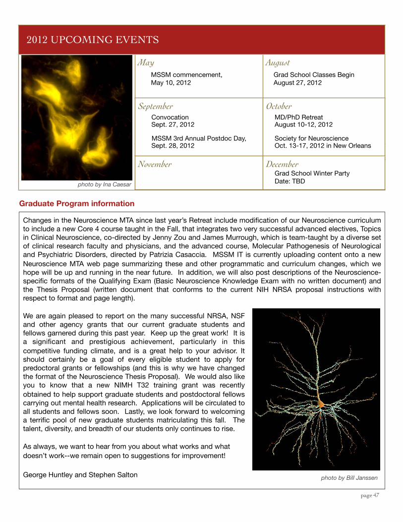
page 47
2012 UPCOMING EVENTS2012 UPCOMING EVENTS2012 UPCOMING EVENTS
May August
September October
November December
Grad School Classes BeginAugust 27, 2012
MD/PhD RetreatAugust 10-12, 2012
Grad School Winter PartyDate: TBD
Changes in the Neuroscience MTA since last year’s Retreat include modification of our Neuroscience curriculum to include a new Core 4 course taught in the Fall, that integrates two very successful advanced electives, Topics in Clinical Neuroscience, co-directed by Jenny Zou and James Murrough, which is team-taught by a diverse set of clinical research faculty and physicians, and the advanced course, Molecular Pathogenesis of Neurological and Psychiatric Disorders, directed by Patrizia Casaccia. MSSM IT is currently uploading content onto a new Neuroscience MTA web page summarizing these and other programmatic and curriculum changes, which we hope will be up and running in the near future. In addition, we will also post descriptions of the Neuroscience-specific formats of the Qualifying Exam (Basic Neuroscience Knowledge Exam with no written document) and the Thesis Proposal (written document that conforms to the current NIH NRSA proposal instructions with respect to format and page length).
We are again pleased to report on the many successful NRSA, NSF and other agency grants that our current graduate students and fellows garnered during this past year. Keep up the great work! It is a significant and prestigious achievement, particularly in this competitive funding climate, and is a great help to your advisor. It should certainly be a goal of every eligible student to apply for predoctoral grants or fellowships (and this is why we have changed the format of the Neuroscience Thesis Proposal). We would also like you to know that a new NIMH T32 training grant was recently obtained to help support graduate students and postdoctoral fellows carrying out mental health research. Applications will be circulated to all students and fellows soon. Lastly, we look forward to welcoming a terrific pool of new graduate students matriculating this fall. The talent, diversity, and breadth of our students only continues to rise.
As always, we want to hear from you about what works and what doesn’t work--we remain open to suggestions for improvement!
George Huntley and Stephen Salton
Graduate Program information
Society for NeuroscienceOct. 13-17, 2012 in New Orleans
photo by Bill Janssen
photo by Ina Caesar
MSSM commencement, May 10, 2012
November
SeptemberConvocationSept. 27, 2012
MSSM 3rd Annual Postdoc Day, Sept. 28, 2012
OctoberMD/PhD RetreatAugust 10-12, 2012
Society for NeuroscienceOct. 13-17, 2012 in New Orleans
DecemberGrad School Winter PartyDate: TBD