Embed Size (px)
Citation preview
Transatrial Intrapericardial Tricuspid Annuloplasty
Toby Rogers, BM, BCh*, Kanishka Ratnayaka, MD*,†, Merdim Sonmez, PhD*, Dominique N. Franson, BS*, William H. Schenke, BA*, Jonathan R. Mazal, MS*, Ozgur Kocaturk, PhD*,‡, Marcus Y. Chen, MD*, Anthony Z. Faranesh, PhD*, and Robert J. Lederman, MD*
*Cardiovascular and Pulmonary Branch, Division of Intramural Research, National Heart, Lung, and Blood Institute, National Institutes of Health, Bethesda, Maryland †Department of Cardiology, Children’s National Medical Center, Washington, DC ‡Institute of Biomedical Engineering, Bogazici University, Istanbul, Turkey.
Abstract
OBJECTIVES—This study sought to demonstrate transcatheter deployment of a circumferential
device within the pericardial space to modify tricuspid annular dimensions interactively and to
reduce functional tricuspid regurgitation (TR) in swine.
BACKGROUND—Functional TR is common and is associated with increased morbidity and
mortality. There are no reported transcatheter tricuspid valve repairs. We describe a transcatheter
extracardiac tricuspid annuloplasty device positioned in the pericardial space and delivered by
puncture through the right atrial appendage. We demonstrate acute and chronic feasibility in
swine.
METHODS—Transatrial intrapericardial tricuspid annuloplasty (TRAIPTA) was performed in 16
Yorkshire swine, including 4 with functional TR. Invasive hemodynamics and cardiac magnetic
resonance imaging (MRI) were performed at baseline, immediately after annuloplasty and at
follow-up.
RESULTS—Pericardial access via a right atrial appendage puncture was uncomplicated. In 9
naïve animals, tricuspid septal-lateral and anteroposterior dimensions, the annular area and
perimeter, were reduced by 49%, 31%, 59%, and 24% (p < 0.001), respectively. Tricuspid leaflet
coaptation length was increased by 53% (p < 0.001). Tricuspid geometric changes were
maintained after 9.7 days (range, 7 to 14 days). Small effusions (mean, 46 ml) were observed
immediately post-procedure but resolved completely at follow-up. In 4 animals with functional
TR, severity of regurgitation by intracardiac echocardiography was reduced.
CONCLUSIONS—Transatrial intrapericardial tricuspid annuloplasty is a transcatheter
extracardiac tricuspid valve repair performed by exiting the heart from within via a transatrial
puncture. The geometry of the tricuspid annulus can interactively be modified to reduce severity
of functional TR in an animal model.
REPRINT REQUESTS AND CORRESPONDENCE: Dr. Robert J. Lederman, National Heart, Lung, and Blood Institute, National Institutes of Health, Building 10, Room 2c713, Bethesda, Maryland 20892-1538. [email protected].
All other authors have reported that they have no relationships relevant to the contents of this paper to disclose.
APPENDIX For supplemental material and video, please see the online version of this article.
HHS Public AccessAuthor manuscriptJACC Cardiovasc Interv. Author manuscript; available in PMC 2015 September 01.
Published in final edited form as:JACC Cardiovasc Interv. 2015 March ; 8(3): 483–491. doi:10.1016/j.jcin.2014.10.013.
Author M
anuscriptA
uthor Manuscript
Author M
anuscriptA
uthor Manuscript

Keywords
annuloplasty; structural heart interventions; transcatheter repair; tricuspid regurgitation
Tricuspid regurgitation (TR) is a predictor of mortality, increasing with regurgitation
severity and independent of age, biventricular function or dimensions, or pulmonary artery
pressure (1). In patients with mitral regurgitation (2) or undergoing left-sided valve surgery
(3), TR is associated with heart failure and worse outcome. Persistent TR after left-sided
valve surgery predicts poor outcome (4). Isolated tricuspid valve surgery is associated with
high mortality (5,6) and, therefore, is seldom justifiable.
Most clinical TR is “functional” and is caused by dilation of the right ventricle and tricuspid
annulus (7), resulting from volume or pressure overload. Less commonly, dilation results
from primary right ventricular (RV) pathology such as RV infarction or cardiomyopathy.
The severity of TR corresponds to tricuspid annulus dimension (8) and is dependent on
individual patient preload and afterload conditions, which vary with time.
Surgical annuloplasty restores annular geometry and improves leaflet coaptation (9).
Replacement tricuspid valves are more susceptible to thrombosis compared with prostheses
in the mitral or aortic positions. Transcatheter repairs have been developed for functional
mitral regurgitation (10–12) but not functional TR.
Transatrial intrapericardial tricuspid annuloplasty (TRAIPTA) is a novel transcatheter
tricuspid valve repair, guided by x-ray fluoroscopy. The pericardium is accessed via a
puncture of the right atrial appendage (RAA) from within. A circumferential implant, which
exerts compressive force over the tricuspid annulus, is delivered along the atrioventricular
groove within the pericardial space. Tension on the implant is adjusted interactively to
modify tricuspid annular geometry and reduce TR. The RAA puncture is sealed using off-
the-shelf nitinol closure devices.
In this study, we tested pre-clinical feasibility of transcatheter extracardiac tricuspid
annuloplasty in swine and showed that substantial modification of tricuspid annular
geometry is achieved and sustained for at least 7 days. In animals with induced functional
TR, we showed that TRAIPTA effectively reduces the severity of tricuspid regurgitation.
We also explored the feasibility of clinical translation through human cardiac computed
tomography (CT) analysis.
METHODS
Procedures were approved by the institutional animal care and use committee. TRAIPTA
was performed in 16 Yorkshire swine (mean 48 ± 14 kg) under general anesthesia and
mechanical ventilation. Of these, 9 naïve animals were survived for 7 to 14 days, 4 with
functional TR were survived for up to 48 days and 3 underwent nonsurvival characterization
of TRAIPTA tension and coronary artery protection elements. Hemodynamics and geometry
were characterized at each time point by angiography, magnetic resonance imaging (MRI) at
Rogers et al. Page 2
JACC Cardiovasc Interv. Author manuscript; available in PMC 2015 September 01.
Author M
anuscriptA
uthor Manuscript
Author M
anuscriptA
uthor Manuscript

mid-systole at 1.5-T, and/or 320-detector row CT. Selective coronary angiography was
performed before and after TRAIPTA.
TRAIPTA SYSTEM CONFIGURATION
We designed and built a catheter system comprising a delivery device and a permanent
implant (Figure 1). The delivery device is formed from an interrupted 0.035-inch nitinol
wire, pre-shaped into a self-expanding loop to open inside the pericardium and encircle the
heart. The TRAIPTA implant comprises a 2.5-mm diameter hollow tube of braided 0.005-
inch nitinol wire. The braided design allows the implant to shorten longitudinally by more
than 50%. A braided polyester suture (Ti-Cron size 2, Covidien, Waltham, Massachusetts)
runs within the hollow implant with a pre-tied sliding Roeder knot that allows interactive
tightening after deployment and that ensures continuous myocardial apposition. The implant
is pre-mounted on the delivery device loop and is pre-loaded into a 12-French braided
sheath (Cook Medical, Bloomington, Indiana) for deployment.
TRANSATRIAL PERICARDIAL ACCESS
The RAA was engaged from the femoral vein with a 4-French multipurpose catheter (Figure
2) and punctured using the back end of a 0.035-inch guidewire. The catheter was exchanged
for a 14-French sheath (Cook Medical); 10 ml of 50% iopamidol/saline was injected into the
pericardial sheath to visualize epicardial structures (Online Appendix, Online Figure 1).
DELIVERY AND TIGHTENING OF TRAIPTA IMPLANT
The nitinol loop of the TRAIPTA system opened inside the pericardium to encircle the heart
along the atrioventricular groove (Figure 1). The delivery device was then withdrawn,
leaving the TRAIPTA implant in place. The suture was tightened interactively during real-
time 1.5-T MRI (Aera, Siemens, Erlangen, Germany) to achieve the desired tension.
In animals with functional TR, implant tension was adjusted during intracardiac
echocardiography. In 2 naïve animals, we assessed the tension-geometry relationship by
applying progressive tension measured with a force meter, 3 times in each animal, during
real-time MRI.
CLOSURE OF THE RAA PUNCTURE
After TRAIPTA deployment, pericardial contrast was washed out and 2 mg/kg
triamcinolone was infused to impede pericardial adhesions (13). Alongside a 0.014-inch
“buddy” guidewire (Ironman, Abbott Vascular, Santa Clara, California) for emergency
bailout, a 5- or 6-mm nitinol atrial septal occluder (Amplatzer, St. Jude Medical, St. Paul,
Minnesota or Lepu Medical, Bejing, China) closed the appendage puncture site. A final
RAA angiogram confirmed hemostasis before all wires and sheaths were withdrawn.
ANIMAL MODEL OF FUNCTIONAL TR
Ten naïve Yorkshire swine underwent procedures to cause right ventricular (RV) volume
and/or pressure overload or tricuspid papillary restriction. This included combinations of (1)
isolated RV infarction by ethanol infusion into the right coronary artery during balloon
protection of the posterior descending artery (n = 8); (2) interatrial septostomy with a 20-
Rogers et al. Page 3
JACC Cardiovasc Interv. Author manuscript; available in PMC 2015 September 01.
Author M
anuscriptA
uthor Manuscript
Author M
anuscriptA
uthor Manuscript

mm balloon (n = 4); (3) pulmonary artery infusion of 8 ml of 300- to 700-μm polyvinyl-
alcohol spheres to induce pulmonary hypertension (n = 5); (4) ethanol needle injection
directly into the RV papillary muscles (n = 1); and (5) stenting open the pulmonary valve to
cause severe regurgitation (n = 1). Of these 10 animals, 4 were euthanized for intractable
ventricular fibrillation or severe right heart failure after RV infarction. The remaining 6
developed a dilated right ventricle, but only 4 of these developed moderate-severe TR after a
mean of 117 days. These 4 animals underwent TRAIPTA with intracardiac
echocardiography and were survived for up to 48 days.
HUMAN IMAGING FOR SUITABILITY
We studied human cardiac CT angiograms of patients with RV enlargement in the
anonymized and delinked National Heart, Lung, and Blood Institute database. This does not
constitute human subjects research under US45CFR§46.102(f). Suitability criteria for
transatrial access were the presence of a discrete lobe from which to puncture and anterior
orientation. Patients were evaluated for the presence of a clearly demarcated atrioventricular
groove suitable for TRAIPTA. Epicardial coronary arteries at risk of compression were
identified.
STATISTICAL ANALYSIS
Data were analyzed using SPSS 19.0 (IBM, Armonk, New York) and reported as mean ±
SD. Differences were examined by 1-way repeated-measures analysis of variance, with
Bonferroni post-hoc tests as appropriate. A p ≤ 0.05 was considered significant.
RESULTS
TRAIPTA was performed in a total of 16 animals (9 naïve survived for 7 to 14 days, 4 with
functional TR survived up to 48 days, and 3 were not survived to characterize tension-
geometry and coronary protection elements). Transatrial pericardial access was successful in
a single pass, and the TRAIPTA device was consistently delivered to the atrioventricular
groove in 16 of 16 animals. These 2 stages required <10 min to perform (Online Video).
SURVIVAL EXPERIMENTS IN 9 NAÏVE ANIMALS
Significant tricuspid annular geometric reduction was achieved by tightening the TRAIPTA
implant (Table 1, Figures 3 and 4), with 49% (p < 0.001) reduction in the septal-lateral
dimension and 31% (p < 0.001) in the anteroposterior dimension. The tricuspid annular area
and perimeter were reduced by 59% (p < 0.001) and 24% (p < 0.001), respectively.
Tricuspid leaflet coaptation length (Figure 4) was significantly increased by 53% (p <
0.001). Lesser mitral annular geometric change was observed with a mean reduction in the
septal-lateral and anteroposterior dimensions of 15% (p < 0.05) and 15% (p < 0.05),
respectively. No significant hemodynamic changes and no sustained arrhythmias were
observed (Online Table 1). Coronary artery compression was not seen in any animal (Online
Figure 2).
The RAA puncture was consistently closed using a nitinol closure device in all 9 animals.
Post-procedure MRI demonstrated small pericardial effusions (mean, 46 ± 44 ml), but there
Rogers et al. Page 4
JACC Cardiovasc Interv. Author manuscript; available in PMC 2015 September 01.
Author M
anuscriptA
uthor Manuscript
Author M
anuscriptA
uthor Manuscript

was no evidence of tamponade (Online Table 1). No animal required pericardial drainage.
At follow-up (mean, 9.7 days; range, 7 to 14 days), the TRAIPTA implant remained in place
without migration, and pericardial effusions had resolved. On necropsy (Figure 5), the
implant was encased in fibrous tissue and fused to the myocardium along its entire course.
There were no adhesions between the visceral and parietal pericardial layers.
SURVIVAL EXPERIMENTS IN 4 ANIMALS WITH FUNCTIONAL TR
TRAIPTA successfully reduced the severity of TR by intracardiac echocardiography (Figure
4). This was maintained up to 48 days of follow-up. After euthanasia, the TRAIPTA implant
remained intact in the correct anatomic position on necropsy and was completely
endothelialized and fused to the myocardium (Figure 5).
NONSURVIVAL EXPERIMENTS IN 3 NAÏVE ANIMALS
Implant tension correlated with changes in annular geometry (Figure 3), and the feasibility
of coronary artery protection was demonstrated (Online Figure 2), although, in fact, no
coronary compression was observed in any of these experiments.
ANATOMIC SUITABILITY IN HUMANS
We reviewed 14 cardiac CT angiograms for anatomic suitability from patients with an
intracardiac shunt and a dilated right heart. RAA morphology and anatomic position were
consistent across subjects. All 14 met both suitability criteria for transatrial pericardial
access. Thirteen of 14 patients (93%) had a clearly defined atrioventricular groove. Of those,
all had at least 1 epicardial coronary artery that crossed the projected course of the
TRAIPTA implant.
DISCUSSION
To our knowledge, this is the first description of a transcatheter tricuspid valve repair and
the first mechanical intervention accomplished via a right atrial exit to the pericardium. In
contrast with the mitral valve, where primary leaflet or subvalvular apparatus pathology
often contributes to regurgitation, most symptomatic TR is caused by annular dilation with
intact valvular apparatus. For this reason, annuloplasty is the preferred surgical repair. We
showed a dose-response relationship between TRAIPTA tension and tricuspid geometry and
leaflet coaptation; we showed that TRAIPTA treats functional TR in a clinically relevant
animal model, that the right atrial exit port is reliably closed, that the procedure is rapid and
reproducible, and that eligible humans appear to have suitable anatomy.
TRANSATRIAL PERICARDIAL ACCESS
Transatrial pericardial access was first described by Verrier et al. (14) to sample pericardial
fluid, drain effusions, and deliver drugs. A number of interventional (15) and
electrophysiology procedures require pericardial access, which is usually obtained through a
“dry” subxiphoid puncture and which risks hemopericardium from right ventricular or
coronary artery laceration.
Rogers et al. Page 5
JACC Cardiovasc Interv. Author manuscript; available in PMC 2015 September 01.
Author M
anuscriptA
uthor Manuscript
Author M
anuscriptA
uthor Manuscript

TRAIPTA is the first mechanical intervention performed by exiting the heart from within
via a transatrial puncture. This approach provides direct access to the base of the heart in the
plane of the atrioventricular groove. The puncture was sealed with off-the-shelf nitinol
closure devices, and hemostasis was consistently achieved in all animals. However, right
atrial pressures were low in these animals, and the risk of bleeding could be higher in
patients with severe TR or coagulopathy. Small post-procedural pericardial effusions were
observed but without tamponade, whereas in patients, a temporary pericardial drain would
likely be placed. Complete resolution of these effusions was observed in all animals at
follow-up, even with the permanent TRAIPTA implant in situ. Our current technique
requires inversion of the appendage to ensure proper nitinol closure device positioning. We
believe that the device redistributes force adequately to protect against appendage injury.
INTRAPERICARDIAL TRICUSPID ANNULOPLASTY
Aortic and mitral valve disease commonly coexist with tricuspid valve disease. Despite the
adoption of transcatheter aortic and mitral valve interventions, there remains an unmet need
for transcatheter tricuspid valve repair (16), particularly because untreated TR after aortic or
mitral surgery confers a poor outcome. Percutaneous orthotopic (17,18) or heterotopic
(19,20) prosthetic tricuspid valve replacement has been described, but these risk thrombosis
without anticoagulation. TRAIPTA would not require anticoagulation because the implant is
extravascular, although some patients may have other indications (e.g., atrial fibrillation).
Transcatheter tricuspid replacement valves are contraindicated in the presence of
transvenous pacing or defibrillator leads, but TRAIPTA is possible because it is
extravascular. Transatrial exit is possible in the presence of right atrial pacing leads because
the puncture can be performed distant to the lead insertion point. TRIAPTA may also be a
treatment strategy for pediatric and adult congenital systemic single right ventricle patients
with TR.
In this study, we correlated implant tension with reduction in tricuspid annular dimensions,
especially in the septal-lateral dimension, which is the main axis of annular dilation (7) and
the principal target for surgical annuloplasty. The magnitude of geometric modification
easily exceeded ranges reported for surgical annuloplasty (21). We observed lesser
geometric modification of the mitral annulus, likely reflecting the higher pressures and
increased myocardial thickness of the left heart. Nevertheless, this technique could
potentially be adapted to treat both atrioventricular valves. Unlike surgery, TRAIPTA can be
performed in the beating heart and titrated in real time under varying loading conditions
imposed by hemodynamic provocations such as exercise and volume. Importantly, we found
geometric change was maintained over the follow-up period.
ANIMAL MODEL
TR has been induced in animals by surgical annular disruption (22,23), but there are no
transcatheter large animal models of functional TR. In this study, we found that to create TR
required multiple insults, including volume and pressure overload, as well as considerable
time to allow the right ventricle to remodel.
Rogers et al. Page 6
JACC Cardiovasc Interv. Author manuscript; available in PMC 2015 September 01.
Author M
anuscriptA
uthor Manuscript
Author M
anuscriptA
uthor Manuscript

STUDY LIMITATIONS AND POTENTIAL FAILURE MODES
TRAIPTA requires the pericardial space to be free of adhesions, which could impede
implant delivery. This likely precludes TRAIPTA in patients with previous pericardiotomy
or pericarditis. However, the technique might be useful in conjunction with transcatheter
aortic valve replacement and/or mitral valve repair. Because of its circumferential position
around the heart, 1 potential complication is coronary artery and coronary sinus
compression. At the level of tension exerted to achieve 40% reduction in the tricuspid
septal-lateral dimension, we did not observe any coronary artery or sinus compression
(Online Figure 2), albeit in animals without severe RV hypertension. To address this
potential complication, we demonstrated that a protection element could be incorporated
into the TRAIPTA implant (Online Figure 2) and that its position along the TRAIPTA
implant was adjustable interactively in situ. This bridgelike element, originally developed in
our lab for mitral cerclage annuloplasty (12), directs compressive force away from an
entrapped coronary artery or sinus. Finally, in common with all permanent implants,
potential failure modes are implant migration and tissue erosion. We did not observe any
implant migration, and on necropsy, the TRAIPTA device was fully adhered to the
myocardium with no macroscopic evidence of tissue erosion (Figure 5A). Similar
macroscopic findings were observed for the nitinol closure device at the right atrial puncture
site. No pericarditis or pericardial adhesions were observed, perhaps because we
administered intrapericardial glucocorticoids after deployment. These finding suggests a
very low likelihood of late device migration or tissue erosion, but this will be evaluated in a
future long-term animal study.
CLINICAL TRANSLATION AND SUITABILITY OF HUMAN ANATOMY
RAA morphology was suitable for transatrial pericardial access in all patients, and most had
a clearly defined atrioventricular groove suitable for TRAIPTA. Most patients also had
epicardial coronary arteries crossing the projected course of the TRAIPTA implant, and we
therefore anticipate that protection elements would be required in human implementation
(Online Figure 2).
CONCLUSIONS
We report a novel transcatheter tricuspid annuloplasty technique using transatrial pericardial
access and pericardial deployment of a permanent implant. We demonstrate interactive
adjustment of tricuspid annular and leaflet geometry in naïve swine, comparable to that
achieved with surgical annuloplasty. In animals with functional tricuspid regurgitation, this
geometric adjustment reduces the severity of regurgitation.
Supplementary Material
Refer to Web version on PubMed Central for supplementary material.
ACKNOWLEDGMENTS
The authors thank Katherine Lucas and Joni Taylor for animal experiments and Irena Cich for help with device prototyping.
Rogers et al. Page 7
JACC Cardiovasc Interv. Author manuscript; available in PMC 2015 September 01.
Author M
anuscriptA
uthor Manuscript
Author M
anuscriptA
uthor Manuscript

This work was supported by the Division of Intramural Research, NHLBI, NIH (Z01-HL006040). Dr. Rogers, Dr. Ratnayaka, Dr. Sonmez, Ms. Franson, Dr. Kocaturk, and Dr. Lederman are coinventors on patents, assigned to NIH, for TRAIPTA devices.
ABBREVIATIONS AND ACRONYMS
CT computed tomography
MRI magnetic resonance imaging
RAA right atrial appendage
RV right ventricular
TR tricuspid regurgitation
TRAIPTA transatrial intrapericardial tricuspid annuloplasty
REFERENCES
1. Nath J, Foster E, Heidenreich PA. Impact of tricuspid regurgitation on long-term survival. J Am Coll Cardiol. 2004; 43:405–9. [PubMed: 15013122]
2. Agricola E, Stella S, Gullace M, et al. Impact of functional tricuspid regurgitation on heart failure and death in patients with functional mitral regurgitation and left ventricular dysfunction. Eur J Heart Fail. 2012; 14:902–8. [PubMed: 22552182]
3. Turina J, Stark T, Seifert B, Turina M. Predictors of the long-term outcome after combined aortic and mitral valve surgery. Circulation. 1999; 100:II48–53. [PubMed: 10567278]
4. Ruel M, Rubens FD, Masters RG, Pipe AL, Bedard P, Mesana TG. Late incidence and predictors of persistent or recurrent heart failure in patients with mitral prosthetic valves. J Thorac Cardiovasc Surg. 2004; 128:278–83. [PubMed: 15282466]
5. Mangoni AA, DiSalvo TG, Vlahakes GJ, Polanczyk CA, Fifer MA. Outcome following isolated tricuspid valve replacement. Eur J Cardiothorac Surg. 2001; 19:68–73. [PubMed: 11163563]
6. Kwon DA, Park JS, Chang HJ, et al. Prediction of outcome in patients undergoing surgery for severe tricuspid regurgitation following mitral valve surgery and role of tricuspid annular systolic velocity. Am J Cardiol. 2006; 98:659–61. [PubMed: 16923456]
7. Ton-Nu TT, Levine RA, Handschumacher MD, et al. Geometric determinants of functional tricuspid regurgitation: insights from 3-dimensional echocardiography. Circulation. 2006; 114:143–9. [PubMed: 16818811]
8. Sugimoto T, Okada M, Ozaki N, Hatakeyama T, Kawahira T. Long-term evaluation of treatment for functional tricuspid regurgitation with regurgitant volume: characteristic differences based on primary cardiac lesion. J Thorac Cardiovasc Surg. 1999; 117:463–71. [PubMed: 10047648]
9. Shinn SH, Schaff HV. Evidence-based surgical management of acquired tricuspid valve disease. Nat Rev Cardiol. 2013; 10:190–203. [PubMed: 23399976]
10. Siminiak T, Wu JC, Haude M, et al. Treatment of functional mitral regurgitation by percutaneous annuloplasty: results of the TITAN Trial. Eur J Heart Fail. 2012; 14:931–8. [PubMed: 22613584]
11. Whitlow PL, Feldman T, Pedersen WR, et al. Acute and 12-month results with catheter-based mitral valve leaflet repair: the EVEREST II (Endovascular Valve Edge-to-Edge Repair) High Risk Study. J Am Coll Cardiol. 2012; 59:130–9. [PubMed: 22222076]
12. Kim JH, Kocaturk O, Ozturk C, et al. Mitral cerclage annuloplasty, a novel transcatheter treatment for secondary mitral valve regurgitation: initial results in swine. J Am Coll Cardiol. 2009; 54:638–51. [PubMed: 19660696]
13. Maisch B, Ristic AD, Pankuweit S. Intrapericardial treatment of autoreactive pericardial effusion with triamcinolone; the way to avoid side effects of systemic corticosteroid therapy. Eur Heart J. 2002; 23:1503–8. [PubMed: 12242070]
Rogers et al. Page 8
JACC Cardiovasc Interv. Author manuscript; available in PMC 2015 September 01.
Author M
anuscriptA
uthor Manuscript
Author M
anuscriptA
uthor Manuscript

14. Verrier RL, Waxman S, Lovett EG, Moreno R. Transatrial access to the normal pericardial space: a novel approach for diagnostic sampling, pericardiocentesis, and therapeutic interventions. Circulation. 1998; 98:2331–3. [PubMed: 9826322]
15. Bartus K, Bednarek J, Myc J, et al. Feasibility of closed-chest ligation of the left atrial appendage in humans. Heart Rhythm. 2011; 8:188–93. [PubMed: 21050893]
16. Agarwal S, Tuzcu EM, Rodriguez ER, Tan CD, Rodriguez LL, Kapadia SR. Interventional cardiology perspective of functional tricuspid regurgitation. Circ Cardiovasc Interv. 2009; 2:565–73. [PubMed: 20031775]
17. Pott D, Malasa M, Urban U, et al. A novel approach to an anatomical adapted stent design for the percutaneous therapy of tricuspid valve diseases: preliminary experiences from an engineering point of view. ASAIO J. 2012; 58:568–73. [PubMed: 22990286]
18. Boudjemline Y, Agnoletti G, Bonnet D, et al. Steps toward the percutaneous replacement of atrioventricular valves an experimental study. J Am Coll Cardiol. 2005; 46:360–5. [PubMed: 16022968]
19. Lauten A, Ferrari M, Hekmat K, et al. Heterotopic transcatheter tricuspid valve implantation: first-in-man application of a novel approach to tricuspid regurgitation. Eur Heart J. 2011; 32:1207–13. [PubMed: 21300731]
20. Laule M, Stangl V, Sanad W, Lembcke A, Baumann G, Stangl K. Percutaneous transfemoral management of severe secondary tricuspid regurgitation with Edwards Sapien XT bioprosthesis: first-in-man experience. J Am Coll Cardiol. 2013; 61:1929–31. [PubMed: 23500268]
21. Min SY, Song JM, Kim JH, et al. Geometric changes after tricuspid annuloplasty and predictors of residual tricuspid regurgitation: a real-time three-dimensional echocardiography study. Eur Heart J. 2010; 31:2871–80. [PubMed: 20601392]
22. Otaki M, Lust RM. Modification of De Vega’s tricuspid annuloplasty for experimental tricuspid regurgitation. J Cardiac Surg. 1994; 9:399–404.
23. Walter EMD, Delmo Walter E, Vasilyev N, et al. Creation of a tricuspid valve regurgitation model from tricuspid annular dilatation using the cardioport video-assisted imaging system. J Heart Valve Dis. 2011; 20:184–8. [PubMed: 21560820]
Rogers et al. Page 9
JACC Cardiovasc Interv. Author manuscript; available in PMC 2015 September 01.
Author M
anuscriptA
uthor Manuscript
Author M
anuscriptA
uthor Manuscript

FIGURE 1. TRAIPTA System and Procedural Steps(A) Braided suture (packaging and spool), transatrial intrapericardial tricuspid annuloplasty
(TRAIPTA) implant and delivery device. (B) TRAIPTA implant loaded onto delivery
device. The suture is housed within the hollow TRAIPTA implant with a pre-tied Roeder
sliding knot. (C) The system is advanced into the pericardium through the right atrial
appendage. (D) The nitinol delivery device ensures that the system opens into a loop inside
the pericardium and reaches the atrioventricular groove. (E) The delivery device is
withdrawn. (F) The implant is tightened. See Online Video for demonstration of TRAIPTA
procedure. TRAIPTA = transatrial intrapericardial tricuspid annuloplasty.
Rogers et al. Page 10
JACC Cardiovasc Interv. Author manuscript; available in PMC 2015 September 01.
Author M
anuscriptA
uthor Manuscript
Author M
anuscriptA
uthor Manuscript

FIGURE 2. TRAIPTA Procedure In Vivo(A) Right atrial appendage (RAA) angiogram. (B) RAA puncture with 0.035-inch
guidewire. (C) A 14-French sheath introduced into the pericardium. (D) The transatrial
intrapericardial tricuspid annuloplasty (TRAIPTA) implant is deployed around the heart
within the contrast-filled pericardial space. (E) Delivery system (arrows) withdrawal
leaving the TRAIPTA implant in the atrioventricular groove. (F) The TRAIPTA implant is
tightened by a sliding Roeder knot. (G) Closure of the RAA puncture with a nitinol closure
device (arrow). (H) RAA angiogram 7 days later (arrow indicates the closure device).
Rogers et al. Page 11
JACC Cardiovasc Interv. Author manuscript; available in PMC 2015 September 01.
Author M
anuscriptA
uthor Manuscript
Author M
anuscriptA
uthor Manuscript
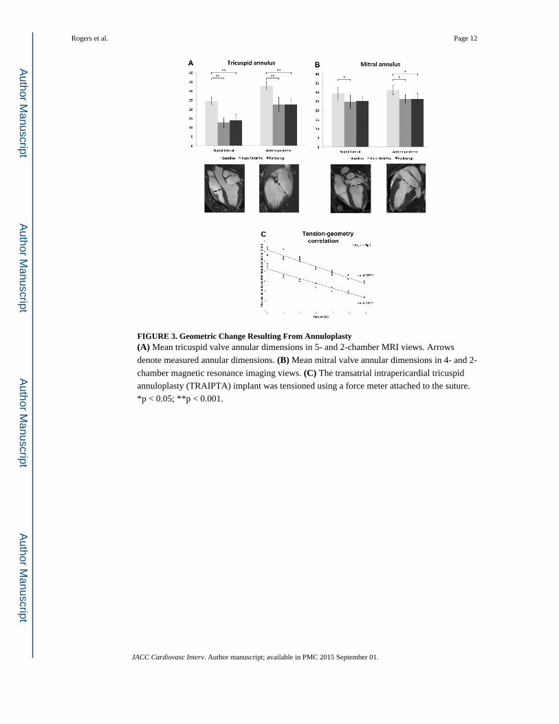
FIGURE 3. Geometric Change Resulting From Annuloplasty(A) Mean tricuspid valve annular dimensions in 5- and 2-chamber MRI views. Arrows
denote measured annular dimensions. (B) Mean mitral valve annular dimensions in 4- and 2-
chamber magnetic resonance imaging views. (C) The transatrial intrapericardial tricuspid
annuloplasty (TRAIPTA) implant was tensioned using a force meter attached to the suture.
*p < 0.05; **p < 0.001.
Rogers et al. Page 12
JACC Cardiovasc Interv. Author manuscript; available in PMC 2015 September 01.
Author M
anuscriptA
uthor Manuscript
Author M
anuscriptA
uthor Manuscript

FIGURE 4. Imaging of Tricuspid Valve and Annulus Before and After Annuloplasty(A) Magnetic resonance imaging 5-chamber view at baseline. Arrow denotes tricuspid
annulus. (B) A 5-chamber view after transatrial intrapericardial tricuspid annuloplasty
(TRAIPTA). (C) A 2-chamber view at baseline. (D) A 2-chamber view after TRAIPTA. (E) Tricuspid leaflet coaptation at baseline. Arrow denotes coaptation length. (F) Tricuspid
leaflet coaptation after TRAIPTA. Arrow denotes coaptation length. (G) Intracardiac
echocardiography of the tricuspid valve before deployment of the TRAIPTA implant. (H) Color Doppler shows a central jet of moderate-severe tricuspid regurgitation (peak velocity
1.8 m/s). (I) Tensioned TRAIPTA implant at the level of the tricuspid annulus (arrow). (J) Color Doppler showing significant reduction in tricuspid regurgitation severity after
TRAIPTA. Ao = aorta; LA = left atrium; LV = left ventricle; RA = right atrium; RV = right
ventricle.
Rogers et al. Page 13
JACC Cardiovasc Interv. Author manuscript; available in PMC 2015 September 01.
Author M
anuscriptA
uthor Manuscript
Author M
anuscriptA
uthor Manuscript
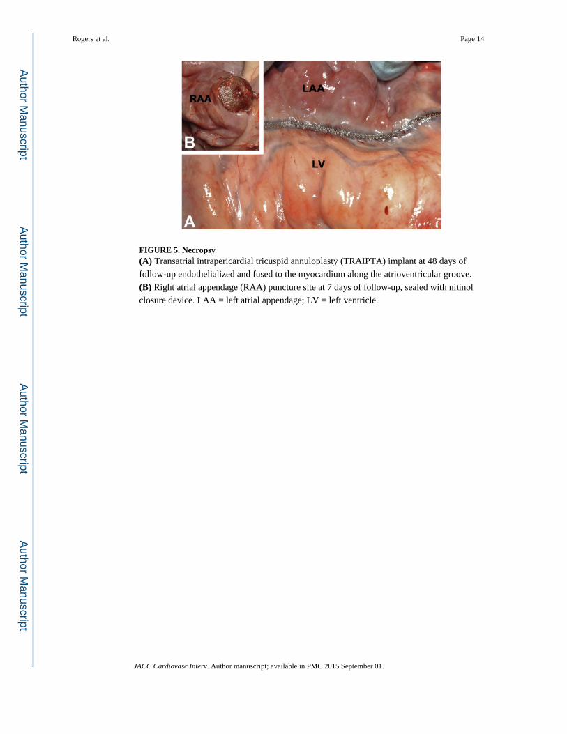
FIGURE 5. Necropsy(A) Transatrial intrapericardial tricuspid annuloplasty (TRAIPTA) implant at 48 days of
follow-up endothelialized and fused to the myocardium along the atrioventricular groove.
(B) Right atrial appendage (RAA) puncture site at 7 days of follow-up, sealed with nitinol
closure device. LAA = left atrial appendage; LV = left ventricle.
Rogers et al. Page 14
JACC Cardiovasc Interv. Author manuscript; available in PMC 2015 September 01.
Author M
anuscriptA
uthor Manuscript
Author M
anuscriptA
uthor Manuscript

Author M
anuscriptA
uthor Manuscript
Author M
anuscriptA
uthor Manuscript
Rogers et al. Page 15
TABLE 1
MRI Findings (N = 9)
Baseline Post-TRAIPTAFollow-up
(mean, 9.7 days)
Cardiac chambers
RA area, cm2 11.8 ± 1.6 12.0 ± 1.2 11.4 ± 2.1
RV end-diastolic volume, ml 84.5 ± 12.3 65.6 ± 20.9 73.9 ± 20.4
RV end-systolic volume, ml 42.5 ± 14.8 35.8 ± 16.9 36.8 ± 17.8
RV stroke volume, ml 42.0 ± 7.5 29.9 ± 5.9* 37.1 ± 6.5
RV ejection fraction, % 50.8 ± 11.6 47.6 ± 10.3 52.1 ± 10.4
LA area, cm2 12.3 ± 2.6 11.1 ± 1.1 12.1 ± 1.9
LV end-diastolic volume, ml 93.3 ± 14.7 66.9 ± 13.3* 79.2 ± 8.7
LV end-systolic volume, ml 51.5 ± 13.9 36.6 ± 11.3* 41.9 ± 5.6
LV stroke volume, ml 41.8 ± 8.8 30.3 ± 5.6* 37.3 ± 8.0
LV ejection fraction, % 45.3 ± 9.6 46.1 ± 8.3 46.8 ± 6.5
TV annulus‡
TV septal-lateral, mm 24.4 ± 2.1 12.6 ± 2.8† 13.9 ± 3.1†
TV anteroposterior, mm 32.8 ± 2.4 22.6 ± 4.0† 22.6 ± 3.6†
TV area, cm2 11.8 ± 1.9 5.3 ± 1.8† 4.4 ± 1.5†
TV annular perimeter, cm 13.2 ± 1.1 9.6 ± 2.4† 9.8 ± 1.2†
TV leaflet coaptation length, mm 3.1 ± 0.9 6.8 ± 0.8† 6.1 ± 0.8†
MV annulus‡
MV septal-lateral, mm 29.2 ± 3.7 24.8 ± 3.8* 25.2 ± 2.4
MV anteroposterior, mm 31.1 ± 2.5 26.3 ± 2.4* 26.1 ± 3.3*
MV area, cm2 9.3 ± 2.0 6.0 ± 1.3† 5.6 ± 1.2†
MV annular perimeter, cm 11.9 ± 1.1 9.3 ± 1.2† 9.1 ± 0.9†
Pericardium
Pericardial effusion, ml 0.0 ± 0.0 45.8 ± 43.7† 0.0 ± 0.0
Values are mean ± SD.
*p < 0.05.
†p < 0.001 compared with baseline.
‡Annular measurements were performed in mid-systole.
LA = left atrium; LV = left ventricle; MRI = magnetic resonance imaging; MV = mitral valve; RA = right atrium; RV = right ventricle; TRAIPTA = transatrial intrapericardial tricuspid annuloplasty; TV = tricuspid valve.
JACC Cardiovasc Interv. Author manuscript; available in PMC 2015 September 01.