Embed Size (px)
Citation preview

http://jdr.sagepub.com/Journal of Dental Research
http://jdr.sagepub.com/content/82/2/136The online version of this article can be found at:
DOI: 10.1177/154405910308200212
2003 82: 136J DENT RESJ. De Munck, B. Van Meerbeek, Y. Yoshida, S. Inoue, M. Vargas, K. Suzuki, P. Lambrechts and G. Vanherle
Four-year Water Degradation of Total-etch Adhesives Bonded to Dentin
Published by:
http://www.sagepublications.com
On behalf of:
International and American Associations for Dental Research
can be found at:Journal of Dental ResearchAdditional services and information for
http://jdr.sagepub.com/cgi/alertsEmail Alerts:
http://jdr.sagepub.com/subscriptionsSubscriptions:
http://www.sagepub.com/journalsReprints.navReprints:
http://www.sagepub.com/journalsPermissions.navPermissions:
What is This?
- Feb 1, 2003Version of Record >>
at UNIV OF SOUTHERN CALIFORNIA on April 6, 2014 For personal use only. No other uses without permission.jdr.sagepub.comDownloaded from
International and American Associations for Dental Research
at UNIV OF SOUTHERN CALIFORNIA on April 6, 2014 For personal use only. No other uses without permission.jdr.sagepub.comDownloaded from
International and American Associations for Dental Research
INTRODUCTION
Most current total-etch adhesives perform well in bond strength tests, atleast when tested shortly after application and under controlled in vitro
conditions (Inoue et al., 2001). However, the oral cavity—with temperaturechanges, chewing loads, and chemical attacks by acids and enzymes—forms arather severe challenge for tooth-composite bonds to survive for a reasonablylong time. Clinically, marginal deterioration of composite restorations remainsproblematic and forms the major reason that dramatically shortens the lifetimeof adhesive restorations (Van Meerbeek et al., 1998a). A factor known todegrade tooth-composite bonds is exposure to water (Gwinnett and Yu, 1995;Sano et al., 1999; Armstrong et al., 2001b). Among different forms ofmarginal leakage, nano-leakage, or the ingress of oral fluids throughnanometer-sized channels along collagen fibrils within the hybrid layer, isconsidered very detrimental to bond integrity (Sano et al., 1995; Hashimoto etal., 2000, 2002). As part of a total-etch procedure, the rather aggressivephosphoric-acid-etching nearly completely deprives collagen ofhydroxyapatite (Van Meerbeek et al., 1998b). Consequently, adequateinfiltration into, wetting of, and molecular interaction with hydroxyapatite-depleted collagen by resin monomers is challenging. It may result inincomplete hybridization, leaving collagen unprotected and vulnerable tohydrolytic degeneration (Hashimoto et al., 2000). Other degradation-promoting factors are, e.g., residual solvent of the adhesive or insufficientlyremoved surface water. Eventually, resin itself degrades over time and leachesout, causing the restoration-tooth bond to deteriorate (Santerre et al., 2001).
The objective of this laboratory study was to test the hypotheses that: (1)two-step total-etch adhesives resist water degradation as well as do three-step total-etch adhesives, and that (2) an adjacent composite-enamel bondprotects the composite-dentin bond against degradation. Therefore, themicro-tensile bond strength (mTBS) to dentin of 2 three-step total-etchadhesives was compared with that of 2 two-step total-etch adhesives after 4yrs of storage in water. Quantitative and qualitative failure analysis wasconducted correlating field-emission scanning (Fe-SEM) and transmissionelectron microscopy (TEM).
MATERIALS & METHODS
Micro-tensile Bond Strength TestingTwenty-eight non-carious human third molars (gathered following informedconsent approved by the Commission for Medical Ethics of the CatholicUniversity of Leuven) were stored in 0.5% chloramine in water at 4°C and usedwithin 1 mo after extraction. The occlusal third of the molar crowns was removedby means of an Isomet diamond saw (Isomet 1000, Buehler, Lake Bluff, IL,USA). We produced a standard smear layer by wet-sanding (60 sec) the dentinsurface with 600-grit silicon-carbide paper. All specimens were randomly dividedinto 4 groups of 7 teeth each, and subjected to a bonding treatment strictly
ABSTRACTResin-dentin bonds degrade over time. Theobjective of this study was to evaluate theinfluence of variables like hybridizationeffectiveness and diffusion/elution of interfacecomponents on degradation. Hypotheses testedwere: (1) There is no difference in degradationover time between two- and three-step total-etchadhesives; and (2) a composite-enamel bondprotects the adjacent composite-dentin bondagainst degradation. The micro-tensile bondstrength (mTBS) to dentin of 2 three-step total-etch adhesives was compared with that of 2 two-step total-etch adhesives after 4 years of storage inwater. Quantitative and qualitative failure analyseswere conducted correlating Fe-SEM and TEM.Indirect exposure to water did not significantlyreduce the mTBS of any adhesive, while directexposure resulted in a significantly reduced mTBSof both two-step adhesives. It is concluded thatresin bonded to enamel protected the resin-dentinbond against degradation, while direct exposure towater for 4 years affected bonds produced by two-step total-etch adhesives.
KEY WORDS: adhesion, dentin, total-etch, bondstrength, durability.
Received May 1, 2002; Last revision August 29, 2002;Accepted October 24, 2002
A supplemental appendix to this article is publishedelectronically only at http://www.dentalresearch.org.
Four-year Water Degradation of Total-etch Adhesives Bonded to Dentin
J. De Munck1, B. Van Meerbeek1*, Y. Yoshida2, S. Inoue3, M. Vargas4, K. Suzuki2, P. Lambrechts1, and G. Vanherle1
1Leuven BIOMAT Research Cluster, Department ofConservative Dentistry, School of Dentistry, OralPathology and Maxillo-Facial Surgery, Catholic Universityof Leuven, Kapucijnenvoer 7, B-3000 Leuven, Belgium;2Department of Biomaterials, Okayama UniversityGraduate School of Medicine and Dentistry, 2-5-1 Shikata-cho, Okayama 700-8525, Japan; 3Department of OralHealth Science, Hokkaido University Graduate School ofDental Medicine, Kita 13 Nishi 7, Kita-ku, Sapporo 060-8586, Japan; and 4Department of Operative Dentistry, TheUniversity of Iowa, Iowa City, USA; *correspondingauthor, [email protected]
J Dent Res 82(2):136-140, 2003
RESEARCH REPORTSBiomaterials & Bioengineering
136 at UNIV OF SOUTHERN CALIFORNIA on April 6, 2014 For personal use only. No other uses without permission.jdr.sagepub.comDownloaded from
International and American Associations for Dental Research
J Dent Res 82(2) 2003 Four-year Water Degradation of Resin-Dentin Bonds 137
according to the manufacturer's instructions, with 2 two-step total-etch adhesives, Optibond Solo (Kerr, Orange, CA, USA) andScotchbond 1 (Single Bond outside Europe, 3M ESPE, St. Paul,MN, USA), and their three-step precursors, respectively, OptibondDual-Cure (Kerr) and Scotchbond Multi-Purpose (3M ESPE). Thesurface was built up with resin composite (Z100, 3M ESPE) in 3 or4 layers to a height of 5 to 6 mm. From each group, 3 teeth weresubjected to mTBS-testing after 24 hours' storage in water (indirectexposure of resin-dentin interface, 24hr-IE) at 37°C. The 4remaining teeth were stored for 4 yrs at 37°C in water thatcontained 0.5% chloramine to prevent bacterial growth (Burrow etal., 1996). Prior to water storage, 2 of these teeth were sectioned inhalf for direct exposure of the resin-dentin interface to water (4yr-DE). The remaining teeth were kept intact, with the resin-dentininterface entirely surrounded by resin bonded to the outer enamelrim, and consequently only indirectly exposed to water (4yr-IE).After storage, the teeth were sectioned perpendicular to theadhesive-tooth interface, by means of the Isomet saw, yieldingrectangular sticks (2 x 2 mm wide; 8-9 mm long). Specimens weretrimmed at the biomaterial-tooth surface to a cylindrical hourglassshape (diameter of about 1.2 mm), by means of the MicroSpecimenFormer (De Munck et al., 2003) and fine cylindrical diamond burs(835KREF, Komet, Lemgo, Germany) under continuous air/waterspray. Specimens were then fixed to Ciucchi's jig withcyanoacrylate glue (Model Repair II Blue, Dentsply-Sankin,Ohtawara, Japan) and stressed at a crosshead speed of 1 mm/minuntil failure in an LRX testing device (LRX, Lloyd, Hampshire,UK). The mTBS was expressed in MPa, as we derived fromdividing the imposed force (N) at the time of fracture by the bondarea (mm2). The means were evaluated by a two-way ANOVA test,with the type of product and the degree of water exposure aspredicting factors. An additional random factor, with the mTBS-samples from the same tooth grouped, was added to the statisticalmodel as correction for the multiple samples gathered from onetooth. The means of all groups were compared by a Tukey-Kramermultiple-comparisons test. All statistical analyses were carried outwith Statistica software (StatSoft, Tulsa, OK, USA).
Failure AnalysisAll mTBS-specimens exhibiting mixed adhesive-cohesive failureswere processed for Fe-SEM (Philips XL30, Eindhoven, The
Netherlands) by common specimen-processing procedures,including fixation, dehydration, chemical drying, and gold-sputter-coating (Perdigão et al. , 1995). The proportionalprevalence of different fracture modes was determined for eachmTBS-specimen by image analysis (Image Pro Plus, MediaCybernetics, Silver Spring, MD, USA) applied to digitallyrecorded Fe-SEM images. For statistical analysis, only thepercentage of adhesive failures was taken into account. The datawere re-ordered dichotomously and evaluated by logisticregression with the type of product and the degree of waterexposure as predicting factors. We also used logistic regression totest for association of mTBS and failure mode.
After Fe-SEM, representative mTBS samples of eachadhesive were further processed for TEM. The mTBS sampleswere immersed for 12 hrs in epoxy resin prior to being embeddedin molds (Robinson and Gray, 1996). Non-demineralized 70- to90-nm sections through the fracture plane were cut by means of adiamond knife (Diatome, Bienne, Switzerland) in anultramicrotome (Ultracut UCT, Leica, Vienna, Austria). Forevaluation of collagen, TEM sections were positively stained with5% uranyl acetate (UA) for 20 min and saturated lead citrate (LC)for 3 min prior to TEM examination (Philips CM10, Eindhoven,The Netherlands).
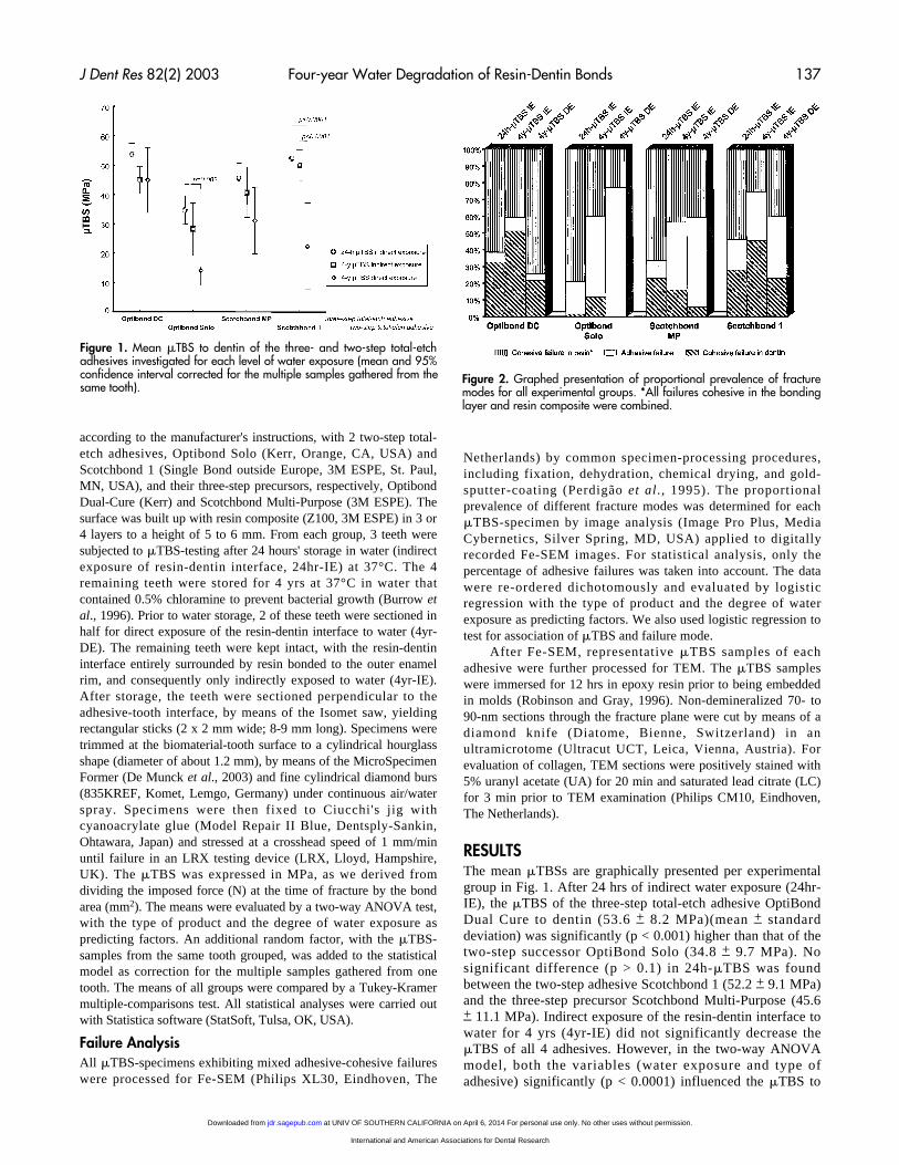
RESULTSThe mean mTBSs are graphically presented per experimentalgroup in Fig. 1. After 24 hrs of indirect water exposure (24hr-IE), the mTBS of the three-step total-etch adhesive OptiBondDual Cure to dentin (53.6 + 8.2 MPa)(mean + standarddeviation) was significantly (p < 0.001) higher than that of thetwo-step successor OptiBond Solo (34.8 + 9.7 MPa). Nosignificant difference (p > 0.1) in 24h-mTBS was foundbetween the two-step adhesive Scotchbond 1 (52.2 + 9.1 MPa)and the three-step precursor Scotchbond Multi-Purpose (45.6+ 11.1 MPa). Indirect exposure of the resin-dentin interface towater for 4 yrs (4yr-IE) did not significantly decrease themTBS of all 4 adhesives. However, in the two-way ANOVAmodel, both the variables (water exposure and type ofadhesive) significantly (p < 0.0001) influenced the mTBS to
Figure 1. Mean mTBS to dentin of the three- and two-step total-etchadhesives investigated for each level of water exposure (mean and 95%confidence interval corrected for the multiple samples gathered from thesame tooth).
Figure 2. Graphed presentation of proportional prevalence of fracturemodes for all experimental groups. *All failures cohesive in the bondinglayer and resin composite were combined.
at UNIV OF SOUTHERN CALIFORNIA on April 6, 2014 For personal use only. No other uses without permission.jdr.sagepub.comDownloaded from
International and American Associations for Dental Research

138 De Munck et al. J Dent Res 82(2) 2003
dentin. The p-value for the interaction term of the model wasalso significant (p < 0.0001), indicating that not all 4 adhesiveswere sensitive to the same degree of degradation. Directexposure to water did not significantly (p = 0.62) affect themTBS of OptiBond Dual Cure, while the decrease in mTBS ofScotchbond Multi-Purpose was nearly significant (p = 0.069):The mTBS (4yr-DE) of both two-step total-etch adhesivesdropped significantly (Fig. 1).
Low mTBSs are significantly associated with higherpercentages of adhesive failures (p = 0.008). The number ofadhesive failures (Fig. 2) also significantly (p = 0.03)increased with increasing degree of water exposure,suggesting that the interface itself degraded over time. Onlyfor OptiBond Dual Cure, indirect or direct exposure of theinterface to water for 4 yrs changed neither the proportion ofadhesive failures nor the mTBS.
Correlative Fe-SEM and TEM examination of specimenfailures revealed that adhesive failures occurred either at thetop or at the base of the hybrid layer, often both within onesection (Figs. 3, 4). Optibond Dual Cure typically presentedmore with cohesive failures within the adhesive resin or within
dentin (Fig. 4). Characteristic of Scotchbond systems was theexposure of collagen fibrils at the fracture planes, suggestingthat the hydroxyapatite-depleted collagen was ineffectivelyenveloped by resin (Fig. 3).
DISCUSSIONIn this study, the long-term degradation of resin-dentin bondswas studied by means of a mTBS-testing methodology throughexposure to water for 4 yrs, either directly or indirectly, whenthe resin-dentin interface was surrounded by resin bonded toenamel. Qualitative Fe-SEM and TEM examination of thefracture planes combined with quantitative fractographicanalysis substantiated the bond strength data very well.
Direct exposure to water resulted in a significant decreaseof the mTBS of only the two- but not the three-step total-etchadhesives. Consequently, the first hypothesis was rejected, withthe adhesives involving simplified application to performsignificantly worse on a long-term perspective. The decrease inmTBS was in accordance with the increase in percentage ofadhesive failures. While the mTBS of Optibond Dual Cure
Figure 3. Fractographic failure analysis of Scotchbond MP andScotchbond 1 (3M Espe). (a) Fe-SEM overview photomicrographs of thefracture surfaces (left = dentin side; right = composite counterpart) of arepresentative mTBS sample prepared with Scotchbond MP that wasstored for 4 yrs with the resin-dentin interface directly exposed to water(4yr-DE). Scratches remaining from smear-layer preparation confirmedthat the interface failed adhesively (A) at the level between dentin andthe bonding layer for an area of 0.84 mm2 or 84% of the total surfacearea. A small area of 0.16 mm2 or 16% of the total surface arearepresents a cohesive (C) failure in the bonding resin. (b) Magnificationof the adhesive failure area at the composite side of the same sample asin (a) shows a typical pattern of islands of hybrid layer (H) fragmentsstill attached to the composite (Comp.) and detached from dentin. (c)TEM photomicrograph (non-demineralized, unstained section) of theadhesive failure area sectioned from the same sample as in (a). The thinblack line covering the fracture plane (arrows) and underneath theembedding resin (E) represents the gold coating applied for the Fe-SEMexamination conducted beforehand. The hybrid layer (H) was pulledfrom unaffected dentin (U) either at the base (left) or close to the top(right). (d) High-magnification Fe-SEM photomicrograph of thecomposite site of a fractured four-year-stored Scotchbond 1 sample withdirect exposure of the interface to water (4yr-DE). The sample failedwithin the hybrid layer, part of which remained attached to thecomposite. A resin tag (T) within a dentinal tubule is surrounded byloosely organized collagen fibrils (Coll.), the typical cross-banding ofwhich can be observed. This suggests either that this hybrid layercollagen was inadequately enveloped by resin or that resin was eluted.
Figure 4. Fractographic failure analysis of Optibond DC andOptibond Solo (Kerr). (a). Fe-SEM overview photomicrographs of thefracture surfaces (left = dentin side, right = composite counterpart) ofa representative mTBS sample prepared with Optibond Dual Curethat was stored for 4 yrs with the resin-dentin interface indirectlyexposed to water (4yr-IE). A small area of 0.11 mm2 or 10% of thetotal surface area represents an adhesive (A, marked with black line)failure, while the major part (1.0 mm2 or 90% of the total surfacearea) failed cohesively (C) in resin. (b) TEM photomicrograph (non-demineralized, stained section) of a typical failure of Optibond DualCure after 4 yrs of water storage (4yr-DE). A thin layer of a few mmof the particle-filled adhesive resin remained attached to the hybridlayer (H), indicating that the adhesive layer failed cohesively.Although this section was stained by heavy metals (UA/LC), collagenseemed not to have picked up much of the staining solution. E =embedding resin; T = resin tag (T) packed with filler; U = unaffecteddentin; arrows = gold coating. (c) TEM photomicrograph (non-demineralized, unstained section) of a sample that was preparedwith Optibond solo and stored for 4 yrs with the resin-dentininterface directly exposed to water (4yr-DE). The hybrid layer (H)remained attached to unaffected dentin (U). E = embedding resin; T =resin tag (T). (d) TEM photomicrograph (non-demineralized,unstained section) of the same sample as in (c), but now at a sitewere the sample failed at the base of the hybrid layer. Small hybrid-layer fragments (arrows) remained attached to unaffected dentin (U).The resin tag (T) was fractured at the same level. E = embeddingresin.
at UNIV OF SOUTHERN CALIFORNIA on April 6, 2014 For personal use only. No other uses without permission.jdr.sagepub.comDownloaded from
International and American Associations for Dental Research

J Dent Res 82(2) 2003 Four-year Water Degradation of Resin-Dentin Bonds 139
remained quite stable despite the water storage, fewer than 8%of the failure modes were recorded as adhesive. Cohesivefailure of the adhesive resin above the hybrid layer typicallyoccurred. The superior results obtained with Optibond DualCure were not unexpected, since this adhesive performedrepeatedly favorably in several laboratory (Pilo and Ben-Amar,1999; Armstrong et al., 2001a,b; Inoue et al., 2001; Meiers andYoung, 2001; De Munck et al., 2003) as well as clinical trials(Boghosian, 1996; Van Meerbeek et al., 2001). Among otheras-yet-unknown features, the three-step application procedurewith a low technique-sensitive application of, successively,etchant, primer, and adhesive (Van Meerbeek et al., 2001), theapparent favorable composition with regard to hybridizationefficiency (Van Meerbeek et al., 1996), the particle-filledadhesive providing elastic shock-absorbing potential (VanMeerbeek et al., 1993), the formation of a separate couplingresin layer, and the lower hydrophilicity of the cured resin ascompared with the two-step version may have resulted in thislow sensitivity to water degradation.
The mTBS of all other adhesives dropped when theirrespective interfaces with dentin were directly exposed to waterduring 4 yrs. The percentage of adhesive failures significantlyincreased accordingly. Although the basic ingredients betweenthe three-step adhesive Optibond Dual Cure and the two-stepadhesive Optibond Solo are comparable, the simplifiedapplication procedure with the less-concentrated combinedprimer/adhesive resin appeared to make the Optibond-Solo-produced hybrid layers more sensitive to aging. TEM disclosedadhesive failures to prevail at different depths within the hybridlayer. The results are in total agreement with those from aprevious ultra-morphological study (Van Meerbeek et al.,1999) that revealed that Optibond Dual Cure more uniformlyand completely infiltrated the collagen fibril network, incontrast to Optibond Solo. Such less-optimal hybridizationmight explain, to a large extent, why the hybrid layer producedby Optibond Solo is more prone to degradation than thatproduced by Optibond Dual Cure.
The mTBS of the two-step total-etch adhesive Scotchbond1 decreased significantly more than that of its three-stepprecursor in cases of direct exposure to water. However, incontrast to Optibond Dual Cure, the mTBS of the ScotchbondMulti-purpose to dentin was also reduced, thereby approachinga statistically significant difference. For both adhesives, thiseffect should be partly attributed to the incorporation of a high-molecular-weight (MW) polyalkenoic acid copolymer.Previously, phase separation was shown to occur with thecopolymer being filtered out by the collagen network anddeposited as a distinct gel on the exposed collagen network(Van Meerbeek et al., 1996; Eliades et al., 2001). In theextreme case, the gel hinders adequate resin-interdiffusion, bywhich the hybrid layer would be constituted of collageninfiltrated by the low-MW 2-hydroxyethylmethacrylate(HEMA) that was polymerized to linear poly-HEMA chains,and any residual water (solvent) that was insufficientlyremoved. Indeed, analysis of the failure planes showedabundant, unprotected collagen fibrils.
Worth mentioning is also the reduced stainability of TEMsections. In positively stained sections, the heavy metal stain(UA/LC) binds to regions along the collagen fibril that are richin polar amino acids. The staining pattern reflects thesummation of charged residues along the fibril (Weiss, 1988).
The reduced stainability of collagen in the hybrid layer maythen reflect a decreased quantity of polar groups caused bydegeneration of collagen during the four-year water storage.Accordingly, a further in-depth study on degradation ofcollagen as well as degradation/leaching of resin from theinterface is required.
The second hypothesis could not be rejected. Directexposure to water significantly affected bond integrity (at leastfor the two-step adhesives), while the effect of indirectexposure was negligible for all 4 adhesives tested. This must beattributed to the retarding role of the longer diffusion path inthe indirect-exposure groups, and/or to the protective role ofthe surrounding resin-enamel bond against degradation. Thesealing effect at enamel must have been most determiningbased on the following: First, no significant difference in bondstrength (p < 0.05) was found for the 4yr-IE samples at areascloser to the enamel rim (outer sample area closest to the watersource) and at the central area (most remote from the watersource) for all adhesives tested. This means that the length ofdiffusion must have been less important than the protectiongathered from bonding to surrounding enamel. One could arguethat regional differences in bond strength (peripheral vs. mid-coronal dentin) must have been involved as well; however,these are considered negligible (Tay et al., 2000). Second, fouryears is long enough to expect diffusion to have occurredthroughout the entire sample. Third, for the 4yr-DE samples, atendency existed to higher bond strengths at areas more remotefrom the exposure plane (no statistical analysis was done due tosmall sample size). This suggests that, in the absence of enamelbonding, diffusion may play a more significant role. Last, evenin cases where diffusion is involved, the difference in long-term bonding performance between the three- and the two-stepadhesives remains.
This means that, in the clinical situation, one can rely ondurable dentin bonding using three- or two-step total-etchadhesives if all cavity margins are located in enamel. Forcavities with margins ending in dentin, three-step total-etchadhesives are preferred.
In conclusion: (1) The resin-dentin bond formed by total-etch adhesives is prone to water degradation; (2) two-step total-etch adhesives are more susceptible to water degradation thanthree-step total-etch adhesives; and (3) a surrounding resin-enamel bond protects the resin-dentin interface against waterdegradation.
ACKNOWLEDGMENTSThis study was supported in part by a Research Grant from theFund for Scientific Research-Flanders (F.W.O.-grant "Kredietaan Navorsers" 1.5.054.99) and by a fund of the Toshio NakaoChair for Adhesive Dentistry inaugurated at the CatholicUniversity of Leuven, with G. Vanherle awarded asChairholder. We thank Kerr and 3M Espe for the generousdonation of materials. A preliminary report was presented atthe IADR General Session in San Diego, CA, March, 2002.
REFERENCESArmstrong SR, Keller JC, Boyer DB (2001a). Mode of failure in the
dentin-adhesive resin-resin composite bonded joint as determinedby strength-based (mTBS) and fracture-based (CNSB) mechanicaltesting. Dent Mater 17:201-210.
at UNIV OF SOUTHERN CALIFORNIA on April 6, 2014 For personal use only. No other uses without permission.jdr.sagepub.comDownloaded from
International and American Associations for Dental Research
140 De Munck et al. J Dent Res 82(2) 2003
Armstrong SR, Keller JC, Boyer DB (2001b). The influence of waterstorage and C-factor on the dentin-resin composite microtensilebond strength and debond pathway utilizing a filled and unfilledadhesive resin. Dent Mater 17:268-276.
Boghosian A (1996). Clinical evaluation of a filled adhesive system inClass 5 restorations. Compend Contin Educ Dent 7:750-752, 754-757.
Burrow MF, Satoh M, Tagami J (1996). Dentin bond durability afterthree years using a dentin bonding agent with and withoutpriming. Dent Mater 12:302-307.
De Munck J, Van Meerbeek B, Inoue S, Vargas M, Yoshida Y,Armstrong S, et al. (2003). Micro-tensile bond strengths of one-and two-step self-etch adhesives to bur-cut enamel and dentin. AmJ Dent (in press).
Eliades G, Vougiouklakis G, Palaghias G (2001). Heterogeneousdistribution of single-bottle adhesive monomers in the resin-dentininterdiffusion zone. Dent Mater 17:277-283.
Gwinnett AJ, Yu S (1995). Effect of long-term water storage on dentinbonding. Am J Dent 8:109-111.
Hashimoto M, Ohno H, Kaga M, Endo K, Sano H, Oguchi H (2000).In vivo degradation of resin-dentin bonds in humans over 1 to 3years. J Dent Res 79:1385-1391.
Hashimoto M, Ohno H, Sano H, Tay FR, Kaga M, Kudoi Y, et al.(2002). Micromorphological changes in resin-dentin bonds after 1year of water storage. J Biomed Mater Res 63:306-311.
Inoue S, Vargas MA, Abe Y, Yoshida Y, Lambrechts P, Vanherle G,et al. (2001). Microtensile bond strength of eleven contemporaryadhesives to dentin. J Adhes Dent 3:237-245.
Meiers JC, Young D (2001). Two-year composite/dentin bondstability. Am J Dent 14:141-144.
Perdigão J, Lambrechts P, Van Meerbeek B, Vanherle G, Lopes AL(1995). Field emission SEM comparison of four postfixationdrying techniques for human dentin. J Biomed Mater Res 29:1111-1120.
Pilo R, Ben Amar A (1999). Comparison of microleakage for threeone-bottle and three multiple-step dentin bonding agents. JProsthet Dent 82:209-213.
Robinson G, Gray T (1996). Electron microscopy 2: practicalprocedures. In: Theory and practice of histological techniques.
Bancroft JD, Stevens A, editors. New York: ChurchillLivingstone, pp. 585-626.
Sano H, Takatsu T, Ciucchi B, Horner JA, Matthews WG, Pashley DH(1995). Nanoleakage: leakage within the hybrid layer. Oper Dent20:18-25.
Sano H, Yoshikawa T, Pereira PN, Kanemura N, Morigami M, TagamiJ, et al. (1999). Long-term durability of dentin bonds made with aself-etching primer, in vivo. J Dent Res 78:906-911.
Santerre JP, Shajii L, Leung BW (2001). Relation of dental compositeformulations to their degradation and the release of hydrolyzedpolymeric-resin-derived products. Crit Rev Oral Biol Med 12:136-151.
Tay FR, Carvalho R, Sano H, Pashley DH (2000). Effect of smearlayers on the bonding of a self-etching primer to dentin. J AdhesDent 2:99-116.
Van Meerbeek B, Willems G, Celis JP, Roos JR, Braem M,Lambrechts P, et al. (1993). Assessment by nano-indentation ofthe hardness and elasticity of the resin-dentin bonding area. J DentRes 72:1434-1442.
Van Meerbeek B, Conn LJ Jr, Duke ES, Eick JD, Robinson SJ,Guerrero D (1996). Correlative transmission electron microscopyexamination of nondemineralized and demineralized resin-dentininterfaces formed by two dentin adhesive systems. J Dent Res75:879-888.
Van Meerbeek B, Perdigão J, Lambrechts P, Vanherle G (1998a). Theclinical performance of adhesives. J Dent 26:1-20.
Van Meerbeek B, Yoshida Y, Lambrechts P, Vanherle G, Duke ES,Eick JD, et al. (1998b). A TEM study of two water-based adhesivesystems bonded to dry and wet dentin. J Dent Res 77:50-59.
Van Meerbeek B, Yoshida Y, Snauwaert J, Hellemans L, LambrechtsP, Vanherle G, et al. (1999). Hybridization effectiveness of a two-step versus a three-step smear layer removing adhesive systemexamined correlatively by TEM and AFM. J Adhes Dent 1:7-23.
Van Meerbeek B, Vargas M, Inoue S, Yoshida Y, Peumans M,Lambrechts P, et al. (2001). Adhesives and cements to promotepreservation dentistry. Oper Dent 26(Suppl 6):S119-S144.
Weiss L, editor (1988). Cell and tissue biology. A textbook ofhistology. 6th ed. Baltimore: Urban & Schwarzenberg, pp. 160-170.
at UNIV OF SOUTHERN CALIFORNIA on April 6, 2014 For personal use only. No other uses without permission.jdr.sagepub.comDownloaded from
International and American Associations for Dental Research


![State of the art etch-and-rinse adhesiveswebdelprofesor.ula.ve/odontologia/robertramirez/PDF/2.pdf · 1. Introduction to state of the art etch-and-rinse adhesives Buonocore [1] was](https://img.dokumen.tips/doc/110x75/5f02c0337e708231d405d42c/state-of-the-art-etch-and-rinse-adh-1-introduction-to-state-of-the-art-etch-and-rinse.jpg)


![Dentin bonding systems: Fromdentin collagen …...of adhesives cannot infiltrate to the full depth of demineral-ized dentin created by phosphoric acid in the E&R strategy [21]. In](https://img.dokumen.tips/doc/110x75/5e7dc93ba39c2e29b845f9c1/dentin-bonding-systems-fromdentin-collagen-of-adhesives-cannot-iniltrate.jpg)


