Embed Size (px)
Citation preview
Four-year water degradation of a total-etch and twoself-etching adhesives bonded to dentin
Ali I. Abdalla a,*, Albert J. Feilzer b
aDepartment of Restorative Dentistry, Faculty of Dentistry, University of Tanta, Tanta, EgyptbDepartment of Dental Materials Science (ACTA), Universiteit van Amsterdam and Vrije Universiteit, Amsterdam, The Netherlands
j o u r n a l o f d e n t i s t r y 3 6 ( 2 0 0 8 ) 6 1 1 – 6 1 7
a r t i c l e i n f o
Article history:
Received 18 November 2007
Received in revised form
18 April 2008
Accepted 21 April 2008
Keywords:
Self-etch adhesives
Water storage
Bond strength
Dentin
a b s t r a c t
Objectives: To evaluate effect of direct and indirect water storage on the microtensile dentin
bond strength of one total-etch and two self-etching adhesives.
Methods: The adhesive materials were: one total-etch adhesive; ‘Admira Bond’ and two self-
etch adhesives; ‘Clearfil SE Bond’ and ‘Hybrid Bond’. Freshly extracted human third molar
teeth were used. In each tooth, a Class I cavity (4 mm � 4 mm) was prepared in the occlusal
surface with the pulpal floor extending approximately 1 mm into dentin. The teeth were
divided into three groups (n = 18). Each group was restored with the resin composite ‘Clearfil
APX’ using one of the tested adhesives. For each experimental group 3 test procedures (n = 6)
were carried out: Procedure A: the teeth were stored in water for 24 h (control), then
sectioned longitudinally, buccolingually and mesiodistally to get rectangular slabs of 1.0–
1.2 mm thickness on which a microtensile test was carried out. Procedure B: the teeth were
also sectioned; however, the slabs were stored in water at 37 8C for 4 years before micro-
tensile testing (direct water storage). Procedure C: the teeth were kept in water at 37 8C 4
years before sectioning and microtensile testing (Indirect water storage). During micro-
tensile testing the slabs were placed in a universal testing machine and load was applied at
cross-head speed of 0.5 mm/min.
Results: For the 24 h control, there was no significant difference in bond strength between
the three tested adhesives. After 4 years of indirect water storage, the bond strength
decreased but the reduction was not significantly different from those of 24 h. After 4 years
of direct water storage, the bond strengths of all tested adhesives were significantly reduced
compared to their 24 h results.
Conclusion: All the tested adhesives showed no reduction in bond strength after indirect
water exposure for 4 years. After 4-year direct water exposure, the bond produced by all
tested adhesives was unable to resist deterioration.
# 2008 Elsevier Ltd. All rights reserved.
avai lab le at www.sc iencedi rec t .com
journal homepage: www. int l .e lsev ierhea l th .com/ journa ls / jden
1. Introduction
The durability of the adhesive bond between resin and tooth
structure is of significant importance for longevity of adhe-
sive restorations. Clinically, marginal deterioration of resin
composite remains problematic and forms the major factor
* Corresponding author. Tel.: +20 40 3336654; fax: +20 40 331800.E-mail address: [email protected] (A.I. Abdalla).
0300-5712/$ – see front matter # 2008 Elsevier Ltd. All rights reserveddoi:10.1016/j.jdent.2008.04.011
that dramatically shorten the lifetime of composite–tooth
bond.
The immediate bonding effectiveness of most current
adhesive systems is quite favorable1 regardless of the adhesive
used. However, when these adhesives are tested in a clinical
trial, the bonding effectiveness of some materials appears
.

j o u r n a l o f d e n t i s t r y 3 6 ( 2 0 0 8 ) 6 1 1 – 6 1 7612
dramatically low, whereas the bonds of other materials are
more stable.2,3
Bond strength tests are the most frequently used tests to
screen adhesives. The rationale behind this testing method is
that the stronger the adhesion between tooth and adhesives,
the better it will resist stress imposed by resin polymerization
and oral function. Different bond strength tests have been
developed. Currently, the shear and microtensile bond strength
test methods are used the most. In addition, different artificial
aging techniques were used to reveal valuable clinical informa-
tion. The most commonly used artificial aging technique is
water storage. In this technique, the bonded specimens are
stored in fluid at 37 8C for a specific period. This period may vary
from a few months4 up to 4–5 years5–7 or even longer. Most of
these studies report significant decreases in bond strengths,
even after relatively short storage periods.8–15 The decrease in
bonding effectiveness after water storage was supposed to be
caused by degradation of interface components by hydrolysis
(mainly resin and/or collagen). Also, water can infiltrate and
decrease the mechanical properties of the polymer matrix, by
swelling and reducing thefrictional forces between thepolymer
chains, a process known as ‘plasticization’.16,17
Most in vitro bond strength studies use flat surfaces to test
the bonding effectiveness of dental adhesives. Clinically,
however, adhesives are applied in cavities, which result in
higher polymerization contraction stress. This stress puts the
resin–tooth interfaces under severe tension during the critical
setting of the adhesive, particularly when restoring cavities
with a high C-factor.18 Such prestressed interfaces are more
susceptible to degradation19 by gaps and micro-voids that
facilitate fluid exchange along the interface.
In this study, the degradation of resin–dentin bonds formed
in Class I cavities was studied by exposure to water for 4 years
at 37 8C. In addition, the restored teeth were either stored in
water intact or after sectioning. The first case represents a
clinical situation in which the occlusal seal produced by
bonding to the enamel margin may protect the bond of the
adhesive to cavity dentin against degradation. The second
case represents a situation in which degradation of bond
occurs in cavity with margin entirely in dentin.
The present study was designed to evaluate the influence of
direct and indirect water storage on the microtensile bond
strength of one total-etch adhesive and two self-etching
adhesives to dentin.
Table 1 – Composition of the adhesive systems used in the st
Adhesive system Component Com
Admira Bond Acid 36% phosphoric acid
Bond Acetone, bonding ormocer, dim
methacrylates, initiators, stabi
Clearfil SE Bond Primer HEMA, hydrophilic dimethacry
camphorquinone, water
Adhesive 10-MDP, BIS-GMA, HEMA, hydr
microfiller
Hybrid Bond Brush Sodium p-toluenesulfinate, Sod
Adhesive Methylmethacrylate (MMA), 4-
acid anhydride tri(2-hydroxyet
HEMA, acetone, water
2. Materials and methods
The materials used in this study (Table 1) include a total-etch
adhesive; Admira Bond; and two self-etch Adhesives; Clearfil
SE Bond and Hybrid Bond. Clearfil AP-X was used as restorative
resin composite.
2.1. Test methods
Fifty-four extracted human sound lower molar teeth were
collected and stored in 0.5% chloramine solution in water. The
teeth were used within 1 month after extraction.
The root of each tooth was embedded in a cylindrical
plastic tube up to 1 mm from cemento-enamel junction with
cold curing acrylic resin. A standard box-type Class I cavity
(4 mm � 4 mm) was prepared in the occlusal surface of all
teeth using a #56 carbide fissure bur at high speed with water
coolant and finished with a straight fissure bur at low speed
handpiece. The pulpal floor of the cavity was created
approximately 1 mm into dentin. The enamel margins were
beveled (458, 1 mm) using fine diamond points. The teeth were
divided into 3 groups of 18 teeth. Each group was restored with
resin composite using one of the adhesives.
The adhesive materials were applied following the man-
ufacturers’ instructions, as follows.
2.1.1. Admira BondThe entire cavity preparation was etched for 15 s with 36%
phosphoric acid (Vococid, Voco, Cuxhaven, Germany),
rinsed with water spray. Excess water was removed with
air blast for 3 s leaving the dentin moist. Admira Bond (Voco)
was applied with a disposable brush, thinned with mild air
for 2–3 s and light cured for 20 s using a visible light curing
device (Heliolux DLX, Ivoclar Vivadent, Schaan, Liechten-
stein). The output of the light curing unit was regularly
checked periodically with radiometer (Demetron Research
Corp., Danbury, CT, USA) to ensure that the light was always
about 500 mW/mm2.
2.1.2. Clearfil SE BondClearfil SE primer (Kuraray Medical Inc., Tokyo, Japan) was
applied to the cavity for 20 s using a disposable brush and air
thinned. Clearfil SE Bond (Kuraray) was then applied an,
thinned with gentle stream of air and light cured for 20 s.
udy
position Manufacturer
Voco, Cuxhaven, Germany
ethacrylate, functionizing
lizer
late, 10-MDP toluidine, Kuraray, Tokyo, Japan
ophilic dimethacrylate,
ium N-phenylglycine (NPG-Na) Sun-Medical, Shiga, Japan
methacryloxyethyltrimetillic
hyl)-isocyanurat-triacrylate (THIT),
j o u r n a l o f d e n t i s t r y 3 6 ( 2 0 0 8 ) 6 1 1 – 6 1 7 613
2.1.3. Hybrid BondHybrid Bond (Sun Medical Inc., Shiga, Japan) was dispensed
into the mixing well. A Hybrid Bond brush was dipped into the
solution, stirred shortly and then applied to the cavity for 20 s.
The adhesive was thinned with gentle blast of air for 5 s and
light cured for 20 s.
The cavities were restored with resin composite using an
incremental condensation technique. Each increment was
(thickness <2 mm) light cured for 40 s. All restorations were
finished with a set of carbide finishing burs (Komet, Gebr,
Brasseler, Germany) and polished with polishing discs (Sof-
Lex Pop On, 3M Espe, AG, Seefeld, Germany).
Three test procedures were carried out for each adhesive
including sex teeth for each procedure:
Procedure A: After preparation and resin composite place-
ment, the teeth were stored in water at 37 8C for 1 day, and
then microtensile bond strength measurements were
carried out (24 h indirect).
Procedure B: The teeth were stored for 4 years at 37 8C in
water that contained 0.5% chloramine to prevent bacterial
growth. Then, microtensile bond strength measurements
were carried out (4-year indirect water storage).
Procedure C: The restored teeth were sectioned, and the
slabs were stored in water containing 0.5 chloramine at
37 8C for 4 years, where after microtensile bond strength
measurements were carried out (4-year direct water
storage).
2.2. Specimen preparation for microtensile bond strength
Therestored teethweresectioned longitudinally, perpendicular
to adhesive interface, buccolingually and mesiodistally with
low speed water cooled diamond saw (Isomet, Buehler, Ltd.
Lake Bluff, IL, USA), then the mounted tooth are rotated 908 and
sectioned at its cervical portion to separate the micro-speci-
mens. This serial sectioning leads to the formation of numerous
rectangular ‘‘beams’’ or ‘‘sticks’’ with approximately 1–1.2 mm2
of cross-sectional area. Only beams from the central portion of
the restoration were selected as peripheral beams may not have
had the same dentin thickness. Four-six beams were obtained
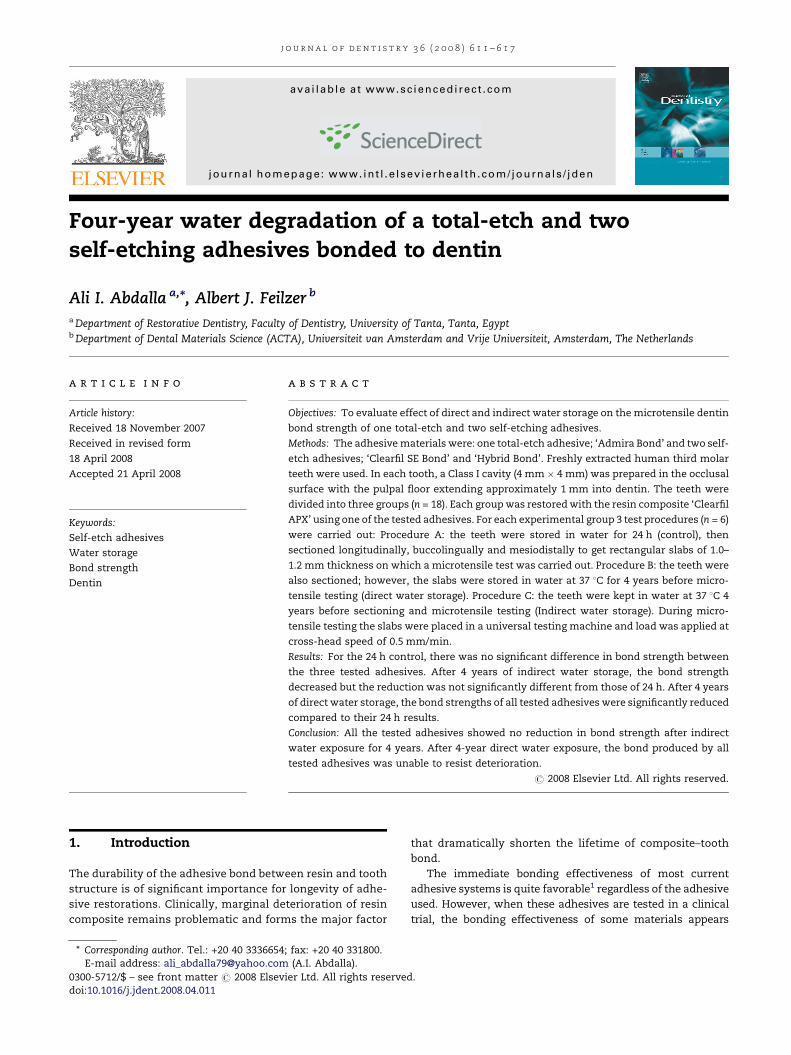
Table 2 – Bond strength of the tested materials (MPa W S.D.)
Adhesive system Number of slabs 24 h (control) Four-y
Admira Bond 20 39 � 5.2
Clearfil SE Bond 20 41 � 3.7
Hybrid Bond 20 37 � 3.4
Table 3a – Fracture patterns of bonded specimens after 24 h
Adhesive system Mixed Cohesive/de
Admira Bond (n = 20) 7 2
Clearfil SE Bond (n = 20) 5 3
Hybrid Bond (n = 20) 9 1
21 (35%) 6 (10%)
n: number of slabs tested.
from each tooth and there cross-sectional areas were measured
with a digital calliper (Mitutoyo Corp., Japan) before testing. For
each test procedures 20 beams were prepared.
For microtensile testing, the beams were glued to a testing
device as previously described by El Zohairy et al.20 using a
light curing adhesive (Clearfil SE Bond, Kuraray Co., Japan) and
placed in a universal testing machine (Instron, Corp., High
Wycombe, UK). Tensile load was applied at cross-head speed
of 0.5 mm/min until failure occurred.
2.3. Statistical analysis
The results were analyzed using a two-way ANOVA, with the
adhesive system and testing procedure as the main factors.
When the F-factor was significant, the Student–Newman–
Keuls multiple comparison test was used.
2.4. Fracture surfaces observation
After microtensile testing, the fractured surface of the speci-
men was inspected by stereomicroscope (Olympus, Tokyo,
Japan) to evaluate the mode of failure. In addition, for some
specimens of each test group impressions of the fractured
surfaces were made using a light-body polyvinylsiloxane
impression material (Extrude, Kerr Gmbh, Karlsruhe, Ger-
many). The impressions were casted in epoxy resin (Epoxy
Die), then mounted on aluminum stubs, sputter-coated with
gold and observed by using SEM (Philips XL30, Eindhoven, the
Netherlands) operating at 15 kV.
3. Results
3.1. Microtensile bond strength test
The mean bond strengths are shown in Table 2. After 24 h
water storage, there were no significant differences (P > 0.05)
for the different adhesives tested. After 4 years of indirect
water storage, the bond strength of each adhesive was
decreased but this reduction was not significant (P > 0.05).
Also there was no significant difference between the different
ear indirect water storage Four-year direct water storage
36 � 4.1 22 � 4.7
39 � 5.1 21 � 2.9
32 � 2.9 12 � 2.5
ntin Cohesive/resin composite Adhesive
3 8
2 10
2 8
7 (11.6%) 26 (43.3%)

Table 3b – Fracture patterns of bonded specimens after 4-year indirect water storage
Adhesive system Mixed Cohesive/dentin Cohesive/resin composite Adhesive
Admira Bond (n = 20) 4 1 2 13
Clearfil SE Bond (n = 20) 3 1 1 15
Hybrid Bond (n = 20) 5 0 1 14
12 (20%) 2 (3.3%) 4 (6.6%) 42 (70%)
n: number of slabs tested.
Table 3c – Fracture patterns of bonded specimens after 4-year direct water storage
Adhesive system Mixed Cohesive/dentin Cohesive/resin composite Adhesive
Admira Bond (n = 20) 5 0 1 14
Clearfil SE Bond (n = 20) 2 0 1 17
Hybrid Bond (n = 20) 0 0 0 20
7 (11.6%) 0 (0%) 2 (3.4%) 51 (85%)
n: number of slabs tested.
j o u r n a l o f d e n t i s t r y 3 6 ( 2 0 0 8 ) 6 1 1 – 6 1 7614
adhesive. After 4-year direct water storage, the bond strength
of all adhesives were significantly (P < 0.05) reduced compared
to their 24 h results and to their 4-year indirect water storage.
3.2. Fracture surface observation
The fractured pattern of bonded specimens is shown in
Tables 3a–3c and in Figs. 1–3. At 24 h water storage, 21.6% of
the samples failed either cohesive in dentin or in composite,
43.4% failed purely adhesively or 35% showed mixed type of
failure. After indirect water storage for 4 years 70% of the
samples failed adhesively at the interface between adhesive
and resin composite or between adhesive and dentin while
10% failed cohesively and 20% showed mixed failure pattern.
After 4 years of direct water storage, 85% of samples showed
adhesive failure at the interface, while 3.4% showed cohesive
failure and 11.6% showed mixed failure.
Fig. 1 – (a) SEM photograph of the fractured surface of
Admira Bond specimen after 4-year indirect water storage
showed a dense hybrid layer that consisted of resin
enveloped collagen fibrils and resin matrix. (b) SEM
photograph of the fractured surface of Admira Bond
specimen after 4 years of direct water storage showed
exposed collagen fibrils with loss of resin contents.
4. Discussion
In the present study the effect of by water storage on the bond
strength of two self-etching adhesives and one total-etch
adhesive to dentin was evaluated. Under clinical situation,
cycling masticatory function has been reported to fatigue the
integrity of resin enamel bond, thereby permitting micro- or
nanoleakage of the peripheral enamel seal.21 This in turn
could lead to degradation of both resin and exposed collagen
fibrils by indirect exposure to water, saliva and enzymes
attack.22 In addition restorations with margins that extend
into the cementum are more susceptible to degradation by
direct water contact.3 In the present study, both these
situations were represented by direct and indirect exposure
of the bonded interface to water in order to visualize the
possible behavior of the tested materials under similar clinical
condition.
With the total-etch system, Admira Bond, bond strength of
37 � 4.9 MPa was found at 24 h. The primary bonding
mechanism of Admira Bond was thought to be diffusion-
based and depends on hybridization or infiltration of resin

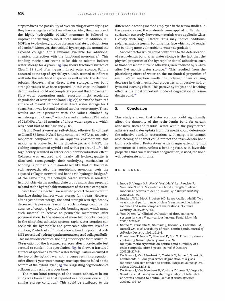
Fig. 3 – (a) Fractured surface of specimen of hybrid bond
after 24 h water storage. Failure occurred at the top of the
hybrid layer with a dense resin impregnation. (b)
Fractured surface of Hybrid Bond specimen after direct
water storage for 4 years. Resin seemed to be extracted
from the hybrid layer with increase in the size of the
interfibrillar spaces.
Fig. 2 – (a) Fractured surface of Clearfil SE Bond after 4-year
indirect water storage. Failure occurred at the top of
Hybrid layer. Resin seemed to infiltrate well into the
interfibrillar spaces as well as into the dentinal tubules. (b)
Fractured surface of Clearfil SE Bond after direct water
storage for 4 years. Resin was lost and dentinal tubules
were empty.
j o u r n a l o f d e n t i s t r y 3 6 ( 2 0 0 8 ) 6 1 1 – 6 1 7 615
within the exposed collagen mesh as well as into dentinal
tubules. After polymerization, the hybrid layer provides
micromechanical retention. Accordingly, exposed collagen
fibrils seemed to be well enveloped with resinous component.
The resin will protect the fibrils from hydrolysis. After 4 years
of indirect water storage, the bond strength was not
significantly affected. Again, the protective role of surround-
ing resin–enamel bond against degradation and the optimal
dentin hybridization of Admira Bond could explain such
findings.23 SEM observation of the fractured surface of speci-
men showed good resin impregnation (Fig. 1a).
After direct water exposure for 4 years, the bond strength of
Admira Bond was significantly reduced compared with those
after 24 h control or after 4-year indirect water storage. Several
investigations24–27 have the attributed degradation of resin-
bond strength for total-etch system to the disintegration of
collagen fibrils and the loss of associated resin in the area of
exposed collagen fibrils in the deminerlized zone of dentin.
This area was created by the discrepancy between the depth of
acid etching and resin infiltration. If infiltration depth is less
than the deminerlized depth, a zone of hydroxyapatite
depleted collagen fibrils is left exposed and unsupported.
These naked collagen fibrils will undergo more strain than the
overlying well resin infiltrated hybrid layer27 since the
modulus of elasticity of the deminerlized dentin collagen
matrix is far lower than that of the hybrid layer.28 Thus, the
deminerlized dentin at the bottom of hybrid layer would
became a weak link in the bonding interface over time. SEM
observation of the fractured surface showed exposed collagen
fibrils with loss of resin contents (Fig. 1b).
The bond strength of Clearfil SE Bond showed no
deterioration by indirect water storage. Clearfil SE Bond is a
two-step mild self-etching adhesive. The primer of Clearfil SE
Bond contains 10-MDP as functional monomer dissolved in
water to result in a pH around 2.29 On dentin, Clearfil SE Bond
does not remove the smear layer. It impregnates the smear
plugs, fixing it at the tubules. The bonding mechanism of
Clearfil SE Bond was suggested to result from the simulta-
neous demineralization and infiltration of enamel and dentin
to form a continuum in the substrate incorporating the smear
plug in the resin tag.30 This will led to a shallow but uniform
resin infiltrated dentin layer. Besides to a simplification of the
bonding technique, the elimination of both rinsing and drying
j o u r n a l o f d e n t i s t r y 3 6 ( 2 0 0 8 ) 6 1 1 – 6 1 7616
steps reduces the possibility of over-wetting or over-drying as
they have a negative effect on adhesion. Also, the presence of
the highly hydrophilic 10-MDP monomer is believed to
improve the wetting to moist tooth surface. In addition, 10-
MDP has two hydroxyl groups that may chelate to calcium ions
of dentin.31 Moreover, the residual hydroxyapatite around the
exposed collagen fibrils remains available for additional
chemical interaction with the functional monomers.31 This
bonding mechanism seems to be able to tolerate indirect
water storage for 4 years. Fig. 2(a) shows fractured surface of
Clearfil SE Bond after 4-year indirect water storage. Failure
occurred at the top of Hybrid layer. Resin seemed to infiltrate
well into the interfibrillar spaces as well as into the dentinal
tubules. However, after direct water storage, lower bond
strength values have been reported. In this case, the bonded
dentin surface could not completely prevent fluid movement.
Slow water penetration under pressure might accelerate
degradation of resin dentin bond. Fig. 2(b) shows the fractured
surface of Clearfil SE Bond after direct water storage for 4
years. Resin was lost and dentinal tubules were empty. These
results are in agreement with the values obtained by
Armstrong and others,12 who observed a median mTBS value
of 21.6 MPa after 15 months of direct water exposure, which
was about half of the baseline mTBS.
Hybrid Bond is one-step self-etching adhesive. In contrast
to Clearfil SE Bond, Hybrid Bond contains 4-META as an active
monomer component. In an aqueous environment this
monomer is converted to the dicarboxylic acid 4-MET, the
etching component of Hybrid Bond with a pH around 1.32 This
high acidity resulted in rather deep demineralization effect.
Collagen was exposed and nearly all hydroxyapatite is
dissolved, consequently, their underlying mechanism of
bonding is primarily diffusion-based like that of the total-
etch approach. Also the amphiphilic monomer wets the
exposed collagen network and bonds via hydrogen bridges.17
At the same time, the collagen coated surface is rendered
hydrophobic via the methacrylate group and is thus prepared
to bond to the hydrophobic monomers of the resin composite.
Such bonding mechanism seems to protect the resin–dentin
interface during indirect water storage for 4 years. However,
after 4-year direct storage, the bond strength was significantly
decreased. A possible reason for such findings could be the
absence of coupling hydrophobic bonding agent, which made
such material to behave as permeable membranes after
polymerization. In the absence of more hydrophobic coating
in the simplified adhesive system, rapid water sorption can
occur via the hydrophilic and permeable adhesive layer.8 In
addition, Yoshida et al.31 found a lower bonding potential of 4-
MET to residual hydroxyapatite around exposed collagen fibrils.
This means low chemical bonding efficiency to tooth structure.
Observation of the fractured surfaces after microtensile test
seemed to confirm this speculation. Fig. 3a shows a fractured
surface of specimen after 24 h water storage. Failure occurred at
the top of the hybrid layer with a dense resin impregnation.
After direct 4-year water storage most specimens failed at the
bottom of the hybrid layer (Fig. 3b), suggesting degeneration of
collagen and resin parts over time.
The mean bond strength of the tested adhesives in our
study was lower than that reported in a previous one with a
similar storage condition.7 This could be attributed to the
difference in testing method employed in these two studies. In
the previous one, the materials were applied to flat dentin
surface. In our study, however, materials were applied in Class
I cavity with high C-factor. This may induce additional
polymerization stress in bonding interface which could render
the bonding more vulnerable to water degradation.
Another factor which could contribute to the deterioration
of resin–dentin bond after water storage is the fact that the
physical properties of the hydrophilic dental adhesives, such
as those present in current adhesives, were reduced by 30–40%
after 3–6 month water storage.33 This resulted from the
plasticizing effect of water on the mechanical properties of
resin. Water sorption swells the polymer chain causing
decrease in their mechanical properties with passive hydro-
lysis and leaching effect. This passive hydrolysis and leaching
effect is the most important mode of degradation of resin–
dentin bond.34
5. Conclusion
This study showed that water sorption could significantly
affect the durability of the resin–dentin bond for certain
adhesives. Both the residual water within the polymerized
adhesive and water uptake from the media could deteriorate
the adhesive bond. In restorations with margins in enamel
acid etching of enamel could protect the resin–dentin bond
from such effect. Restorations with margin extending into
cementum or dentin, unless a bonding resin with favorable
properties that can resist water degradation, is used, the bond
will deteriorate with time.
r e f e r e n c e s
1. Inoue S, Vargas MA, Abe Y, Yoshida Y, Lambrechts P,Vanherle G, et al. Micro-tensile bond strength of elevenmodern adhesives to dentin. Journal of Adhesive Dentistry2001;3:237–46.
2. Brackett WW, Dib A, Brackett MG, Reyes AA, Estrada BE. Twoyear clinical performance of class V resin-modified glass-ionomer and resin composite restorations. OperativeDentistry 2003;28:477–81.
3. Van Dijken JW. Clinical evaluation of three adhesivesystems in class V non-carious lesions. Dental Materials2000;16:285–91.
4. Shono Y, Terashita M, Shimada J, Kozono Y, Carvalho RM,Russell CM, et al. Durability of resin–dentin bonds. Journal ofAdhesive Dentistry 1999;1:211–8.
5. Fukushima T, Inoue Y, Miyazaki K, Itoh T. Effect of primerscontaining N-methylolacrylamide or N-methylolmethacrylamide on dentin bond durability of aresin composite after 5 years. Journal of Dentistry2001;29:227–34.
6. De Munck J, Van Meerbeek B, Yoshida Y, Inoue S, Suzuki K,Lambrechts P. Four-year water degradation of a glass-ionomer adhesive bonded to dentin. European Journal of OralScience 2004;112:73–83.
7. De Munck J, Van Meerbeek B, Yoshida Y, Inoue S, Vargas M,Suzuki K, et al. Four-year water degradation of total-etchadhesives bonded to dentin. Journal of Dental Research2003;82:136–40.
j o u r n a l o f d e n t i s t r y 3 6 ( 2 0 0 8 ) 6 1 1 – 6 1 7 617
8. Kato G, Nakabayashi N. The durability of adhesion tophosphoric acid etched, wet dentin substrates. DentalMaterials 1998;14:347–52.
9. Sano H, Shono T, Sonoda H, Takatsu T, Ciucchi B, CarvalhoR, et al. Relationship between surface area for adhesion andtensile bond strength-evaluation of a micro-tensile bondtest. Dental Materials 1994;10:236–40.
10. Kitasako Y, Burrow MF, Nikaido T, Tagami J. The influenceof storage solution on dentin bond durability of resincement. Dental Materials 2000;16:1–6.
11. Armstrong SR, Keller JC, Boyer DB. The influence of waterstorage and C-factor on the dentin–resin compositemicrotensile bond strength and debonded pathway utilizinga filled and unfilled adhesive resin. Dental Materials2001;17:268–76.
12. Armstrong SR, Vargas MA, Fang Q, Laffoon JE. Microtensilebond strength of a total-etch 3-step, total-etch 2-step, self-etch 2-step, and a self-etch 1-step dentin bonding systemthrough 15-month water storage. Journal of Adhesive Dentistry2003;5:47–56.
13. Meiers JC, Young D. Two-year composite/dentin bondstability. American Journal of Dentistry 2001;14:141–4.
14. Burrow MF, Satoh M, Tagami J. Dentin bond durability afterthree years using a dentin bonding agent with and withoutpriming. Dental Materials 1996;12:302–7.
15. Giannini M, Seixas CAM, Reis AF, Pimenta LAF. Six-monthstorage-time evaluation of one-bottle adhesive systems todentin. Journal of Esthetic Restorative Dentistry 2003;15:43–9.
16. Ferracane JL, Berge HX, Condon JR. In vitro aging of dentalcomposites in water—effect of degree of conversion, fillervolume, and filler/matrix coupling. Journal of Biomedical andMaterials Research 1998;42:465–547.
17. Santerre JP, Shajii L, Leung BW. Relation of dental compositeformulations to their degradation and the release ofhydrolyzed polymeric-resin-derived products. CriticalReviews in Oral Biology and Medicine 2001;12:136–51.
18. Feilzer AJ, de Gee AJ, Davidson CL. Setting stress incomposite resin in relation to configuration of therestoration. Journal of Dental Research 1987;66:1636–9.
19. Hashimoto M, Ohno H, Kaga M, Endo K, Sano H, Oguchi H. Invivo degradation of resin-dentin bonds in humans over 1 to3 years. Journal of Dental Research 2000;79:1385–91.
20. El Zohairy AA, de Gee AJ, de Jager N, van Ruijven LJ, FeilzerAJ. The influence of specimen attachment and dimensionon microtensile strength. Journal of Dental Research2004;83:420–4.
21. Frankenberger R, Tay FR. Self-etch vs etch-and-rinseadhesives: effect of thermo-mechanical fatigue loading on
marginal quality of bonded resin composite restorations.Dental Materials 2005;21:397–812.
22. Hashimoto M, Ohno H, Kaga M, Endo K, Sano H, Oguchi H. Invivo degradation of resin-dentin bonds in humans over 1–3years. Journal of Dental Research 2000;79:1385–91.
23. Hannig M, Reinhardt KJ, Bott B. Self-etching primer vsphosphoric acid: an alternative concept for composite-to-enamel bonding. Operative Dentistry 1999;24:172–80.
24. Van Meerbeek B, Dhem A, Goret-Nicaise M, Braem M,Lambrechts P, Vanherle G. Comparative SEM and TEMexamination of the ultra structure of resin–dentininterdiffusion zone. Journal of Dental Research 1993;72:495–501.
25. Kitasako Y, Burrow MF, Nikaido T, Tagami J. The influenceof storage solution on bond durability of resin cement.Dental Materials 2000;16:1–6.
26. Sato M, Miyazaki M. Comparison of depth of dentin etchingand resin infiltration with single step adhesive system.Journal of Dentistry 2005;33:475–84.
27. Tagtekin DA, Yanikoglu FC, Bozkurt FO, Kologlu B, Sur H.Selected characteristics of an Ormocer and aconventional hybrid resin composite. Dental Materials2004;20:487–97.
28. Ajlouni R, Bishara SE, Soliman MM, Oonsombat C, LaffoonJF, Warren J. The use of Ormocer as an alternative materialfor bonding orthodontic brackets. The Angle Orthodontist2005;75:106–8.
29. De Munck J, Vargas M, Iracki J, Van Landuyt K, Poitevin A,Lambrechts P, et al. One day bonding effectiveness of newself-etch adhesives to bur-cut enamel and dentin. OperativeDentistry 2005;30:39–49.
30. Oliveira SS, Pugach MK, Hilton JF, Watanabe LG, MarshallSJ, Marshall Jr GW. The influence of the dentin smear layeron adhesion: a self-etching primer vs. a total-etch system.Dental Materials 2003;19:758–67.
31. Yoshida Y, Nagakane K, Fukuda R, Nakayama Y, Okazaki M,Shintani H, et al. Comparative study on adhesiveperformance of functional monomers. Journal of DentalResearch 2004;83:454–8.
32. Misra DN. Adsorption from solutions on synthetichydroxyapatite: nonaqueous vs. aqueous solvents. Journal ofBiomedical and Materials Research 1999;48:848–55.
33. Carrilho MRO, Carvalho RM, Tay FR, Pashley DH.Effect of storage media on mechanical properties ofadhesive systems. American Journal of Dentistry 2004;17:104–8.
34. Gopferich A. Mechanism of polymer degradation anderosion. Biomaterials 1996;17:103–17.