Embed Size (px)
Citation preview

Acta Biomaterialia 101 (2020) 26–42
Contents lists available at ScienceDirect
Acta Biomaterialia
journal homepage: www.elsevier.com/locate/actbio
Review article
Four-dimensional bioprinting: Current developments and applications
in bone tissue engineering
Zhuqing Wan, Ping Zhang, Yunsong Liu, Longwei Lv
∗, Yongsheng Zhou
∗
Department of Prosthodontics, Peking University School and Hospital of Stomatology, National Engineering Laboratory for Digital and Material Technology
of Stomatology, National Clinical Research Center for Oral Diseases, Beijing Key Laboratory of Digital Stomatology, 22 Zhongguancun Avenue South, Haidian
District, Beijing 10 0 081, PR China
a r t i c l e i n f o
Article history:
Received 21 June 2019
Revised 20 October 2019
Accepted 25 October 2019
Available online 28 October 2019
Keywords:
3D bioprinting
4D bioprinting
Tissue engineering
Shape memory polymers
Shape memory hydrogels
Bone regeneration
a b s t r a c t
Four-dimensional (4D) bioprinting, in which the concept of time is integrated with three-dimensional
(3D) bioprinting as the fourth dimension, has currently emerged as the next-generation solution of tissue
engineering as it presents the possibility of constructing complex, functional structures. 4D bioprinting
can be used to fabricate dynamic 3D-patterned biological architectures that will change their shapes un-
der various stimuli by employing stimuli-responsive materials. The functional transformation and matu-
ration of printed cell-laden constructs over time are also regarded as 4D bioprinting, providing unprece-
dented potential for bone tissue engineering. The shape memory properties of printed structures cater
to the need for personalized bone defect repair and the functional maturation procedures promote the
osteogenic differentiation of stem cells. In this review, we introduce the application of different stimuli-
responsive biomaterials in tissue engineering and a series of 4D bioprinting strategies based on functional
transformation of printed structures. Furthermore, we discuss the application of 4D bioprinting in bone
tissue engineering, as well as the current challenges and future perspectives.
Statements of significance
In this review, we have demonstrated the 4D bioprinting technologies, which integrate the concept of
time within the traditional 3D bioprinting technology as the fourth dimension and facilitate the fabri-
cations of complex, functional biological architectures. These 4D bioprinting structures could go through
shape or functional transformation over time via using different stimuli-responsive biomaterials and a se-
ries of 4D bioprinting strategies. Moreover, by summarizing potential applications of 4D bioprinting in the
field of bone tissue engineering, these emerging technologies could fulfill unaddressed medical require-
ments. The further discussions about future challenges and perspectives will give us more inspirations
about widespread applications of this emerging technology for tissue engineering in biomedical field.
© 2019 Acta Materialia Inc. Published by Elsevier Ltd.
This is an open access article under the CC BY-NC-ND license.
( http://creativecommons.org/licenses/by-nc-nd/4.0/ )
Contents
1. Introduction . . . . . . . . . . . . . . . . . . . . . . . . . . . . . . . . . . . . . . . . . . . . . . . . . . . . . . . . . . . . . . . . . . . . . . . . . . . . . . . . . . . . . . . . . . . . . . . . 27
2. The overview of published articles on 4D bioprinting . . . . . . . . . . . . . . . . . . . . . . . . . . . . . . . . . . . . . . . . . . . . . . . . . . . . . . . . . . . . . . 28
3. 4D bioprinting based on shape-transformation mechanism . . . . . . . . . . . . . . . . . . . . . . . . . . . . . . . . . . . . . . . . . . . . . . . . . . . . . . . . . . 28
3.1. CTF for 4D bioprinting . . . . . . . . . . . . . . . . . . . . . . . . . . . . . . . . . . . . . . . . . . . . . . . . . . . . . . . . . . . . . . . . . . . . . . . . . . . . . . . . . . . 28
3.2. Potential stimuli-responsive materials for 4D bioprinting . . . . . . . . . . . . . . . . . . . . . . . . . . . . . . . . . . . . . . . . . . . . . . . . . . . . . . 28
3.2.1. Physical stimuli-responsive materials . . . . . . . . . . . . . . . . . . . . . . . . . . . . . . . . . . . . . . . . . . . . . . . . . . . . . . . . . . . . . . . . 29
∗ Corresponding author.
E-mail addresses: [email protected] (L. Lv), [email protected] (Y. Zhou).
https://doi.org/10.1016/j.actbio.2019.10.038
1742-7061/© 2019 Acta Materialia Inc. Published by Elsevier Ltd. This is an open access article under the CC BY-NC-ND license.
( http://creativecommons.org/licenses/by-nc-nd/4.0/ )

Z. Wan, P. Zhang and Y. Liu et al. / Acta Biomaterialia 101 (2020) 26–42 27
3.2.2. Chemical stimuli-responsive materials . . . . . . . . . . . . . . . . . . . . . . . . . . . . . . . . . . . . . . . . . . . . . . . . . . . . . . . . . . . . . . . 33
3.2.3. Biological stimuli-responsive materials . . . . . . . . . . . . . . . . . . . . . . . . . . . . . . . . . . . . . . . . . . . . . . . . . . . . . . . . . . . . . . . 33
4. 4D bioprinting based on functional transformation mechanism. . . . . . . . . . . . . . . . . . . . . . . . . . . . . . . . . . . . . . . . . . . . . . . . . . . . . . . 34
5. Applications of 4D printing in bone tissue engineering . . . . . . . . . . . . . . . . . . . . . . . . . . . . . . . . . . . . . . . . . . . . . . . . . . . . . . . . . . . . . 35
5.1. 4D printing of bone tissue based on injectable stimuli-responsive hydrogels . . . . . . . . . . . . . . . . . . . . . . . . . . . . . . . . . . . . . . . 35
5.2. 4D printing of bone tissue based on shape memory scaffolds. . . . . . . . . . . . . . . . . . . . . . . . . . . . . . . . . . . . . . . . . . . . . . . . . . . 36
5.3. 4D printing of bone tissue based on functional transformation mechanism. . . . . . . . . . . . . . . . . . . . . . . . . . . . . . . . . . . . . . . . 37
5.4. 4D-printed bone constructs with blood vessels and nervous networks . . . . . . . . . . . . . . . . . . . . . . . . . . . . . . . . . . . . . . . . . . . 37
6. Future perspectives and current challenges . . . . . . . . . . . . . . . . . . . . . . . . . . . . . . . . . . . . . . . . . . . . . . . . . . . . . . . . . . . . . . . . . . . . . . . 37
7. Conclusions . . . . . . . . . . . . . . . . . . . . . . . . . . . . . . . . . . . . . . . . . . . . . . . . . . . . . . . . . . . . . . . . . . . . . . . . . . . . . . . . . . . . . . . . . . . . . . . . . 38
Declaration of Competing Interest . . . . . . . . . . . . . . . . . . . . . . . . . . . . . . . . . . . . . . . . . . . . . . . . . . . . . . . . . . . . . . . . . . . . . . . . . . . . . . . . . . 38
Acknowledgment . . . . . . . . . . . . . . . . . . . . . . . . . . . . . . . . . . . . . . . . . . . . . . . . . . . . . . . . . . . . . . . . . . . . . . . . . . . . . . . . . . . . . . . . . . . . . . . . 38
References . . . . . . . . . . . . . . . . . . . . . . . . . . . . . . . . . . . . . . . . . . . . . . . . . . . . . . . . . . . . . . . . . . . . . . . . . . . . . . . . . . . . . . . . . . . . . . . . . . . . . 38
1
H
t
e
e
s
s
c
s
t
w
t
d
i
w
a
s
m
a
a
o
3
d
m
s
r
t
G
o
f
t
(
[
o
c
s
t
p
a
i
o
a
f
s
t
w
b
c
g
H
a
t
a
d
s
d
d
[
a
p
w
b
o
p
f
s
i
t
n
r
a
t
s
p
b
i
t
i
b
t
p
f
i
r
(
t
i
i
n
w
u
a
o
a
s
j
t
a
. Introduction
Three-dimensional (3D) printing, which was first proposed by
ull and co-workers in 1986 [1] , has garnered considerable atten-
ion in the tissue engineering and biomedical fields [2–4] . Differ-
nt 3D bioprinting technologies have been used to fabricate differ-
nt kinds of biological structures such as blood vessels, liver tis-
ue, bone, and heart tissue [5–7] . However, 3D bioprinting has a
ignificant limitation that 3D bioprinting only considers the initial
ondition of a printed objectand assumes it to be inanimate and
tatic. Natural tissue regeneration involves sophisticated 3D struc-
ures, microarchitectures, and extracellular matrix compositions, as
ell as generating tissue that possesses unique functions achieved
hrough dynamic changes in tissue conformation. Most of these
ynamic functional conformational changes are caused by built-
n mechanisms that respond to intrinsic or/and external stimuli,
hich cannot be mimicked through 3D bioprinting [8] .
In 2014, Skylar Tibbits, the director of the Self-Assembly Lab
t the Massachusetts Institute of Technology (MIT), first demon-
trated four-dimensional (4D) printing as a technology entailed
ulti-material prints with the capability to transform over time, or
customized material system that can change from one shape to
nother [9] . This technology has been quickly applied to the field
f tissue engineering, the concept of time can be integrated within
D bioprinting technology as the fourth dimension, leading to the
evelopment of 4D bioprinting [10] . By using stimuli-responsive
aterials, 4D bioprinting can be used to fabricate various 3D de-
igned biologically active architectures capable of dynamic configu-
ation transformations in response to different desired stimuli over
ime, addressing the limitations of 3D bioprinting [8] . Furthermore,
ao et al. defined the 4D bioprinting not only as the generation
f cell-laden 3D-printed structures able to respond to internal cell
orces or external stimuli, but also as the maturation and func-
ionalization of cells or tissues in 3D-printed constructs with time
i.e., the configuration of the printed structures does not change)
10] . The shape and functionalities changes of printed constructs
ver time are the two main strategies for 4D bioprinting. These
haracteristics are essential for maintaining long-term homeosta-
is and self-renewal of biosynthetic constructs. However, the con-
rolled degradation of materials in 3D-printed constructs that com-
letely disappear in the dynamic process, should not be considered
s 4D printing. Most 3D-printed constructs remain integrated dur-
ng shape or functional transformation [11] . Thus, the configuration
r function of 4D-printed constructs should be stable before and
fter stimulation during the 4D printing process [12] .
Bone fractures and osteo-degenerative diseases lead to bone de-
ects, necessitating bone regeneration to replace the damaged tis-
ues [13] . Significant progress has been made in 3D bioprinting
echnology for bone tissue engineering over the last two decades
ith numerous researches demonstrating that how to combine
iomaterials, cells and bioactive factors to engineer bone tissue
onstructs [14–18] . These technologies have promoted bone re-
eneration with controlled patterns and biomimetic architectures.
owever, there are still a series of challenges for further clinical
pplications of 3D bioprinting in bone tissue engineering, such as
he reconstruction of large and irregular bone tissues for person-
lized needs, vascularization and neural regeneration in large bone
efect repair, as well as the mechanical properties of 3D-printed
tructures [15 , 19 , 20] .
The 4D-printed constructs are able to change over time under
ifferent stimulus and adapt to the native microenvironments of
efect areas, providing new strategies for bone tissue engineering
21] . A series of progressive 4D strategies have been proposed to
ddress current challenges in bone tissue engineering. For exam-
le, various stimuli-responsive shape-recovery polymers have been
idely studied as suitable scaffolds and injectable hydrogels for
one tissue engineering [22–24] . The shape-transformation feature
f 4D-printed bone tissue constructs could cater to the need of
ersonalized bone regeneration, especially the irregular bone de-
ects. In addition, the mechanical properties of 4D printed con-
tructs could be modulated through the programmed crosslink-
ng or reassembly of stimuli-responsive materials [25] . Meanwhile,
he 4D-printed self-folding micro-tubes could be designed to engi-
eer vascularized bone constructs. The stimuli-responsive biomate-
ials could make it possible to realize spatiotemporal distributions
nd release of bioactive cues and cells for complex heterogeneous
issue regeneration, containing both bone, vascular and nerve tis-
ues [26] . Furthermore, the over-time functional maturation of 4D-
rinted bone structures could contribute to the establishment of
iomimetic microenvironments, which influences the cell behav-
ors during the post-printing stage and enhanced the differentia-
ion of stem cells [27–29] . These developments of 4D bioprinting
n bone tissue engineering could modify the traditional 3D-printed
one constructs with enhanced shape or/and functional adaptabili-
ies, providing additional potentials for the fabrication of elaborate
rinted bone constructs to fit the defect areas dynamically in the
uture clinical applications [30] .
However, 4D bioprinting technology is still in its infancy, and
ts concepts and mechanisms are not yet widely understood by
esearchers. In this review, publications based on three databases
Pubmed, MEDLINE, Web of Science) have been overviewed by
wo researchers respectively. Based on these published articles, we
ntroduce a series of stimuli-responsive materials and their cell-
nherent features, as well as their shape-transformation mecha-
isms for 4D bioprinting, as reported in current literatures. Mean-
hile, several strategies to realize the functionalization and mat-
ration of cells or tissues in 3D-printed constructs over time have
lso been reviewed. Furthermore, this review focuses on the devel-
pment of bone tissue 4D bioprinting and introduces its advanced
pplications in bone tissue regeneration, combining the current
tudies with potential clinical insights. Finally, we discuss the ma-
or obstacles to the development of 4D bioprinting and consider
he future directions and perspectives for this revolutionary, valu-
ble, and fascinating technology.
28 Z. Wan, P. Zhang and Y. Liu et al. / Acta Biomaterialia 101 (2020) 26–42
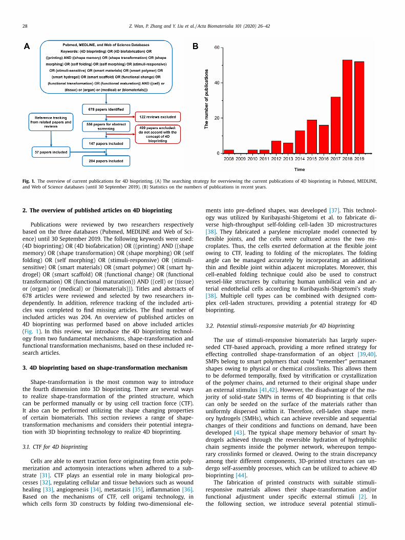
Fig. 1. The overview of current publications for 4D bioprinting. (A) The searching strategy for overviewing the current publications of 4D bioprinting in Pubmed, MEDLINE,
and Web of Science databases (until 30 September 2019). (B) Statistics on the numbers of publications in recent years.
m
o
v
[
fl
c
o
a
t
c
v
t
[
p
b
3
s
e
S
s
t
o
a
j
c
u
o
c
d
d
c
r
a
d
b
r
f
t
2. The overview of published articles on 4D bioprinting
Publications were reviewed by two researchers respectively
based on the three databases (Pubmed, MEDLINE and Web of Sci-
ence) until 30 September 2019. The following keywords were used:
(4D bioprinting) OR (4D biofabrication) OR ((printing) AND ((shape
memory) OR (shape transformation) OR (shape morphing) OR (self
folding) OR (self morphing) OR (stimuli-responsive) OR (stimuli-
sensitive) OR (smart materials) OR (smart polymer) OR (smart hy-
drogel) OR (smart scaffold) OR (functional change) OR (functional
transformation) OR (functional maturation)) AND ((cell) or (tissue)
or (organ) or (medical) or (biomaterials))). Titles and abstracts of
678 articles were reviewed and selected by two researchers in-
dependently. In addition, reference tracking of the included arti-
cles was completed to find missing articles. The final number of
included articles was 204. An overview of published articles on
4D bioprinting was performed based on above included articles
( Fig. 1 ). In this review, we introduce the 4D bioprinting technol-
ogy from two fundamental mechanisms, shape-transformation and
functional transformation mechanisms, based on these included re-
search articles.
3. 4D bioprinting based on shape-transformation mechanism
Shape-transformation is the most common way to introduce
the fourth dimension into 3D bioprinting. There are several ways
to realize shape-transformation of the printed structure, which
can be performed manually or by using cell traction force (CTF).
It also can be performed utilizing the shape changing properties
of certain biomaterials. This section reviews a range of shape-
transformation mechanisms and considers their potential integra-
tion with 3D bioprinting technology to realize 4D bioprinting.
3.1. CTF for 4D bioprinting
Cells are able to exert traction force originating from actin poly-
merization and actomyosin interactions when adhered to a sub-
strate [31] . CTF plays an essential role in many biological pro-
cesses [32] , regulating cellular and tissue behaviors such as wound
healing [33] , angiogenesis [34] , metastasis [35] , inflammation [36] .
Based on the mechanisms of CTF, cell origami technology, in
which cells form 3D constructs by folding two-dimensional ele-
ents into pre-defined shapes, was developed [37] . This technol-
gy was utilized by Kuribayashi-Shigetomi et al. to fabricate di-
erse high-throughput self-folding cell-laden 3D microstructures
38] . They fabricated a parylene microplate model connected by
exible joints, and the cells were cultured across the two mi-
roplates. Thus, the cells exerted deformation at the flexible joint
wing to CTF, leading to folding of the microplates. The folding
ngle can be managed accurately by incorporating an additional
hin and flexible joint within adjacent microplates. Moreover, this
ell-enabled folding technique could also be used to construct
essel-like structures by culturing human umbilical vein and ar-
erial endothelial cells according to Kuribayashi-Shigetomi’s study
38] . Multiple cell types can be combined with designed com-
lex cell-laden structures, providing a potential strategy for 4D
ioprinting.
.2. Potential stimuli-responsive materials for 4D bioprinting
The use of stimuli-responsive biomaterials has largely super-
eded CTF-based approach, providing a more refined strategy for
ffecting controlled shape-transformation of an object [39 , 40] .
MPs belong to smart polymers that could “remember” permanent
hapes owing to physical or chemical crosslinks. This allows them
o be deformed temporally, fixed by vitrification or crystallization
f the polymer chains, and returned to their original shape under
n external stimulus [41 , 42] . However, the disadvantage of the ma-
ority of solid-state SMPs in terms of 4D bioprinting is that cells
an only be seeded on the surface of the materials rather than
niformly dispersed within it. Therefore, cell-laden shape mem-
ry hydrogels (SMHs), which can achieve reversible and sequential
hanges of their conditions and functions on demand, have been
eveloped [43] . The typical shape memory behavior of smart hy-
rogels achieved through the reversible hydration of hydrophilic
hain segments inside the polymer network, whereupon tempo-
ary crosslinks formed or cleaved. Owing to the strain discrepancy
mong their different components, 3D-printed structures can un-
ergo self-assembly processes, which can be utilized to achieve 4D
ioprinting [44] .
The fabrication of printed constructs with suitable stimuli-
esponsive materials allows their shape-transformation and/or
unctional adjustment under specific external stimuli [2] . In
he following section, we introduce several potential stimuli-

Z. Wan, P. Zhang and Y. Liu et al. / Acta Biomaterialia 101 (2020) 26–42 29
Fig. 2. Schematic illustrations of physical stimuli-responsive biomaterials. (A) Thermoresponsive properties of electrospun pNIPAM-PCL bilayer. No folding is observed for
the bilayer at room temperature. The self-fold microtubes form at body temperature. (B) Schematic illustrations of humidity-responsive self-fold bilayer in water. The bio-
compatible bilayer consisting of two layers with different swelling characteristics in aqueous solutions, constructing spontaneous deformation to transform a tubular tissue
architecture. (C) 3D cell culture with magnetic-based levitation. F m : magnetic force. F g : gravity. Magnetic force and gravity guide cells to assemble at the levitation height
and form 3D cell spheriods. (D) Thermal gelation of chitosan solution in the presence of β-glycerophosphate (GP). The solution exhibits liquid state at 25 °C and solidifies to
forma gel at 37 °C. Cationic chitosan chains are linked closer to the negatively charged GP molecules. The decrease in electrostatic repulsion amongst CS–CS chains, increase
in electrostatic attraction between CS–GP and formation of hydrogen bonds are responsible for gelation without precipitation. (E) Schematic illustrations of the UV-assisted
light-responsive polymer cross-linking process. The cross-linked composites networks invoke the shape-transformation of printed structures.
r
t
t
c
s
3
q
b
t
g
c
s
t
t
T
e
o
d
p
t
a
c
o
i
u
a
g
A
s
p
m
w
p
p
t
c
e
m
c
a
esponsive materials, their self-assembly or shape memory abili-
ies, and their potential uses in 4D bioprinting. According to the
ype of the stimuli to arouse 4D procedure, these smart materials
an be classified into physical, chemical, and biological responsive
timuli-responsive materials Table 1 .
.2.1. Physical stimuli-responsive materials
Temperature-responsive materials : Temperature is the most fre-
uently used physical stimulus to achieve shape-transformation in
ioprinted structures [45–47] . A series of thermoresponsive ma-
erials have been developed based on the mechanism of thermal
elation. When the environmental temperature is below the low
ritical solution temperature, the polymeric chains adopt exten-
ion mode and transform into solution phase. However, contrac-
ion of polymeric chains turns the polymer into a gel state when
emperature is above the low critical solution temperature [48–50] .
his mechanism has been used to fabricate cell-laden bilayers that
xhibit reversible folding/unfolding deformation under decreased
r elevated temperature, respectively [51–53] . For example, Hen-
rikson’s group fabricated a thermo-controlled 3D shape-memory
olyurethane scaffold that can change shape with time during cul-
ure [54] . The transformation temperature of polyurethane is 32 °C
rnd a predetermined mechanical strain can be applied to the seed
ells during the shape recovery procedure, directing the behaviours
f cells seeded on the scaffolds.
A classical thermoresponsive material is poly( N -
sopropylacrylamide) (pNIPAM) [47] . For instance, Luo et al.
sed 4-hydroxybutyl acrylate (4HBA) as a crosslinker to fabricate
n elastic porous pNIPAM hydrogel that exhibits rapid and pro-
rammable locomotion prompted by external stimuli [55] , and
psite et al. fabricated self-folding multi-layer porous electrospun
caffolds using thermoresponsive poly(caprolactone) (PCL) and
NIPAM ( Fig. 2 (A) [56] . These pNIPAM-based thermoresponsive
aterials provide a wide range of possibilities for bioprinting,
hile they still have essential limitations, such as poor biocom-
atibility, hydrophobicity, and undegradability [57] . Moreover,
NIPAM generally requires relatively high transformation tempera-
ures, leading to insufficient cell viability and function. Employing
opolymers with pNIPAM may address these limitations. For
xample, cell viability can be maintained by reducing the transfor-
ation temperature to around 37 °C, and cell adhesion and growth
an be improved by combining with bioactive peptides, such as
rginine-glycine-aspartic acid (RGD) [8] .
Another transformation principle of thermoresponsive mate-
ials is based on changes in their wettability and solubility

30 Z. Wan, P. Zhang and Y. Liu et al. / Acta Biomaterialia 101 (2020) 26–42
l
c
p
a
c
p
p
n
g
s
t
i
t
e
t
s
d
u
t
e
s
a
r
s
s
p
s
a
b
m
c
(
a
c
F
c
g
t
n
p
h
f
t
i
b
l
c
d
h
p
3
o
i
l
b
c
m
i
c
o
t
o
with temperature [58 , 59] . For example, Inonv’s group utilized the
sol-gel transition of gelatin to fabricate a temperature-triggered
gelatin-polycaprolactone(PCL) bilayer [60] . The self-rolled poly-
mer bilayer folds at room temperature (22 °C). The photoinitiator-
modified gelatin does not undergo dissolution and is held by non-
photocrosslinked gelatin, preventing the bilayer from folding at
37 °C. Inonv’s group also demonstrated another gelatin-PCL bilayer
in which the gelatin layer swells and generates stress in aqueous
environments [61] . When the PCL layer is in crystalline state at
room temperature, the gelatin layer cannot bend it, while increas-
ing temperature softens the PCL layer and the bilayer undergoes a
folding process. These mechanisms, based on the reversible struc-
ture switching of PCL upon melting and crystallization, is similar to
the reaction of SMPs under increasing temperature, and mechani-
cal stress is induced by swelling of the hydrogel (gelatin) layers.
Any hydrogel can be used to replace gelatin and any hydropho-
bic polymer with an appropriate softening point can replace the
PCL. Furthermore, all the polymers used in these studies are com-
patible and biodegradable [57] . The cells adsorbed or encapsulated
in these constructs remain alive for a considerable period of time,
providing new options for 4D bioprinting.
Humidity-responsive materials : Humidity-responsiveness widely
exists in natural systems, where plants such as pinecones and
wheat utilize humidity shifts to trigger structural transformations
in order to disperse their seeds under beneficial conditions [62 , 63] .
These phenomena have inspired the development of humidity-
responsive materials that change their shapes and sizes during
swelling and shrinking processes upon variations in humidity
( Fig. 2 (B)) [64 , 65] .
For instance, Jamal et al. have reported the spontaneous defor-
mation of a poly(ethylene glycol) biocompatible bilayer consisting
of two cell-laden layers with different swelling characteristics in
aqueous solutions, constructing a series of anatomical microscale
geometrical structures [66] . Zhang et al. combined a poly(ethylene
glycol)-conjugated azobenzene derivative (PCAD) with agarose (AG)
(PCAD@AG) to fabricate a new biomaterial that was capable of
shape reconfiguration upon humidity change [67] . After combin-
ing pure agarose with PCAD, its rate of absorption decreased while
the rate of desorption enhanced. This property is critical to the fast
locomotion of PCAD@AG.
Cellulose stearoly esters (CSEs) also can be used to fabricate
moisture-responsive, self-standing films [68] . As a result of the ab-
or desorption of water molecules, CSE films with the low degree of
stearoyl substitution of 0.3%, labeled CSE0.3, could fold or unfold to
exhibit rhythmical bending motions, while CSE3 (stearoyl substitu-
tion of 3%) is hydrophobic and exhibits thermoresponsive proper-
ties. Humidity-responsive bilayer films with hydrophobic surfaces
were obtained by spraying CSE3 nanoparticles onto the surface of
CSE0.3 films. This kind of bilayer can perform fast reversible bend-
ing motions and continuous shape-transformation in solution.
These technologies represent the earliest versions of 4D print-
ing, where the concept of multilayer structures was compati-
ble with the future design of 4D bioprinting using biocompati-
ble humidity-responsive materials. However, the culture environ-
ment of cells should be maintained under constantly high humid-
ity and specific osmotic pressure. Therefore, the degree of shape-
transformation may be limited due to humidity/osmotic pressure
limitations. These challenges could be addressed by adjusting the
sensitivity of humidity-responsive materials to within the range of
cell endurance [8] .
Electro-responsive materials : Most electro-responsive materials
are polyelectrolyic polymers, which can swell, shrink, or fold un-
der an external electric field. The properties of these materials
can be regulated by the direction or strength of the electric field
[69] . These developed electrically conductive biomaterials could
provide new insights into biomedical applications and drug de-
ivery [70–73] . Moreover, some hydrogels containing electrically
onductive polymers, such as poly(pyrrole)s, poly(aniline)s, and
oly(thiophene)s, can exhibit favorable biocompatibility and print-
bility, giving potential for 4D bioprinting [74 , 75] . For example, a
onductive electroactive hydrogel was fabricated by combining 3D
rinting with polypyrrole interfacial polymerization has been re-
orted [76] . The printed constructs could be applied for developing
ew bioelectronics interfaces and neuroprosthetic devices.
The electro-responsive carbon-based nano-biomaterials, such as
raphene and carbon nanotubes (CNTs), have also attracted exten-
ive attention in recent years as tools to investigate and control
he biology and fate of stem cells due to their unique mechan-
cal properties, adjustable surface chemistry, and favorable elec-
rical conductivity, which have more advantages for nerve tissue
ngineering [77 , 78] . Nanoparticles of these materials have the po-
ential to be used as bioinks for 4D bioprinting [79–81] . For in-
tance, Servant et al. designed a graphene-based macroporous hy-
rogel matrix that is able to control the release of small molecules
nder particular electrical stimulation via reversible deswelling of
he hybrid gel [82] . Moreover, CNTs also show desired mechanical,
lectrical, and cytocompatible properties for 4D bioprinting. For in-
tance, Shin’s group has dispersed CNTs into gelatin methacryloyl
nd hyaluronic acid bioinks to facilitate the manufacture of elabo-
ate foldable biosensors and functionalized tissue-engineering con-
tructs [81] . The in vitro and in vivo studies showed that printed
caffolds coated with graphene and carbon nanotubes nanocom-
osites could accelerate the osteogenic differentiation of seeded
tem cells obviously [83–85] . In conclusion, these biocompatible
nd electrically conductive carbon-based nano-biomaterials could
e used to fabricate stimuli-responsive 4D architectures, providing
ore possibilities for neural and bone tissue regeneration [86] .
Magnetic responsive materials : Magnetic responsive materials
onsist of ferromagnetic or paramagnetic magnetic nanoparticles
MNPs) that can respond to magnetic fields [87] . These materi-
ls have been widely used in biomedical applications, such as
ontrolled drug release system and tissue engineering [88–92] .
or example, a magnetic responsive drug delivery system can be
onstructed by combining Fe 3 O 4 nanoparticles with polyethylene
lycol/agar hydrogel networks, representing a promising alterna-
ive for soft tissue injury treatment [93] . In addition, the Fe 3 O 4
anoparticles can also be encapsulated into the organogel to pre-
are a magnetic gel with remarkable magnetic responsive and self-
ealing properties, which shows favorable rheological properties
or bioprinting [94] .
Magnetic 3D bioprinting technology, which controls the orien-
ation and assembly of anisotropic micro-biomaterials as build-
ng blocks during the printing process, has been proposed as a
iofabrication strategy for generating innervated secretory epithe-
ial organoids [2 , 95] . By magnetizing with magnetic nanoparticles,
ells in monolayers arranged spatially with magnet dots to pro-
uce 3D spheroids. These magnetic 3D bioprinting spheroids ex-
ibit higher cell viability and steadier intracellular activity com-
ared with magnetic-nanoparticle-free spheroids. This magnetic
D bioprinting technology also was used to pattern human my-
metrium cells into rings that then were monitored for contractil-
ty and function over time [96] .
Recently, the magnetic assembly of cells or tissue spheroids, or
iving materials has been studied, providing great potential for 4D
ioprinting ( Fig. 2 (C)) [97] . The paramagnetic properties of MNPs
ould be used to realize magnetic levitational assembly of cells or
icro-tissues. For example, Souza et al. utilized the magnetic lev-
tation of cells to establish a 3D tissue culture system in which
ells or a matrix are labeled with gold, iron oxide nanoparticles,
r filamentous phages. The geometry of the cell spheroids is con-
rolled via the spatial control of the magnetic field, and co-cultures
f different types of cells could be realized [98] . Furthermore, Sun

Z. Wan, P. Zhang and Y. Liu et al. / Acta Biomaterialia 101 (2020) 26–42 31
Table 1
Stimuli-responsive shape-memory materials and methods.
Stimulus Materials/methods Mechanisms Application Pros Cons References
Temperature poly( N -
isopropylacrylamide)
(pNIPAM)
Sol-gel transition Cell-laden bilayers
for soft tissue
Easy availability;
High sensitivity
to temperature
Not well
biocompatibility;
Hydrophobicity;
Low
biodegradability;
Low control
precision.
Wei et al. [48]
Bakarich et al.
[47]
Breger et al. [49]
Pei et al. [50]
Zakharchenko
et al. [51]
Luo et al. [55]
Apsite et al. [56]
Polyurethane Shape
transformation
Cell-laden scaffolds
for bone, muscle,
cardiovascular
tissue repair
Suitable shape
transition
temperature
Complex
mechanical
stimuli control
Hendrikson et al.
[54]
Gelatin-
Polycaprolactone(PCL)
Sol-gel transition;
Wettability and
solubility
differences
between two
materials
Bilayers,
Cell-laden
bioscaffolds for
tissue
engineering
Compatible;
Biodegradable
Unsuitable for
complex shape
transformation
Stroganov et al.
[60 , 61]
Ionov L et al.
[57]
Humidity Poly-ethylene glycol (PEG) Swelling properties Cell-laden bilayers Biocompatibility Humidity/osmotic
limitation
Jamal et al. [66]
Zhang et al. [67]
Agarose Water
sorption/water
desorption
Hybrid films Native; Strong
hygroscopic
Humidity/osmotic
limitation
Zhang et al. [67]
Cellulose stearoly esters
(CSEs)
Water
sorption/water
desorption
Bio-sensors or
bio-actuators
Biocompatible Humidity/osmotic
limitation
Zhang et al. [68]
Electric field Polypyrrole(PPy);
Polyaniline;
Polythiophene
Electrically
conductive
properties
Drug delivery;
Biomimetic or
bioinspired
systems
Improved
conductive
characteristics
Undesirable
biocompatibility
Green et al. [74]
Song et al. [75]
Fantino et al.
[76]
Carbon-based
nanobiomaterials
(graphene, carbon
nanotubes)
Electro-responsive
properties
Drug delivery;
Biosensors;
Cell-laden
scaffolds for
bone and nerve
tissue
engineering
Suitable
mechanical
properties;
Adjustable
surface
chemistry;
Excellent
electrical
conductivity;
Cytocompatibility.
Restricted
electrical
stimulation
Ahadian et al. [77]
Ramon et al. [78]
Shin et al. [81]
Servant et al.
[82]
Miao et al. [184]
Magnetic
field
Magnetic nanoparticles
(MNPs)
Magnetic-
responsive
properties;
Self-assembly
Drug delivery;
Cell/tissue
spheroids
(epithelial
organoids;
myomethrium
rings; 3D
embryoid body;
microvascular-
like
structures)
Remote control;
High control
precision;
Harmlessness to
cells
Complex control
system;
Challenging
sufficient
magnetic field
gradient
achievement
Zhang et al. [90]
Lalitha et al. [94]
Souza et al.
[96 , 98]
Du et al. [97]
Sun et al. [99]
Adine et al. [95]
Gadolinium (Gd 3 + ) Magnetic levitation Cell/Tissue
spheroids;
Drug delivery;
Cancer therapy
Low toxicity;
Noninvasiveness;
Easy-to-use
Undesirable
cytotoxic effect
at high
concentration
Tocchio et al. [100]
Parfenov et al.
[102]
Fattah et al.
[103]
Turker et al.
[104]
light Photoinitiators
(e.g.: benzophenone,
hydrazone bonds,
methylene blue)
Photocrosslink;
Dynamic
covalent
chemistry
Soft robotics;
Cell delivery;
in-situ
photo-curing
structures
High shape
fidelity;
Stability
relaxation;
Cytocompatibility
Low light tissue
penetration
Wei et al. [113]
Kuang et al.
[114]
Photodegradable
moieties(e.g.: coumarin,
o-nitrobenzyl ether
groups)
Programmable
photodegrada-
tion
Cell delivery;
Cell-laden
structures;
3D vascular
networks
Precise multistaged
light program
Low tissue
penetration;
Phototoxicity.
Griffin et al. [116]
Arakawa et al.
[117]
Acoustic Surface acoustic waves
(SAWs)
Acoustic force
assembly
3D cellular
patterns and
constructs
Non-invasive;
Cytocompatible;
Biocompatible
Limited line
patterns;
Micro
dimensions only
Nasser et al. [119]
( continued on next page )

32 Z. Wan, P. Zhang and Y. Liu et al. / Acta Biomaterialia 101 (2020) 26–42
Table 1 ( continued )
Stimulus Materials/methods Mechanisms Application Pros Cons References
Near field standing waves Acoustic radiation
forces
3D brain-like
constructs
Easy to use;
Cytocompatible
Limited cell
populations;
Homogenous
tissues only
Bouyer et al. [122]
Ultrasound Ultrasound-
disrupted
ionically
crosslink
Drug release;
Chemotherapy
Non-invasive;
Biocompatible
Need further test
mimicking
Huebsch et al.
[121]
pH Amino acids (e.g.:
polypeptide, I-arginine
grafted alginate)
Noncovalent
crosslink;
pH determined
swelling
properties
β-sheet structure;
Protein delivery
Tunable
morphology and
mechanical
properties
Specific pH
environments;
Unsuitability for
complex shape-
transformation
Clarke et al. [128]
Eldin et al. [129]
Chitosan-based polymers
(N-succinyl chitosan
grafted polyacrylamide,
chitosan-based
tripolyphosphate)
pH determined
swelling
properties;
Electrostatic
crosslink
Drug delivery;
Bone
regenerative
therapies
Better controlled
release of drugs;
Tunable
mechanical
properties
Limited pH change
range
Mukhopadhyay
et al. [131]
Xu et al. [132]
Ion Zn 2 + /Ca 2 + -responsive
hydrogels
Reversible
chemical
crosslink
Cell-laden shape
memory
structures;
Hollow
self-folding
tubes (blood
vessels)
Suitable strength;
Tunable
mechanical
properties;
Biocompatibility
Difficult to control
in vivo
Liu et al. [135 , 136]
Lonov et al.
[140]
Biological Polypeptides;
Polynucleotides
Hydrogen bond
crosslinks or
ionic interactions
Shape memory
hydrogels for
drug delivery,
programmed cell
adhesion
matrices
Biodegradable and
biocompatible
Low mechanical
strength
Skrzeszewska et al.
[142]
Hao et al. [143]
Guo et al. [144]
Todhunter et al.
[145]
Enzymes (e.g.: matrix
metalloproteinase,
thrombin, Sortase A,
horseradish
peroxidase)
Enzymatic
mediated
crosslink
Drug delivery;
Bioinspired
multi-activities
object for blood
vessels /bone
/cartilage tissue
regeneration
Biological
substance; Cyto-
compatibility;
Suitable
degradation;
Tunable
morphology
Complex
regulation;
Unidentified
immune
response
Kim et al. [146]
Broguiere et al.
[147]
Costa et al. [148]
Yan et al. [149]
c
b
i
h
t
a
m
4
a
t
e
t
b
s
b
t
P
t
r
m
h
A
d
n
c
h
g
T
et al. presented an innovative strategy based on magnetic alginate
microfibers as scaffolding elements to fabricate microvascular-like
structures, allowing direct cell-to-cell interaction, which is essen-
tial for the formation of vessel-like structures [99] .
Another universal system for levitating and assembling cells
using a gadolinium (Gd
3 + )-based nonionic paramagnetic agent
has been reported for fabricating scaffold-free living architectures
[100 , 101] . Parfenov et al. developed a prototype device equipped
with magnetic levitation capabilities using gadolinium in culture
media [102] , Similarly, Fattah et al. demonstrated a new technol-
ogy for fabricating 3D cellular structures, co-culturing breast can-
cer MCF-7 cells and human umbilical vein endothelial cells utiliz-
ing a magnet array to manipulate diamagnetic cells in a param-
agnetic gadopentatic acid medium [103] . Three different gadolin-
ium chelates were utilized to magnetize the cell culture envi-
ronment, thus realizing scaffold-free levitation and assembly of
cells [104] .
Moreover, Tasoglu’s group manipulated and assembled a cell-
encapsulating poly(ethylene glycol) hydrogel in magnetic fields us-
ing permanent magnets by exploiting the paramagnetic character-
istic of free radicals without using magnetic nanoparticles [105] .
They also fabricated tunable and magnetic self-assembly microgels
using a cell-laden gelatin methacryloyl hydrogel that is paramag-
netized upon submerging in a stable free-radical solution [106] .
In conclusion, these innovative magnetic levitational assembly sys-
tems show enormous promise for complex cellular assemblies and
tissue engineering, providing a new paradigm for the application
of 4D bioprinting.
Photoresponsive materials : Photoresponsive materials could cap-
ture externally applied optical signals and convert them into me-
hanical responses. Photoresponsive biomaterials can be activated
y light in a relatively wide wavelength range, including near-
nfrared (NIR), infrared (IR), and ultraviolet (UV) regions, which
ave been broadly applied in biomedical applications such as con-
rolled drug release and tissue engineering [107–112] .
Photoisomerization and photodegradation of polymer chains
re the most common response mechanisms for light-responsive
aterials, which have been widely applied to fabricate active
D shape-changing structures. For instance, Wei et al. printed
tubular shape-memory poly(lactic acid) (PLA) structure by in-
roducing a UV crosslinking agent ( Fig. 2 (E)) [113] , while Kuang
t al. reported a UV-light-assisted printing bioink containing ure-
hane diacrylate and a linear semi-crystalline polymer [114] . The
ioink showed capability of high strain shape memory as well as
elf-healing feature, paving the way for the development of 4D
ioprinting.
In addition, photodegradation of biomaterials provides real-
ime temporal and spatial control during hydrogel degradation.
hotodegradation of biomaterials can be induced by exploiting
heir photoresponsive features, producing dynamic hydrogel envi-
onments [115] For instance, Griffin et al. added photodegradable
oieties, such as o-nitrobenzyl ether groups and coumarin, into
ydrogels to tune the rate of its biodegradation [116] . Furthermore,
rakawa et al. exploited a programmable biomaterial photodegra-
ation strategy to construct 3D multicellular endothelial vascular
etworks within cell-laden hydrogels [117] . Networks of mi-
rochannels with similar sizes and scales to those of the native
uman vascular systems could be easily generated through pro-
rammable 4D control using multiphoton lithography technology.
herefore, these photoresponsive 4D bioinks show the potential to

Z. Wan, P. Zhang and Y. Liu et al. / Acta Biomaterialia 101 (2020) 26–42 33
m
d
i
u
p
l
t
c
m
p
a
i
b
f
s
a
b
s
u
F
m
o
a
a
d
a
4
t
w
M
e
3
c
c
p
p
c
i
t
e
p
v
h
r
p
t
[
q
[
l
t
d
h
e
o
r
p
a
h
[
s
t
s
r
a
a
s
p
m
i
t
p
s
o
[
s
o
u
fl
c
c
b
a
s
i
e
F
p
a
m
p
C
c
a
s
p
d
I
n
s
3
m
h
o
m
b
w
f
s
g
c
s
a
s
s
i
n
s
f
b
[
s
e
imic the dynamic characteristics of natural extracellular matrix
egradation.
However, the strong attenuation of light by biological tissues
s a challenge. This challenge may be addressed by exploring the
se of ultra-IR light, which has lower living tissue absorbency and
hototoxicity as well more effective tissue penetration than UV
ight. Meanwhile, multiple kinds of photo-initiators have limita-
ions in applications of tissue engineering due to their undesired
ytotoxicity. New photo-polymerized systems with significant
itigated adverse effects on cellular metabolic activities and
roliferative capacities have been developed, giving potential for
pplications including cell encapsulation and biofabrication of
njectable hydrogels [118] .
Acoustic responsive materials : Acoustic responsive materials have
een used in drug release systems and tissue engineering. Acoustic
orce patterning is a potential technology that can be used to con-
truct location-controlled cell platforms in a contactless, rapid, and
ccurate manner [119 , 120] . Huebsch et al. developed an alginate-
ased, self-healing, ultrasound-responsive hydrogel for near-digital
patiotemporal control of drug delivery and used ultrasound stim-
lation to disrupt the ionic crosslinks within the hydrogels [121] .
urthermore, Naseer et al. reported a biological acoustic-force-
ediated micro-patterning technique to achieve rapid arrangement
f cells within gelatin methacryloyl hydrogels by using surface
coustic waves [119] . A multilayered 3D brain-like construct was
lso fabricated via levitating neuroprogenitors through acoustic ra-
iation forces based on near field standing waves, indicating that
coustic responsive hydrogels have the potential to be applied to
D bioprinting in tissue engineering [122] . These bioacoustic levi-
ational assembly technologies are non-invasive and biocompatible,
hile limited to line patterns and homogenous cell populations.
ore sophisticated methods are needed to fabricate complex het-
rogeneous tissue cellular architectures.
.2.2. Chemical stimuli-responsive materials
pH-responsive materials : pH-responsive materials containing
hemical groups (carboxyl, pyridine, sulfonic, phosphate, etc.) that
an release or accept protons with changing pH have been ap-
lied to the fabrication of self-assembled structures [123] . These
H-responsive materials exhibit globule-to-coil transition at a criti-
al pH value. When the functional group of the polymer is neutral-
zed, the polymer chains are converted into globule structures from
heir coil forms in electrostatically repulsion states [124] . Nadgorny
t al. described a 3D-printed pH-responsive construct by using
oly(2-vinylpyridine) [125] . These 3D-printed structures exhibit re-
ersible and dynamic pH-dependent swelling characteristics. The
ydrogel can be used as a flow-regulating valve to regulate flow
ate by controlling the changes of pH. Few synthetic pH-responsive
olymers have been reported in the field of bioprinting, while
he applications of natural proteins have gained great attention
124 , 126 , 127] . For instance, three different charge pentapeptide se-
uences were used to fabricate a robust pH-responsive hydrogel
128] . The mechanical properties of the hydrogel could be regu-
ated significantly by tuning the charge distribution and concentra-
ion of the pentapeptide sequences. Moreover, a pH-responsive hy-
rogel using L -arginine-grafted alginate (Arg-g-Alg) hydrogel beads
as been synthesized as a new type of carrier for protein deliv-
ry at specific pH environments [129] . The swelling characteristics
f the hydrogel beads were determined by pH changes. These pH-
esponsive hydrogels with tunable stiffness and adjustable mor-
hology are promising for applications in injectable drug delivery
nd tissue engineering.
Meanwhile, a series of pH-responsive chitosan hydrogels also
ave been widely used in drug delivery and bioprinting ( Fig. 3 (A))
130 , 131] . Xu’s group reported a chitosan-based tripolyphosphate
caffold with regulated primary amine content, which influenced
he pH-responsive resorption and made the scaffold enable to ab-
orb or release water under the changes of pH [132] . These pH-
esponsive materials modified scaffolds with tunable morphology
nd mechanical properties possess the possibility for biomedical
pplications, providing new possibilities for 4D bioprinting.
Ion-sensitive hydrogels : Many recent studies have reported
trategies for the fabrication of sufficiently strong scaffolds to sup-
ort clinical-scale cell-laden structures [133] . Crosslinking with
ultivalent ions such as Ca 2 + and Zn
2 + has been exploited
n bioprinting to obtain scaffolds with suitable strength and
unable mechanical properties ( Fig. 3 (B)) [134 , 135] . For exam-
le, Liu et al. fabricated a reversible shape-memory ultrahigh
trength cell-laden hydrogel utilizing the dipole-dipole interactions
f poly(acrylonitrile) chains that are reversible in response to Zn
2 +
136] . They also utilized imidazole-zinc ion coordination to synthe-
ize another shape memory hydrogel [137] . The permanent shapes
f these printed constructs can be restored by extracting zinc ions
sing chelating agents, and this memory process is reversible. The
at cell-laden hydrogel sheet can be fold into a temporary tubular
onstruct and fixed in culture medium containing zinc ions.
In addition, a crosslinking mechanism involving sodium-
alcium ion exchange between alginate and calcium chloride has
een widely applied in bioprinting [138 , 139] . Hydrogels based on
hydrogen bonding/calcium ion crosslinking mechanism exhibited
hape memory abilities in response to reversible Ca 2 + crosslink-
ng. Such hydrogels with weaker hydrogen bonding interactions
xhibit sharp volume changes triggered by calcium ions [135] .
urthermore, Lonov et al. have reported an advanced 4D bio-
rinting approach to cell-laden, stimuli-triggered, shape-changing
lginate and hyaluronic acid hydrogels, which were modified with
ethacrylate groups to endow them with photo-crosslinkable
roperties [140] . The photo-crosslinking hydrogel exhibited strong
a 2 + -ion-concentration dependent rheological properties. Cells
an tolerate calcium ion changes in the extracellular environment,
llowing Ca 2 + ions to act as biocompatible shape-transformation
timuli. This bioprinting strategy based on Ca 2 + stimuli-triggered
olymers could be used to print hollow tubular structures with
iameters equivalent to those of the narrowest blood vessels.
n a word, these ion-sensitive crosslinking hydrogels pave the
ew ways for presentation of tunable cell-laden shape-morphing
tructures for tissue engineering and 4D bioprinting applications.
.2.3. Biological stimuli-responsive materials
Besides gelation through ion-crosslinking processes, shape-
emory hydrogels exhibit self-assembly abilities via reversible
ydrogen bond crosslinks or ionic interactions with polypeptides
r polynucleotides [141] . For example, a thermally induced shape-
emory hydrogel was formed by utilizing biocompatible and
iodegradable recombinant telechelic polypeptides that combine
ith random coil-like middle blocks and collagen-like end blocks,
orming of triple helices and allowing the fixation of temporary
hapes upon cooling [142] . The programmed shape of hydro-
els containing lysine residues was achieved through chemical
rosslinking of random coils. The triple helix led to permanent
hape recovery upon opening of the crosslinks during heating
nd melting. Similarly, by utilizing DNA as a programmable and
equence-specific glue, shape-controlled hydrogel units can be
elf-assembled into diverse prescribed structures in aqueous or
nterfacial agitation systems [143] . In addition, the cytosine-rich
ucleotide sequences generated an i-motif construct through
elf-assembly at low pH and dissociated into random coil con-
ormations at pH 8, leading to a reversible shape-transformation
etween a “quasi-liquid” state and a predesigned shape structure
144] . Furthermore, Todhunter’s group performed multicellular tis-
ue organizations utilizing a DNA-programmed cell-assembly strat-
gy [145] . This technology exploits dissociated cells functionalized

34 Z. Wan, P. Zhang and Y. Liu et al. / Acta Biomaterialia 101 (2020) 26–42
Fig. 3. Schematic illustrations of chemical stimuli-responsive biomaterials. (A) Schematic presentation of swelling and drug release pattern of the pH-responsive hydrogel. (B)
A schematic diagram depicting the mechanism underlying a small number of calcium/zinc ions triggered reversible shape memory behavior. The functional groups coordinate
with Ca 2 + /Zn 2 + , while the Ca 2 + /Zn 2 + functional group linkages are dissociated in EDTA solution.
p
c
u
m
a
f
c
c
4
m
o
o
p
3
o
r
s
a
t
chemically with degradable oligonucleotides that can adhere to
other cell surfaces labeled with complementary DNA sequences.
The rapid, reversible, and specific cell adhesion induces layer-by-
layer DNA-programmed assembly, fabricating 3D tissue constructs
with programmed shapes, sizes, and spatial heterogeneities above
a template, providing a potential approach for 4D bioprinting.
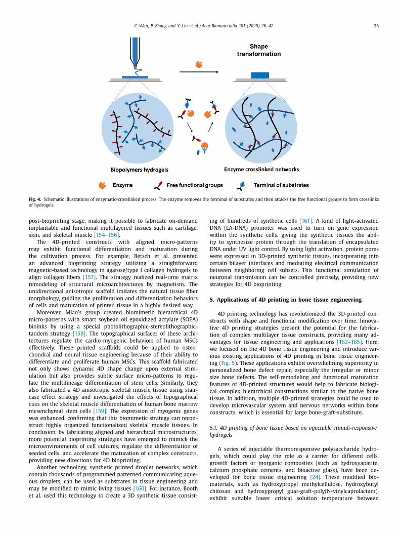
Moreover, active biological molecules, such as antibodies and
enzymes, can be entrapped in 3D objects to obtain 4D structures
( Fig. 4 ). Enzymes play important roles in processes of many
biochemical reactions. The human body has multiple kinds of en-
zymes that can be utilized as triggering factors for shape memory
properties of hydrogels. The substrates of enzymes should serve as
functional side groups or crosslinkers of hydrogels [73] . For exam-
ple, matrix metalloproteinase is an important protease related to
the degradation of extracellular matrix components. Matrix met-
alloproteinase sensitive hyaluronic acid-based hydrogels showed
tunable swelling and degradation capacities with dramatic cell
attachment [146] . In addition, the bacterial ligase sortase A (SA)
and its mutated variants have been used as crosslinking enzymes
for hydrogel-based tissue engineering [147] . Modifying hyaluronan
(HA) with SA-substrate peptides achieved near-instantaneous gel
formation of HA. Similarly, Costa et al. demonstrated a fast-gelling
silk fibroin bioink capable of enzymatic crosslinking for 3D bio-
printing [148] . The hydrogels can transform from random coil
onfigurations into β-sheet conformations over time, and can be
sed to fabricate different fine-tuned structures, such as shape-
emory patient-specific implants, with good reliability, resolution,
nd reproducibility [149] . These enzyme-sensitive hydrogels with
ast-crosslinking, suitable degradation and tunable morphology
haracteristics could be applied for tissue defect regeneration and
omplex tissue engineering.
. 4D bioprinting based on functional transformation
echanism
Advanced biology studies have extended the original definition
f 4D bioprinting, which was limited to the geometric change
f 3D-printed objects, to include the transformation of shape,
roperties, and physical, chemical, or biological compositions of
D constructs [150] . The functional transformation and maturation
f 3D-printed cell/tissue constructs over time have also been
egarded as constituting 4D bioprinting [10 , 151 , 152] . Natural tis-
ues and organs are structurally anisotropic and highly organized
rchitectures [3 , 153] . The establishment of biomimetic constructs
hat mimic the native extracellular matrix could guide and sup-
ort the growth and differentiation of stem cells during the
Z. Wan, P. Zhang and Y. Liu et al. / Acta Biomaterialia 101 (2020) 26–42 35
Fig. 4. Schematic illustrations of enzymatic-crosslinked process. The enzyme removes the terminal of substrates and then attacks the free functional groups to form crosslinks
of hydrogels.
p
i
s
m
t
a
m
a
r
u
m
o
m
b
t
t
e
c
d
n
u
l
a
c
c
m
w
s
c
m
m
s
p
c
o
m
e
i
D
w
i
D
w
c
b
n
s
5
s
t
t
v
w
i
i
p
s
f
c
t
d
c
5
h
g
g
c
v
m
c
ost-bioprinting stage, making it possible to fabricate on-demand
mplantable and functional multilayered tissues such as cartilage,
kin, and skeletal muscle [154–156] .
The 4D-printed constructs with aligned micro-patterns
ay exhibit functional differentiation and maturation during
he cultivation process. For example, Betsch et al. presented
n advanced bioprinting strategy utilizing a straightforward
agnetic-based technology in agarose/type I collagen hydrogels to
lign collagen fibers [157] . The strategy realized real-time matrix
emodeling of structural microarchitectures by magnetism. The
nidirectional anisotropic scaffold imitates the natural tissue fiber
orphology, guiding the proliferation and differentiation behaviors
f cells and maturation of printed tissue in a highly desired way.
Moreover, Miao’s group created biomimetic hierarchical 4D
icro-patterns with smart soybean oil epoxidized acrylate (SOEA)
ioinks by using a special photolithographic-stereolithographic-
andem strategy [158] . The topographical surfaces of these archi-
ectures regulate the cardio-myogenic behaviors of human MSCs
ffectively. These printed scaffolds could be applied to osteo-
hondral and neural tissue engineering because of their ability to
ifferentiate and proliferate human MSCs. This scaffold fabricated
ot only shows dynamic 4D shape change upon external stim-
lation but also provides subtle surface micro-patterns to regu-
ate the multilineage differentiation of stem cells. Similarly, they
lso fabricated a 4D anisotropic skeletal muscle tissue using stair-
ase effect strategy and investigated the effects of topographical
ues on the skeletal muscle differentiation of human bone marrow
esenchymal stem cells [159] . The expression of myogenic genes
as enhanced, confirming that this biomimetic strategy can recon-
truct highly organized functionalized skeletal muscle tissues. In
onclusion, by fabricating aligned and hierarchical microstructures,
ore potential bioprinting strategies have emerged to mimick the
icroenvironments of cell cultures, regulate the differentiation of
eeded cells, and accelerate the maturation of complex constructs,
roviding new directions for 4D bioprinting.
Another technology, synthetic printed droplet networks, which
ontain thousands of programmed patterned communicating aque-
us droplets, can be used as substrates in tissue engineering and
ay be modified to mimic living tissues [160] . For instance, Booth
t al. used this technology to create a 3D synthetic tissue consist-
eng of hundreds of synthetic cells [161] . A kind of light-activated
NA (LA-DNA) promoter was used to turn on gene expression
ithin the synthetic cells, giving the synthetic tissues the abil-
ty to synthesize protein through the translation of encapsulated
NA under UV light control. By using light activation, protein pores
ere expressed in 3D-printed synthetic tissues, incorporating into
ertain bilayer interfaces and mediating electrical communication
etween neighboring cell subsets. This functional simulation of
euronal transmission can be controlled precisely, providing new
trategies for 4D bioprinting.
. Applications of 4D printing in bone tissue engineering
4D printing technology has revolutionized the 3D-printed con-
tructs with shape and functional modification over time. Innova-
ive 4D printing strategies present the potential for the fabrica-
ion of complex multilayer tissue constructs, providing many ad-
antages for tissue engineering and applications [162–165] . Here,
e focused on the 4D bone tissue engineering and introduce var-
ous existing applications of 4D printing in bone tissue engineer-
ng ( Fig. 5 ). These applications exhibit overwhelming superiority in
ersonalized bone defect repair, especially the irregular or minor
ize bone defects. The self-remodeling and functional maturation
eatures of 4D-printed structures would help to fabricate biologi-
al complex hierarchical constructions similar to the native bone
issue. In addition, multiple 4D-printed strategies could be used to
evelop microvascular system and nervous networks within bone
onstructs, which is essential for large bone-graft-substitute.
.1. 4D printing of bone tissue based on injectable stimuli-responsive
ydrogels
A series of injectable thermoresponsive polysaccharide hydro-
els, which could play the role as a carrier for different cells,
rowth factors or inorganic composites (such as hydroxyapatite,
alcium phosphate cements, and bioactive glass), have been de-
eloped for bone tissue engineering [24] . These modified bio-
aterials, such as hydroxypropyl methylcellulose, hydroxybutyl
hitosan and hydroxypropyl guar-graft-poly(N-vinylcaprolactam),
xhibit suitable lower critical solution temperature between

36 Z. Wan, P. Zhang and Y. Liu et al. / Acta Biomaterialia 101 (2020) 26–42
Fig. 5. Applications of 4D printing in bone tissue engineering. (A) Injectable thermosensitive hydrogels for 4D bone tissue regeneration: the hydrogel could be injected into
the irregular defect area and transform to gel state under body temperature. (B) 4D printing of bone tissue based on shape-transformation mechanism: a shape memory
scaffold changes its size to occupy the void space, realizing personalized bone defect repair. (C) 4D printing of bone tissue based on the establishment of biomimetic
microenvironment: the 4D printed biomimetic scaffold with modified architectures can induce the functional maturation of neo-bone tissue and promote the osteogenesis
of stem cells, enhancing the formation of new bone tissue.
e
b
c
p
5
e
s
w
p
e
t
t
f
i
i
d
a
a
m
physiological temperature and manipulative room temperature and
could translate into gel state under body temperature [166–169] .
The classical thermoresponsive material, pNIPAM, has been incor-
porated with hyaluronic acid and chitosan to fabricate an injectable
hydrogel for bone tissue regeneration [170–173] . Similarly, chitosan
could form a thermoresponsive hydrogel by combining with β-
glycerophosphate salt, which modulates the hydrogen bonding and
electrostatic, hydrophobic crosslinkings during the gel formation
[22 , 174] . This kind of chitosan hydrogel exhibited desirable in-
jectability and rapid gelation at body temperature ( Fig. 2 (D)). An-
other kind of available thermosensitive material is a copolymer
of poly(lactic acid), poly(ethylene glycol) and poly(glycolic acid),
which converts to a gel state under physiological conditions [175] .
The mechanical strength and load-bearing capacity of these hy-
drogels were improved by including different mineral components,
such as tricalcium phosphate, nano-hydroxyapatite and bioactive
glass [176–179] . These injectable hybrid hydrogels with desirable
rheological feature and in vivo self-setting ability served as de-
sirable carriers for osteoblast cells with improved alkaline phos-
phatase activity and calcium deposition, making them enable to
fill small, irregular-shaped defects and form gel at body tempera-
ture and providing a significant potential in mini-invasive repair of
bone defects. Bioactive cues, such as osteogenic and angiogenetic
growth factors, have been involved into the hydrogel systems to
inhance the differentiations of MSCs, providing new strategies for
one defect repair [180–182] . Therefore, the composite material
an be used as an injectable osteogenic material for orthopedic ap-
lications, providing new insights for clinical translation.
.2. 4D printing of bone tissue based on shape memory scaffolds
4D printing has been used to print hard-tissue constructs. For
xample, shape-recovery polylactide and hydroxyapatite porous
caffolds were obtained by fused filament fabrication [23] , in
hich direct heating stimulated the shape-memory effect. The
olylactide/hydroxyapatite hybrid porous scaffolds with high lev-
ls of shape recovery ability could be used as self-fitting implants
o repair small bone defects. Thus, the shape-transformation fea-
ures of 4D printing constructs may realize personalized bone de-
ect repair. Such biomaterials could be used to repair bone defects
n which the scaffold shape changes to occupy the void space after
mplantation [21] .
Moreover, Miao et al. utilized PCL and crosslinkers with pre-
etermined amounts of castor oil to synthesize smart renew-
ble bio-scaffolds, which exhibit favorable shape-memory effects
nd shaperecovery at physiological temperature [183] . The surface
orphology, shape memory, mechanical properties, biocompatibil-
ty and biodegradability of the synthesized smart polymers were

Z. Wan, P. Zhang and Y. Liu et al. / Acta Biomaterialia 101 (2020) 26–42 37
d
f
s
a
p
m
b
p
t
5
m
o
n
u
m
c
i
n
t
c
T
f
t
p
[
i
v
o
a
m
p
s
5
n
i
t
p
l
s
M
d
t
t
b
w
l
b
i
T
s
t
m
t
b
t
s
c
w
6
w
c
a
e
i
d
g
t
e
s
s
[
t
t
r
o
e
a
i
s
i
[
m
b
s
s
w
t
f
e
d
d
i
e
v
a
o
p
w
t
t
m
p
t
n
i
r
m
h
a
h
m
a
a
a
t
p
o
emonstrated to be satisfactory [184] . Similarly, the authors also
abricated a biocompatible temperature-responsive shape-memory
caffold comprising epoxidized acrylate materials based on renew-
ble soybean oil using a 3D laser printing technique [185] . The
orous scaffolds are biocompatible and exhibit comparable attach-
ent and proliferation abilities to those of multipotent human
one marrow mesenchymal stem cells. Thus, these studies pro-
osed renewable biomedical scaffolds that could potentially con-
ribute to the development of 4D constructs in bone engineering.
.3. 4D printing of bone tissue based on functional transformation
echanism
The biomimetic bony microenvironment can improve the bi-
logical functionality of 3D-printed scaffolds and drive osteoge-
esis of stem cells during the post-bioprinting stage. It inspires
s whether the establishment of this biomimetic microenviron-
ent, which enhanced the functional maturation of 3D-printed
onstructs, would be considered as 4D printing in tissue engineer-
ng. Pati et al. [186] reformed polymeric 3D-printed scaffolds by or-
amenting them with a cell-laden mineralized extracellular matrix
o mimic bony microenvironments. The printed bone structures be-
ame mature after culturing in a rotary flask bioreactor over time.
he results showed that the extracellular-matrix-ornamented scaf-
olds exhibit better osteoinductive and osteoconductive properties
han those of bare 3D-printed scaffolds.
Moreover, the complex hierarchical structures of bone tissue
ossess anisotropic mechanical and electromechanical properties
187] . The smart biomaterial with piezoelectric effect, such as bar-
um titanate, could stimulate the physiological electrical microen-
ironment in response to applied stress and promote the growth of
steoblasts, showing favorable biocompatibility and bone-inducing
bilities [188–190] . This promising development of piezoelectric
aterials could be used to enhance the functional maturation of
rinted constructs during the post-printing stage, presenting new
trategies for 4D bone tissue bioprinting.
.4. 4D-printed bone constructs with blood vessels and nervous
etworks
The major challenge in large bone-graft-substitute engineering
s the regeneration of microvasculature and nervous networks in
he substitute [191–193] . A series of 4D strategies have been pro-
osed to fabricate microvasculature constructs. For example, hol-
ow self-folding tubes with diameters comparable to those of the
mallest blood vessels have been fabricated by combining mouse
SCs with methacrylate alginate and hyaluronic acid hybrid hy-
rogels [140] . In addition, localized and pre-programmed calcifica-
ion and direct fibrin biofilm formation could be triggered through
he entrapment of enzymes within the 4D hydrogel during the
ioprinting processes. The bioinspired 3D constructs in this study
ere composed of bone-like structures surrounding a blood-vessel-
ike structure, making it possible to fabricate vascularized alveolar
one constructs [194] . These enzymes could be used alone or co-
mmobilized to create bioinspired constructs with multi-activity.
his technology was the first to demonstrate the fabrication of
uch 4D-printed constructs with multi-activity, providing a poten-
ial approach for complex bone tissue engineering.
Furthermore, the 4D bioprinting of the electro-responsive bio-
aterials have shown great potential for nerve tissue regenera-
ion. Miao’s group demonstrated a multi-responsive graphene hy-
rid 4D-printed architecture providing multiple nerve regenera-
ion characteristics, such as physical guidance, chemical cues, and
eamless integration [195] . This stimuli-responsive 4D technique
ould be combined with the bone tissue fabrications, paving the
ay for repairing bone defects with nerve damage.
. Future perspectives and current challenges
4D bioprinting, incorporating “time” as the fourth dimension
ithin 3D bioprinting, is expected to allow the creation of compli-
ated structures with on-demand dynamically controllable shapes
nd functions, considering to be the next generation of tissue
ngineering technology [8] . In the last few years, with emerg-
ng development of stimuli-responsive biomaterials and better un-
erstanding of tissue regeneration, 4D bioprinting technology has
ained lots of attention in biomedical area and clinical applica-
ions [53 , 196–201 ]. For example, 4D bioprinting technology provide
normous application prospects in the field of personalized tis-
ue regeneration. The 4D-printed implantation with programmed
hape and size would fit the defect sites with precise geometry
202–205] . The functional transformation of implantation during
he post-printing stage would show bio-mimicking features, facili-
ating the tissue remodeling and maturation. At the same time, the
ecent progress of computational model system has provided new
pportunities to program neo-tissue growth in personalized tissue
ngineering [73 , 206] .
Meanwhile, the transformation features of 4D bioprinting could
lso benefit the treatment of adolescent patients through fabricat-
ng self-growing constructs. For example, Morrison et al. demon-
trated the successful application of personalized 4D-printed med-
cal device for the treatment of pediatric tracheobronchomalacia
164] . The 4D-printed PCL airway splints exhibited optimal adjust-
ent to tissue growth with designed mechanical and degradation
ehaviors over time, which indicates a self-growing feature. The
elf-transformation and self-maturation abilities of 4D-printed con-
tructs would give a new perspective to produce specific implants
ith time-dependent growth behavior, which shows great advan-
ages in the treatment of adolescent patients with congenital mal-
ormation [207] .
In addition, stimuli-responsive cell-assembly and tissue remod-
ling technologies could be used in the clinical applications of drug
elivery and cell therapy [208–210] . An effective magnetic force
riven stem cells delivery system has been applied in spinal cord
njury repair [211] . Similarly, these stimuli-responsive cell carri-
rs could exhibit directional migration and cell homing features in
ivo , serving as a clinically applicable vehicle for the injury repair
t special site [212] .
To summarize, the shape and function transformation features
f 4D bioprinting could be utilized to design and control the
rinted constructs with special shapes, sizes, functional time and
orking sites over time, which will meet the requirements of
issue engineering and clinical applications. These 4D bioprinting
echnologies could provide great potential for personalized treat-
ent and precision medicine, which have been regarded as the
aramount trend in the field of tissue engineering [213 , 214] . Al-
hough a series of stimuli-responsive biomaterials and multiple in-
ovative strategies have been developed, 4D bioprinting is still in
ts infancy and multiple challenges are needed to be addressed.
Firstly, it is still challenging to make the existing stimuli-
esponsive biomaterials printable and transforming them to opti-
ized bioinks [8] . Although several stimuli-responsive biomaterials
ave been thoroughly investigated for biofabrication approaches,
nd the cytocompatibility and in vivo suitability of SMP materials
ave been verified [215–219] , their direct application into bioinks
ay not be simple. In addition, more researches are required to
ddress the multiple challenges in 4D bioprinting, such as the neg-
tive effects of the printing procedures on cell-laden bio-scaffolds
nd the possibilities for scaling-up and high-throughput produc-
ions.
Secondly, the existing shape-transformation procedures of 4D-
rinted structures are still simple deformations, such as folding
r assembling, which can not meet complex needs in clinical ap-
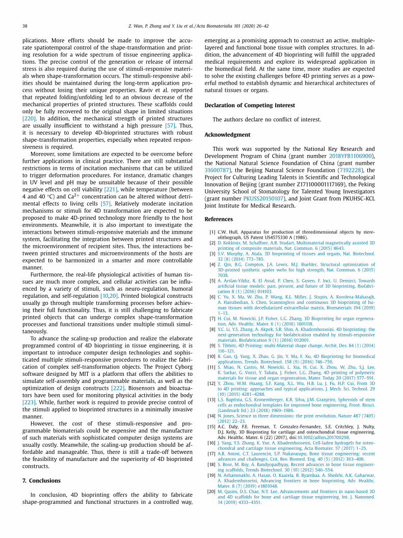
38 Z. Wan, P. Zhang and Y. Liu et al. / Acta Biomaterialia 101 (2020) 26–42
e
l
d
m
t
t
e
n
D
A
D
t
3
P
I
U
(
J
R
plications. More effort s should be made to improve the accu-
rate spatiotemporal control of the shape-transformation and print-
ing resolution for a wide spectrum of tissue engineering applica-
tions. The precise control of the generation or release of internal
stress is also required during the use of stimuli-responsive materi-
als when shape-transformation occurs. The stimuli-responsive abil-
ities should be maintained during the long-term application pro-
cess without losing their unique properties. Raviv et al. reported
that repeated folding/unfolding led to an obvious decrease of the
mechanical properties of printed structures. These scaffolds could
only be fully recovered to the original shape in limited situations
[220] . In addition, the mechanical strength of printed structures
are usually insufficient to withstand a high pressure [57] . Thus,
it is necessary to develop 4D-bioprinted structures with robust
shape-transformation properties, especially when repeated respon-
siveness is required.
Moreover, some limitations are expected to be overcome before
further applications in clinical practice. There are still substantial
restrictions in terms of incitation mechanisms that can be utilized
to trigger deformation procedures. For instance, dramatic changes
in UV level and pH may be unsuitable because of their possible
negative effects on cell viability [221] , while temperature (between
4 and 40 °C) and Ca 2 + concentration can be altered without detri-
mental effects to living cells [57] . Relatively moderate incitation
mechanisms or stimuli for 4D transformation are expected to be
proposed to make 4D-prined technology more friendly to the host
environments. Meanwhile, it is also important to investigate the
interactions between stimuli-responsive materials and the immune
system, facilitating the integration between printed structures and
the microenvironment of recipient sites. Thus, the interactions be-
tween printed structures and microenvironments of the hosts are
expected to be harmonized in a smarter and more controllable
manner.
Furthermore, the real-life physiological activities of human tis-
sues are much more complex, and cellular activities can be influ-
enced by a variety of stimuli, such as neuro-regulation, humoral
regulation, and self-regulation [10 , 20] . Printed biological constructs
usually go through multiple transforming processes before achiev-
ing their full functionality. Thus, it is still challenging to fabricate
printed objects that can undergo complex shape-transformation
processes and functional transitions under multiple stimuli simul-
taneously.
To advance the scaling-up production and realize the elaborate
programmed control of 4D bioprinting in tissue engineering, it is
important to introduce computer design technologies and sophis-
ticated multiple stimuli-responsive procedures to realize the fabri-
cation of complex self-transformation objects. The Project Cyborg
software designed by MIT is a platform that offers the abilities to
simulate self-assembly and programmable materials, as well as the
optimization of design constructs [222] . Biosensors and bioactua-
tors have been used for monitoring physical activities in the body
[223] . While, further work is required to provide precise control of
the stimuli applied to bioprinted structures in a minimally invasive
manner.
However, the cost of these stimuli-responsive and pro-
grammable biomaterials could be expensive and the manufacture
of such materials with sophisticated computer design systems are
usually costly. Meanwhile, the scaling-up production should be af-
fordable and manageable. Thus, there is still a trade-off between
the feasibility of manufacture and the superiority of 4D bioprinted
constructs.
7. Conclusions
In conclusion, 4D bioprinting offers the ability to fabricate
shape-programmed and functional structures in a controlled way,
merging as a promising approach to construct an active, multiple-
ayered and functional bone tissue with complex structures. In ad-
ition, the advancement of 4D bioprinting will fulfill the upgraded
edical requirements and explore its widespread application in
he biomedical field. At the same time, more studies are expected
o solve the existing challenges before 4D printing serves as a pow-
rful method to establish dynamic and hierarchical architectures of
atural tissues or organs.
eclaration of Competing Interest
The authors declare no conflict of interest.
cknowledgment
This work was supported by the National Key Research and
evelopment Program of China (grant number 2018YFB1106900 ),
he National Natural Science Foundation of China (grant number
1600787 ), the Beijing Natural Science Foundation ( 7192228 ), the
roject for Culturing Leading Talents in Scientific and Technological
nnovation of Beijing (grant number Z17110 0 0 01117169), the Peking
niversity School of Stomatology for Talented Young Investigators
grant number PKUSS20150107 ), and Joint Grant from PKUHSC-KCL
oint Institute for Medical Research.
eferences
[1] C.W. Hull, Apparatus for production of threedimensional objects by stere-
olithograph, US Patent US4575330 A (1986).
[2] D. Kokkinis , M. Schaffner , A.R. Studart , Multimaterial magnetically assisted 3Dprinting of composite materials, Nat. Commun. 6 (2015) 8643 .
[3] S.V. Murphy , A. Atala , 3D bioprinting of tissues and organs, Nat. Biotechnol.32 (8) (2014) 773–785 .
[4] Z. Qin , B.G. Compton , J.A. Lewis , M.J. Buehler , Structural optimization of3D-printed synthetic spider webs for high strength, Nat. Commun. 6 (2015)
7038 .
[5] A. Arslan-Yildiz , R. El Assal , P. Chen , S. Guven , F. Inci , U. Demirci , Towardsartificial tissue models: past, present, and future of 3D bioprinting, Biofabri-
cation 8 (1) (2016) 014103 . [6] C. Yu , X. Ma , W. Zhu , P. Wang , K.L. Miller , J. Stupin , A. Koroleva-Maharajh ,
A. Hairabedian , S. Chen , Scanningless and continuous 3D bioprinting of hu-man tissues with decellularized extracellular matrix, Biomaterials 194 (2019)
1–13 .
[7] H. Cui , M. Nowicki , J.P. Fisher , L.G. Zhang , 3D Bioprinting for organ regenera-tion, Adv. Healthc. Mater. 6 (1) (2016) 1601118 .
[8] Y.C. Li , Y.S. Zhang , A. Akpek , S.R. Shin , A. Khademhosseini , 4D bioprinting: thenext-generation technology for biofabrication enabled by stimuli-responsive
materials, Biofabrication 9 (1) (2016) 012001 . [9] S. Tibbits , 4D Printing: multi-Material shape change, Archit. Des. 84 (1) (2014)
116–121 .
[10] B. Gao , Q. Yang , X. Zhao , G. Jin , Y. Ma , F. Xu , 4D Bioprinting for biomedicalapplications, Trends. Biotechnol. 158 (9) (2016) 746–756 .
[11] S. Miao , N. Castro , M. Nowicki , L. Xia , H. Cui , X. Zhou , W. Zhu , S.J. Lee ,K. Sarkar , G. Vozzi , Y. Tabata , J. Fisher , L.G. Zhang , 4D printing of polymeric
materials for tissue and organ regeneration, Mater. Today 20 (2017) 577–591 .[12] Y. Zhou , W.M. Huang , S.F. Kang , X.L. Wu , H.B. Lu , J. Fu , H.P. Cui , From 3D
to 4D printing: approaches and typical applications, J. Mech. Sci. Technol. 29
(10) (2015) 4281–4288 . [13] L.S. Baptista , G.S. Kronemberger , K.R. Silva , J.M. Granjeiro , Spheroids of stem
cells as endochondral templates for improved bone engineering, Front. Biosci.(Landmark Ed.) 23 (2018) 1969–1986 .
[14] N. Jones , Science in three dimensions: the print revolution, Nature 487 (7405)(2012) 22–23 .
[15] A.C. Daly, F.E. Freeman, T. Gonzalez-Fernandez, S.E. Critchley, J. Nulty,
D.J. Kelly, 3D Bioprinting for cartilage and osteochondral tissue engineering,Adv. Healthc. Mater. 6 (22) (2017), doi: 10.10 02/adhm.20170 0298 .
[16] J. Yang , Y.S. Zhang , K. Yue , A. Khademhosseini , Cell-laden hydrogels for osteo-chondral and cartilage tissue engineering, Acta Biomater. 57 (2017) 1–25 .
[17] A.R. Amini , C.T. Laurencin , S.P. Nukavarapu , Bone tissue engineering: recentadvances and challenges, Crit. Rev. Biomed. Eng. 40 (5) (2012) 363–408 .
[18] S. Bose , M. Roy , A. Bandyopadhyay , Recent advances in bone tissue engineer-ing scaffolds, Trends Biotechnol. 30 (10) (2012) 546–554 .
[19] N. Ashammakhi , A. Hasan , O. Kaarela , B. Byambaa , A . Sheikhi , A .K. Gaharwar ,
A. Khademhosseini , Advancing frontiers in bone bioprinting, Adv. Healthc.Mater. 8 (7) (2019) e1801048 .
[20] M. Qasim , D.S. Chae , N.Y. Lee , Advancements and frontiers in nano-based 3Dand 4D scaffolds for bone and cartilage tissue engineering, Int. J. Nanomed.
14 (2019) 4333–4351 .

Z. Wan, P. Zhang and Y. Liu et al. / Acta Biomaterialia 101 (2020) 26–42 39
[21] L. Zhang , G. Yang , B.N. Johnson , X. Jia , Three-dimensional (3D) printed scaf-fold and material selection for bone repair, Acta Biomater. 84 (2019) 16–33 .
[22] S. Saravanan , S. Vimalraj , P. Thanikaivelan , S. Banudevi , G. Manivasagam , A re-view on injectable chitosan/beta glycerophosphate hydrogels for bone tissue
regeneration, Int. J. Biol. Macromol. 121 (2019) 38–54 . [23] F.S. Senatov , K.V. Niaza , M.Y. Zadorozhnyy , A.V. Maksimkin , S.D. Kaloshkin ,
Y.Z. Estrin , Mechanical properties and shape memory effect of 3D-printedPLA-based porous scaffolds, J. Mech. Behav. Biomed. Mater. 57 (2016)
139–148 .
[24] S. Graham , P.F. Marina , A. Blencowe , Thermoresponsive polysaccharides andtheir thermoreversible physical hydrogel networks, Carbohydr. Polym. 207
(2019) 143–159 . [25] H. Suo , D. Zhang , J. Yin , J. Qian , Z.L. Wu , J. Fu , Interpenetrating polymer net-
work hydrogels composed of chitosan and photocrosslinkable gelatin withenhanced mechanical properties for tissue engineering, Mater. Sci. Eng. C
Mater. Biol. Appl. 92 (2018) 612–620 .
[26] C. Li , J.P. Armstrong , I.J. Pence , W. Kit-Anan , J.L. Puetzer , S. Correia Carreira ,A.C. Moore , M.M. Stevens , Glycosylated superparamagnetic nanoparticle gra-
dients for osteochondral tissue engineering, Biomaterials 176 (2018) 24–33 . [27] H.D. Kim, S. Amirthalingam, S.L. Kim, S.S. Lee, J. Rangasamy, N.S. Hwang,
Biomimetic materials and fabrication approaches for bone tissue engineering,Adv. Healthc. Mater. 6 (23) (2017), doi: 10.10 02/adhm.20170 0612 .
[28] J. Vaithilingam, P. Sanjuan-Alberte, S. Campora, G.A. Rance, L. Jiang, J. Thorpe,
L. Burroughs, C.J. Tuck, C. Denning, R.D. Wildman, R.J.M. Hague, M.R. Alexan-der, F.J. Rawson, Multifunctional bioinstructive 3D architectures to modu-
late cellular behavior, Adv. Funct. Mater. 29 (38) (2019), doi: 10.1002/adfm.201902016 .
[29] Z. Luo , S. Zhang , J. Pan , R. Shi , H. Liu , Y. Lyu , X. Han , Y. Li , Y. Yang , Z. Xu ,Y. Sui , E. Luo , Y. Zhang , S. Wei , Time-responsive osteogenic niche of stem
cells: a sequentially triggered, dual-peptide loaded, alginate hybrid system for
promoting cell activity and osteo-differentiation, Biomaterials 163 (2018) 25–42 .
[30] Q. Ge , A.H. Sakhaei , H. Lee , C.K. Dunn , N.X. Fang , M.L. Dunn , Multimaterial 4Dprinting with tailorable shape memory polymers, Sci. Rep. 6 (2016) 31110 .
[31] J.L. Tan , J. Tien , D.M. Pirone , D.S. Gray , K. Bhadriraju , C.S. Chen , Cells lying ona bed of microneedles: an approach to isolate mechanical force, Proc. Natl.
Acad. Sci. USA 100 (4) (2003) 1484–1489 .
[32] K. Franze , The mechanical control of nervous system development, Develop-ment 140 (15) (2013) 3069–3077 .
[33] A. Brugués , E. Anon , V. Conte , J.H. Veldhuis , M. Gupta , J. Colombelli ,J.J. Muñoz , G.W. Brodland , B. Ladoux , X. Trepat , Forces driving epithelial
wound healing, Nat. Phys. 10 (9) (2014) 683–690 . [34] C.J. Underwood , L.T. Edgar , J.B. Hoying , J.A. Weiss , Cell-generated traction
forces and the resulting matrix deformation modulate microvascular align-
ment and growth during angiogenesis, Am. J. Physiol. Heart Circ. Physiol. 307(2) (2015) H152–H164 .
[35] C.M. Kraningrush , J.P. Califano , C.A. Reinhartking , Cellular traction stresses in-crease with increasing metastatic potential, PLoS ONE 7 (2) (2012) e32572 .
[36] R.T. Tranquillo , J.D. Murray , Continuum model of fibroblast-driven woundcontraction: inflammation-Mediation, J. Theor. Biol. 158 (2) (1992) 135–172 .
[37] N. Ashammakhi , S. Ahadian , F. Zengjie , K. Suthiwanich , F. Lorestani , G. Orive ,S. Ostrovidov , A. Khademhosseini , Advances and future perspectives in 4D
bioprinting, Biotechnol. J. 13 (12) (2018) 1800148 .
[38] K. Kuribayashi-Shigetomi , H. Onoe , S. Takeuchi , Cell origami: self-Folding ofthree-dimensional cell-laden microstructures driven by cell traction force,
PLoS ONE 7 (12) (2012) e51085 . [39] A.C. Schone , B. Schulz , A. Lendlein , Stimuli responsive and multifunctional
polymers: progress in materials and applications, Macromol. Rapid Commun.37 (23) (2016) 1856–1859 .
[40] A .S. Gladman , E.A . Matsumoto , R.G. Nuzzo , L. Mahadevan , J.A. Lewis ,
Biomimetic 4D printing, Nat. Mater. 15 (4) (2016) 413–418 . [41] J.W. Su , X. Tao , H. Deng , C. Zhang , S. Jiang , Y. Lin , J. Lin , 4D printing of a
self-morphing polymer driven by a swellable guest medium, Soft Matter 14(5) (2018) 765–772 .
[42] C. Liu , H. Qin , P.T. Mather , Review of progress in shape-memory polymers, J.Mater. Chem. 17 (16) (2007) 1543–1558 .
[43] C. LãWenberg , M. Balk , C. Wischke , M. Behl , A. Lendlein , Shape-Memory hy-
drogels: evolution of structural principles to enable shape switching of hy-drophilic polymer networks, Acc. Chem. Res. 50 (4) (2017) 723–732 .
[44] S. Naficy , R. Gately , R. Gorkin , H. Xin , G.M. Spinks , 4D Printing of re-versible shape morphing hydrogel structures, Macromol. Mater. Eng. 302
(2017) 1600212 . [45] L. Klouda , A.G. Mikos , Thermoresponsive hydrogels in biomedical applica-
tions, Eur. J. Pharm. Biopharm. 68 (1) (2008) 34–45 .
[46] X. Wang , Y. Sun , C. Peng , H. Luo , R. Wang , D. Zhang , Transitional suspen-sions containing thermosensitive dispersant for three-dimensional printing,
ACS Appl. Mater. Interfaces 7 (47) (2015) 26131–26136 . [47] S.E. Bakarich , R. Gorkin 3rd. , M. in het Panhuis , G. Spinks , 4D Printing with
mechanically robust, thermally actuating hydrogels, Macromol. Rapid Com-mun. 36 (12) (2015) 1211–1217 .
[48] H. Wei , S.X. Cheng , X.Z. Zhang , R.X. Zhuo , Thermo-sensitive polymeric mi-
celles based on poly(N -isopropylacrylamide) as drug carriers, Prog. Polym.Sci. 34 (9) (2009) 893–910 .
[49] J.C. Breger , C. Yoon , R. Xiao , H.R. Kwag , M.O. Wang , J.P. Fisher , T.D. Nguyen ,D.H. Gracias , Self-folding thermo-magnetically responsive soft microgrippers,
ACS Appl. Mater. Interfaces 7 (5) (2016) 3398–3405 .
[50] Y. Pei , J. Chen , L. Yang , L. Shi , Q. Tao , B. Hui , J. Li , The effect of pH on the LCSTof poly(N-isopropylacrylamide) and poly(N-isopropylacrylamide-co-acrylic
acid), J. Biomater. Sci. Polym. Ed. 15 (5) (2004) 585–594 . [51] S. Zakharchenko , N. Puretskiy , G. Stoychev , M. Stamm , L. Ionov , Temperature
controlled encapsulation and release using partially biodegradable thermo–magneto-sensitive self-rolling tubes, Soft Mater. 6 (12) (2010) 2633–2636 .
[52] Y. Gao , M. Wei , X. Li , W. Xu , A. Ahiabu , J. Perdiz , Z. Liu , M.J. Serpe , Stimuli-re-sponsive polymers: fundamental considerations and applications, Macromol.
Res. 6 (25) (2017) 513–527 .
[53] S.H. Hsiao , S.H. Hsu , Synthesis and characterization of dual stimuli-sensitivebiodegradable polyurethane soft hydrogels for 3D cell-laden bioprinting, ACS
Appl. Mater. Interfaces 10 (35) (2018) 29273–29287 . [54] W.J. Hendrikson , J. Rouwkema , F. Clementi , C.A. van Blitterswijk , S. Farè,
L. Moroni , Towards 4D printed scaffolds for tissue engineering: exploiting3D shape memory polymers to deliver time-controlled stimulus on cultured
cells, Biofabrication 9 (3) (2017) 031001 .
[55] R. Luo , J. Wu , N.D. Dinh , C.H. Chen , Hydrogels: gradient porous elastic hy-drogels with shape-memory property and anisotropic responses for pro-
grammable locomotion, Adv. Funct. Mater. 25 (47) (2016) 7272–7279 . [56] I. Apsite , G. Stoychev , W. Zhang , D. Jehnichen , J. Xie , L. Ionov , Porous stim-
uli-responsive self-folding electrospun mats for 4D biofabrication, Biomacro-molecules 18 (10) (2017) 3178–3184 .
[57] L. Ionov , 4D Biofabrication: materials, methods, and applications, Adv.
Healthc. Mater. 7 (17) (2018) e1800412 . [58] N.A. Peppas , P. Bures , W. Leobandung , H. Ichikawa , Hydrogels in pharmaceu-
tical formulations, Eur. J. Pharm. Biopharm. 50 (1) (20 0 0) 27–46 . [59] L. Li , H. Shan , C.Y. Yue , Y.C. Lam , K.C. Tam , X. Hu , Thermally induced associ-
ation and dissociation of methylcellulose in aqueous solutions, Langmuir 18(20) (2002) 7291–7298 .
[60] V. Stroganov , S. Zakharchenko , E. Sperling , A.K. Meyer , O.G. Schmidt , L. Ionov ,
Biodegradable self-folding polymer films with controlled thermo-triggeredfolding, Adv. Funct. Mater. 24 (27) (2014) 4357–4363 .
[61] V. Stroganov , M. Alhussein , J.U. Sommer , A. Janke , S. Zakharchenko , L. Ionov ,Reversible thermosensitive biodegradable polymeric actuators based on con-
fined crystallization, Nano Lett. 15 (3) (2015) 1786–1790 . [62] P. Fratzl , F.G. Barth , Biomaterial systems for mechanosensing and actuation,
Nature 462 (7272) (2009) 4 42–4 48 .
[63] R. Elbaum , L. Zaltzman , I. Burgert , P. Fratzl , The role of wheat awns in theseed dispersal unit, Science 316 (5826) (2005) 884–886 .
[64] M. Dai , O.T. Picot , J.M. Verjans , L.T.D. Haan , A.P. Schenning , T. Peijs , C.W. Bas-tiaansen , Humidity-responsive bilayer actuators based on a liquid-crystalline
polymer network, ACS Appl. Mater. Interfaces 5 (11) (2013) 4 945–4 950 . [65] L.T. de Haan , J.M. Verjans , D.J. Broer , C.W. Bastiaansen , A.P. Schenning , Hu-
midity-responsive liquid crystalline polymer actuators with an asymmetry in
the molecular trigger that bend, fold, and curl, J. Am. Chem. Soc. 136 (30)(2014) 10585–10588 .
[66] M. Jamal , S.S. Kadam , R. Xiao , F. Jivan , T.M. Onn , R. Fernandes , T.D. Nguyen ,D.H. Gracias , Bio-Origami hydrogel scaffolds composed of photocrosslinked
peg bilayers, Adv. Healthc. Mater. 2 (8) (2013) 1042–1050 . [67] L. Zhang , H. Liang , J. Jacob , P. Naumov , Photogated humidity-driven motility,
Nat. Commun. 6 (2015) 7429 . [68] K. Zhang , A. Geissler , M. Standhardt , S. Mehlhase , M. Gallei , C. Marie Thiele ,
Moisture-responsive films of cellulose stearoyl esters showing reversible
shape transitions, Sci. Rep. 5 (2015) 11011 . [69] O.V. Borisova , L. Billon , R.P. Richter , E. Reimhult , O.V. Borisov , pH-
and electro-responsive properties of poly(acrylic acid) and poly(acrylicacid)-block-poly(acrylic acid-grad-styrene) brushes studied by quartz crys-
tal microbalance with dissipation monitoring, Langmuir 31 (27) (2015)7684–7694 .
[70] H. Okuzaki , T. Kuwabara , K. Funasaka , T. Saido , Humidity-sensitive polypyrrole
films for electro-active polymer actuators, Adv. Funct. Mater. 23 (36) (2013)4 400–4 407 .
[71] S. Li , K. Shu , C. Zhao , C. Wang , Z. Guo , G. Wallace , H.K. Liu , One-step synthe-sis of graphene/polypyrrole nanofiber composites as cathode material for a
biocompatible zinc/polymer battery, ACS Appl. Mater. Interfaces 6 (19) (2014)16679–16686 .
[72] N. Sood , A. Bhardwaj , S. Mehta , A. Mehta , Stimuli-responsive hydrogels
in drug delivery and tissue engineering, Drug Deliv. 23 (3) (2016) 758–780 .
[73] Y. Wang , Programmable hydrogels, Biomaterials 178 (2018) 663–680 . [74] R.A. Green , S. Baek , L.A. Poolewarren , P.J. Martens , Conducting polymer-hydro-
gels for medical electrode applications, Sci. Technol. Adv. Mater. 11 (1) (2010)014107 .
[75] E. Song , J.W. Choi , Conducting polyaniline nanowire and its applications in
chemiresistive sensing, Nanomaterials (Basel) 3 (3) (2013) 498–523 . [76] E. Fantino , I. Roppolo , D. Zhang , J. Xiao , A. Chiappone , M. Castellino , Q.Q. Guo ,
C. Fabrizio Pirri , J. Yang , 3D printing/interfacial polymerization coupling forthe fabrication of conductive hydrogel, Macromol. Mater. Eng. 303 (4) (2018)
1700356 . [77] S. Ahadian , R. Obregón , J. Ramónazcón , G. Salazar , H. Shiku , M. Ramalingam ,
T. Matsue , Carbon nanotubes and graphene-based nanomaterials for stem
cell differentiation and tissue regeneration, J. Tissue Eng. Regen. Med. 16 (9)(2016) 8862–8880 .
[78] J. Ramón-Azcón , S. Ahadian , R. Obregón , H. Shiku , M. Ramalingam , T. Matsue ,Applications of carbon nanotubes in stem cell research, J. Biomed. Nanotech-
nol. 10 (10) (2014) 2539–2561 .

40 Z. Wan, P. Zhang and Y. Liu et al. / Acta Biomaterialia 101 (2020) 26–42
[79] C.W. Foster , M.P. Down , Y. Zhang , X. Ji , S.J. Rowleyneale , G.C. Smith , P.J. Kelly ,C.E. Banks , 3D Printed graphene based energy storage devices, Sci. Rep. 7
(2017) 42233 . [80] J.H. Kim , W.S. Chang , D. Kim , J.R. Yang , J.T. Han , G.W. Lee , J.T. Kim , S.K. Seol ,
3D printing of reduced graphene oxide nanowires, Adv. Mater. 27 (1) (2015)157–161 .
[81] S.R. Shin , R. Farzad , A. Tamayol , V. Manoharan , P. Mostafalu , Y.S. Zhang ,M. Akbari , S.M. Jung , D. Kim , M. Comotto , N. Annabi , F.E. Al-Hazmi , M.R. Dok-
meci , A. Khademhosseini , A bioactive carbon nanotube-based ink for printing
2D and 3D flexible electronics, Adv. Mater. 28 (17) (2016) 3280–3289 . [82] A. Servant , V. Leon , D. Jasim , L. Methven , P. Limousin , E.V. Fernandezpacheco ,
M. Prato , K. Kostarelos , Graphene-based electroresponsive scaffolds as poly-meric implants for on-demand drug delivery, Adv. Healthc. Mater. 3 (8)
(2015) 1334–1343 . [83] Y. Zhang , D. Zhai , M. Xu , Q. Yao , H. Zhu , J. Chang , C. Wu , 3D-printed bioce-
ramic scaffolds with antibacterial and osteogenic activity, Biofabrication 9 (2)
(2017) 025037 . [84] B. Huang , C. Vyas , I. Roberts , Q.A. Poutrel , W.H. Chiang , J.J. Blaker , Z. Huang ,
P. Bartolo , Fabrication and characterisation of 3D printed MWCNT compositeporous scaffolds f or bone regeneration, Mater. Sci. Eng. C Mater. Biol. Appl.
98 (2019) 266–278 . [85] E.M. Goncalves , F.J. Oliveira , R.F. Silva , M.A. Neto , M.H. Fernandes , M. Amaral ,
M. Vallet-Regi , M. Vila , Three-dimensional printed PCL-hydroxyapatite scaf-
folds filled with CNTs for bone cell growth stimulation, J. Biomed. Mater. Res.B Appl. Biomater. 104 (6) (2016) 1210–1219 .
[86] K. Dan , M.J. Tan , M.P. Prabhakaran , B.Q.Y. Chan , S.S. Liow , S. Ramakrishna ,X.J. Loh , Biocompatible electrically conductive nanofibers from inorganic-or-
ganic shape memory polymers, Colloids Surf. B Biointerfaces 148 (2016)557–565 .
[87] G. Filipcsei , I. Csetneki , A. Szilágyi , M.J. Zrínyi , Magnetic field-responsive
smart polymer composites, Oligom.Polym. Compos. Mol. Imprint. 206 (1)(2007) 137–189 .
[88] K. Ulbrich , K. Hola , V. Subr , A. Bakandritsos , J. Tucek , R. Zboril , Targeteddrug delivery with polymers and magnetic nanoparticles: covalent and non-
covalent approaches, release control, and clinical studies, Chem. Rev. 116 (9)(2016) 5338–5431 .
[89] Q.I. Zhang , J. Liu , K. Yuan , Z. Zhang , X. Zhang , X. Fang , A multi-controlled
drug delivery system based on magnetic mesoporous Fe 3 O 4 nanopaticles anda phase change material for cancer thermo-chemotherapy, Nanotechnology
28 (40) (2017) 405101 . [90] F.H. Zhang, L.L. Wang, Z.C. Zheng, Y.J. Liu, J.S. Leng, Magnetic programming
of 4D printed shape memory composite structures, Compos. A – Appl. Sci.Manuf. 125 (2019), doi: 10.1016/j.compositesa.2019.105571 .
[91] L.Q. Ren , B.Q. Li , Z.Y. Song , Q.P. Liu , L. Ren , X.L. Zhou , Bioinspired fiber-regu-
lated composite with tunable permanent shape and shape memory proper-ties via 3d magnetic printing, Compos. B – Eng. 164 (2019) 458–466 .
[92] D. Wang , S. Zheng , H. Liu , J. Tang , W. Miao , H. Wang , Y. Tian , H. Yang ,L. Jiang , A magnetic gated nanofluidic based on the integration of a super-
hydrophilic nanochannels and a reconfigurable ferrofluid, Adv. Mater. 31 (7)(2019) e1805953 .
[93] L. Zhang , X. Zuo , S. Li , M. Sun , H. Xie , K. Zhang , J. Zhou , L. Che , J. Ma , Z. Jia ,F. Yang , Synergistic therapy of magnetism-responsive hydrogel for soft tissue
injuries, Bioact. Materr. 4 (2019) 160–166 .
[94] K. Lalitha , Y.S. Prasad , V. Sridharan , C.U. Maheswari , G. John , S. Nagarajan ,A renewable resource-derived thixotropic self-assembled supramolecular gel:
magnetic stimuli responsive and real-time self-healing behaviour, RSC Adv. 5(95) (2015) 77589–77594 .
[95] C. Adine , K.K. Ng , S. Rungarunlert , G.R. Souza , J.N. Ferreira , Engineering in-nervated secretory epithelial organoids by magnetic three-dimensional bio-
printing for stimulating epithelial growth in salivary glands, Biomaterials 180
(2018) 52–66 . [96] G.R. Souza , H. Tseng , J.A. Gage , A. Mani , P. Desai , F. Leonard , A. Liao , M. Longo ,
J.S. Refuerzo , B. Godin , Magnetically bioprinted human myometrial 3D cellrings as a model for uterine contractility, Int. J. Mol. Sci. 18 (4) (2017) e683 .
[97] V. Du , N. Luciani , S. Richard , G. Mary , C. Gay , F. Mazuel , M. Reffay ,P. Menasché, O. Agbulut , C. Wilhelm , A 3D magnetic tissue stretcher for re-
mote mechanical control of embryonic stem cell differentiation, Nat. Com-
mun. 8 (1) (2017) 400 . [98] G.R. Souza , J.R. Molina , R.M. Raphael , M.G. Ozawa , D.J. Stark , C.S. Levin ,
L.F. Bronk , J.S. Ananta , J. Mandelin , M.M. Georgescu , J.A. Bankson , J.G. Gelo-vani , T.C. Killian , W. Arap , R. Pasqualini , Three-dimensional tissue culture
based on magnetic cell levitation, Nat. Nanotechnol. 5 (4) (2010) 291–296 . [99] T. Sun , Q. Shi , Q. Huang , H. Wang , X. Xiong , C. Hu , et al. , Magnetic alginate
microfibers as scaffolding element s f or the fabrication of microvascular-like
structures, Acta Biomater. 66 (2018) 272–281 . [100] A. Tocchio , N.G. Durmus , K. Sridhar , V. Mani , B. Coskun , R. El Assal ,
U. Demirci , Magnetically guided self-assembly and coding of 3D living archi-tectures, Adv. Mater. 30 (4) (2018) 1–15 .
[101] S. Tasoglu , C.H. Yu , V. Liaudanskaya , S. Guven , C. Migliaresi , U. Demirci , Mag-netic levitational assembly for living material fabrication, Adv. Healthc. Mater.
4 (10) (2015) 1469–1476 1422 .
[102] V.A. Parfenov , E.V. Koudan , E.A. Bulanova , P.A. Karalkin , F.D. Pereira ,N.E. Norkin , A.D. Knyazeva , A.A. Gryadunova , O.F. Petrov , M.M. Vasiliev ,
M.I. Myasnikov , V.P. Chernikov , V.A. Kasyanov , A.Y. Marchenkov , K. Brakke ,Y.D. Khesuani , U. Demirci , V.A. Mironov , Scaffold-free, label-free and noz-
zle-free biofabrication technology using magnetic levitational assembly, Bio-fabrication 10 (2018) 034104 .
[103] A.R. Abdel Fattah , S. Mishriki , T. Kammann , R.P. Sahu , F. Geng , I.K. Puri ,3D cellular structures and co-cultures formed through the contactless mag-
netic manipulation of cells on adherent surfaces, Biomater. Sci. 6 (3) (2018)683–694 .
[104] E. Turker , N. Demircak , A. Arslan-Yildiz , Scaffold-free three-dimensional cellculturing using magnetic levitation, Biomater. Sci. 6 (7) (2018) 1745–1753 .
[105] S. Tasoglu , D. Kavaz , U.A. Gurkan , S. Guven , P. Chen , R. Zheng , U. Demirci ,
Hydrogels: paramagnetic levitational assembly of hydrogels, Adv. Mater. 25(8) (2013) 1137–1143 .
[106] S. Tasoglu , C.H. Yu , H.I. Gungordu , S. Guven , T. Vural , U. Demirci , Guidedand magnetic self-assembly of tunable magnetoceptive gels, Nat. Commun.
5 (2014) 4702 . [107] Y. Qin , J. Chen , Y. Bi , X. Xu , H. Zhou , J. Gao , Y. Hu , Y. Zhao , Z. Chai ,
Near-infrared light remote-controlled intracellular anti-cancer drug deliv-
ery using thermo/pH sensitive nanovehicle, Acta Biomater. 17 (2015) 201–209 .
[108] A. Bagheri , H. Arandiyan , C. Boyer , M. Lim , Lanthanide-Doped upconver-sion nanoparticles: emerging intelligent light-activated drug delivery systems,
Adv. Sci. (Weinh) 3 (7) (2016) 1500437 . [109] M. Nakayama , W.Q. Lim , S. Kajiyama , A. Kumamoto , Y. Ikuhara , T. Kato ,
Y. Zhao , Liquid-crystalline hydroxyapatite/polymer nanorod hybrids: potential
bioplatform for photodynamic therapy and cellular scaffolds, ACS Appl. MaterInterfaces 11 (19) (2019) 17759–17765 .
[110] S.A. Kumar , M. Alonzo , S.C. Allen , L. Abelseth , V. Thakur , J. Akimoto ,Y. Ito , S.M. Willerth , L. Suggs , M. Chattopadhyay , B. Joddar , A visible
light-cross-linkable, fibrin-gelatin-based bioprinted construct with humancardiomyocytes and fibroblasts, ACS Biomater. Sci. Eng. 5 (9) (2019) 4551–
4563 .
[111] Y. Luo , X. Lin , B. Chen , X. Wei , Cell-laden four-dimensional bioprinting usingnear-infrared-triggered shape-morphing alginate/polydopamine bioinks, Bio-
fabrication 11 (4) (2019) 045019 . [112] H.T. Cui , S.D. Miao , T. Esworthy , S.J. Lee , X. Zhou , S.Y. Hann , T.J. Webster ,
B.T. Harris , L.G. Zhang , A novel near-infrared light responsive 4D printednanoarchitecture with dynamically and remotely controllable transformation,
Nano Res. 12 (6) (2019) 1381–1388 .
[113] H. Wei , Q. Zhang , Y. Yao , L. Liu , Y. Liu , J. Leng , Direct-write fabrication of 4Dactive shape-changing structures based on a shape memory polymer and its
nanocomposite, ACS Appl. Mater. Interfaces 9 (1) (2017) 876–883 . [114] X. Kuang , K. Chen , C.K. Dunn , J. Wu , V.C.F. Li , H.J. Qi , 3D printing of highly
stretchable, shape-memory and self-healing elastomer toward novel 4D print-ing, ACS Appl. Mater. Interfaces 10 (8) (2018) 7381–7388 .
[115] T.E. Brown , K.S. Anseth , Spatiotemporal hydrogel biomaterials for regenerative
medicine, Chem. Soc. Rev. 46 (21) (2017) 6532–6552 . [116] D.R. Griffin , A.M. Kasko , Photodegradable macromers and hydrogels for
live cell encapsulation and release, J. Am. Chem. Soc. 134 (31) (2012)13103–13107 .
[117] C.K. Arakawa , B.A. Badeau , Y. Zheng , C.A. Deforest , Multicellular vascular-ized engineered tissues through user-programmable biomaterial photodegra-
dation, Adv. Mater. 29 (37) (2017) 201703156 . [118] K.S. Lim , B.J. Klotz , G.C.J. Lindberg , F.P.W. Melchels , G.J. Hooper , J. Malda ,
D. Gawlitta , T.B.F. Woodfield , Visible light cross-linking of gelatin hydrogels
offers an enhanced cell microenvironment with improved light penetrationdepth, Macromol. Biosci. 19 (6) (2019) e190 0 098 .
[119] S.M. Naseer , A. Manbachi , M. Samandari , P. Walch , Y. Gao , Y.S. Zhang ,F. Davoudi , W. Wang , K. Abrinia , J.M. Cooper , A. Khademhosseini , S.R. Shin ,
Surface acoustic waves induced micropatterning of cells in gelatin methacry-loyl (GelMA) hydrogels, Biofabrication 9 (1) (2017) 015020 .
[120] K. Melde, E. Choi, Z. Wu, S. Palagi, T. Qiu, P. Fischer, Acoustic fabrication via
the assembly and fusion of particles, Adv. Mater. 30 (3) (2018), doi: 10.1002/adma.201704507 .
[121] N. Huebsch , C.J. Kearney , X. Zhao , J. Kim , C.A. Cezar , Z. Suo , D.J. Mooney , Ul-trasound-triggered disruption and self-healing of reversibly cross-linked hy-
drogels for drug delivery and enhanced chemotherapy, Proc. Natl. Acad. Sci.USA 111 (27) (2014) 9762–9767 .
[122] C. Bouyer , P. Chen , S. Guven , T.T. Demirtas , T.J. Nieland , F. Padilla , U. Demirci ,
A bio-acoustic levitational (BAL) assembly method for engineering of multi-layered, 3D brain-like constructs, using human embryonic stem cell derived
neuro-progenitors, Adv. Mater. 28 (1) (2016) 161–167 . [123] G. Kocak , C. Tuncer , V. Bütün , pH-Responsive polymers, Polym. Chem. 8 (1)
(2016) 144–176 . [124] M. Nadgorny , A. Ameli , Functional polymers and nanocomposites for 3D
printing of smart structures and devices, ACS Appl. Mater. Interfaces 10 (21)
(2018) 17489–17507 . [125] M. Nadgorny , Z. Xiao , C. Chen , L.A. Connal , Three-dimensional printing of
pH-Responsive and functional polymers on an affordable desktop printer, ACSAppl. Mater. Interfaces 8 (42) (2016) 28946–28954 .
[126] C. Chen , M.L. Zhao , R.K. Zhang , G. Lu , C.Y. Zhao , F. Fu , H.T. Sun , S. Zhang ,Y. Tu , X.H. Li , Collagen/heparin sulfate scaffolds fabricated by a 3D bioprinter
improved mechanical properties and neurological function after spinal cord
injury in rats, J. Biomed. Mater. Res. A 105 (5) (2017) 1324–1332 . [127] V. Lee , G. Singh , J.P. Trasatti , C. Bjornsson , X. Xu , T.N. Tran , S.S. Yoo , G. Dai ,
P. Karande , Design and fabrication of human skin by three-dimensional bio-printing, Tissue Eng. C Methods 20 (6) (2014) 473–484 .

Z. Wan, P. Zhang and Y. Liu et al. / Acta Biomaterialia 101 (2020) 26–42 41
[128] D.E. Clarke , C.D.J. Parmenter , O.A. Scherman , Tunable pentapeptide self--assembled beta-Sheet hydrogels, Angew. Chem. Int. Ed. Engl. 57 (26) (2018)
7709–7713 . [129] M.S.M. Eldin , E.A. Kamoun , M.A. Sofan , S.M. Elbayomi , l -Arginine grafted algi-
nate hydrogel beads: a novel pH-sensitive system for specific protein delivery,Arab. J. Chem. 8 (3) (2015) 355–365 .
[130] M. Kashi , F. Baghbani , F. Moztarzadeh , H. Mobasheri , E. Kowsari , Green syn-thesis of degradable conductive thermosensitive oligopyrrole/chitosan hydro-
gel intended for cartilage tissue engineering, Int. J. Biol. Macromol. 107 (Pt B)
(2018) 1567–1575 . [131] P. Mukhopadhyay , K. Sarkar , S. Bhattacharya , A. Bhattacharyya , R. Mishra ,
P.P. Kundu , pH sensitive N-succinyl chitosan grafted polyacrylamide hydrogelfor oral insulin delivery, Carbohydr. Polym. 112 (2) (2014) 627–637 .
[132] Y. Xu , J. Han , Y. Chai , S. Yuan , H. Lin , X. Zhang , Development of porous chi-tosan/tripolyphosphate scaffolds with tunable uncross-linking primary amine
content for bone tissue engineering, Mater. Sci. Eng. C Mater. Biol. Appl. 85
(2018) 182–190 . [133] L.A. Hockaday , K.H. Kang , N.W. Colangelo , P.Y.C. Cheung , B. Duan , E. Mal-
one , J. Wu , L.N. Girardi , L.J. Bonassar , H. Lipson , C.C. Chu , J.T. Butcher , Rapid3D printing of anatomically accurate and mechanically heterogeneous aortic
valve hydrogel scaffolds, Biofabrication 4 (3) (2012) 035005 . [134] A.G. Tabriz , M.A. Hermida , N.R. Leslie , W. Shu , Three-dimensional bioprinting
of complex cell laden alginate hydrogel structures, Biofabrication 7 (4) (2015)
045012 . [135] Z. Ren , Y. Zhang , Y. Li , B. Xu , W. Liu , Hydrogen bonded and ionically
crosslinked high strength hydrogels exhibiting Ca 2 + -triggered shape memoryproperties and volume shrinkage for cell detachment, J. Mater. Chem. B 3 (30)
(2015) 6347–6354 . [136] T. Bai , Y. Han , P. Zhang , W. Wang , W. Liu , Zinc ion-triggered two-way
macro-/microscopic shape changing and memory effects in high strength hy-
drogels with pre-programmed unilateral patterned surfaces, Soft Matter. 8(25) (2012) 6 846–6 852 .
[137] W. Nan , W. Wang , H. Gao , W. Liu , Fabrication of a shape memory hydro-gel based on imidazole–zinc ion coordination for potential cell-encapsulating
tubular scaffold application, Soft Matter. 9 (1) (2012) 132–137 . [138] T. Jungst , W. Smolan , K. Schacht , T. Scheibel , J. Groll , Strategies and molec-
ular design criteria for 3D printable hydrogels, Chem. Rev. 116 (3) (2016)
1496–1539 . [139] A . Skardal , A . Atala , Biomaterials for integration with 3-D bioprinting, Ann.
Biomed. Eng. 43 (3) (2015) 730–746 . [140] A. Kirillova, R. Maxson, G. Stoychev, C.T. Gomillion, L.J. Ionov, 4D Biofabri-
cation using shape-morphing hydrogels, Adv. Mater. 29 (46) (2017), doi: 10.1002/adma.201703443 .
[141] P. Camacho , H. Busari , K.B. Seims , P. Schwarzenberg , H.L. Dailey , L.W. Chow ,
3D printing with peptide-polymer conjugates for single-step fabricationof spatially functionalized scaffolds, Biomater. Sci. 7 (10) (2019) 4237–
4247 . [142] P.J. Skrzeszewska , L.N. Jong , F.A. de Wolf , M.A. Cohen Stuart , d.G.J. Van ,
Shape-memory effects in biopolymer networks with collagen-like transientnodes, Biomacromolecules 12 (6) (2011) 2285–2292 .
[143] Q. Hao , M. Ghodousi , Y. Du , C. Grun , H. Bae , Y. Peng , A. Khademhosseini ,DNA-directed self-assembly of shape-controlled hydrogels, Nat. Commun. 4
(9) (2013) 2275 .
[144] W. Guo , C.H. Lu , R. Orbach , F. Wang , X.J. Qi , A. Cecconello , D. Seliktar , I. Will-ner , pH-stimulated DNA hydrogels exhibiting shape-memory properties, Adv.
Mater. 27 (1) (2015) 73–78 . [145] M.E. Todhunter , N.Y. Jee , A.J. Hughes , M.C. Coyle , A. Cerchiari , J. Farlow ,
J.C. Garbe , M.A. LaBarge , T.A. Desai , Z.J. Gartner , Programmed synthesis ofthree-dimensional tissues, Nat. Methods 12 (10) (2015) 975–981 .
[146] J. Kim , Y. Park , G. Tae , K.B. Lee , S.J. Hwang , I.S. Kim , I. Noh , K. Sun , Syn-
thesis and characterization of matrix metalloprotease sensitive-low molecu-lar weight hyaluronic acid based hydrogels, J. Mater. Sci. Mater. Med. 19 (11)
(2008) 3311–3318 . [147] N. Broguiere , F. Formica , G. Barreto , M. Zenobi-Wong , Sortase a as a
cross-linking enzyme in tissue engineering, Acta Biomater. 77 (2018)182–190 .
[148] J.B. Costa , J. Silva-Correia , J.M. Oliveira , L.R. Rui , Fast setting silk fibroin bioink
for bioprinting of patient-specific memory-shape implants, Adv. Healthc.Mater. 6 (22) (2017) 1701021 .
[149] L.P. Yan , J.M. Oliveira , A.L. Oliveira , R.L. Reis , R. Medicine , Core-shell silk hy-drogels with spatially tuned conformations as drug-delivery system, J. Tissue
Eng. Regen. Med. 11 (11) (2016) 3168–3177 . [150] C.A. Mandon , L.J. Blum , C.A. Marquette , Adding biomolecular recognition ca-
pability to 3D printed objects, Anal. Chem. 88 (21) (2017) 10767–10772 .
[151] N.J. Castro , C. Meinert , P. Levett , D.W. Hutmacher , Current developments inmultifunctional smart materials for 3D/4D bioprinting, Curr. Opin. Biomed.
Eng. 2 (2017) 67–75 . [152] A .S. Gladman , E.A . Matsumoto , R.G. Nuzzo , L. Mahadevan , J.A. Lewis ,
Biomimetic 4D printing, Nat. Mater. 15 (4) (2016) 413–418 . [153] J. Malda , J. Visser , F.P. Melchels , T. Jüngst , W.E. Hennink , W.J.A. Dhert , J. Groll
J , D.W. Hutmacher , 25th Anniversary article: engineering hydrogels for bio-
fabrication, Adv. Mater. 25 (36) (2013) 5011–5028 . [154] B. Lanfer , F.P. Seib , U. Freudenberg , D. Stamov , T. Bley , M. Bornhäuser ,
C. Werner , The growth and differentiation of mesenchymal stem and progen-itor cells cultured on aligned collagen matrices, Biomaterials 30 (30) (2009)
5950–5958 .
[155] M. Costantini , S. Testa , P. Mozetic , A. Barbetta , C. Fuoco , E. Fornetti , F. Tamiro ,S. Bernardini , J. Jaroszewicz , W. Swi eszkowski , M. Trombetta , L. Castagnoli ,
D. Seliktar , P. Garstecki , G. Cesareni , S. Cannata , A. Rainer , C. Gargioli , Mi-crofluidic-enhanced 3D bioprinting of aligned myoblast-laden hydrogels leads
to functionally organized myofibers in vitro and in vivo, Biomaterials 131(2017) 98–110 .
[156] J. Koffler , W. Zhu , X. Qu , O. Platoshyn , J.N. Dulin , J. Brock , L. Graham , P. Lu ,J. Sakamoto , M. Marsala , S.C. Chen , M.H. Tuszynski , Biomimetic 3D-printed
scaffolds f or spinal cord injury repair, Nat. Med. 25 (2) (2019) 263–269 .
[157] M. Betsch , C. Cristian , Y.Y. Lin , A. Blaeser , J. Schöneberg , M. Vogt , E.M. Buhl ,H. Fischer , D.F. Duarte Campos , Incorporating 4D into bioprinting: real-time
magnetically directed collagen fiber alignment for generating complex multi-layered tissues, Adv. Healthc. Mater. 7 (21) (2018) e1800894 .
[158] S. Miao , H. Cui , M. Nowicki , S.J. Lee , J. Almeida , X. Zhou , W. Zhu , X. Yao , F. Ma-sood , M.W. Plesniak , M. Mohiuddin , L.G. Zhang , Photolithographic-stereolitho-
graphic-tandem fabrication of 4D smart scaffolds for improved stem cell car-
diomyogenic differentiation, Biofabrication 10 (3) (2018) 035007 . [159] S. Miao , M. Nowicki , H. Cui , S.J. Lee , X. Zhou , D.K. Mills , L.G. Zhang , 4D
anisotropic skeletal muscle tissue constructs fabricated by staircase effectstrategy, Biofabrication 11 (3) (2019) 035030 .
[160] G. Villar , A.D. Graham , H. Bayley , A tissue-like printed material, Science 340(6128) (2013) 48–52 .
[161] M.J. Booth , V.R. Schild , A.D. Graham , S.N. Olof , H. Bayley , Light-activated com-
munication in synthetic tissues, Sci. Adv. 2 (4) (2016) e160 0 056 . [162] A .K. Miri , A . Khalilpour , B. Cecen , S. Maharjan , S.R. Shin , A. Khademhos-
seini , Multiscale bioprinting of vascularized models, Biomaterials 198 (2019)204–216 .
[163] C. Norotte , F. Marga , L. Niklason , G. Forgacs , Scaffold-Free vascular tissue en-gineering using bioprinting, Biomaterials 30 (30) (2009) 5910–5917 .
[164] R.J. Morrison , S.J. Hollister , M.F. Niedner , M.G. Mahani , A.H. Park , D.K. Mehta ,
R.G. Ohye , G.E. Green , Mitigation of tracheobronchomalacia with 3D-printedpersonalized medical devices in pediatric patients, Sci. Transl. Med. 7 (285)
(2015) 285ra264 . [165] J. Park , Y.V. Kalinin , S. Kadam , C.L. Randall , D.H. Gracias , Design for a litho-
graphically patterned bioartificial endocrine pancreas, Artif. Organs 37 (12)(2013) 1059–1067 .
[166] T. Wang , L. Chen , T. Shen , D. Wu , Preparation and properties of a novel
thermo-sensitive hydrogel based on chitosan/hydroxypropyl methylcellu-lose/glycerol, Int. J. Biol. Macromol. 93 (Pt A) (2016) 775–782 .
[167] Y. Cai , J. Cao , C. Xu , J. Zhou , Thermo-responsive behaviors and bioactivities ofhydroxybutyl chitosans prepared in alkali/urea aqueous solutions, Carbohydr.
Polym. 215 (2019) 90–98 . [168] C. Li , K. Wang , X. Zhou , T. Li , Y. Xu , L. Qiang , M. Peng , Y. Xu , L. Xie , C. He ,
B. Wang , J. Wang , Controllable fabrication of hydroxybutyl chitosan/oxidized
chondroitin sulfate hydrogels by 3D bioprinting technique for cartilage tissueengineering, Biomed. Mater. 14 (2) (2019) 025006 .
[169] A. Parameswaran-Thankam , C.M. Parnell , F. Watanabe , A.B. RanguMagar ,B.P. Chhetri , P.K. Szwedo , A .S. Biris , A . Ghosh , Guar-Based injectable thermore-
sponsive hydrogel as a scaffold f or bone cell growth and controlled drug de-livery, ACS Omega 3 (11) (2018) 15158–15167 .
[170] J. Yang , R. van Lith , K. Baler , R.A. Hoshi , G.A. Ameer , A thermorespon-sive biodegradable polymer with intrinsic antioxidant properties, Biomacro-
molecules 15 (11) (2014) 3942–3952 .
[171] J.P. Chen , M.J. Tsai , H.T. Liao , Incorporation of biphasic calcium phosphatemicroparticles in injectable thermoresponsive hydrogel modulates bone cell
proliferation and differentiation, Colloids Surf. B Biointerfaces 110 (2013)120–129 .
[172] X. Chen , Y. Zhao , S. Geng , R.J. Miron , Q. Zhang , C. Wu , Y. Zhang , In vivoexperimental study on bone regeneration in critical bone defects using PIB
nanogels/boron-containing mesoporous bioactive glass composite scaffold,
Int. J. Nanomed. 10 (2015) 839–846 . [173] C. Zhao , Z. Zeng , N.T. Qazvini , X. Yu , R. Zhang , S. Yan , Y. Shu , Y. Zhu , C. Duan ,
E. Bishop , J. Lei , W. Zhang , C. Yang , K. Wu , Y. Wu , L. An , S. Huang , X. Ji ,C. Gong , C. Yuan , L. Zhang , W. Liu , B. Huang , Y. Feng , B. Zhang , Z. Dai , Y. Shen ,
X. Wang , W. Luo , L. Oliveira , A. Athiviraham , M.J. Lee , J.M. Wolf , G.A. Ameer ,R.R. Reid , T.C. He , W. Huang , Thermoresponsive citrate-based graphene oxide
scaffold enhances bone regeneration from BMP9-Stimulated adipose-derived
mesenchymal stem cells, ACS Biomater. Sci. Eng. 4 (8) (2018) 2943–2955 . [174] J. Wu , K. Zheng , X. Huang , J. Liu , H. Liu , A.R. Boccaccini , Y. Wan , X. Guo ,
Z. Shao , Thermally triggered injectable chitosan/silk fibroin/bioactive glassnanoparticle hydrogels for in-situ bone formation in rat calvarial bone de-
fects, Acta Biomater. 91 (2019) 60–71 . [175] L. Vojtova, L. Michlovska, K. Valova, M. Zboncak, M. Trunec, K. Castkova,
M. Krticka, V. Pavlinakova, P. Polacek, M. Dzurov, V. Lukasova, M. Rampichova,
T. Suchy, R. Sedlacek, M.P. Ginebra, E.B. Montufar, The effect of the ther-mosensitive biodegradable plga(-)peg(-)plga copolymer on the rheological,
structural and mechanical properties of thixotropic self-hardening tricalciumphosphate cement, Int. J. Mol. Sci. 20 (2) (2019), doi: 10.3390/ijms20020391 .
[176] A.S. Azevedo , M.J. Sa , M.V. Fook , P.I. Neto , O.B. Sousa , S.S. Azevedo , M.W. Teix-eira , F.S. Costa , A.L. Araujo , Use of chitosan and beta-tricalcium phosphate,
alone and in combination, for bone healing in rabbits, J. Mater. Sci. Mater.
Med. 25 (2) (2014) 4 81–4 86 . [177] M. Dessi , A. Borzacchiello , T.H. Mohamed , W.I. Abdel-Fattah , L. Ambrosio ,
Novel biomimetic thermosensitive beta-tricalcium phosphate/chitosan-based hydrogels for bone tissue engineering, J. Biomed. Mater. Res. A 101 (10)
(2013) 2984–2993 .
42 Z. Wan, P. Zhang and Y. Liu et al. / Acta Biomaterialia 101 (2020) 26–42
[178] C.D. Moreira , S.M. Carvalho , H.S. Mansur , M.M. Pereira , Thermogelling chi-tosan-collagen-bioactive glass nanoparticle hybrids as potential injectable
systems for tissue engineering, Mater. Sci. Eng. C Mater. Biol. Appl. 58 (2016)1207–1216 .
[179] T.T. Demirtas , G. Irmak , M. Gumusderelioglu , A bioprintable form of chitosanhydrogel for bone tissue engineering, Biofabrication 9 (3) (2017) 035003 .
[180] X. Liu , B. Yu , Q. Huang , R. Liu , Q. Feng , Q. Cai , S. Mi , In vitro BMP-2 peptiderelease from thiolated chitosan based hydrogel, Int. J. Biol. Macromol. 93 (Pt
A) (2016) 314–321 .
[181] G. Chang , Y. Cai , X. Kong , G. Han , J. Yao , Development and characterizationof injectable chitosan-based hydrogels containing dexamethasone/rhBMP-2
loaded hydroxyapatite nanoparticles, Mater. Lett. 93 (1) (2013) 312–315 . [182] N. Nafee , M. Zewail , N. Boraie , Alendronate-loaded, biodegradable smart hy-
drogel: a promising injectable depot formulation for osteoporosis, J. Drug Tar-get 26 (7) (2018) 563–575 .
[183] S. Miao , P. Wang , Z. Su , S. Zhang , Vegetable-oil-based polymers as future
polymeric biomaterials, Acta Biomater. 10 (4) (2014) 1692–1704 . [184] S. Miao , W. Zhu , N.J. Castro , J. Leng , L.G. Zhang , Four-dimensional printing
hierarchy scaffolds with highly biocompatible smart polymers for tissue en-gineering applications, Tissue Eng. C Methods 22 (10) (2016) 952–963 .
[185] S. Miao , W. Zhu , N.J. Castro , M. Nowicki , X. Zhou , H. Cui , J.P. Fisher , L.G. Zhang ,4D printing smart biomedical scaffolds with novel soybean oil epoxidized
acrylate, Sci. Rep. 6 (2016) 27226 .
[186] F. Pati , T.H. Song , G. Rijal , J. Jang , S.W. Kim , D.W. Cho , Ornamenting 3D printedscaffolds with cell-laid extracellular matrix for bone tissue regeneration, Bio-
materials 37 (2015) 230–241 . [187] H. Kang , Z. Hou , Q.H. Qin , Experimental study of time response of bending
deformation of bone cantilevers in an electric field, J. Mech. Behav. Biomed.Mater. 77 (2018) 192–198 .
[188] B. Tandon , J.J. Blaker , S.H. Cartmell , Piezoelectric materials as stimulatory
biomedical materials and scaffolds for bone repair, Acta Biomater. 73 (2018)1–20 .
[189] J. Jacob, N. More, K. Kalia, G. Kapusetti, Piezoelectric smart biomaterials forbone and cartilage tissue engineering, Inflamm. Regen. 38 (2018) 2, doi: 10.
1186/s41232-018-0059-8 . [190] Y. Tang, C. Wu, Z. Wu, L. Hu, W. Zhang, K. Zhao, Fabrication and in vitro bio-
logical properties of piezoelectric bioceramics for bone regeneration, Sci. Rep.
7 (2017) 43360, doi: 10.1038/srep43360 . [191] A.S. Neto , J.M.F. Ferreira , Synthetic and marine-derived porous scaffolds for
bone tissue engineering, Materials 11 (9) (2018) E1702 . [192] G.D. Barabaschi , V. Manoharan , Q. Li , L.E. Bertassoni , Engineering pre-vas-
cularized scaffolds f or bone regeneration, Adv. Exp. Med. Biol. 881 (2015)79–94 .
[193] B.P. Dos Santos, B. Garbay, M. Fenelon, M. Rosselin, E. Garanger, S. Lecomman-
doux, H. de Oliveira, J. Amedee, Development of a cell-free and growth factor-free hydrogel capable of inducing angiogenesis and innervation after subcu-
taneous implantation, Acta Biomater. (2019), doi: 10.1016/j.actbio.2019.08.028 .[194] C.D. Devillard , C.A. Mandon , S.A. Lambert , L.J. Blum , C.A.J.B.J. Marquette ,
Bioinspired multi-activities 4D printing objects: a new approach toward com-plex tissue engineering, Biotechnol. J. 13 (12) (2018) e180 0 098 .
[195] S. Miao, H. Cui, M. Nowicki, L. Xia, X. Zhou, S.J. Lee, W. Zhu, K. Sarkar,Z. Zhang, L.G. Zhang, Stereolithographic 4D bioprinting of multiresponsive
architectures for neural engineering, Adv. Biosyst. 2 (9) (2018), doi: 10.1002/
adbi.201800101 . [196] I. Apsite, J.M. Uribe, A.F. Posada, S. Rosenfeldt, S. Salehi, L. Ionov, 4D biofab-
rication of skeletal muscle microtissues, Biofabrication (2019), doi: 10.1088/1758-5090/ab4cc4 .
[197] T.J. Esworthy , S.D. Miao , S.J. Lee , X. Zhou , H.T. Cui , Y.Y. Zuo , L.J.G. Zhang , Ad-vanced 4D-bioprinting technologies for brain tissue modeling and study, Int.
J. Smart Nano Mater. 10 (3) (2019) 177–204 .
[198] D. Grinberg , S. Siddique , M.Q. Le , R. Liang , J.F. Capsal , P.J. Cottinet , 4D Print-ing based piezoelectric composite for medical applications, J. Polym. Sci. B –
Polym. Phys. 57 (2) (2019) 109–115 . [199] E. Tomaskovic-Crook , P. Zhang , A. Ahtiainen , H. Kaisvuo , C.Y. Lee , S. Beirne ,
Z. Aqrawe , D. Svirskis , J. Hyttinen , G.G. Wallace , J. Travas-Sejdic , J.M. Crook ,Human neural tissues from neural stem cells using conductive biogel and
printed polymer microelectrode arrays for 3D electrical stimulation, Adv.
Healthc. Mater. 8 (15) (2019) e1900425 . [200] M. Ali , A.K. Pr , J.J. Yoo , F. Zahran , A. Atala , S.J. Lee , A photo-crosslinkable
kidney ECM-Derived bioink accelerates renal tissue formation, Adv. Healthc.Mater. 8 (7) (2019) e1800992 .
[201] Y. Wang , Y. Miao , J. Zhang , J.P. Wu , T.B. Kirk , J. Xu , D. Ma , W. Xue , Three--dimensional printing of shape memory hydrogels with internal structure for
drug delivery, Mater. Sci. Eng. C Mater. Biol. Appl. 84 (2018) 44–51 .
[202] J.W. Su , W.X. Gao , K. Trinh , S.M. Kendere , E.T. Pulatsu , C. Zhang , A. Whittingto ,M.S. Lin , J. Lin , 4D printing of polyurethane paint-based composites, Int. J.
Smart Nano Mater. 10 (3) (2019) 237–248 . [203] D. Kim, T. Kim, Y.G. Lee, 4D Printed bifurcated stents with kirigami-inspired
structures, J. Vis. Exp. (149) (2019), doi: 10.3791/59746 . [204] M. Bodaghi, W.H. Liao, 4D printed tunable mechanical metamaterials with
shape memory operations, Smart Mater. Struct. 28 (4) (2019), doi: 10.1088/1361-665X/ab0b6b .
[205] L. Wu , J. Virdee , E. Maughan , A. Darbyshire , G. Jell , M. Loizidou , M. Emberton ,
P. Butler , A. Howkins , A. Reynolds , I.W. Boyd , M. Birchall , W. Song , Stiffnessmemory nanohybrid scaffolds generated by indirect 3D printing for biologi-
cally responsive soft implants, Acta Biomater. 80 (2018) 188–202 . [206] S.J. Bryant, F.J. Vernerey, Programmable hydrogels for cell encapsulation and
neo-tissue growth to enable personalized tissue engineering, Adv. Healthc.Mater. 7 (1) (2018), doi: 10.10 02/adhm.20170 0605 .
[207] M. Invernizzi , S. Turri , M. Levi , R. Suriano , Processability of 4D printable
modified polycaprolactone with self-healing abilities, Mater. Today – Proc. 7(2019) 508–515 .
[208] X. Zhao , C.X. Yang , L.G. Chen , X.P. Yan , Dual-stimuli responsive and reversiblyactivatable theranostic nanoprobe for precision tumor-targeting and fluores-
cence-guided photothermal therapy, Nat. Commun. 8 (2017) 14998 . [209] F. Zhang , N. Zhang , H.X. Meng , H.X. Liu , Y.Q. Lu , C.M. Liu , Z.M. Zhang , K.Y. Qu ,
N.P. Huang , Easy applied gelatin-based hydrogel system for long-term func-
tional cardiomyocyte culture and myocardium formation, ACS Biomater. Sci.Eng. 5 (6) (2019) 3022–3031 .
[210] Q. Tang , T.N. Plank , T. Zhu , H. Yu , Z. Ge , Q. Li , L. Li , J.T. Davis , H. Pei ,Self-Assembly of metallo-nucleoside hydrogels for injectable materials that
promote wound closure, ACS Appl. Mater Interfaces 11 (22) (2019) 19743–19750 .
[211] D. Tukmachev , O. Lunov , V. Zablotskii , A. Dejneka , M. Babic , E. Sykova , S. Ku-
binova , An effective strategy of magnetic stem cell delivery for spinal cordinjury therapy, Nanoscale 7 (9) (2015) 3954–3958 .
[212] W.S. Yun, J.S. Choi, H.M. Ju, M.H. Kim, S.J. Choi, E.S. Oh, Y.J. Seo, J. Key,Enhanced homing technique of mesenchymal stem cells using iron oxide
nanoparticles by magnetic attraction in olfactory-injured mouse models, Int.J. Mol. Sci. 19 (5) (2018), doi: 10.3390/ijms19051376 .
[213] I.R. Konig, O. Fuchs, G. Hansen, E. von Mutius, M.V. Kopp, What is precision
medicine? Eur. Resspir. J. 50 (4) (2017), doi: 10.1183/139930 03.0 0391-2017 . [214] M.E. Gomes , M.T. Rodrigues , R.M.A. Domingues , R.L. Reis , Tissue engineering
and regenerative medicine: new trends and directions-a year in review, Tis-sue Eng. B Rev. 23 (3) (2017) 211–224 .
[215] P. Rychter , E. Pamula , A. Orchel , U. Posadowska , M. Krokborkowicz , A. Kaps ,N. Smigielgac , A. Smola , J. Kasperczyk , W. Prochwicz , P. Dobrzynski , Scaf-
folds with shape memory behavior for the treatment of large bone defects, J.
Biomed. Mater. Res. A 103 (11) (2015) 3503–3515 . [216] D. Zhang , O.J. George , K.M. Petersen , A.C. Jimenezvergara , M.S. Hahn ,
M.A. Grunlan , A bioactive "self-fitting" shape memory polymer scaffold withpotential to treat cranio-maxillo facial bone defects, Acta Biomater. 10 (11)
(2014) 4597–4605 . [217] L.F. Tseng , J. Wang , R.M. Baker , G. Wang , P.T. Mather , J.H. Henderson , Os-
teogenic capacity of human adipose-derived stem cells is preserved follow-ing triggering of shape memory scaffolds, T issue Eng. A 22 (15–16) (2016)
1026–1035 .
[218] R.M. Baker , L.F. Tseng , M.T. Iannolo , M.E. Oest , J.H. Henderson , Self-deployingshape memory polymer scaffolds for grafting and stabilizing complex bone
defects: a mouse femoral segmental defect study, Biomaterials 76 (2016)388–398 .
[219] X. Liu , K. Zhao , T. Gong , J. Song , C. Bao , E. Luo , J. Weng , S. Zhou , Deliveryof growth factors using a smart porous nanocomposite scaffold to repair a
mandibular bone defect, Biomacromolecules 15 (3) (2014) 1019–1030 .
[220] D. Raviv , W. Zhao , C. Mcknelly , A. Papadopoulou , A. Kadambi , B. Shi , S. Hirsch ,D. Dikovsky , M. Zyracki , C. Olguin , R. Raskar , S. Tibbits , Active printed mate-
rials for complex self-evolving deformations, Sci. Rep. 4 (2014) 7422 . [221] C.R. Kruse , M. Singh , S. Targosinski , I. Sinha , J.A. Sørensen , E. Eriksson , K. Nuu-
tila , The effect of pH on cell viability, cell migration, cell proliferation, woundclosure, and wound reepithelialization: in vitro and in vivo study, Wound Re-
pair. Regen. 25 (2) (2017) 260–269 .
[222] S. Tibbits , 4D Printing: multi-Material shape change, Archit. Des. 84 (1) (2014)116–121 .
[223] S. Wu , J. Zhang , R.B. Ladani , A.R. Ravindran , A.P. Mouritz , A.J. Kinloch ,C.H. Wang , Novel electrically conductive porous PDMS/Carbon nanofiber com-
posites for deformable strain sensors and conductors, ACS Appl. Mater. Inter-faces 9 (16) (2017) 14207–14215 .
![Bioprinting technologyand its applications · PDF filedue to its ability to spatially control the placement of cells, bioma- ... [35]. (E) Three-dimensional cardiac tissue with biomimetic](https://img.dokumen.tips/doc/110x75/5aa21a087f8b9a1f6d8cc86f/bioprinting-technologyand-its-applications-to-its-ability-to-spatially-control-the.jpg)


![PAPER ...dimensional (3D) bioprinting technology will even-tually enable the biofabrication of functional and vas-cularized tissue from autologous human cells suitable forclinicaltransplantation[4].Theoriginalconceptof](https://img.dokumen.tips/doc/110x75/60b9bd3455b3043ef571ef26/paper-dimensional-3d-bioprinting-technology-will-even-tually-enable-the-biofabrication.jpg)