Embed Size (px)
Citation preview

University of Szeged, Faculty of Pharmacy Department of Pharmaceutical Technology
Head: Prof. Dr. Habil. István Erős Ph.D., D. Sc.
PhD Thesis
Formulation of rectal suppositories containing diuretic drugs and their biopharmaceutical studies
By
Szilvia Berkó
Supervisor: Dr. Géza Regdon jr. Ph.D.
Assistant Professor
Szeged 2002

2
TABLE OF CONTENTS
1. Introduction..........................................................................................................................3
2. Literature survey ..................................................................................................................3
2.1. Rectal absorption........................................................................................................3
2.2. Biopharmacy of the suppositories ..............................................................................4
2.3. Rectal route of drug application nowadays ................................................................5
2.4. Influencing factors of drug liberation and rectal absorption ......................................6
2.4.1. Properties of suppository bases.........................................................................6
2.4.2. Effect of surfactants ..........................................................................................7
2.4.3. Increase of drug solubility by cyclodextrin.......................................................8
2.4.4. Influence of pH change on drug release............................................................9
2.5. Examination of suppositories ...................................................................................10
3. Aims...................................................................................................................................13
4. Experimental work.............................................................................................................14
4.1. Materials ...............................................................................................................14
4.1.1. Active agents...................................................................................................14
4.1.2. Suppository bases............................................................................................16
4.1.3. Surfactants.......................................................................................................16
4.1.4. Random-methyl-β-cyclodextrin......................................................................19
4.2. Methods ....................................................................................................................19
4.2.1. Preparing of the cyclodextrin complexes........................................................19
4.2.2. Suppository formulation methods...................................................................20
4.2.3. In vitro release study .......................................................................................21
4.2.4. In vivo study....................................................................................................21
4.3. Experimental conditions...........................................................................................22
5. Results and Discussion ......................................................................................................24
5.1 Preliminary examination ..........................................................................................24
5.1.1. Plotting of calibration lines.............................................................................24
5.1.2. Powder diffusion studies.................................................................................24

3
5.2. Influence of pH change on ethacrynic acid release from different suppository
bases ......................................................................................................................25
5.3. Influence of surfactant concentration on ethacrynic acid release from Witepsol
H 15 base ..................................................................................................................28
5.4. Results of ethacrynic acid and random-methyl-β-cyclodextrin complex release from
different suppository bases .......................................................................................30
5.5. In vitro membrane diffusion of furosemide from different suppository bases.........31
5.6. Diuretic effect of furosemide from different suppository compositions ..................32
5.7. Influence of surfactants on furosemide release and diuretic effect ..........................33
5.8. Mathematical evaluation of experimental data.........................................................36
6. Summary............................................................................................................................40
7. References..........................................................................................................................43
4
1. INTRODUCTION
Nowadays one of the basic tasks of drug formulation is to develop an already existing
dosage form in a way which makes drug release the best possible under the given
circumstances, that is to enhance bioavailability in this way [1-4]. The other important aim is
to widen the choice of products with respect to dosage, that is to make a given drug available
in as many dosage forms as possible [5-10].
In view of the above the future objective of research can be to formulate a diuretic rectal
suppository of proper biological effectiveness, which is missing from present pharmaceutical
trade in spite of the fact that internists expressed concrete therapeutic need for the formulation
of a rectal preparation containing furosemide. Regdon et al. were the first to deal with this task
[11]. The formulation of this dosage form would add to the choice of existing treatment
methods and would also improve the possibilities of individual cure in cases when oral and
intravenous administration should be avoided (vomiting, shock, patient with bad compliance,
injury of oesophagus, diseases of liver).
2. LITERATURE SURVEY
2.1. Rectal absorption
The lowest section of the intestinal tract is the 16-20 cm-long rectum, which is moistened
by about 1-3 ml mucus and the pH value of which varies between 6.8-7.9 [12-13].
Anastomoses are found between the arteries and veins of the rectal area. The absorbed drug is
transported by blood in two different directions. From the anal region the absorbed drugs enter
the blood circulation bypassing the liver, which yields useful advantages in certain cases: on
the one hand the onset of the effect is very rapid, it can equal even the speed of an intravenous
injection [14-15], and on the other hand drugs enter the organism bypassing the first-pass
metabolising effect of the liver, which can be a therapeutic advantage in the case of liver
diseases and also in the case of drugs which are biotransformed by the liver into ineffective
products [16-18]. Drugs absorbed from the upper part of the rectum enter the circulation

5
through the liver, thus the rate and intensity of the effect of the administered drugs can be
characterized similarly to oral administration [12, 19-20].
In view of the fact that the melted (or dissolved) rectal suppository spreads in the rectum,
the lower few centimetres of which are not separated sharply from the pelvic upper part with
respect to blood paths either, if a drug is administered rectally into the body, the rate of a drug
administered in the form of an intramuscular injection can be expected [12-13, 21].
2.2. Biopharmacy of the suppositories
Modern drug administration today requires that not only the quantity and stability of the
active agent have to be ensured but the subsequent fate of the administered drug should also be
known in the organism. According to Ritschel's definition [22] biopharmacy deals with the
physical and chemical properties of both drugs and drug preparations, as well as with the
biological effectiveness after application, that is with the availability of the drug from a given
dosage form in the human or animal organism.
Similarly it was Ritschel who pointed out the importance of liberation [21]. Absorption
and thereby therapeutic effect can take place only after liberation. Thus it is indispensable to
be familiar with the factors influencing drug liberation, the major ones of which are the
following [22-25] :
- the properties of the drug to be used (chemical structure, solubility, particle size,
polymorphy, etc. )
- properties of the vehicle (melting point, lipophilic or hydrophilic nature, spreadability,
HLB value, hydroxyl value, etc.)
- use of various additives (additives increasing solubility, viscosity, melting point,
consistence softening additives, etc.)
- the relationship of the drug with the vehicle (solubility, distribution quotient, dissolved
or suspended form, concentration used, interactions)
- properties of the medium used (pH, temperature, quantity, solubility distribution
between the acceptor phase and the base).

6
Thus it must be emphasized that the fate of the drug in the living organism depends
largely on the dosage form and on the production technology used. The primary task of
pharmaceutical technology is to select the bases and additives which suit the
biopharmaceutical purpose the best and also work out the best composition from among the
available and ever-increasing choice [26-28].
2.3. Rectal route of drug application nowadays
Rectal drug administration has undergone major changes for the last decades, which is
partly shown by the increased number of rectal dosage forms. In Ph. Eur. 3 and in Ph. Eur. 4
official as of 2002 the following rectal dosage forms are official: suppositories, rectal capsules,
rectal solutions and suspensions, powders and tablets for rectal solutions and suspensions,
semi-solid rectal preparations, rectal foams, rectal tampons [29-30].
In addition to the traditionally formulated rectal suppositories, rectal drug carrier systems
providing sustained effect [14, 31-33] and controlled drug release [34-37] have come to the
foreground. Several studies have reported layered suppositories [38-39], coated suppositories
[40], Suppo-Kap [41-43] and rectal dosage forms such as:
♦“thermo-reversible liquid suppositories” which are easy to administer to the anus, since
they are in a liquid form at room temperature and turn into a gel instantly at physiological
temperature and are also mucoadhesive to the rectal tissues without leakage after the dose [36,
44-45],
♦ “solid-reversed-micellar-solutions (SRMS) suppositories” after contact with water or
any physiological aqueous media exhibit an application induced transformation into a
semisolid system of liquid crystalline microstructure [34],
♦“pre-microemulsified suppositories” are mixtures of oil, surfactants and co-surfactants,
they are solid at room temperature, and they self-emulsify into water (at 37 °C) under
moderate stirring [46].
Moreover, in addition to the frequently used traditional active agent groups (antipyretic,
analgesic, spasmolytic, antiemetic, purgative, anti-haemorrhoidal), several new drugs have
proved to be suitable for rectal administration (Table 1).

7
Table 1 Some drugs used rectally [13,48]
Therapeutic
indications, purpose
of effect
Active agents
Literature
sources
Narcotics Morphine Hydrochloride, Pethidine, Methadone 35, 48-51
Cardiacs Nitroglycerin, Lidocaine 14, 52
Hormones Progesterone, Testosterone, Insulin, hCG 44, 53-59
Diuretics Furosemide, Spironolactone 11, 60-61
Anticoagulants Heparin 62
Antihypertensive Propranolol 45, 63-64
Antibiotics Beta-lactam antibiotics, cephalosporins 65-66
Chemotherapeutics Amoxicillin, Ampicillin, Chloramphenicol,
Erythromycin, Gentamicin, Trimethoprim,
Sulfonamide derivatives, Metronidazole
67-71
Anxiolytics Diazepam, Nitrazepam 72-75
2.4. Influencing factors of drug liberation and rectal absorption
2.4.1. Properties of suppository bases
Drug liberation can be greatly enhanced, decreased, delayed or almost entirely prevented
by the vehicle. [12-13]. For this reason the choice of the proper vehicle is of primary
importance in developing a new suppository composition with a given drug.
Hydrophilic (water-soluble) and lipophilic (water-insoluble) suppository bases are
distinguished according to solubility.

8
Water necessary for dissolution is very often taken by the hydrophilic base from the
surrounding interstitial space. This process is on the one hand opposite to absorption, and on
the other hand it can induce local irritation and thereby a stimulus of defecation. Therefore
hydrophilic suppositories cannot be expected to give a rapid effect. Their application is
indicated in cases when this base is specifically prescribed by the doctor, is recommended by
FoNo or when so-called tropics-resistant suppositories are formulated [76-77].
The melting of lipophilic suppository bases is a faster process. Besides the melting point,
the rheological behaviour of the melt is extremely important as it determines the extent of
spreading and consequently the area of the contact surface with the rectal fluid. With respect to
absorption, the HLB value of the suppository base is also essential, absorption is generally
worse from a purely triglyceride base than from a base which contains a certain amount of
monoglyceride, too.
The active agent can be present in both types of bases in a dissolved or suspended form.
Experiments have shown that in the case of both bases faster drug release can be expected
from the suspension from as here passing from the base to the rectal fluid takes place
depending on the distribution coefficient.
Further requirements expected from an „ideal” suppository base are listed by Rácz and
Selmeczi [12, 78], such as e.g. that it should melt under 37°C or dissolve in the rectal juice, it
should solidify fast, should not have a polymorphic modification, should have proper
viscosity, etc. Unfortunately, the available choice of Hungarian suppository bases is very
limited, there are only 5 official bases partly in the pharmacopoeia [79] and partly in the FoNo
[80]. In the fourth enlarged edition of Fiedler published in 1996 [81] approximately 200
suppository bases are mentioned. Therefore it is worth obtaining information about other
suppository bases widely used in European countries, such as e.g. about the French Suppocire
or German Witepsol products.
2.4.2. Effect of surfactants
Lipophilic suppository bases can be turned into lipohydrophilic by adding a few per cent
of surfactants. Their characteristic property is that they do not dissolve in water but they

9
moisten. The surfactant component has a favourable effect on consistency, shortens the
disintegration time and frequently accelerates drug liberation, which is mainly due to the
change in the moistening ability of the drug, and at the same time the spreading of the melt on
the rectal mucosa is also influenced favourably [82-83].
The rate of absorption is usually enhanced, too, with the exception of cases when large-
molecular surfactants form a stable complex with the drug molecule, or if the dissociation
conditions change unfavourably due to the effect of the additive. Besides the moistening of the
drug, there can be two reasons for the increase of the absorption rate by surfactants: one reason
is that they denature the proteins on the intestinal mucosa and thus disrupt its integrity, the
other is that by cleaning the surface of the mucosa they increase the number of the absorption
places (Fig. 1) [12, 84-87].
However, some of their unfavourable properties must also be mentioned. Surfactants are
not inert biologically, if their concentration is too high, they may damage epithelial cells. They
may cause irritation and topical hyperaemia, which may lead to a stimulus of defecation or to
effects on absorption. However, today fourth-generation non-ionic surfactants are available
(BASF, SEPPIC products), which have a negligible mucosa-irritating effect [88-89].
Fig. 1 Effect of surfactants
dosage form dissolved drug
effect on disintegration,
releasesolubilization
mem
bra
ne
effect on membrane
permeability
drug in the blood
site of effect
effect on binding receptor
elimination
effect on metabolising
enzymes
dosage form dissolved drug
effect on disintegration,
releasesolubilization
mem
bra
ne
effect on membrane
permeability
drug in the blood
site of effect
effect on binding receptor
elimination
effect on metabolising
enzymes
2.4.3. Increase of drug solubility by cyclodextrin
The solubility of the drug is a decisive factor with respect to liberation and absorption,
therefore it may be important to make substances insoluble or poorly soluble in water to be
soluble. [2, 90-91]. One possible method for this is to form inclusion complexes. Inclusion

10
complexes are molecule compounds in which the molecule of the other component (guest
substance) is found in the inner cavity of the structure carrier (host molecule) without any
change in the structure of the latter one. The size and shape of the available cavity typically
remains unchanged. The best-known host molecules are cyclodextrins (CD). CDs can form an
inclusion complex with every compound which has a molecular size suitable for the size of the
cavity. Complexes can be formed with larger molecules, too, but in this case only a certain
group of the molecule or its side chain is built into the cavity.
The principle of complex formation with cyclodextrins is the following: the outer surface
of the CD ring is polar and as such is surrounded by a hydrate envelope in an aqueous
medium. However, due to the apolar cavity wall, the water molecules in the ring cavity are in
an unfavourable energetic situation. During complex formation the apolar part of the guest
molecule enters the cavity of the ring and thus an apolar-apolar interaction results, which is
more favourable energetically. The dissolution rate largely depends on the crystalline structure
of the substance and on the size of the crystal. The cyclodextrin complexes of substances
which have poor water solubility and consequently are absorbed to a limited extent can be
dissolved better because the complex separating the molecules of the drug has to be broken
during dissolution. This represents a much weaker bond than the crystal lattice of the drug [92-
94].
2.4.4. Influence of pH change on drug release
In the gastrointestinal tract the pH increases gradually from the stomach towards the
rectum. The pH of the fluid in the lumen of the rectum ranges between 6.8-7.9 [12-13].
However, experimental results indicate that immediately before the epithelic membrane a
mucus layer of pH 5.4 is found, which the drug has to pass through in order to be absorbed
[13]. The distribution behaviour of substances with various pK values and the laws of their
passing through the membrane can be explained only if the pH of the membrane is 5.4. The
buffer capacity of the rectal fluid is small so substances with an acidic or alkaline character can
change the pH of the rectum, which may elicit irritation and may influence absorption.

11
75 % of the materials available in commerce is a weak base, 20 % is a weak acid and 5 %
has a non-ionic, amphoteric character, thus the bioavailability of the majority of drugs depends
on the pH of the site of application to a great extent. The solubility and membrane
permeability of the drug is decisive with respect to bioavailability [82, 91].
The proportion of the dissociated and non-dissociated forms of weak acids and bases is
determined by the dissociation constant and by the pH of the medium according to the
Henderson-Hasselbach equation [12]:
1. For weak acid
(dissociated form) pH-pKa =log ------------------------------ (1) (non-dissociated form)
2. For weak base
(non-dissociated form) pH-pKa=log ------------------------------ pKa=14-pKb (2) (dissociated form)
The solubility of a slightly acidic substance can be increased with pH increase as the
dissociation equilibrium is shifted towards the dissociated form. In the stomach, however, it is
present mainly in a non-dissociated form, its solubility is generally poor, but it has good
permeability. In this case the decrease of the particle size can improve solubility and thereby
increase the quantity of the drug available for absorption. After entering the duodenum, its
solubility is considerably improved as a result of salt formation, but at the same time the
absorption ratio will decrease due to the pronounced ionic character, as only a small part of the
drug molecules is present in a non-dissociated lipoid-soluble form.
In view of the above the drug with a weak acidic character can be expected to have good
solubility and bad permeability on the rectal pH, while weak bases can be expected to exhibit
bad solubility and good absorption. However, it must be pointed out here that the pH of a
given absorption area may be different from the general pH of the content of the
gastrointestinal tract. Consequently, absorption from an ionic solution is not necessarily so
limited as could be expected according to the principle of pH distribution [12, 84].

12
2.5. Examination of suppositories
Table 2 presents the examinations concerning the rectal preparations official in the
Hungarian Pharmacopoeia VII [79] and in the fourth edition of the European Pharmacopoeia
[30] published in 2002:
Table 2 Official pharmacopoeial examinations of rectal suppositories
Ph. Hg. VII. (1986) Ph. Eur. 4. (2002) Quality requirements
♦Dosage form examination ♦Uniformity of content ♦Individual and average mass ♦Uniformity of mass ♦Composition examination
Methods for controlling physical parameters ♦Melting point ♦Melting point ♦Drop point ♦Drop point ♦Solidification temperature ♦Freezing point ♦Penetrometric examination ♦Measurement of
consistency by penetrometry Biopharmaceutical examinations
♦Dissolution, melting ♦Disintegration test ♦Dissolution test
The comparison of the data of the two pharmacopoeias reveals that only the
biopharmaceutical examinations have been extended. The in vitro determination of drug
liberation was not official in the Hungarian Pharmacopoeia VII although various models have
been set up based on different principles for studying the influence of various bases and
additives on drug liberation during formulation (agar diffusion method, test tube shaking
method, membrane diffusion method, Cox model, Dibbern-Wirbitzki rotation cell, flow-
through cell method, etc.) [13, 95-96]. All of them essentially determine the first kinetic
concept of the LADMER system, that is liberation, according to Ritschel’s viewpoints. From

13
among them the flow-through cell method is recommended by the fourth edition of the
European Pharmacopoeia for the examination and classification of rectal suppositories [30].
The examination of drug liberation forms part of the biopharmaceutical assessment of
suppositories but the results published in various sources of literature cannot be compared
even if the drug is the same. Yet, as biopharmaceutical preliminary experiments, they can give
reliable predictions about in vivo results [13, 97-100].
The release apparatus used for determining the active agent liberated from various
suppository bases is presented by Bornschein et al [101] and the in vitro / in vivo correlation
coefficients are determined. They found that the applicability order of suppository bases for a
given drug can be given with proper in vitro examinations. The correlation of in vitro / in vivo
results was reported by Regdon et al in several publications, e.g.: sodium-salicylate [102-103],
sulphadimidine [104], theobromine-sodium-salicylate [61], diazepam [105-106]. They found
that in vitro examinations constituted proper grounds for choosing the suppository bases which
later gave good in vivo results. (Table 3).
Table 3 Possibilities for determining bioavailability [22]
Phenomenon Method Example 1. liberation and
dissolution rate of dissolution in vitro:
water, buffer, artificial gastric juice, saliva, rectal fluid
2. free drug in the systemic circulation
blood level curve, blood level peak, time to reach this, AUC
in vivo: whole blood, plasma, serum
3. pharmacological effect onset of effect, duration of effect, intensity of effect
in vivo: distinctive measurement of pharmacological effect
4. clinical response controlled clinical blind or double blind test
in vivo: evaluation of clinical response
5. elimination entire quantity of the selected drug
in vivo: urine
14
3. AIMS
1. In order to extend the therapeutic possibilities, the formulation of diuretic rectal
suppositories from which the liberation and absorption of the two studied active agents
(ethacrynic acid and furosemide) is to the greatest extent possible.
2. Formulation of the active agents in suppository bases with various physical-chemical
properties, examination of several vehicles not official but obtainable in Hungary, such
as e.g. Witepsol bases (CONDEA Chemie GmbH) or Suppocire products (Gattefossé).
3. Examination of in vitro drug release as the function of the pH of the acceptor phase.
4. Improvement of drug liberation by adding various surfactants, with special respect to
examining how the concentration of the additives influences in vitro drug liberation.
5. Examination of in vitro drug liberation of the ethacrynic acid+cyclodextrin inclusion
complex with good water solubility, comparison of the results with those of poorly
water-soluble ethacrynic acid.
6. Determination of the in vitro / in vivo correlation of furosemide in the case of rectal
suppositories.

15
4. EXPERIMENTAL WORK
4.1. Materials
4.1.1. Active agents
Ethacrynic acid and furosemide, which belong to the group of loop-diuretics, are very
effective (high-ceiling) in draining all kinds of oedemas (of cardiac, hepatic or renal origin), in
mild or moderate hypertension (in itself or combined with other antihypertensive drugs), or
used in greater doses in acute and chronic renal failure, in oliguria [84]. Loop diuretics block
the Na+/K+/2Cl- carrier at the luminal side, thus inhibiting the absorption of sodium,
potassium and chloride ions in the thick ascending limb of the loop [107]. Currently they are
available as oral and parenteral solutions, tablets, capsules or granules for oral administration
[108].
ETHACRYNIC ACID (UREGYT) [109]
Physical properties: white or almost white, odourless, crystalline powder, very slightly
soluble in water
Chemical structure (Fig. 2): phenoxyacetic acid derivative, weak acid, pKa=3.5 [12]
Fig. 2 Structural formula of ethacrynic acid
O
O
CH2 CO
OHC C
CH2
CH3
H2C
Cl Cl
Dosage form: tablet, injection
Dose: 50 mg - 200 mg
Pharmacokinetics: The absorption of ethacrynic acid is very rapid. When administered orally,
its effect can be detected in half an hour and it lasts for 6-8 hours, with intravenous
administration the onset of the effect is within 5 minutes and lasts for 2 hours. It is largely

16
bound to proteins, it is secreted by the proximal tubules of the kidney. Animal experiments
have confirmed that it is decomposed into its active metabolite in the liver. Its 30-65 % is
excreted in the urine, 35-40 % is secreted in the bile in the form of active metabolite.
When given orally, ethacrynic acid may cause watery diarrhoea and other gastrointestinal
disturbances. Gastrointestinal bleeding occurred in some patients during intravenous therapy.
For example abnormal results of liver function tests have been reported rarely [107].
FUROSEMIDE (FURON, FUROSEMID PHARMAVIT, FUROSEMID, HUMA-
SEMIDE) [109]
Physical properties: white or slightly yellow, odourless, almost tasteless crystalline powder,
practically insoluble in water
Chemical structure (Fig. 3): sulphonamide, weak acid, pKa=3.9 [12]
Fig. 3 Structural formula of furosemide
NH
Cl
SO 2 NH 2
HOOC
CH 2 O
Dosage form: tablet, injection, infusion
Dose: 20 mg - 600 mg
Pharmacokinetics: it is absorbed from the gastrointestinal tract extremely well, diuresis arises
within 30 minutes after oral administration and its effect lasts for 6-8 hours. With intravenous
administration diuresis is elicited within 5 minutes, peak diuresis is reached in 30 minutes and
the diuretic effect lasts for about 2 hours. It circulates in the blood bound to proteins (mainly to
albumin). The half-life of furosemide is approximately 2 hours, it is excreted rapidly with
glomerular filtration and tubular secretion.
The bioavailability of oral furosemide is 60% to 69% in normal subjects but is reduced to 43%
to 46% in patients with end-stage renal disease. Some generic products may show lower

17
bioavailability. Food slows the rate of absorption but does not alter the total amount of
furosemide absorbed [107, 110-111].
4.1.2. Suppository bases
Table 4 contains the properties of Witepsol and Massa Estarinum type bases produced by
the German CONDEA Chemie GmbH and those of the Suppocire suppository bases of the
French Gattefossé. More than 20 types of Witepsol suppository bases are commercially
available in Germany, while in Hungary only Witepsol W 35 and Massa Estarinum 299 are
official from among them under the name of "Adeps solidus 50" and "Adeps solidus 3". Adeps
solidus compositus is a lipohydrophilic suppository base official in FoNo VI, it contains not
only Witepsol W 35 base but Polysorbatum 20 and Polysorbatum 61 as well in a concentration
of 10 % each. Macrogolum 1540 is a suppository base official in Ph.Hg. VII.
4.1.3. Surfactants
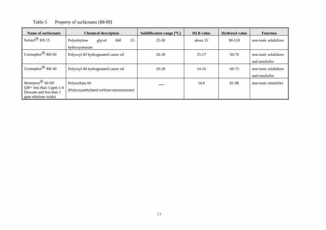
Four surfactants were tested for enhancing the liberation of poorly water-soluble drugs.
Solutol HS 15, Cremophor RH 40, Cremophor RH 60 (BASF, Germany) Montanox 60 DF
(SEPPIC, France) non-ionic surfactants were added to suppository bases. These are all well-
known additives which had not been used in the dosage form of rectal suppositories before.
These surfactants have good physiological tolerance and considerable efficiency as
regards solubilization and emulsification. Solutol HS 15 is recommended as a non-ionic
solubilizing agent to be added to injection solutions, while the use of Cremophor products is
proposed to make fat-soluble vitamins, essential oils, hydrophobic drugs, cosmetics water-
soluble and to improve bioavailability in solid dosage forms. The Montanox products are used
to obtain oil/water emulsion, for the dispersion or solubilization of essential oils or vitamins,
for some problems of gelification, in cosmetic and the pharmaceutical industries (Table 5).

18
Table 4 Property of suppository bases [79-80, 112-113]
Name of bases Chemical description Melting range (°°°°C) Hydroxyl value Function
Witepsol H 15 Triglycerides (C10-C18) 33.5-35.5 5-15 lipophilic base
Witepsol S 58 Higher proportion of mono- and diglycerides (C10-C18)
with the presence of cetostearyl alcohol
31.5-33 60-70 lipophilic base
Witepsol W 35 Higher proportion of mono- and diglycerides (C10-C18) 33.5-35.5 40-50 lipophilic base
Adeps solidus compositus Witepsol W 35 with the presence of two non-ionic
emulsifying additives
32-36 50-60 lipohydrophilic base
Massa Estarinum 299 Triglycerides (C10-C18) 33.5-35.5 max. 5 lipophilic base
Massa Estarinum B Higher proportion of mono- and diglycerides (C12-C18) 33.5-35.5 20-30 lipophilic base
Massa Estarinum BC Higher proportion of mono- and diglycerides (C12-C18) 33.5-35.5 30-40 lipophilic base
Suppocire AML Triglycerides (C8-C18) with the presence of a
phospholipid
35-36.5 max. 6 lipophilic base
Suppocire AP Saturated polyglycolysed glycerides 33-35 30-50 amphiphilic base
Suppocire AS2X Higher proportion of mono- and diglycerides (C8-C18)
with the presence of a non-ionic emulsifying additive
35-36.5 15-25 lipophilic base
Macrogolum 1540 Polyethylen glycol (n 28-36) Solidification range (°°°°C)
40-50
70-80 hydrophilic base
19
Table 5 Property of surfactants [88-89]
Name of surfactants Chemical description Solidification range (°°°°C) HLB value Hydroxyl value Function
Solutol HS 15 Polyethylene glycol 660 12-
hydroxystearate
25-30 about 15 90-110 non-ionic solubilizer
Cremophor RH 60 Polyoxyl 60 hydrogenated castor oil
20-28 15-17 50-70 non-ionic solubilizer
and emulsifier
Cremophor RH 40 Polyoxyl 40 hydrogenated castor oil 20-28 14-16 60-75 non-ionic solubilizer
and emulsifier
Montanox 60 DF (DF= less than 3 ppm 1-4 Dioxane and less than 1 ppm ethylene oxide)
Polysorbate 60
(Polyoxyaethylated sorbitan monostearate)
14.9 81-96 non-ionic emulsifier

20
4.1.4 Random-methyl-ββββ-cyclodextrin (RAMEB)
β-cyclodextrin is cyclic, non-reducting oligosaccharide built up from seven glucopyranose
units. Degree of substitution: DS: ∼ 12-13 methyl groups / CD ring.
Formula: C55H95O35
Molecular Weight: 1318.4
Appearance: White or slightly yellow powder
Melting point: 177-182 °C
Solubility: (in 100 cm3 solvent, at 25 °C)
Water >40 g
Methanol >25 g
Chloroform >25 g
Acetone < 5 g
Cavity diameter: 0.78 nm
Diameter of outer periphery: 1.53 nm
Height of torus: 0.78 nm
Number of water molecules filling the cavity: 11
Crystal water content: 13.2-14.5 % [114].
4.2. Methods
4.2.1. Preparing of the cyclodextrin (CD) complexes
The effect of the different CD derivatives on the solubility of ethacrynic acid was
determined: a mixture of 0.1 g of ethacrynic acid and 0.5 of CD derivative diluted to 20.0 g
with water was stirred for 10 min with a magnetic mixer. Suspension systems were filtered
through filter paper and, after suitable dilution, the UV spectra were recorded. A system
without CD was used as a control. Dimethyl-β-CD, methyl-β-CD, random-methyl-β-
cyclodextrin (RAMEB) had the highest influence on the solubility of the active agent.

21
RAMEB was chosen for further examinations on the bases of the costs and the solubility-
increasing effect: the solubility was increased by a factor of 9.33 [115].
The two-component products were prepared in four different mole ratios (drug:CD mole
ratio = 2:1, 1:1, 1:2 and 1:3) The ethacrynic acid content of the products was 35.91%, 21.88%,
12.29% and 8.54%.
Physical mixture: The ground components were mixed in a mortar and sieved through a
100 µm sieve.
Kneaded products: Physical mixtures of the drug and CD were mixed (Erweka LK5) in
the same quantity of ethanol + water (1:1). They were kneaded until the bulk of the solvent
mixture had evaporated. After this, they were dried at room temperature and then at 105 °C,
and were next pulverized and sieved through a 100 µm sieve.
Products were stored under normal conditions at room temperature in closed glass
containers.
The 1:1 kneaded product was selected for further investigations on the bases of the
dissolution and in vitro membrane diffusion results. This high active agent-containing
composition with improved solubility and diffusibility is suitable for incorporation into
lipophilic suppository bases [115].
4.2.2. Suppository formulation methods
Suppositories were formulated by moulding. In the case of in vitro experiments the drug
content was 2.5 w/w%, which corresponded to the therapeutic dose, that is a 2 g adult
suppository contained 50 mg drug. For the animal experiments 0.3 g suppositories were
prepared, adjusted to the anatomical size of rats, the drug content was 15 mg/suppository. The
additives were incorporated in the suppository base in a concentration of 1, 3, 5 or 10 %.
Suppositories were stored under normal conditions at room temperature and examined after
one week.

22
4.2.3. In vitro release study
Experiments were performed with the method of dynamic membrane diffusion [116],
which is a useful method for following the rate of drug release and membrane diffusion from
the powder without excipient and from the different suppository compositions, too. The
acceptor phase was distilled water at a pH 6.8 or phosphate buffer at a pH 7.5. The
suppositories were individually packed in a kidney dialysing membrane (VISKING
Dialysis
Tubing 36/32 SERVA, Germany [117]) and placed into 20 ml (lipophilic base) or 40 ml
(hydrophilic base) acceptor phase of body temperature (37 ± 0.5 °C). The samples were placed
into VIBROTHERM shake bath [118] and exposed to slight shaking (50/min.). The acceptor
phase was replaced after 30, 60, 120, 240 min. The quantity of drug in these samples was
measured with a spectrophotometer at λ= 278 nm in case of ethacrynic acid and at λ= 274 nm
in case of furosemide, using the absorbance value. [119]. The results were evaluated and
analysed statistically with the Prism 2.01 (GraphPad Software, USA) computer program. The
data are the averages of the results of five experiments ± S.E.M. (*p<0.05; **p<0.01;
***p<0.001 versus control, analysis of variance Newman-Kleus test).
Composition of the phosphate buffer pH=7.5:
Sodium Hydroxide 2,445 g
Potassium dihydrogen phosphate 10,569 g
Distilled water up to 1000 ml
4.2.4. In vivo study *
Animal investigations were carried out with the approval of the Ethical Committee for
Animal Research, University of Szeged (Registration number: 23/1999).

23
*The animal investigations were carried out in the Department of Pharmacodynamics and Biopharmacy of the University of Szeged. I would like to thank Prof. Dr. György Falkay, Head of Department, and Eszter Ducza, assistant lecturer, for their co-operation and assistance in evaluation.
The animal studies were performed with Sprague-Dawley male rats of 280-300 g. After 6
hours’ fasting the oral administration was done with oral tube and the suppository was placed
in the animals in ether anaesthesia, then they received 20 ml/kg water per rat. They were
placed in special cages where urine was collected every 10 minutes during 150 minutes. The
control rats received only 20 ml/kg water. The results were evaluated and analysed statistically
with the Prism 2.01 (GraphPad Software, USA) computer program. The data are the averages
of the results of six experiments ± S.E.M. (*p<0.05; **p<0.01; ***p<0.001 versus control,
analysis of variance Newman-Kleus test).
4.3. Experimental conditions
The conditions of the experiments carried out with the two drugs are summarized in
Table 6. In the case of ethacrynic acid 11 various suppository bases were examined in two
acceptor phases with different pH values. 3 non-ionic surfactants were tested for enhancing
the membrane diffusion of the drug, and liberation was increased by making the drug water-
soluble. In addition to in vitro experiments, in vivo studies were also performed, but no
evaluable dose-effect relationship was found in the studied rats.
In the course of furosemide examinations 7 different suppository bases were examined in
phosphate buffer of pH = 7.5. 3 non-ionic surfactants were used to facilitate drug liberation. In
vitro membrane diffusion examinations were accompanied by in vivo animal investigations.

24
Table 6 Summary of experimental conditions
Drugs In vitro membrane diffusion In vivo study
Acceptor phase Suppository bases Surfactants Cyclodextrin
Ethacrynic acid
distilled water
pH=6.8
Witepsol H 15; S 58; W 35;
Adeps solidus compositus;
Massa Estarinum 299; B; BC;
Solutol HS 15
Cremophor RH 40
Cremophor RH 60
Random-
methyl-β-
cyclodextrin
no dose-effect curve
could be evaluated
phosphate buffer
pH=7.5
Suppocire AML; AS2X; AP;
Macrogolum 1540
(RAMEB)
Furosemide
phosphate buffer
pH=7.5
Witepsol H 15; W 35;
Massa Estarinum B; BC;
Solutol HS 15
Cremophor RH 60
no results In vitro / in vivo
correlation
Suppocire AML; AS2X; AP Montanox 60 DF

25
5. RESULTS AND DISCUSSION
5.1 Preliminary examinations
5.1.1. Plotting of calibration lines
A stock solution of known concentration was prepared from the active agents and
measurements were made from dilutions made from this stock solution at λ=278 [120] and
λ=274 [120] nm in the case of ethacrynic acid and furosemide, respectively. Based on the six
parallel measurements, a linear relationship was found between the concentrations of the
active agent and the extinctions measured. The slope and intercept of the lines as well as the
value of the correlation coefficient confirming the closeness of the correlation were
determined with the help of a computer; these values are shown in Table 7.
Table 7 Characteristics of the calibration lines
Active agent Slope Intercept Correlation Coefficient Ethacrynic acid 0.0128 0.0085 0.9992
Furosemide 0.0616 0.0123 0.9998
5.1.2. Powder diffusion studies
50 mg of the active agents was measured on an analytical balance into the previously
prepared kidney dialysing membrane and five parallel measurements were carried out in each
case according to the method of dynamic membrane diffusion described above. The quantity
of the diffused drug was determined in % (Table 8). The powder diffusion results were taken
as control in subsequent examinations. It was found that the diffusion of the drug increased
considerably in the phosphate buffer of pH=7.5, a significant difference could be observed
with the change of the acceptor phase at the significance level of p<0.001, which can be
explained by the better solubility of the drug at alkaline pH.

26
Table 8 Power diffusion results of drugs
Ethacrynic acid (%) Furosemide (%)
time (min.) distilled water pH=6.8
phosphate buffer pH=7.5
phosphate buffer pH=7.5
30 1.84 13.24 7.37
60 2.06 25.84 15.29
120 4.64 48.42 34.15
240 7.69 79.88 65.07
5.2. Influence of pH change on ethacrynic acid release from different
suppository bases
The pH of the rectum varies between 6.8-7.9 [13]. The experiments were carried out in
distilled water (pH=6.8) and phosphate buffer (pH=7.5). The membrane diffusion of the
powder without a suppository base was regarded as control. Release values obtained with the
hydrophilic Macrogolum 1540 (***p<0.001) base in aqueous medium were manifold higher
then those determined with lipophilic bases or powder (Fig. 4). This can be due to the fact that
poorly water-soluble drugs are better released from hydrophilic suppositories, and the base
may moisten or solubilize the drug, therefore drug solubility and membrane diffusion were
increased. Results of membrane diffusion were 7-8 % from lipophilic bases, which were near
the membrane diffusion of the powder (Fig. 5).
It, however, the results obtained in aqueous medium and buffer medium are compared, it
can be seen that the change of the acceptor phase did not have a significant influence on drug
release from Macrogolum 1540, but from lipophilic bases it was increased about tenfold in the
acceptor phase of pH= 7.5 (Fig 6). This result can be explained by the change of the solubility
of the drug, as ethacrynic acid is a weak acid so its solubility will increase with the increase of
pH, which facilitates drug liberation from lipophilic bases, and the membrane diffusion of the
drug will also be enhanced. As concerns lipophilic bases, bases with a small hydroxyl value

27
Fig. 4 Ethacrynic acid release from different suppository bases after 240 minutes
Acceptor phase: Distilled water
0
25
50
75powderWitepsol H 15Witepsol S 58Witepsol W 35Adeps sol. comp.Massa Estarin. 299Massa Estarin. BMassa Estarin. BCSuppocire AMLSuppocire AS2XSuppocire APMacrogolum 1540
***
dru
g r
elea
se (
%)
Fig. 5 Ethacrynic acid release from different lipophilic suppository bases after 240 minutes
Acceptor phase: Distilled water
0.0
2.5
5.0
7.5
10.0powderWitepsol H 15Witepsol S 58Witepsol W 35Adeps sol. comp.Massa Estarin. 299Massa Estarin. BMassa Estarin. BCSuppocire AMLSuppocire AS2XSuppocire AP
*****
***
**
***dru
g r
elea
se (
%)

28
Fig. 6 Ethacrynic acid release from different suppository bases after 240 minutes
Acceptor phase: Phosphate buffer
0
25
50
75
100powderWitepsol H 15Witepsol S 58Witepsol W 35Adeps sol. comp.Massa Estarin. 299Massa Estarin. BMassa Estarin. BCSuppocire AMLSuppocire AS2XSuppocire APMacrogolum 1540
* *
*** ***
** ***
dru
g r
elea
se (
%)
gave better results. In the case of Massa Estarinum 299, Massa Estarinum B and Suppocire
AML there was no significant decrease (p>0.05) compared to the membrane diffusion values
of the powder either in aqueous or buffer medium, so the suppository bases did not have a
retaining effect. Witepsol W 35 (***p<0.001), Adeps solidus compositus (***p<0.001) and
Suppocire AP (***p<0.001) with a greater hydroxyl value gave the worst results in both
acceptor phases. Adeps solidus compositus contains Witepsol W 35 and two non-ionic
surfactants, too, so drug diffusion could be expected to increase with the moistening,
solubilization of the drug and by making the base lipohydrophilic. The membrane diffusions of
Witepsol W 35 and Adeps solidus compositus showed no significant difference, which can
probably be explained by the fact that the joint quantity of 20 % of the two surfactants has an
unfavourable influence on drug liberation (see the figures in 5.2).
It is obvious that the kinetics of release from lipophilic and hydrophilic bases differ, as
drug diffusion from the hydrophilic base showed a considerable increase only after the first
hour. This finding is related to the longer dissolving time of hydrophilic bases. As the
efficiency of the two active agents used in the study is not independent of time, in order to

29
achieve faster and better effect, the combinations of lipophilic bases and various additives
(surfactants, cyclodextrins) were used to further improve the results.
5.3. Influence of surfactant concentration on ethacrynic acid release
from Witepsol H 15 base
The surfactants were incorporated in the Witepsol H 15 base in a concentration of 1, 3, 5
and 10 %. The Witepsol H 15 suppository base was chosen because it did not yield maximum
results in the two acceptor phases, so the use of additives was expected to enhance drug
liberation. The membrane diffusion of the drug from Witepsol H 15 base was regarded as a
control. The diffusion of the drug was found to vary with their concentration. When distilled
water was used as the acceptor phase, the concentration of 3 % yielded the best results in the
case of all the three surfactants, this led to about a twofold increase in liberation. Except for
5% of Cremophor RH 60, their use in a concentration of 1-5-10 % did not change or decrease
drug liberation (Fig 7), which can probably be explained by the concentration of surfactants
accumulated on the boundary surface as the quantity of the diffused drug is increased by
proper saturation, while a too small or too great amount of surfactants may lead to its decrease.
When the same examinations were performed in a buffer medium, 1, 3, 5 % of Solutol HS
15 (***p <0.001) and Cremophor RH 40 (**p<0.01) led to increase in diffusion, while the use
of Cremopor RH60 (p>0.05) (which gave the best results in distilled water) did not bring about
a change in the extent of drug release (Fig. 8).
Consequently, it can be established that the increase of the pH of the acceptor phase
decreased the drug liberation-increasing effect of Cremophor RH 60 surfactant, while Solutol
HS 15 and Cremophor RH 40 were more effective in a buffer medium. However, in the
phosphate buffer 1 % of the given additive was sufficient for eliciting the required effect.

30
Fig. 7 Influence of additives on drug release after 240 minutes
Acceptor phase: Distilled water
0 % 1 % 3 % 5 % 10 %0
5
10
15
Witepsol H 15+Solutol HS 15Witepsol H 15+Cremophor RH 40Witepsol H 15+Cremophor RH 60
******
*
**
*
surfactant concentration
dru
g r
elea
se (
%)
Fig. 8 Influence of additives on drug release after 240 minutes
Acceptor phase: Phosphate buffer
0 % 1 % 3 % 5 % 10 %0
25
50
75
100
Witepsol H 15+Solutol HS 15Witepsol H 15+Cremophor RH 40Witepsol H 15+Cremophor RH 60
****** ***
** ** **
surfactant concentration
dru
g r
elea
se (
%)

31
5.4. Results of ethacrynic acid and random-methyl-ββββ-cyclodextrin
complex release from different suppository bases
Ethacrynic acid and the previously selected ethacrynic acid + RAMEB 1:1 kneaded
product were incorporated into 5 different, previously examined lipophilic suppository bases
(Witepsol H 15, Witepsol W 35, Massa Estarinum 299, Suppocire AML, Suppocire AP). The
membrane diffusion of ethacrynic acid without a suppository base was regarded as control.
The amount of ethacrynic acid released in distilled water was under 10%. This can be
explained by the aqueous solubility of the active agent, resulting in an unsatisfactory liberation
from lipophilic suppository bases. Witepsol H 15, Suppocire AML and Massa Estarinum 299
afforded the best results as concerns the investigated suppository bases. The diffusion of the
drug from all the suppository bases was higher when the CD complex of ethacrynic acid was
used. A 10-fold increase in liberation was experienced in the cases of Witepsol H 15,
Suppocire AML and Massa Estarinum 299 (***p<0,001) (Fig 9).
Fig. 9 Solubility and diffusibility increasing effect of RAMEB in distilled water
0
25
50
75powderWitepsol H 15Witepsol H 15 (CD)Witepsol W 35Witepsol W 35(CD)Massa Estarin. 299Massa Estarin. 299(CD)Suppocire AMLSuppocire AML(CD)Suppocire APSuppocire AP(CD)
***
******
dru
g r
elea
se (
%)
The solubility of ethacrynic acid increased with the pH increase of the acceptor phase, and
so did the diffusibility through the membrane (Fig 10). The best suppository bases in the

32
distilled water experiments (Witepsol H 15, Suppocire AML and Massa Estarinum 299) were
also the best in the phosphate buffer medium. The diffusion results for the suppositories
containing CD complexes were poorer than those for the suppository containing pure
ethacrynic acid, which can be explained by the higher solubility of ethacrynic acid in the
phosphate buffer. The rectal pH range is 6.8-7.9. As the liberation and diffusion of the active
agent are pH-dependent processes, the diuretic effect can fail if the rectal pH lies out of the
physiological range. The CD complex of ethacrynic acid was found to be appropriate for the
production of suppositories that are effective independently of the pH of the surrounding
media.
Fig. 10 Solubility and diffusibility increasing effect of RAMEB in phosphate buffer
0
25
50
75
100powderWitepsol H 15Witepsol H 15(CD)Witepsol W 35Witepsol W 35(CD)Massa Estarin. 299Massa Estarin. 299(CD)Suppocire AMLSuppocire AML(CD)Suppocire APSuppocire AP(CD)
***
***
***
*********
*
dru
g r
elea
se (
%)
5.5. In vitro membrane diffusion of furosemide from different
suppository bases
The membrane diffusion of the powder without a suppository base was regarded as
control during the in vitro experiments. It can be stated that drug diffusion from Suppocire
AS2X (***p<0.001), Massa Estarinum B (**p<0.01) and Witepsol H 15 (*p<0.05) was about

33
the same as from the powder without a suppository base. Suppocire AML (***p<0.001),
Massa Estarinum BC (**p<0.01) and Suppocire AP (***p<0.001) decreased drug release to a
smaller extent, while Witepsol W 35 (***p<0.001), which has a relatively high hydroxyl
value, decreased drug release with orders of magnitude (Fig. 11). This is contradicted by the
fact that the hydroxyl value of Suppocire AP is approximately the same as that of Witepsol W
35, nevertheless furosemide liberation shows a significant difference. This is probably due to
the amphiphilic properties of Suppocire AP, which - for most drugs - lead to increased
bioavailability compared to traditional lipophilic suppository bases.
Fig. 11 Furosemide in vitro release study from different suppository bases
0
25
50
75powderWitepsol H 15Witepsol W 35Massa Estarin. BMassa Estarin. BCSuppocire AMLSuppocire AS2XSuppocire AP
****
***
***
***
***
*
dru
g r
elea
se (
%)
5.6. Diuretic effect of furosemide from different suppository
compositions
In the course of the in vivo trials the dose-effect relationship was examined after the
administration of furosemide orally and rectally (suppository with the Witepsol H 15 base)
(Fig. 12). The ED50 value was calculated from the figure in both cases (ED50 supp=15.39 mg,
ED50 per os=19.03 mg), which revealed that rectal administration is slightly more effective than
oral administration. In the case of furosemide the hepatic first-pass effect is almost negligible,
the major site for the first-pass metabolism of the drug in rats is probably the GI tract.
Gastrointestinal and intestinal first-pass effect has been described in rats concerning

34
furosemide, where 20-40 % of the administered drug is metabolised [46]. Further
examinations were carried out with the ED50 value calculated from the dose-effect
examinations.
Fig. 12 Dose-dependent effect of furosemide
-1 0 1 20
10
20suppositoryper os
furosemide (log mg)
vol
ume
(ml)
Furosemide was incorporated in suppository bases, and after application in rats urine was
collected for 150 minutes. Compared to the control, a significant increase was observed in the
quantity of urine when Suppocire AP (* p<0.05), Witepsol H 15 (* p<0.05), Witepsol W 35
(** p<0.01), Massa Estarinum B (*** p<0.001) and Suppocire AS2X (*** p<0.001)
suppository bases were used. The use of Suppocire AML and Massa Estarinum BC did not
bring about a significant difference in urine quantity compared to the control (Fig. 13). The
effectiveness of Suppocire AS2X and Massa Estarinum B is clearly shown by the fact that the
amount of urine collected for 150 minutes came near to the 24-hour urine quantity of rats
[121].
5.7. Influence of surfactants on furosemide release and diuretic effect
Three non-ionic surfactants were also tested for increasing furosemide liberation. The
surfactants were incorporated in the Witepsol H 15 base in a concentration of 1, 3, 5 and 10 %.

35
The Witepsol H 15 suppository base was chosen because it did not yield maximum result
either during the in vitro or - mainly - in the in vivo examinations, so the use of additives was
expected to enhance drug liberation and diuretic effect. During the in vitro examinations only

36
Fig. 13 Diuretic effect of different suppository bases containing furosemide in rats
0 50 100 1500
10
20
Witepsol W 35Massa Estarin. B
Suppocire AS2X
Witepsol H 15
Suppocire AML
Suppocire AP
Massa Estarin. BC
control
time (min)
volu
me
(ml)
the 1 % concentration of Cremophor RH 60 led to a significant increase, in the other cases no
significant differences were observed, or furosemide diffusion even decreased with the
increase of the surfactant concentration (Fig. 14). The decrease in drug diffusion through the
membrane is due to two causes: 1. The additive, drug and base formed a stable complex, or the
conditions of dissociation were influenced unfavourably by the additive. 2. Although the drug
was released from the suppository base, a certain extent of increase in the surfactant
concentration resulted in the formation of micelles of colloidal size, so it is possible that the
drug molecules closed in the micelles were unable to pass through the dialysing membrane
which had a pore size of 25Å. This latter supposition is confirmed by the results of the in vivo
experiments, in which the diuretic effect was definitely enhanced by the surfactants, and in the
case of Cremophor RH 60 the critical micellar concentration was probably over 1 % so no
aggregate was formed and the drug could diffuse through the membrane.
In the in vivo examinations the use of surfactants led to the significant increase in the
amount of the collected urine (Fig. 15). Their effect is composed of several factors: they
moisten the drug, they denaturate the proteins found on the intestinal mucosa thereby
disrupting the integrity of the membrane, and furthermore they increase the number of
adsorption places by cleaning the membrane surface. Nerurkar et al. [122] suggest that

37
Fig. 14 Influence of additives on drug release after 240 minutes
0 % 1 % 3 % 5 % 10 %0
25
50
75
100
Witepsol H 15+Solutol HS 15Witepsol H 15+Cremophor RH 60Witepsol H 15+Montanox 60 DF
***
***
* ***
***
*** **
***
******
surfactant concentration
dru
g r
elea
se (
%)
Fig. 15 Influence of additives on diuretic effect in rats
0% 1% 3% 5% 10%0
10
20
Witepsol H15+Solutol HS 15Witepsol H 15+Cremophor RH 60Witepsol H15+Montanox 60DF
********* ****** *********
***
******
surfactant concentration
volu
me
(ml)

38
surfactants, which are commonly added to pharmaceutical formulations, may enhance the
intestinal absorption of some drugs by inhibiting an apically polarized efflux system. In the
animal experiments performed with rats all the three additives increased the quantity of the
excreted urine approximately to the same extent, which indicates increased drug liberation.
Figure 15 also shows that the increase of the surfactant concentration was not accompanied
with significant changes, so a concentration of 1 % is enough to achieve the desired effect.
5.8. Mathematical evaluation of experimental data
Linear regression was used to find a relationship between the process of dissolution and
time. The calculations revealed that the process of dissolution could be characterized by a
power function, which is also confirmed by the fact that lines were obtained when the
logarithm of the quantity of the dissolved drug was plotted against the logarithm of the time. If
the parameters of the functions are known, the extent of drug liberation or its diffusion through
the membrane can be calculated at any intermediate time.
log C = K * log t + B (3) where C is the amount of material released after time t, K is the slope and B is the intercept of
the straight line.
The following tables (Table 9-16) show the slope (K) and intercept (B) of the lines, the
values of the correlation coefficients indicating the closeness of the correlation (R), the time
needed for the liberation of 50 % of the drug (t50) and in vitro availability, the values over 90
% are presented in red colour. The slope is the rate constant of the process, the value of the
intercept (liberation belonging to 0 time) should be 0 in principle. The negative values are due
to the fact that first the membrane has to be impregnated with the drug, and diffusion starts
after impregnation.

39
Table 9 Ethacrynic acid in distilled water
slope (K)
intercept (B)
R t50 (min.)
in vitro availability
powder 0.7357 -1.1578 1.0000 2977 100.00 Witepsol H 15 0.7009 -1.1042 0.9994 3714 91.27 Witepsol S 58 0.7596 -1.4462 0.9990 5549 58.10 Witepsol W 35 0.7772 -1.3843 0.9986 3800 70.15 Adeps solidus comp. 0.7865 -1.5221 0.9996 5160 53.99 Massa Est. 299 0.8566 -1.5246 0.9998 2581 80.36 Massa Est. B 0.6991 -1.1566 0.9997 4508 80.03 Massa Est. BC 0.7865 -1.3312 0.9997 2951 83.61 Suppocire AML 0.8075 -1.3366 0.9995 2434 96.53 Suppocire AS2X 0.7126 -1.2410 0.9993 5049 73.34 Suppocire AP 0.5916 -1.3561 0.9990 45210 28.06 Macrogolum 1540 1.2891 -1.3200 0.9778 128 795.31
Table 10 Ethacrynic acid in phosphate buffer
slope (K)
intercept (B)
R t50 (min.)
in vitro availability
powder 0.7724 -0.2184 0.9981 123 100.00 Witepsol H 15 0.8165 -0.3866 0.9843 153 77.81 Witepsol S 58 0.8200 -0.4005 0.9920 156 78.56 Witepsol W 35 0.5091 0.1263 0.9958 314 50.61 Adeps solidus comp. 0.9811 -0.9323 0.9989 237 53.15 Massa Est. 299 0.7149 -0.0763 0.9948 115 95.50 Massa Est. B 0.7975 -0.2485 0.9916 116 98.29 Massa Est. BC 0.7743 -0.3427 0.9995 177 73.41 Suppocire AML 0.8740 -0.4548 0.9935 131 93.06 Suppocire AS2X 0.7492 -0.1524 0.9864 117 91.46 Suppocire AP 0.8006 -0.4079 0.9963 180 70.62 Macrogolum 1540 1.7240 -2.2859 0.9740 137 85.24
Table 11 Influence of additives in distilled water
slope (K)
intercept (B)
R t50 (min.)
in vitro availability
powder 0.7357 -1.1578 1.0000 2977 100.00 Witepsol H 15+Solutol HS 15 1% 0.8208 -1.4113 0.9998 2645 86.35 Witepsol H 15+Solutol HS 15 3% 0.7927 -1.1752 0.9991 1762 137.84 Witepsol H 15+Solutol HS 15 5% 0.8857 -1.4947 0.9991 1844 102.08 Witepsol H 15+Solutol HS 15 10% 0.9526 -1.8391 1.0000 2500 63.41 Witepsol H 15+Cremophor RH 40 1% 0.9361 -1.6892 0.9979 1985 85.33 Witepsol H 15+Cremophor RH 40 3% 0.8532 -1.3351 0.9997 1596 125.61 Witepsol H 15+Cremophor RH 40 5% 0.9413 -1.6885 0.9971 1900 84.86 Witepsol H 15+Cremophor RH 40 10% 0.8754 -1.5535 0.9987 2352 86.82 Witepsol H 15+Cremophor RH 60 1% 0.7951 -1.3355 0.9972 2740 87.99 Witepsol H 15+Cremophor RH 60 3% 0.9208 -1.3924 0.9990 1072 153.61 Witepsol H 15+Cremophor RH 60 5% 1.0336 -1.7984 0.9976 1237 112.24 Witepsol H 15+Cremophor RH 60 10% 0.8657 -1.5947 0.9991 2863 89.14

40
Table 12 Influence of additives in phosphate buffer
slope (K)
intercept (B)
R t50 (min.)
in vitro availability
powder 0.7724 -0.2184 0.9981 123 100.00 Witepsol H 15+Solutol HS 15 1% 0.7329 -0.0780 0.9936 103 103.16 Witepsol H 15+Solutol HS 15 3% 0.7639 -0.2044 0.9978 125 95.88 Witepsol H 15+Solutol HS 15 5% 0.7358 -0.1302 0.9954 119 94.54 Witepsol H 15+Solutol HS 15 10% 0.8778 -0.5965 0.9993 187 73.27 Witepsol H 15+Cremophor RH 40 1% 0.7470 -0.1563 0.9976 120 98.46 Witepsol H 15+Cremophor RH 40 3% 0.7764 -0.2226 0.9958 122 96.58 Witepsol H 15+Cremophor RH 40 5% 0.8297 -0.3568 0.9973 130 95.52 Witepsol H 15+Cremophor RH 40 10% 0.8735 -0.5412 0.9992 165 81.65 Witepsol H 15+Cremophor RH 60 1% 0.7580 -0.2853 0.9979 166 77.07 Witepsol H 15+Cremophor RH 60 3% 0.7783 -0.3711 0.9999 187 72.96 Witepsol H 15+Cremophor RH 60 5% 0.9304 -0.7113 0.9990 184 74.59 Witepsol H 15+Cremophor RH 60 10% 0.8819 -0.6192 0.9994 193 70.78
Table 13 Ethacrynic acid with RAMEB in distilled water
slope (K)
intercept (B)
R t50 (min.)
in vitro availability
powder 0.7357 -1.1578 1.0000 2977 100.00 Witepsol H 15 0.6334 -0.0729 0.9792 209 629.59 Witepsol W 35 0.6287 -0.8194 0.9998 3364 119.41 Massa Est. 299 0.9056 -0.8332 0.9987 290 511.70 Suppocire AML 0.7349 -0.3751 0.9861 258 548.76 Suppocire AP 0.5401 -0.7437 0.9907 9231 82.89
Table 14 Ethacrynic acid with RAMEB in phosphate buffer
slope (K)
intercept (B)
R t50 (min.)
in vitro availability
powder 0.7724 -0.2184 0.9981 123 100.00 Witepsol H 15 0.7619 -0.2315 0.9890 137 85.42 Witepsol W 35 0.5516 -0.3051 0.9922 1223 22.73 Massa Est. 299 0.9114 -0.6119 0.9897 160 79.94 Suppocire AML 0.8965 -0.6637 0.9955 199 68.29 Suppocire AP 0.9120 -0.8208 0.9977 270 51.56

41
Table 15 Furosemide in phosphate buffer
slope (K)
intercept (B)
R t50 (min.)
in vitro availability
powder 1.0782 -1.0414 0.9994 182 100.00 Witepsol H 15 0.7829 -0.3301 0.9965 161 96.24 Witepsol W 35 0.5417 -0.8751 0.9997 15708 7.78 Massa Est. B 0.7797 -0.2971 0.9972 149 102.92 Massa Est. BC 0.7287 -0.2350 0.9988 174 91.86 Suppocire AML 0.6669 -0.1323 0.9963 197 81.19 Suppocire AS2X 1.0197 -0.8398 0.9966 156 107.24 Suppocire AP 0.7444 -0.3142 0.9972 199 80.46
Table 16 Influence of additives in phosphate buffer
slope (K)
intercept (B)
R t50 (min.)
in vitro availability
powder 1.0782 -1.0414 0.9994 182 100.00 Witepsol H 15+Solutol HS 15 1% 0.8646 -0.6586 0.9999 239 75.26 Witepsol H 15+Solutol HS 15 3% 0.9678 -0.8282 0.9985 199 85.79 Witepsol H 15+Solutol HS 15 5% 1.0667 -1.0641 0.9993 203 86.07 Witepsol H 15+Solutol HS 15 10% 1.1462 -1.3408 0.9995 245 60.17 Witepsol H 15+Cremophor RH 60 1% 0.7005 -0.1155 0.9987 144 115.42 Witepsol H 15+Cremophor RH 60 3% 0.7554 -0.2519 0.9975 152 100.15 Witepsol H 15+Cremophor RH 60 5% 0.9276 -0.8776 0.9997 283 51.02 Witepsol H 15+Cremophor RH 60 10% 0.9772 -0.8228 0.9992 187 89.69 Witepsol H 15+Montanox 60 DF 1% 0.8075 -0.4895 0.9989 217 91.33 Witepsol H 15+Montanox 60 DF 3% 0.9797 -0.7562 0.9957 158 97.84 Witepsol H 15+Montanox 60 DF 5% 1.0667 -1.0641 0.9993 203 86.06 Witepsol H 15+Montanox 60 DF 10% 1.1421 -1.3358 0.9955 247 77.78

42
6. SUMMARY
Having considered the characteristics of rectal drug administration, the physiological state
of the rectum, the properties of drugs, bases and additives, I have drawn the following
conclusions and I am proposing the following compositions for the formulation of diuretic
rectal suppositories:
Considerations in the technological formulation of rectal suppositories containing
ethacrynic acid:
1. The solubility of the drug was increased manifold by changing the pH of the acceptor
phase. As a result drug liberation from various suppository bases changed. Liberation
from lipophilic bases was increased about ten times by increasing the pH. The best
results were given by bases with a small hydroxyl value and by lipophilic bases
containing an additive. Hydrophilic Macrogolum 1540 gave good results both in an
aqueous and buffer medium, but because of its long disintegration time it can be
proposed for the formulation of diuretic suppositories only under certain conditions (e.g.
tropics-resistant suppositories).
2. When non-ionic surfactants are used with lipophilic bases, drug liberation increases
independently of the pH due to the base becoming lipohydrophilic. The extent of the
increase was greater in distilled water (pH=6.8) as the surfactant contributed not only to
making the base lipohydrophilic but it also solubilized the poorly soluble drug. The
quantity of the surfactant is one of the most important factors in the formulation of rectal
suppositories. Drug liberation changed according to a maximum function. In aqueous
medium a surfactant concentration of 3-5 % proved to be optimal, while in a buffer
medium 1 % was enough to give the best results. The physical-chemical parameters of
the surfactant were also decisive, which modified the results with pH change.

43
3. The formulation of the cyclodextrin complex of the drug resulted in about a tenfold
increase in the solubility of ethacrynic acid in distilled water, and as a consequence the
membrane diffusion of the drug also improved considerably. The solubility of ethacrynic
acid increases with the pH increase, so the results of cyclodextrin complexes were worse
than those of the membrane diffusion of the pure drug. In this case the retaining effect of
the complex may have to be reckoned with.
In view of the above summary, with the consideration of the pH of the rectum, the following is
proposed for the formulation of rectal suppositories containing ethacrynic acid:
♦♦♦♦Witepsol H 15 base containing 3% Solutol HS 15 additive, or
♦♦♦♦ethacrynic acid + random-methyl-ββββ-cyclodextrin complex incorporated in
Witepsol H 15 suppository base.
Considerations in the technological formulation of rectal suppositories containing
furosemide:
1. When the membrane diffusion examinations are compared with the actual diuretic effect,
it can be stated that drug liberation and pharmacological effect showed the same
tendency in 70 %, that is a greater extent of furosemide liberation was accompanied with
a greater amount of animal urine. The best results were given by the Suppocire AS2X
base in both cases, which means that the liberation of the drug was about 70 % and the
animal produced approximately 15 ml of urine in 150 minutes, which equals the daily
urine quantity of a rat according to literature data.
2. The Witepsol H 15 base yielded better results under in vitro conditions than in the
animal investigations, and in the case of the Witepsol W 35 base the pharmacological
effect proved to be better than the results of the membrane diffusion examinations. This
also confirms that if the best composition is to be chosen, it is essential to supplement in

44
vitro results with in vivo experiments in order to form a clear picture about the
interactions between the active agent-base-living organism.
3. When non-ionic surfactants were used, in vitro examinations revealed a significant
increase only with the use of 1 % Cremophor RH 60 surfactant concentration, in the
other cases there was no significant difference or the diffusion of furosemide decreased
with the increase of the surfactant concentration. In the in vivo experiments diuretic
effect was definitely increased by surfactants, but 1 % of them was sufficient for eliciting
maximum effect.
Based on the results, I have found two compositions suitable for formulating furosemide-
containing suppositories:
♦♦♦♦Suppocire AS2X suppository base in itself, which proved to be the best both in
the membrane diffusion and during the animal experiments, or
♦♦♦♦Witepsol H 15 suppository base with 1% Cremophor RH 60 additive, which
also gave optimal results with both examination methods.

45
7. REFERENCES
1. ifj. Regdon G., Regdon G., Selmeczi B.: Gyógyszerészet 42, 451-461 (1998)
2. Daharjanu, M., D., Kumaran, K. S., Baskaran, T., Moorthy, M. S.: Drug. Dev. Ind.
Pharm. 24, 583-587 (1998)
3. Hermann, T. W.: Int. J. Pharm. 123, 1-11 (1995)
4. Yahagi, R., Machida, Y., Onishi, H., Machida, Y.: Int. J. Pharm. 193, 205-212 (2000)
5. Nagatomi, A., Mishima, M., Tsuzuki, O., Ohdo, S., Higuchi, S.: Biol. Pharm. Bull. 20,
892-896 (1997)
6. VanDenBerg, C. M., Kazmi, Y., Stewart, J., Weidler, D. J., Tenjarla, S. N., Ward, E. S.,
Jann, M. W.: Am. J. Health Syst. Pharm. 57, 1046-1050 (2000)
7. Samy, E. M., Hassan, M. A., Tous, S. S., Rhodes, C. T.: Eur. J. Pharm. Biopharm. 49,
119-127 (2000)
8. Janicki, S., Sznitowska, M., Zebrowska, W., Gabiga, H., Kupiec, M.: Eur. J. Pharm.
Biopharm. 52, 249-254 (2001)
9. Makó S., Stampf G.: Acta Pharm. Hung. 71, 293-299 (2001)
10. Stampf G.: Acta Pharm. Hung. 68, 119-122 (1998)
11. Regdon, G., Fazekas, T., Regdon, G. jr., Selmeczi, B.: Pharmazie 51, 116-119 (1996)
12. Rácz I., Selmeczi B.: Gyógyszertechnológia, Egyetemi tankönyv, 4. kiadás, Medicina
Könyvkiadó Rt. Budapest, 2001
13. Müller, B. W.: Suppositorien. Pharmakologie, Biopharmazie und Galenik rektal und
vaginal anzuwendender Arzneiformen. Wissenschaftlich. Verlag. mbH Stuttgart, 1986
14. Kim, N. S., Umejima, H., Ito, T., Uchida, T., Goto, S.: Chem. Pharm. Bull. 40, 2800-
2804 (1992)
15. Umejima, H., Kim, N. S., Ito, T., Uchida, T., Goto, S.: J. Pharm. Sci. 82, 195-199 (1993)
16. Yahagi, R., Onishi, H., Machida, Y.: J. Control. Release 61, 1-8 (1999)
17. Kato, Y., Matsushita, T., Uchida, H., Egi, S., Yokoyama, T., Mohri, K.: Eur. J. Clin.
Pharmacol. 42, 619-622 (1992)
18. Lievertz, R. W.: Am. J. Obstet. Gynecol. 156, 1289-1293 (1987)
19. Knoll J.: Gyógyszertan, 8. kiadás, Medicina Könyvkiadó, Budapest, 1993

46
20. Pfaff, G., Zimmermann, T., Lach, P., Yeates, R., Simon, G., Wildfeuer, A.: Arzneim. -
Forsch./Drug Res. 43, 391-395 (1993)
21. Ritschel, W. A.: Angewandte Biopharmazie, Wiss. Verlag mbH Stuttgart, 1973
22. Ritschel, W. A.: Handbook of Basic Pharmacokinetics Including Clinical Applications,
Ed. 3. Hamilton USA Drug Intelligence, 1986
23. Hakata, T., Ijima, M., Kimura, S., Sato, H., Watanabe, Y., Matsumoto, M.: Chem.
Pharm. Bull. 41, 351-356 (1993)
24. Bornschein, M., Grohmann, A., Voigt, R.: Pharmazie 35, 772-776 (1980)
25. Schmitt, M., Guentert, T. W.: J. Pharm. Sci. 79, 359-363 (1990)
26. Nishihata, T., Rytting, J.: Adv. Drug Deliv. Rev. 28, 205-228 (1997)
27. Regdon G., Selmeczi B.: A rektális gyógyszerbevitel jelentősége napjainkban. A
gyógyszerészeti tudomány aktuális kérdései 10. szám, MGYT Kiadvány, Budapest, 1992
28. Regdon G.: A kúpkészítés és a kúpok gyógyszerleadását befolyásoló tényezők
tanulmányozása gyógyszertechnológiai és biofarmáciai szempontok figyelembevételével.
Kandidátusi értekezés, Szeged, 1975
29. European Pharmacopoeia Ed. 3. Council of Europe, Strasbourg Cedex 1997
30. European Pharmacopoeia Ed. 4. Council of Europe, Strasbourg Cedex 2002
31. Ermis, D., Tarimci, N.: Int. J. Pharm. 113, 65-71 (1995)
32. Azechi, Y., Ishikawa, K., Mizuno, N., Takahashi, K.: Drug. Dev. Ind. Pharm. 26, 1177-
1183 (2000)
33. Nakajima, T., Takashima, Y., Furuya, A., Ozawa, Y., Kawashima, J.: Chem. Pharm.
Bull. 37, 3145-3147 (1989)
34. Schneeweis, A., Müller-Goymann, C.: Int. J. Pharm. 196, 193-196 (2000)
35. Moolenaar, F., Meyler, P., Frijlink, E., Jauw, T., Visser, J., Proost, H.: Int. J. Pharm.
114, 117-120 (1995)
36. Mijazaki, S., Suisha, F., Kawasaki, N., Shirakawa, M., Yamatoya, K., Attwood, D.: J.
Control. Release 56, 75-83 (1998)
37. Schneeweis, A., Müller-Goymann, C. C.: Pharm. Res. 14, 1726-1729 (1997)
38. Realdon, N., Ragazzi, E., Dal Zotto, M., Dalla Fini, G.: Int. J. Pharm. 148, 155-163
(1997)
47
39. Chicco, D., Grabnar, I., Skerjanec, A., Vojnovic, D., Manrich, V., Realdon, N., Rajavni,
E., Belic, A., Karba, R., Mrhar, A.: Int. J. Pharm. 189, 147-160 (1999)
40. Mawatari, Hu Z., Shimokawa, S., Kimura, T., Yoshikawa, G., Shibata, Y., Takada K,
N.: J. Pharm. Pharmacol. 52, 1187-1193 (2000)
41. Eckert, Th., van Husen, N.: Dtsch. Apoth. Ztg. 120, 55-56 (1980)
42. Sákovits J., Regdon G., Pintyéné Hódi K., Elek B-né, Benkő A., Kraszkó P., Cseke B.:
Gyógyszerészet 34, 529-532 (1990)
43. Sákovics J., Benkő A., Jónás J., Elek B-né, Regdon G., Pintyéné Hódi K., Mézes É.:
Gyógyszerészet 34, 581-585 (1990)
44. Yun, M-O., Choi, H. G., Jung, J. H., Kim, C. K.: Int. J. Pharm. 189, 137-145 (1999)
45. Ryu, J. M., Chung, S. J., Lee, M. H., Kim, C. K., Shim, C. K.: J. Control. Release 59,
163-172 (1999)
46. Kim, E. J., Han, K. S., Lee, M. G.: J. Pharm. Pharmacol. 52, 1337-1343 (2000)
48. Regdon G., ifj. Regdon G., Tariné Gombkötő Zs., Berényi M.: Gyógyszerészet 39, 259-
265 (1995)
48. Watanabe, S., Belzile, M., Kuehn, N., Hanson, J., Bruera, E.: Cancer Treat. Rev. 22,
131-136 (1996)
49. Bruera, E., Watanabe, S., Fainsinger, R. L., Spachynski, K., Suarez-Almazor, M.,
Inturrisi, C.: Pain. 62, 141-146 (1995)
50. Kanamoto, I., Xin Zheng, N., Ueno, M., Koizumi, T., Adachi, I., Horikoshi, I.: Chem.
Pharm. Bull. 40, 1883-1886 (1992)
51. Bredow, V.: Zentralbl. Gynakol. 114, 551-554 (1992)
52. Kamiya, A., Ogata, H., Fung, HL.: J. Pharm. Sci. 71, 621-624 (1982)
53. Watanabe, Y., Matsumoto, Y., Hori, N., Funato, H., Matsumoto, H.: Chem. Pharm. Bull.
39, 3007-3012 (1991)
54. Hosny, E., El-Ahmady, O., El-Shattawy, H., El-M. Nabih, A., El-Damacy, H., Gamal-El-
Deen, S., El-Kabbany, N.: Arzneim. - Forsch./Drug Res. 44, 611-613 (1994)
55. Hacker, E., Kossowicz, J., Milde, K.: Pharmazie 46, 125-127 (1991)
56. Hosny, E. A.: Drug Dev. Ind. Pharm. 25, 745-752 (1999)

48
57. Iwata, M., Komiya, S., Nakamura, K., Kiuchi, M., Andoh, N., Hirahara, F., Maitani, Y.,
Takayama, K.: Drug Dev. Ind. Pharm. 27, 1039-1045 (2001)
58. Babar, A., Fares, H. M., Plakogiannis, F. M.: J. Pharm. Sci. 82, 389-391 (1993)
59. Kowari, K., Hirosawa, I., Kurai, H., Utoguchi, N., Fujii, M., Watanabe, Y.: Biol. Pharm.
Bull. 25, 678-681 (2002)
60. Regdon, G. jr., Deák, D., Regdon, G., Muskó, Zs., Erős, I.: Pharmazie 56, 70-73 (2001)
61. Regdon G., Sztanovics K., Zádeczky S., Selmeczi B.: Gyógyszerészet 33, 297-300
(1989)
62. Nissan, A., Ziv, E., Kidron, M., Bar-On, H., Friedman, G., Hyam, E., Eldor, A.:
Haemostasis 30, 225-232 (2000)
63. Sastri, M. S. P., Satyanarayana, N. V., Krishna, D. R., Diwan, P. V.: Arzneim. -
Forsch./Drug Res. 43, 320-323 (1993)
64. Ogiso, T., Iwaki, M., Kashitani, Y., Yamashita, K.: Chem. Pharm. Bull. 39, 2657-2661
(1991)
65. Ofoefule, S. I., Nwankwo, C., Chukwu, A., Orisakwe, O. E.: Boll. Chim. Farm. 137,
341-344 (1998)
66. Bergogne-Berezin, E., Bryskier, A.: J. Antimicrob. Chemother. 43, 177-185 (1999)
67. Sallai, J., Vernyik, Á., Regdon, G., Gombkötő, S., Németh, J., Regdon, G. jr.: J. Pharm.
Pharmacol. 49, 496-499 (1997)
68. Regdon, G., Dorogi-Jakab, I., Bándi, D., Várföldi, T., Regdon, G. jr., Selmeczi, B.: Eur.
J. Pharm. Biopharm. 38, 150-153 (1992)
69. Hacker, E., Krause, D., Kossowicz, J.: Pharmazie 47, 275-276 (1992)
70. Webster, J. A., Dowse, R., Walker, R. B.: Drug Dev. Ind. Pharm. 24, 395-399 (1998)
71. Lindmark, T., Soderholm, J. D., Olaison, G., Alvan, G., Ocklind, G., Artursson, P.:
Pharm. Res. 14, 930-935 (1997)
72. Sznitowska, M., Gajewska, M., Janicki, S., Radwanska, A., Lukowski G.: Eur. J. Pharm.
Biopharm. 52, 159-163 (2001)
73. Kelemen-Fazekas H., ifj. Regdon G.: Orvostudományi Értesítő 71, 243-249 (1998)
74. ifj. Regdon G., Bácskay I., Kata M., Regdon G.: Gyógyszerészet 35, 219-222 (1991)

49
75. Bácskay I., Keki S., Deák G., Zsuga M., Mezey G.: Acta Pharm. Hung. 71,153-156
(2001)
76. Czetsch-Lindenwald, V. H.: Suppositorien, Ed. Cantor, Aulendorf i. Württ. 1959
77. ifj. Regdon G., Schirm I., Pittmann Á., Regdon G.: Acta Pharm. Hung. 65, 45-50 (1995)
78. Kovács B., Selmeczi B., Szepessy A.: A gyógyszerkészítés segédanyagai, OGYI
Kiadvány, Budapest, 1983
79. Pharmacopoea Hungarica Editio VII., Medicina Könyvkiadó, Budapest, 1986
80. Formulae Normales Editio. VI., Medicina Könyvkiadó, Budapest, 1987
81. Fiedler, H. P.: Lexikon der Hilfsstoffe für Pharmazie Kosmetik und angrenzende
Gebiete, vierte, völlig überarbeitete und erweiterte Auflage, Ed. Cantor, Aulendorf, 1996
82. Jinno, J., Oh, D-M., Crison, J. R., Amidon, G. L.: J. Pharm. Sci. 89, 268-274 (2000)
83. Mezey G., Regdon G.: Acta Pharm. Hung. 41, 193-202 (1971)
84. Goodman and Gilman’s The pharmacological basis of therapeutics 9th Ed.., McGraw-
Hill, New York 1995
85. Buszello, K., Schnier, C., Müller B. W.: Eur. J. Pharm. Biopharm. 47, 247-253 (1999)
86. Scheller, Dworniczak, S., Pogorzelska, T., Rajca, M., Shani, J.: Arzneimittelforschung
50, 72-76 (2000)
87. Buszello, K.,Harnisch, S., Müller, R. H., Müller, B. W.:Eur. J. Pharm. Biopharm. 49,
143-149 (2000)
88. Pharmazeutische Spezialprodukte. Prospekt der BASF AG. Ludwigshafen, Germany
1993
89. Pharmaceutical and Health food. Seppic, Paris, France 1990, 1991
90. Usayapant, A., Iyer, B. R.: Drug Dev. Ind. Pharm. 25, 387-390 (1999)
91. Jarho, P., VanderVelde, D., Stella, V. J.: J. Pharm. Sci. 89, 241-249 (2000)
92. Aigner, Z.: Iomeglamsav- és β-ciklodextrin-tartalmú termékek, tabletták előállítása és
vizsgálata, Egyetemi Doktori Értekezés, Szeged, 1992
93. Aigner, Z., Kata, M.: STP Pharm. Sci. 9, 279-282 (1999)
94. Szejtli, J.: Cyclodextrins and their Inclusion Complexes. Akadémiai kiadó, Budapest,
1982
95. Bozsik E., Kissné Csikós E.: Acta Pharm. Hung. 63, 290-295 (1993)

50
96. USP 23/NF 18. United States Pharmacopeial Convention, Inc. Rockville, 1995
97. Thoma, K.: Arzneiformen zur rektalen und vaginalen Applikation. Werbe- und
Vertriebsgesellschaft Deutscher Apotheker mbH, Frankfurt am Main, 1980
98. Kingsford, M., Eggers, N. J., Soteros, G., Maling, T. J., Shirkey, R. J.: J. Pharm.
Pharmacol. 36, 536-538 (1984)
99. Yen, S. Y., Sung, K. C., Wang, J. J., Yoa-Pu Hu, O.: Int. J. Pharm. 220, 91-99 (2001)
100. Regdon, G., Szikszay, M., Gebri, G., Regdon, G. jr., Erős, I.: Pharmazie 54, 521-523
(1999)
101. Bornschein, M., Hoffmann, K., Voigt, R.: Pharmazie 40, 449-455 (1985)
102. Kapás M., Kedvessy Gy., Regdon G., Minker E., Magyarlaki A.: Acta Pharm. Hung. 51,
161-167 (1981)
103. Magyarlaki A., Minker E., Kapás M., Kedvessy Gy., Regdon G.: Acta Pharm. Hung. 51,
258-263 (1981)
104. Regdon, G., Magyarlaki, A., Kedvessy, Gy., Minker, E., Regdon, E.: Pharmazie 33, 67-
69 (1978)
105. Regdon, G., Bácskay, I., Kata, M., Selmeczi, B., Szikszay, M., Sánta, A., Bálint, G. S.:
Pharmazie 49, 346-349 (1994)
106. Bácskay I., Regdon G., Kata M., Selmeczi B., Szikszay M., Sánta A., Bálint G. S.: Acta
Pharm. Hung. 63, 277-283 (1993)
107. Drug Evaluations Annual 1992, American Medical Association, Chicago, 1992
108. Martindale, in: K. Parfitt (Ed.), The complete drug reference, 32th edn. Pharmaceutical
Press, London, 1999
109. Gyógyszer Kompendium 2001, MediMedia Információs Kft, Budapest, 2001
110. Chungi, V. S., Ditter, L. W., Smith, R. B.: Int. J. Pharm. 4, 27-38 (1979)
111. Hamarlund, M. M., Paalzow, L. K., Odlind, B.: Eur. J. Clin. Pharmacol. 26, 197-207
(1984)
112. Fatty bases for suppositories. Hüls AG Witten, Germany 1994
113. Suppocire brossure, Gattefossé, Saint-Priest, France, 1994
114. Sample Specification, CYCLOLAB R&D Laboratory Ltd., Budapest, 1999
115. Szepesi E.: Etakrinsav-komlexek készítése és vizsgálata. Szakdolgozat, Szeged, 2000

51
116. Regdon G., Kovács B., Pécsiné Szebellédi I., Regdon É.: Acta Pharm. Hung. 59, 23-31
(1989)
117. Brossure of kidney dialysing membranes, Union Carbide Co. Chicago/USA, 1986
118. Vibrotherm shake-bath. Type 609/A, MTA KUTESZ, Budapest, 1990
119. Regdon G., Kovács B., Pécsiné Szebellédi I., Regdon G.: Acta Pharm. Hung. 59, 32-41
(1989)
120. Dibbern, H. W.: UV- und IR - Spektren wichtiger pharmazeutischer Wirkstoffe. Ed.
Cantor Aulendorf, 1978
121. Krinke, G. J. (Ed.): The Laboratory Rat, 1st Ed.., Academic Press London, 2000
122. Nerurkar, M. M., Burton, P. S., Borchardt, R. T.: Pharm. Res. 13, 528-534 (1996)


![[Product Monograph Template - Schedule D] · Pentasa® (mesalazine) Extended Release Tablets, enemas and suppositories Page 4 of 42 Product Monograph PENTASA suppositories are indicated](https://img.dokumen.tips/doc/110x75/5e12e31892df44545d23a5b0/product-monograph-template-schedule-d-pentasa-mesalazine-extended-release.jpg)