Embed Size (px)
Citation preview

Forensic Science International: Genetics xxx (2012) xxx–xxx
G Model
FSIGEN-831; No. of Pages 9
An enzyme-based DNA preparation method for application to forensicbiological samples and degraded stains
Jenny A. Lounsbury a, Natalie Coult a, Daniel C. Miranian a, Stephen M. Cronk a,Doris M. Haverstick c, Paul Kinnon b, David J. Saul b, James P. Landers a,b,c,d,*a Department of Chemistry, McCormick Road, University of Virginia, Charlottesville, VA 22904, United Statesb ZyGEM Corporation, Innovation Park, Hamilton, New Zealandc Department of Pathology, University of Virginia Health Science Center, Charlottesville, VA 22908, United Statesd Department of Mechanical Engineering, University of Virginia, Engineer’s Way, Charlottesville, VA 22904, United States
A R T I C L E I N F O
Article history:
Received 2 August 2011
Received in revised form 4 January 2012
Accepted 29 January 2012
Keywords:
Enzyme-based DNA preparation
Forensic DNA analysis
STR
Degraded samples
A B S T R A C T
Extraction of DNA from forensic samples typically uses either an organic extraction protocol or solid
phase extraction (SPE) and these methods generally involve numerous sample transfer, wash and
centrifugation steps. Although SPE has been successfully adapted to the microdevice, it can be
problematic because of lengthy load times and uneven packing of the solid phase. A closed-tube enzyme-
based DNA preparation method has recently been developed which uses a neutral proteinase to lyse cells
and degrade proteins and nucleases [14]. Following a 20 min incubation of the buccal or whole blood
sample with this proteinase, DNA is polymerase chain reaction (PCR)-ready. This paper describes the
optimization and quantitation of DNA yield using this method, and application to forensic biological
samples, including UV- and heat-degraded whole blood samples on cotton or blue denim substrates.
Results demonstrate that DNA yield can be increased from 1.42 (�0.21) ng/mL to 7.78 (�1.40) ng/mL by
increasing the quantity of enzyme per reaction by 3-fold. Additionally, there is a linear relationship between
the amount of starting cellular material added and the concentration of DNA in the solution, thereby allowing
DNA yield estimations to be made. In addition, short tandem repeat (STR) profile results obtained using DNA
prepared with the enzyme method were comparable to those obtained with a conventional SPE method,
resulting in full STR profiles (16 of 16 loci) from liquid samples (buccal swab eluate and whole blood), dried
buccal swabs and bloodstains and partial profiles from UV or heat-degraded bloodstains on cotton or blue
denim substrates. Finally, the DNA preparation method is shown to be adaptable to glass or poly(methyl
methacrylate) (PMMA) microdevices with little impact on STR peak height but providing a 20-fold reduction
in incubation time (as little as 60 s), leading to a �1 h reduction in DNA preparation time.
� 2012 Elsevier Ireland Ltd. All rights reserved.
Contents lists available at SciVerse ScienceDirect
Forensic Science International: Genetics
jou r nal h o mep ag e: w ww .e lsev ier . co m / loc ate / fs ig
1. Introduction
Generally, DNA extraction is the first step in forensic DNAanalysis for use in human identification, and is essential togenerating short tandem repeat (STR) profiles from forensicbiological samples. DNA must be separated from other compo-nents (proteins, nucleases and other cellular material) that wouldhinder and inhibit downstream analyses, such as STR PCRamplification [1]. As a result, DNA extraction from biologicalsamples is the first and most crucial step in forensic DNA analysis.If the extraction method has a low efficiency, precious quantities ofDNA could be lost. Low efficiency purification may lead to the
* Corresponding author at: Department of Chemistry, University of Virginia,
McCormick Road, P.O. Box 400319, Charlottesville, VA 22904, United States.
Tel.: +1 434 243 8658; fax: +1 434 243 8852.
E-mail address: [email protected] (J.P. Landers).
Please cite this article in press as: J.A. Lounsbury, et al., An enzyme-basamples and degraded stains, Forensic Sci. Int. Genet. (2012), doi:10
1872-4973/$ – see front matter � 2012 Elsevier Ireland Ltd. All rights reserved.
doi:10.1016/j.fsigen.2012.01.011
presence of contaminants that will inhibit PCR. Although there aremany different extraction protocols available, they typically fallinto one of two categories: liquid or solid phase. Table 1 provides acomparison of several commonly used DNA extraction methodsincluding phenol/chloroform [2,3], Chelex1 [4,5] and solid phaseextraction (SPE) [6,7]. Although each method has been validatedfor use in forensic laboratories, each still requires multiple samplehandling steps, creating potential points for ingress of contami-nants into the system.
In this respect, it would be beneficial to perform DNApurification on an automated microfluidic platform wherehandling and contamination can be minimized. Such a platformprovides an alternative, low-volume, closed analysis methodreducing overall analysis costs, sample and reagent consumption,increasing sample throughput and allowing for the development ofpoint-of-care systems with sample-in, answer-out capability [8].Solid phase extraction has been successfully adapted to amicrodevice and provides DNA in a small, concentrated volume
sed DNA preparation method for application to forensic biological.1016/j.fsigen.2012.01.011

Table 1Comparison of different DNA extraction and preparation methods. ‘‘Spins (#)’’ refers to the number of centrifugation steps required. ‘‘Tube open (#)’’ refers to the number of
times the sample tube must be opened to add and/or remove reagents. ‘‘Tips (#)’’ refers to the number of pipette tips that are needed to complete the procedure.
Company Brand name Extraction type Time (min) Spins (#) Tube
open (#)
Tips (#) Time hands-on
(min)
Ease of automation
(1: good; 2:
avg; 3: bad)
ZyGEM forensicGEMTM Enzyme-based 22 0–1z 1 4 2 1
Qiagen QIAmp Silica membrane 45 4–6 5–6 9 35 2
Invitrogen ChargeSwitch1
Forensic
Magnetic silica beads 60–90 4 11 14 30 2
PureLinkTM Silica membrane 45 4–6 5–7 9 35 2
Bio-Rad Chelex1 Chelating resin 40–100 1 4 3 10 2
Promega DNA IQTM Paramagnetic resin 30–60 0–1 6–8 12–14 15 2
Organic Phenol-cholorform 60–180 5 5 5 20 3
Note: Protocols vary from laboratory to laboratory. These are general conditions used.z One spin is needed when using the Blood kit.
J.A. Lounsbury et al. / Forensic Science International: Genetics xxx (2012) xxx–xxx2
G Model
FSIGEN-831; No. of Pages 9
[9–11]. However, microfluidic SPE (mSPE) can be burdened byuneven packing of the solid phase and high backpressure [9,11–13]. In addition, mSPE increases the complexity of the extractionmethod with numerous syringe changes required, makingautomation more difficult. In contrast, liquid-based extractionseliminate the solid phase and are more amenable to automation.
Recently, a liquid-based DNA preparation technique wasdeveloped that uses a neutral proteinase from an Antarctic Bacillus
sp. EA1. The enzyme is stabilized by divalent cations, and can beused to lyse cells and degrade proteins and nucleases at 75 8C,while leaving the nucleic acids intact, in a PCR-compatible buffer[14]. This method provides a closed environment with no sampletransfer steps, reducing the opportunity for cross-contaminationand/or sample loss. Additionally, centrifugation steps and the useof a solid phase are eliminated, making it more easily adaptable toa microdevice and amenable to automation as noted in Table 1.While this method has been demonstrated for numerous forensicsamples [14], no efficiencies were reported with little to nooptimization to improve DNA yield.
Using previously described glass PCR microdevices [15], theenzyme-based method can be adapted to a microfluidic platform.The DNA preparation reagents can be incubated in glass devices,which have a chamber volume of 400 nL, using the same infrared(IR)-mediated heating system used to perform PCR thermal cyclingwith software control of the heating/cooling (halogen lamp andfan) [16]. Concurrently, there has been a shift in the field ofmicrofluidics toward polymer microdevices, which offer advan-tages over glass devices including simple fabrication methods,low-cost and disposability. PCR microdevices can be fabricatedfrom polymeric substrates, such as poly(methyl methacrylate)(PMMA), using laser ablation and thermally bonding. This processremoves the need for etching reagents or bonding solvents.
The work presented here characterizes the enzyme-based DNApreparation method for use with forensic biological samples,specifically, buccal swabs and bloodstains. The DNA yield obtainedusing this procedure was determined for both blood and salivasamples using a fluorescent intercalating dye assay and/orquantitative PCR. In addition, STR PCR was performed using acommercial 16-plex STR amplification kit and the resultscompared with a conventional DNA extraction and purificationmethod. Finally, the new method was adapted to both glass andPMMA microdevices to facilitate future integration with down-stream processes such as PCR and microchip electrophoresis.
2. Materials and methods
2.1. Enzyme-based DNA preparation of non-degraded samples
Two buccal swabs were collected from ten anonymous, healthyvolunteers by using an Institutional Review Board (IRB) approved
Please cite this article in press as: J.A. Lounsbury, et al., An enzyme-basamples and degraded stains, Forensic Sci. Int. Genet. (2012), doi:10
collection method. Swabs were obtained by vigorously rubbinginside both cheeks with a sterile cotton swab for a total of 30 s andswabs were allowed to dry overnight or until needed. Once anaverage buccal swab donor was determined (Person J), thisvolunteer donated numerous swabs (>20) for use in the remainderof the study. Fresh buccal swab eluates were used for each of threereplicates. Venous blood samples were collected through an IRBapproved method from three anonymous, healthy volunteersusing EDTA vacutainers and stored in 1.7 mL microcentrifuge tubesat 4 8C until needed.
Epithelial cells were eluted from the cotton swab by agitatingthe swab in 500 mL of deionized water (dI H2O) in a 1.7 mLmicrocentrifuge tube for 60 s. The eluate was vortexed to ensure ahomogenous suspension of cells and 20 mL added to the DNAextraction solution, which contained 10 mL 1� forensicGEM1 BLUEbuffer (saliva kit), 1 mL forensicGEM1 EA1 enzyme (ZyGEM Corp.,Hamilton, New Zealand) and 69 mL dI H2O in a 0.2 mL PCR tube.After a brief vortex, the sample was incubated in a MyCyclerTM
thermal cycler (Bio-Rad Life Science, Hercules, CA, USA) at 75 8C for15 min, then 95 8C for 5 min.
For whole blood samples, 2.5 mL was added to the DNApreparation reagents, containing 10 mL 1� forensicGEM1 REDbuffer (whole blood kit), 1 mL forensicGEM1 EA1 enzyme, 2 mL10 mM CaCl2 from the ZyGEM RED kit and 86.5 mL dI H2O (totalvolume = 102 mL) in a 0.2 mL PCR tube. After a brief vortex, thesample was incubated using the same conditions as thosedescribed above for epithelial cells. Following incubation, thesample was centrifuged for 3 min at 12,100 � g.
2.2. Enzyme-based preparation of degraded samples
Whole bloodstains were prepared on 200 � 200 (5.08 cm �5.08 cm) clean swatches of either cotton or blue denim by adding1, 2.5, 5 or 10 mL of blood directly onto the fabric and allowing to dryovernight at room temperature. Heat degradation was achieved byplacing the stains in a 56 8C oven for one month, while UVdegradation was completed by exposing the stains to 254 nm UVlight (flux = 0.72 mJ/cm2) at a distance of 1 cm, provided by a 4 WMineralight1 UVGL-25 multiband lamp (UVP, Inc., San Gabriel, CA,USA) for 158 min, corresponding to approximately 12 monthsexposure to noon-time sunlight in Los Angeles [17]. A 0.5 cm2
portion of the substrate, containing the entire stain, was cut intoeight smaller pieces and all the pieces were placed in a 0.2 mLpolypropylene tube. The DNA preparation reagents, were added tothe tube and the sample incubated (the same 100 mL (saliva) or102 mL (blood) volumes were sufficient). The cotton or blue denimfragments were then separated from the supernatant using thepiggy back method [18]. Briefly, a small hole was created in thebottom of the polypropylene tube and the tube was placed in a1.7 mL microcentrifuge tube. The sample was centrifuged for 4 min
sed DNA preparation method for application to forensic biological.1016/j.fsigen.2012.01.011

J.A. Lounsbury et al. / Forensic Science International: Genetics xxx (2012) xxx–xxx 3
G Model
FSIGEN-831; No. of Pages 9
at 12,100 � g and the polypropylene tube containing the substratefragments discarded. DNA recovered from degraded samples wasnot quantitated and, therefore, a 10 mL aliquot was used for PCRanalysis as described below.
2.3. Conventional DNA extraction
Another 0.5 cm2 portion of the substrate was cut into smallerpieces and placed into a 1.7 mL microcentrifuge tube forconventional DNA extraction and purification following exactlythe Qiagen1 QIAamp1 DNA Investigator Handbook (Qiagen,Valencia, CA, USA) protocol specific for body fluid stains exceptfor the following three modifications. First, QIAamp1 Mini Spincolumns were used, as opposed to MinElute columns, as the MiniSpin columns were readily available. Second, the residual lysatewas collected from the substrate using a QIAShredder spin column,centrifuged at 12,100 � g for 2 min and the filtrate was added tothe Mini Spin column to maximize DNA recovery from the cottonor blue denim. Finally, the DNA was eluted in 50 mL of Buffer AErather than the recommended Buffer ATE as Buffer AE was readilyavailable for use. Since the DNA was eluted in 50 mL instead of100 mL (as with the ZyGEM method), only 5 mL of the eluted DNAwas added to PCR as it was thought to be twice as concentrated asthe enzyme-prepared DNA.
2.4. Fabrication and use of microdevices for enzyme-based DNA
preparation
Glass microdevices were fabricated using borofloat glassfollowing previously described photolithographic techniques[19]. The channels and chambers in the PCR devices were etched175 mm deep, resulting in a chamber volume of �400 nL. Reservoirholes were drilled using a 1.1 mm diameter diamond-tip drill bit(Crystalline Corp., Lewis Center, OH, USA) and a borofloat coverplate was thermally bonded to the etched plate. Prior to use, theinternal surfaces of the glass devices were passivated withSigmacote1 (Sigma–Aldrich, St. Louis, MO, USA).
PMMA microdevices were fabricated in 1.5 mm thick PMMA(McMaster-Carr Supply Co., Atlanta, GA, USA) by laser ablationusing a VersaLASER1 3.50 CO2 laser system (l = 10.6 mm, max.power = 25 W; Universal Laser Systems, Inc., Scottsdale, AZ, USA).PCR chambers were etched using a raster cut (60% power, 100%speed), channels were etched using a low power vector cut (3%power, 4% speed) and reservoirs were cut using a high powervector cut (60% power, 3% speed). This resulted in PCR chambersthat were 433 � 18.5 mm deep and 689 � 72 nL in volume. Etchedmicrochips were bonded using a previously described high tempera-ture, low pressure bonding method [20]. After bonding, chips werefilled with blue dye (30 mM erioglaucine) and visualized under astereomicroscope to ensure that the chambers and channels werefluidically sealed.
Epithelial cells were eluted from buccal swabs and prepared inthe enzyme-based DNA preparation solution as described abovefor a final volume of 100 mL (one swab for each replicate). A 2 or6 mL portion of the prepared solution was loaded into a glass orPMMA microdevice, respectively, using a pipette and incubatedusing an IR-mediated PCR system [16,21] for 0.5, 1, 5, 10 or 15 minat 75 8C and 0.5, 1, 3 or 5 min at 95 8C. After incubation, the samplewas removed from the microdevice and added to the conventionalPCR master mix as described below.
2.5. Quantitation protocols
The DNA in enzyme-treated non-degraded samples wasquantified using the Quant-iTTM RiboGreen1 ssDNA assay kit(Invitrogen by Life Technologies, Carlsbad, CA, USA), as 90% of the
Please cite this article in press as: J.A. Lounsbury, et al., An enzyme-basamples and degraded stains, Forensic Sci. Int. Genet. (2012), doi:10
DNA in solution is single-stranded due to the denaturing capabilityof the DNA preparation reagents [22]. However, since RiboGreen1
also quantifies RNA, 5 mL of each sample was treated with 0.25 mLRNase CocktailTM (Ambion, Inc., Austin, TX, USA) and incubated at37 8C for 15 min, followed by 5 min at 95 8C. Samples were thensnap-cooled (placed into an ice bath) to ensure the DNA remainedsingle-stranded. RiboGreen1 was added to the samples and thefluorescence measured using a NanoDropTM 3300 Fluorospect-rometer (Thermo Scientific, Wilmington, DE, USA).
For the enzyme versus no-enzyme studies, after quantitationwith RiboGreen1, quantitative PCR (qPCR) was performed on thesamples using a previously described method [23]. Briefly, astandard curve (from 50 ng to 10 pg DNA), along with samples, wasrun in duplicate using an iCyclerTM with the iQTM5 Multicolor Real-Time PCR Detection System (Bio-Rad Laboratories). The reactionmixture consisted of 2� JumpStartTM Taq ReadyMixTM (Sigma–Aldrich Corp.), 300 nM forward and reverse primers for a 63-bpregion of the human TPOX locus (Invitrogen Corp., Carlsbad, CA,USA), and 250 nM TaqMan1 TPOX probe labeled with VIC(fluorophore) and TAMRA (quencher) (Applied Biosystems). Thethermal cycling conditions were as follows: 50 8C for 2 min, 95 8Cfor 10 min and 40 cycles of 95 8C for 15 s and 60 8C for 1 min.
2.6. PCR amplification
Enzyme-treated samples were amplified in a BioRad MyCy-clerTM using the AmpFlSTR1 Identifiler1 PCR amplification kitaccording to the manufacturer’s instructions. The amount ofsample added to the PCR solution depended on the source of theDNA. Since there were no deviations from the manufacturer’sprotocol for the non-degraded samples, a 1 mL aliquot was used forPCR, which contains 1–1.5 ng DNA. For degraded samples, 10 mLwas added to the PCR to maximize the probability of obtaining highquality STR profiles. For the conventionally extracted degradedsamples, 5 mL was added to PCR since the elution volume was50 mL (half that of the enzyme-based method) so that comparisonscould be made between the two methods. Finally, for samplesincubated on glass microchips, the entire sample (2 mL) was addedto the PCR solution and for those samples incubated on PMMA, a2 mL portion was added to PCR in order to be comparable to thosesamples from the glass microdevices. The amplified product wasseparated and detected using an ABI PRISM1 310 Genetic Analyzer(Applied Biosystems, Carlsbad, CA, USA). The raw data wereanalyzed and deconvoluted using GeneScan1 analysis softwarewith the allele detection threshold set to 100 relative fluorescentunits (RFUs).
3. Results and discussion
3.1. Optimization of the enzyme-based DNA preparation method
Efficient recovery of DNA during purification is essential forsubsequent successful amplification of STR loci; low recoveries(<500 pg DNA) reduce the likelihood of obtaining a full STR profile.Therefore, to gain widespread use in the forensic community, theenzyme-based preparation method must produce consistent DNAyields in a timely manner – even with compromised samples. Forthe characterization and optimization studies, the blood used had awhite blood cell (WBC) count of 7.1 � 103/mL and the blood usedfor the degraded stains study had a WBC count of 2.4 � 103/mL.Since the amount of cheek cells collected with buccal swabs isknown to vary, two buccal swab samples were collected from 10individuals and analyzed with the enzyme-based method. TheDNA yield ranged from 0.72 (�0.09) to 3.78 (�0.07) ng/mL. Four ofthe ten samples gave similar yields and so it was decided to select oneindividual from these, Person J, as a provider of an ‘average’ buccal
sed DNA preparation method for application to forensic biological.1016/j.fsigen.2012.01.011

Table 2DNA yields with 1, 2 or 3 mL EA1 enzyme. Top – DNA yields from buccal swab eluate
using a single donor. Bottom – DNA yields from whole blood from a single donor.
DNA yields following the manufacturer’s recommended protocol are italicized.
Standard deviations represent n = 3 (one buccal swab used for each replicate).
Whole blood samples are not from Person J, but rather collected as described in
Section 2. The incubation times shown refer to the incubation step at 75 8C and then
all samples were incubated for 5 min at 95 8C.
Amt of enzyme (mL) DNA (ng/mL)
15 min 10 min 5 min
Buccal swab eluate
1 1.42 � 0.21 1.26 � 0.22 1.00 � 0.06
2 4.17 � 2.06 3.08 � 0.41 2.87 � 0.54
3 7.78 � 1.40 5.85 � 1.94 5.25 � 1.42
Whole blood
1 1.40 � 0.14 1.06 � 0.06 1.09 � 0.22
2 3.22 � 0.13 2.87 � 0.27 2.62 � 0.26
3 5.36 � 0.30 5.80 � 0.18 5.79 � 0.50
Fig. 1. Scatter plot showing the logarithmic trend between the amount of enzyme
added to the reaction and the DNA yield. Error bars represent the standard deviation
for n = 3 (one buccal swab used for each replicate).
J.A. Lounsbury et al. / Forensic Science International: Genetics xxx (2012) xxx–xxx4
G Model
FSIGEN-831; No. of Pages 9
swab (1.42 (�0.21) ng/mL). These swabs were used for all furtherstudies.
When following the manufacturer’s protocol, the enzyme-based method produced DNA yields of 1.42 (�0.21) and 1.40(�0.14) ng/mL for buccal swabs and whole blood samples, respec-tively, in 20 min (Table 2). To decrease the processing time, shorterincubation times were evaluated to determine whether there was animpact on the DNA yield. Buccal swab eluate and whole bloodsamples were incubated for 10 or 5 min at 75 8C, followed by 5 min at95 8C, and the quantity of DNA recovered from the preparation wasdetermined. The results of these studies (Table 2) show thatincubation for 10 and 5 min, respectively, provided 1.26 (�0.22)and 1.00 (�0.06) ng/mL for buccal swab eluate and 1.06 (�0.06) and1.09 (�0.22) ng/mL for whole blood, respectively, with the recom-mended amount of enzyme. All incubation times tested, for bothbuccal swab eluate and whole blood, resulted in similar DNArecoveries for each sample type. This supports that a 3-fold reductionin incubation time can be used without a significant decrease in theamount of DNA recovered.
To further optimize the enzyme-mediated DNA preparationmethod, modifications were made to improve the DNA yield. First,the amount of enzyme needed in the reaction was tested. DNApreparations were made using enzyme amounts of 1 mL (manu-facturer’s protocol), 2 mL, and 3 mL with buccal swab eluate andwhole blood samples at the incubation times that were discussedabove (5, 10 or 15 min at 75 8C, followed by 5 min at 95 8C).
With a 2- and 3-fold increase in amount of enzyme, anincreasing quantity of DNA can be recovered from both wholeblood and buccal swab lysate (Table 2). Specifically, with a 3-foldreduction in incubation (from 15 to 5 min) and 3-fold increase inenzyme amount (from 1 mL to 3 mL), a 5-fold increase in DNA yieldis observed for both buccal swab lysate (from 1.00 (�0.06) ng/mL to5.25 (�1.42) ng/mL) and whole blood (from 1.09 (�0.22) ng/mL to5.79 (�0.50) ng/mL). These results suggest a linear relationshipbetween the DNA yield and the amount of enzyme added to thereaction, between 1 and 3 mL, given the same amount of startingcellular material, i.e. 20 mL for buccal cell eluate and 2.5 mL for wholeblood. When 4 or 5 mL of enzyme is added, however, a plateau isreached which is most likely the result of the number of cells in thepreparation reaction becoming the limiting factor (Fig. 1). Further-more, these results support the results obtained by Moss et al. forincreased amounts of enzyme, where they showed increased peakheights with higher enzyme amounts.
To yet further optimize the DNA yield, the effect of varying thequantity of starting cellular material was evaluated. The volume ofbuccal swab eluate added to the reaction mixture was increased(from 2.5 mL to 80 mL), while the volume of water added wasreduced to keep the total reaction volume at 100 mL. The amount of
Please cite this article in press as: J.A. Lounsbury, et al., An enzyme-basamples and degraded stains, Forensic Sci. Int. Genet. (2012), doi:10
DNA recovered from buccal swab eluate using the enzyme-basedmethod improved with increasing eluate added to a maximum of40 mL, at which point a plateau was reached (Fig. 2A). The DNAyield could be augmented from the 40 mL data point onward whenusing increasing enzyme concentrations (data not shown).Additionally, between 0 and 10 mL buccal swab eluate a linearrelationship exists (Fig. 2A, inset). This provides a way to estimatethe DNA yield by extrapolating from the known volume of buccalswab eluate added, for a particular sample, as long as a constantvolume of enzyme is used in the reaction. In addition, theunprocessed sample (i.e. the eluted cells) can be placed in a storagebuffer and saved for future testing, if necessary.
Similarly with whole blood, the volume added to the reactionmixture was incrementally increased from 0.5 mL to 90 mL, whilereducing the volume of water (total volume remained 102 mL).Fig. 2B (inset) demonstrates that the yield increases until 2.5 mL ofwhole blood is added, following which the yield begins to decrease.Beyond 10 mL, and up to 90 mL, a steady increase in DNA yield isobserved (Fig. 2B). The results are anomalous in that therelationship does not hold true between 5 and 25 mL but maybe explained by recognizing that measured yields are changed byboth actual yield and inhibition. It is possible that higher quantitiesof blood have a carrier effect assisting in the precipitation ofinhibitors. In addition, the theoretical DNA yield was calculatedusing the WBC count provided and plotted along with theexperimental results. The theoretical and experimental valuestrack well below 5 mL, but above 5 mL, the theoretical yieldremains linear, while the experimental results first decrease thenrebound and significantly increase. The reason for deviation fromthe theoretical values is still under investigation and may berelated to behavior of the EA1 enzyme in the presence of largequantities of blood.
Again the results show that the analyst has some control overthe amount of DNA prepared from a given sample by modulatingthe amount of sample used. As 500 pg–1.5 ng of DNA is required forSTR PCR, it would not be necessary to purify DNA from the entiresample. Hence, some unprocessed sample can be set aside for otheranalyses such as blood drug or alcohol content.
Although the enzyme is responsible for some of the cell lysisthat occurs, the method requires an enzyme deactivation step at95 8C which alone would result in some cell lysis [24,25]. Inaddition, vortexing of the sample can also disrupt cell membranesand cause a small amount of cell lysis. However, without theenzyme present to remove the inhibitors released from the cells(e.g., proteins and nucleases [26,27]), any PCR that is performedwill likely be inhibited and produce little to no product. Todetermine the extent of cell lysis due to temperature andvortexing, buccal swab eluate was incubated for 0–15 min at
sed DNA preparation method for application to forensic biological.1016/j.fsigen.2012.01.011

Fig. 2. Effect of additional starting material in the conventional ZyGEM reaction. (A) Water in the reaction mix was increasingly replaced by buccal swab eluate, until no water
was used. Inset: close-up view of the first five data points. (B) Increasing amounts of whole blood (stored in EDTA) were added to the reaction mix until no water was used.
Inset: close-up view of the first seven data points. All points represent n = 3, with errors bars for the standard deviations (one buccal swab used for each replicate). Whole blood
samples are not from Person J, but rather collected as described in Section 2. Using the WBC count provided, the theoretical yield (if efficiency of enzyme-based method were
100%) of DNA was calculated and is represented as the red dashed line. (For interpretation of the references to color in this figure legend, the reader is referred to the web
version of the article.)
J.A. Lounsbury et al. / Forensic Science International: Genetics xxx (2012) xxx–xxx 5
G Model
FSIGEN-831; No. of Pages 9
75 8C with or without enzyme present, followed by the requisite5 min at 95 8C. In addition to quantitation with RiboGreen1, a qPCRmethod using a TaqMan1 probe was used to determine DNA yieldand/or PCR inhibition from cellular or extracellular materialpresent in the sample [23]. Fig. 3 shows that when no enzyme ispresent in the reaction, the fluorescent assay reveals a DNA yield ofapproximately 1 ng/mL (blue trace). However, when the samesamples are quantified using qPCR, the yield is approximately200 pg/mL (red trace), indicating that even though there is �1 ng/mL DNA present in the solution, only one-fifth of it is available forPCR. The different yield obtained from qPCR may be caused byinhibition from cellular debris and other components still presentin the solution.
When enzyme is added to the reaction, not only does DNA yieldincrease significantly (nearly 4-fold), but the quantitation valuesfrom both the fluorescent and qPCR assays are statistically similar(Fig. 3). Also worthy of note, DNA yield is not significantly alteredfrom 0 to 15 min, corroborating previous studies, and may allow a
Fig. 3. Comparison of samples in the presence or absence of the enzyme EA1. qPCR
(red) and quantitation with RiboGreen1 (blue) show that without the enzyme, cells
are still lysed during the 95 8C incubation (blue), but PCR is significantly inhibited
by cellular materials left in the solution (red). However, qPCR (green) and
quantitation with RiboGreen1 (pink) show that in the presence of the enzyme, DNA
is released from the cells and PCR is not inhibited or hindered by the enzyme
solution. Error bars represent the standard deviation for n = 3 (one buccal swab used
for each replicate). (For interpretation of the references to color in this figure legend,
the reader is referred to the web version of the article.)
Please cite this article in press as: J.A. Lounsbury, et al., An enzyme-basamples and degraded stains, Forensic Sci. Int. Genet. (2012), doi:10
reduction of sample preparation time. This would offer a 15-foldreduction in time needed (from 15 to 1 min) to generate PCR-readyDNA over that suggested by the manufacturer, and a 30-foldreduction in time (from 30 to 1 min) compared to conventionalsilica-based purification columns (i.e., Qiagen).
The optimization and modification of this DNA preparationmethod has shown that two parameters can be altered to effect theDNA yield. DNA yield can be enhanced from the same amount ofstarting cellular material by simply increasing the amount of theEA1 enzyme. Alternatively, holding the enzyme concentrationconstant, the DNA yield from buccal swab eluate can be increasedby supplying more starting cellular material. Concurrently, it wasdemonstrated that there is little to no effect on DNA yield with adecrease in the incubation time at 75 8C. Furthermore, the enzymewas shown to play a critical role in not only the lysis of cells, but inthe removal of PCR inhibitors released from the cells, such asnucleases [28].
3.2. PCR amplification of DNA from enzyme-prepared whole blood
and buccal swab eluate
Following optimization of the enzyme-based DNA prepara-tion method, it was essential to demonstrate that the purifiedDNA was of sufficient quality for forensic analysis, specificallyPCR amplification of STR loci. A common commercial STRamplification kit, Identifiler1, involves the amplification of 15autosomal STR loci and the sex-typing locus, Amelogenin.Processing non-degraded buccal swab eluate and whole blood asdescribed in Section 2, 1 mL of the prepared DNA was amplifiedusing the Identifiler1 PCR amplification kit in the conventionalmanner. Samples without the enzyme in the reaction solutionresulted in partial profiles for all samples (8 of 16 loci [12 of 26alleles]), demonstrating the importance of EA1 in removing PCRinhibitors (data not shown). Full STR profiles (16 of 16 loci) wereobtained from both buccal swab eluates (Fig. 4A) and wholeblood (Fig. 4B). These results demonstrate that not only is theDNA PCR amplifiable from the enzyme-based method but it canalso be used for complex PCR reactions, such as STR amplifica-tions. The average peak heights were 419 (�230) and 353(�185) RFU for buccal swabs and whole blood respectively, withpeak height ratios above 0.7 for all heterozygote loci for both
sed DNA preparation method for application to forensic biological.1016/j.fsigen.2012.01.011

Fig. 4. Identifiler1 profiles (representatives of n = 3) generated using ZyGEM purified DNA. (A) and (B) follow the manufacturer’s recommended protocol. (C) and (D) are
deviations from that protocol, in that the sample was dried on a substrate instead of used as a liquid. (A) DNA from buccal swab eluate from Person J; (B) DNA from whole
blood; (C) DNA from a dried buccal swab from Person J; and (D) DNA from a dried 2.5 mL bloodstain on blue denim. Whole blood samples are not from Person J, but rather
collected as described in Section 2.
J.A. Lounsbury et al. / Forensic Science International: Genetics xxx (2012) xxx–xxx6
G Model
FSIGEN-831; No. of Pages 9
sample types (data not shown). In addition, the inter-color peakbalance for both sample types was above a 20% threshold andthough the intra-color balance for each sample type was below the50% threshold (data not shown), all alleles were still above theallele detection threshold of 100 RFU. Additionally, the method isshown to be sufficiently robust to varying quantities of cellularmaterial (protein content in buccal swab eluate (�0.30 mg protein/ng DNA) [29] versus whole blood (�4.29 mg protein/ng DNA) [30]).
Although the results shown in Fig. 4 demonstrate theapplicability of the enzyme-based method, most samplespresented in forensic laboratories are dried biological stainson a variety of substrates. Therefore, the method was tested ondried samples. A portion of a dried buccal swab (5.0 � 0.1 mg;�1/3 of a swab) or a 0.5 cm2 portion of blue denim, encompassing a2.5 mL dried bloodstain, was cut into pieces, added to 100 mL of theenzyme DNA preparation cocktail and processed using themanufacturer’s protocol as stated in Section 2. Increased amountof enzyme was not used so that the results from the stain could becompared to the results from the liquid blood sample (both used2.5 mL of whole blood). Results show that the enzyme-basedmethod generates full STR profiles (16 of 16 loci) from dried buccalswabs (Fig. 4C) or dried bloodstains (Fig. 4D) without a loss in STRpeak height. With buccal swab samples, only a portion of thesample is processed when the cellular material is eluted first(20 mL). In comparison, the entire sample (�1/3 of the swab) wasprocessed for the dried buccal swab, without eluting any cellularmaterial. As a result, there is an overall increase in average peakheight to 1177 (�669) RFU for buccal swabs. In addition, theresults demonstrate that the method can produce DNA from stainson both cotton and blue denim substrates and produce comparableresults to those obtained using SPE methods (Supplemental Figs. 1and 2). Typically, SPE methods involve the incubation of the stainin lysis buffer for up to 1 h prior to the main extraction procedure.With the enzyme-based method, the DNA from the stain is readyfor direct addition to PCR in 20 min, representing at least a 3-foldreduction in analysis time, with no additional wash or concentra-tion steps required.
Please cite this article in press as: J.A. Lounsbury, et al., An enzyme-basamples and degraded stains, Forensic Sci. Int. Genet. (2012), doi:10
3.3. PCR amplification of DNA from degraded biological samples
In addition to encountering biological samples in the form ofstains, many samples analyzed by forensic laboratories haveexperienced harsh environmental conditions including heat, hu-midity and/or exposure to UV radiation from sunlight. To testwhether the enzyme-based method could be used to purify DNAfrom degraded samples, simulation of outdoor conditions werecreated by placing samples in a 56 8C oven or exposing them toultraviolet-C (UVC) radiation for a prolonged period of time. UVradiation exposure causes numerous photoproducts and DNA strandbreaks, reducing the likelihood of obtaining a full STR profile [17].Specifically, UVC (200–290 nm) radiation, which causes similardamage to UVB (290–320 nm) and UVA (320–400 nm) (the maincomponents of sunlight), was used in this work tocreate the commonphotoproducts seen. 0.5 cm2 cuttings of blue denim or cotton fabricspotted with 10 mL of whole blood were exposed to UVC radiation for158 min (equivalent to �12 months of outdoor exposure). Thematerial was cut into smaller pieces and processed using enzyme-based or conventional SPE methods as described in Section 2.
When purifying DNA from a degraded bloodstain on bluedenim, the enzyme-based method gave a partial profile with 14 of16 detectable loci (15 of 26 alleles), whereas the STR profilegenerated from conventionally purified DNA (SPE method) gaveonly 11 of 16 loci (13 of 26 alleles) (Fig. 5A). However, if the alleledetection threshold can be set to 75 RFU, the STR profile producedfollowing the enzyme-based method has 26 of 27 alleles present,while the STR profile produced from the SPE method has 21 of 27alleles present. A similar result was obtained when DNA waspurified from a bloodstain on cotton, where the enzyme-basedmethod outperformed conventional SPE giving 10 of 16 loci (18 of27 alleles) compared to 4 of 16 loci (11 of 27 alleles) (Fig. 5B).Although full STR profiles were not obtained from these samples,Hall and Ballantyne [17] only demonstrated partial profiles frombloodstains that had been exposed to UV radiation for theequivalent of �4 years outdoors using an organic extractionmethod and concentration step.
sed DNA preparation method for application to forensic biological.1016/j.fsigen.2012.01.011
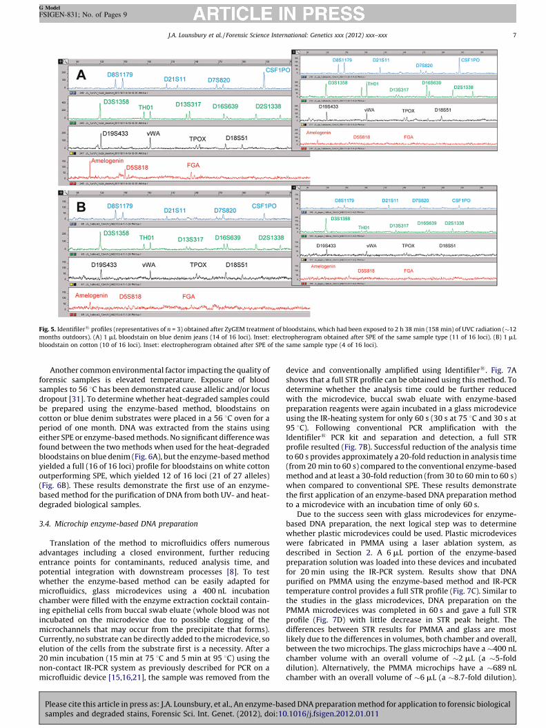
Fig. 5. Identifiler1 profiles (representatives of n = 3) obtained after ZyGEM treatment of bloodstains, which had been exposed to 2 h 38 min (158 min) of UVC radiation (�12
months outdoors). (A) 1 mL bloodstain on blue denim jeans (14 of 16 loci). Inset: electropherogram obtained after SPE of the same sample type (11 of 16 loci). (B) 1 mL
bloodstain on cotton (10 of 16 loci). Inset: electropherogram obtained after SPE of the same sample type (4 of 16 loci).
J.A. Lounsbury et al. / Forensic Science International: Genetics xxx (2012) xxx–xxx 7
G Model
FSIGEN-831; No. of Pages 9
Another common environmental factor impacting the quality offorensic samples is elevated temperature. Exposure of bloodsamples to 56 8C has been demonstrated cause allelic and/or locusdropout [31]. To determine whether heat-degraded samples couldbe prepared using the enzyme-based method, bloodstains oncotton or blue denim substrates were placed in a 56 8C oven for aperiod of one month. DNA was extracted from the stains usingeither SPE or enzyme-based methods. No significant difference wasfound between the two methods when used for the heat-degradedbloodstains on blue denim (Fig. 6A), but the enzyme-based methodyielded a full (16 of 16 loci) profile for bloodstains on white cottonoutperforming SPE, which yielded 12 of 16 loci (21 of 27 alleles)(Fig. 6B). These results demonstrate the first use of an enzyme-based method for the purification of DNA from both UV- and heat-degraded biological samples.
3.4. Microchip enzyme-based DNA preparation
Translation of the method to microfluidics offers numerousadvantages including a closed environment, further reducingentrance points for contaminants, reduced analysis time, andpotential integration with downstream processes [8]. To testwhether the enzyme-based method can be easily adapted formicrofluidics, glass microdevices using a 400 nL incubationchamber were filled with the enzyme extraction cocktail contain-ing epithelial cells from buccal swab eluate (whole blood was notincubated on the microdevice due to possible clogging of themicrochannels that may occur from the precipitate that forms).Currently, no substrate can be directly added to the microdevice, soelution of the cells from the substrate first is a necessity. After a20 min incubation (15 min at 75 8C and 5 min at 95 8C) using thenon-contact IR-PCR system as previously described for PCR on amicrofluidic device [15,16,21], the sample was removed from the
Please cite this article in press as: J.A. Lounsbury, et al., An enzyme-basamples and degraded stains, Forensic Sci. Int. Genet. (2012), doi:10
device and conventionally amplified using Identifiler1. Fig. 7Ashows that a full STR profile can be obtained using this method. Todetermine whether the analysis time could be further reducedwith the microdevice, buccal swab eluate with enzyme-basedpreparation reagents were again incubated in a glass microdeviceusing the IR-heating system for only 60 s (30 s at 75 8C and 30 s at95 8C). Following conventional PCR amplification with theIdentifiler1 PCR kit and separation and detection, a full STRprofile resulted (Fig. 7B). Successful reduction of the analysis timeto 60 s provides approximately a 20-fold reduction in analysis time(from 20 min to 60 s) compared to the conventional enzyme-basedmethod and at least a 30-fold reduction (from 30 to 60 min to 60 s)when compared to conventional SPE. These results demonstratethe first application of an enzyme-based DNA preparation methodto a microdevice with an incubation time of only 60 s.
Due to the success seen with glass microdevices for enzyme-based DNA preparation, the next logical step was to determinewhether plastic microdevices could be used. Plastic microdeviceswere fabricated in PMMA using a laser ablation system, asdescribed in Section 2. A 6 mL portion of the enzyme-basedpreparation solution was loaded into these devices and incubatedfor 20 min using the IR-PCR system. Results show that DNApurified on PMMA using the enzyme-based method and IR-PCRtemperature control provides a full STR profile (Fig. 7C). Similar tothe studies in the glass microdevices, DNA preparation on thePMMA microdevices was completed in 60 s and gave a full STRprofile (Fig. 7D) with little decrease in STR peak height. Thedifferences between STR results for PMMA and glass are mostlikely due to the differences in volumes, both chamber and overall,between the two microchips. The glass microchips have a �400 nLchamber volume with an overall volume of �2 mL (a �5-folddilution). Alternatively, the PMMA microchips have a �689 nLchamber with an overall volume of �6 mL (a �8.7-fold dilution).
sed DNA preparation method for application to forensic biological.1016/j.fsigen.2012.01.011

Fig. 7. Identifiler1 profiles (representatives of n = 3) obtained after incubation on microdevices. (A) Full Identifiler1 profile (16 of 16 loci) after incubation on a glass
microdevice using conventional incubation times (The extra peaks in D19S433 and vWA are pull-up from D3S1358 and TH01, respectively). (B) Full Identifiler1 profile (16 of
16 loci) after incubation on a glass microdevice using reduced incubation times of 30 s at 75 8C and 30 s at 95 8C. (C) Full Identifiler1 profile (16 of 16 loci) after incubation on a
PMMA microdevice using conventional incubation times. (D) Full Identifiler1 profile (16 of 16 loci) after incubation on a PMMA microdevice using reduced incubation times
of 30 s at 75 8C and 30 s at 95 8C.
Fig. 6. Identifiler1 profiles (representatives of n = 3) obtained after ZyGEM treatment of bloodstains, which had been heated in an oven at 56 8C for a period of one month. (A)
1 mL bloodstain on blue denim jeans (13 of 16 loci). Inset: electropherogram obtained after SPE of the same sample type (13 of 16 loci). (B) 1 mL bloodstain on cotton (16 of 16
loci). Inset: electropherogram obtained after SPE of the same sample type (14 of 16 loci).
J.A. Lounsbury et al. / Forensic Science International: Genetics xxx (2012) xxx–xxx8
G Model
FSIGEN-831; No. of Pages 9
Please cite this article in press as: J.A. Lounsbury, et al., An enzyme-based DNA preparation method for application to forensic biologicalsamples and degraded stains, Forensic Sci. Int. Genet. (2012), doi:10.1016/j.fsigen.2012.01.011

J.A. Lounsbury et al. / Forensic Science International: Genetics xxx (2012) xxx–xxx 9
G Model
FSIGEN-831; No. of Pages 9
The increase in this dilution lowers the concentration of DNA in thesample and therefore, in the PCR. These results demonstrate thatthe enzyme-based method is adaptable to both microdevices madefrom glass or plastic, which result in a 30-fold reduction inincubation time compared to conventional SPE methods (1 minversus 30 min).
4. Conclusion
This work demonstrates the utility of an enzyme-based DNApreparation method for forensic DNA analysis of buccal swabs,whole blood and bloodstains on blue denim or cotton. The methodwas shown to produce STR profiles that were comparable to thosefrom conventional SPE, and in many cases outperformed the SPEmethod. In addition, the enzyme-based method produced PCR-ready DNA after a 20 min incubation and requires no centrifuga-tion or sample transfer steps. Furthermore, the DNA yield may becontrolled by increasing the amount of starting material or enzymeadded to the reaction, demonstrating the adaptability of themethod to a wide variety of sample types. The enzyme-basedmethod was also shown to be adaptable to microdevices andallowed for the incubation time to be reduced to 60 s providing a20-fold and 30-fold reduction in analysis time compared toconventional enzyme-based preparation and conventional SPE,respectively. Implementation of this method into the workflow forforensic cases could reduce sample and DNA preparation time,leading to a reduction in overall analysis time. The adaptability ofthis method, due to its simplicity, also allows for the integration ofthis method with downstream processes, such as PCR, on a single,disposable microdevice, providing the quintessential on-siteanalysis tool.
Appendix A. Supplementary data
Supplementary data associated with this article can be found, in
the online version, at doi:10.1016/j.fsigen.2012.01.011.
References
[1] P. Radstrom, R. Knutsson, P. Wolffs, M. Lovenklev, C. Lofstrom, Pre-PCR processing– strategies to generate PCR-compatible samples, Mol. Biotechnol. 26 (2004) 133–146.
[2] C.T. Comey, B.W. Koons, K.W. Presley, J.B. Smerick, C.A. Sobieralski, D.M. Stanley,F.S. Baechtel, DNA extraction strategies for amplified fragment length polymor-phism analysis, J. Forensic Sci. 39 (1994) 1254–1269.
[3] K.L. Opel, D. Chung, B.R. McCord, A study of PCR inhibition mechanisms using realtime PCR, J. Forensic Sci. 55 (2010) 25–33.
[4] P.S. Walsh, D.A. Metzger, R. Higuchi, Chelex-100 as a medium for simple extrac-tion of DNA for PCR-based typing from forensic material, Biotechniques 10 (1991)506–513.
[5] A.M. Divne, H. Edlund, M. Allen, Forensic analysis of autosomal STR markers usingpyrosequencing, Forensic Sci. Int. Genet. 4 (2010) 122–129.
[6] S.A. Greenspoon, M.A. Scarpetta, M.L. Drayton, S.A. Turek, QIAamp spin columns as amethod of DNA isolation for forensic casework, J. Forensic Sci. 43 (1998) 1024–1030.
[7] C.M. Cupples, J.R. Champagne, K.E. Lewis, T.D. Cruz, STR profiles from DNAsamples with ‘‘undetected’’ or low QuantifilerTM results, J. Forensic Sci. 54(2009) 103–107.
[8] C.J. Easley, J.M. Karlinsey, J.M. Bienvenue, L.A. Legendre, M.G. Roper, S.H. Feldman,M.A. Hughes, E.L. Hewlett, T.J. Merkel, J.P. Ferrance, J.P. Landers, A fully integrated
Please cite this article in press as: J.A. Lounsbury, et al., An enzyme-basamples and degraded stains, Forensic Sci. Int. Genet. (2012), doi:10
microfluidic genetic analysis system with sample-in-answer-out capability, Proc.Nat. Acad. Sci. 103 (2006) 19272–19277.
[9] K.A. Hagan, W.L. Meier, J.P. Ferrance, J.P. Landers, Chitosan-coated silica as a solidphase for RNA purification in a microfluidic device, Anal. Chem. 81 (2009) 5249–5256.
[10] L.A. Legendre, J.M. Bienvenue, M.G. Roper, J.P. Ferrance, J.P. Landers, A simple,valveless microfluidic sample preparation device for extraction and amplifi-cation of DNA from nanoliter-volume samples, Anal. Chem. 78 (2006) 1444–1451.
[11] C.R. Reedy, K.A. Hagan, B.C. Strachan, J.J. Higginson, J.M. Bienvenue, S.A. Green-spoon, J.P. Ferrance, J.P. Landers, Dual-domain microchip-based process forvolume reduction solid phase extraction of nucleic acids from dilute, large volumebiological samples, Anal. Chem. 82 (2010) 5669–5678.
[12] J.M. Bienvenue, N. Duncalf, D. Marchiarullo, J.P. Ferrance, J.P. Landers, Microchip-based cell lysis and DNA extraction from sperm cells for application to forensicanalysis, J. Forensic Sci. 51 (2006) 266–273.
[13] Y.C. Chung, M.S. Jan, Y.C. Lin, J.H. Lin, W.C. Cheng, C.Y. Fan, Microfluidic chip forhigh efficiency DNA extraction, Lab Chip 4 (2004) 141–147.
[14] D. Moss, S.A. Harbison, D.J. Saul, An easily automated, closed-tube forensic DNAextraction procedure using a thermostable proteinase, Int. J. Legal Med. 117(2003) 340–349.
[15] C.R. Reedy, K.A. Hagan, D.J. Marchiarullo, A.H. Dewald, A. Barron, J.M. Bien-venue, J.P. Landers, A modular microfluidic system for deoxyribonucleic acididentification by short tandem repeat analysis, Anal. Chim. Acta 687 (2011)150–158.
[16] M.G. Roper, C.J. Easley, L.A. Legendre, J.A.C. Humphrey, J.P. Landers, Infraredtemperature control system for a completely noncontact polymerase chainreaction in microfluidic chips, Anal. Chem. 79 (2007) 1294–1300.
[17] A. Hall, J. Ballantyne, Characterization of UVC-induced DNA damage in blood-stains: forensic implications, Anal. Bioanal. Chem. 380 (2004) 72–83.
[18] D. Kimes, M. Tamir, An extraction procedure for seminal/vaginal stains to elimi-nate streaking in the electrophoresis phosphoglucomutase, Crime Lab. Digest. 12(1985) 32–33.
[19] A. Manz, J.C. Fettinger, E. Verpoorte, H. Ludi, H.M. Widmer, D.J. Harrison, Micro-machining of monocrystalline silicon and glass for chemical-analysis systems – alook into next century technology or just a fashionable craze? Trends Anal. Chem.10 (1991) 144–149.
[20] Y. Sun, Y.C. Kwok, N.T. Nguyen, Low-pressure, high-temperature thermal bondingof polymeric microfluidic devices and their applications for electrophoreticseparation, J. Micromech. Microeng. 16 (2006) 1681–1688.
[21] C.J. Easley, J.A.C. Humphrey, J.P. Landers, Thermal isolation of microchip reactionchambers for rapid non-contact DNA amplification, J. Micromech. Microeng. 17(2007) 1758–1766.
[22] Quick start guide – DNA extraction using forensicGEMTM saliva, Available athttp://www.zygem.com/Products/Products-FG-Saliva.html.
[23] K.M. Horsman, J.A. Hickey, R.W. Cotton, J.P. Landers, L.O. Maddox, Development ofa human-specific real-time PCR assay for the simultaneous quantitation of totalgenomic and male DNA, J. Forensic Sci. 51 (2006) 758–765.
[24] N. Privorotskaya, Y.S. Liu, J.C. Lee, H.J. Zeng, J.A. Carlisle, A. Radadia, L. Millet, R.Bashir, W.P. King, Rapid thermal lysis of cells using silicon-diamond microcanti-lever heaters, Lab Chip 10 (2010) 1135–1141.
[25] J. Membrillo-Hernandez, A. Nunez-De La Mora, T. Del Rio-Albrechtsen, R. Cama-cho-Carranza, M.C. Gomez-Eichelmann, Thermally-induced cell lysis in Escher-ichia coli K12, J. Basic Microbiol. 35 (1995) 41–46.
[26] T. Coolbear, C.W. Eames, Y. Casey, R.M. Daniel, H.W. Morgan, Screening of strainsidentified as extremely thermophilic bacilli for extracellular proteolytic activityand general properties of the proteinases from two of the strains, J. Appl. Bacteriol.71 (1991) 252–264.
[27] T. Coolbear, J.M. Whittaker, R.M. Daniel, The effect of metal ions on the activityand thermostability of the extracellular proteinase from a thermophilic bacillus,strain EA-1, Biochem. J 287 (1992) 367–374.
[28] D.J. Saul, R.M. Daniel, Purification methods and uses thereof, U.S. Patent 2007/0190552 A1 (issued 16 August 2007).
[29] E.J. Lee, G.S. Patten, S.L. Burnard, E.J. McMurchie, Osmotic and other properties ofisolated human cheek epithelial cells, Am. J. Physiol. 267 (1994) C75–C83.
[30] J. Wen, C. Guillo, J.P. Ferrance, J.P. Landers, Microfluidic-based DNA purification ina two-stage, dual-phase microchip containing a reversed-phase and a photo-polymerized monolith, Anal. Chem. 79 (2007) 6135–6142.
[31] S.A. Greenspoon, J.D. Ban, K. Sykes, E.J. Ballard, S.S. Edler, M. Baisden, B.L.Covington, Application of the BioMek1 2000 laboratory automation workstationand the DNA IQTM system to the extraction of forensic casework samples, J.Forensic Sci. 49 (2004) 29–39.
sed DNA preparation method for application to forensic biological.1016/j.fsigen.2012.01.011


![Guidance - assets.publishing.service.gov.uk · 2 R. v. Dlugosz and Ors [2013] EWCA, Crim 2. 3 Clayton et al. (1998) Analysis and interpretation of mixed forensic stains using DNA](https://img.dokumen.tips/doc/110x75/5eb7697a26e8b45ed368407f/guidance-2-r-v-dlugosz-and-ors-2013-ewca-crim-2-3-clayton-et-al-1998.jpg)


![Alternative methodology for extraction of high-quality DNA ... · of DNA from ancient human forensic samples and degraded for forensic identification [10]. Materials and methods The](https://img.dokumen.tips/doc/110x75/5f08c58c7e708231d423a537/alternative-methodology-for-extraction-of-high-quality-dna-of-dna-from-ancient.jpg)