Embed Size (px)
Citation preview

For Review O
nly
Diagnosis of equine penile and preputial masses: A clinical
and pathological perspective
Journal: Equine Veterinary Education
Manuscript ID: Draft
Wiley - Manuscript type: Clinical Commentary (requested)
Date Submitted by the Author: n/a
Complete List of Authors: Scott, Vikki; University of Cambridge, Department of Veterinary Medicine Hughes, Katherine; University of Cambridge, Department of Veterinary Medicine
Discipline: Oncology, Pathology
Body System/Disorder: Reproductive, Integument
Abstract: Clinical commentary so not required.
Equine Veterinary Education

For Review O
nly
Diagnosis of equine penile and preputial masses: A clinical and pathological perspective 1
2
3
4
V. H. L. Scott* and K. Hughes
* 5
6
7
8
9
Department of Veterinary Medicine, University of Cambridge, Madingley Road, Cambridge, 10
Cambridgeshire. CB3 0ES. 11
12
13
* Co-corresponding authors: [email protected]; [email protected] 14
15
16
Keywords: penis; prepuce; tumour; balanitis; balanoposthitis17
Page 1 of 18 Equine Veterinary Education

For Review O
nly
Introduction 18
Masses of the prepuce and penis are common clinical presentations for the equine 19
veterinarian, encompassing a number of differential diagnoses. In such cases, it is important 20
to evaluate the external genitalia thoroughly and devise a methodical diagnostic and 21
treatment plan to allow for accurate prognostication and optimal survival rates. Masses of the 22
prepuce and penis include tumours of epithelial, mesenchymal or round cell origin, such as 23
squamous cell carcinoma (SCC), papillomas, melanocytic tumours, lymphoma, sarcoids, 24
fibrosarcomas (Van Den Top et al. 2010) and fibromas; the latter as described by De Meyer 25
et al. in this issue (De Meyer et al. 2015). Non-neoplastic conditions that can result in masses 26
or enlargement of the external genitalia in male horses include infection or inflammation 27
(primary, or secondary to neoplasia) leading to balanitis or balanoposthitis. Such diseases 28
frequently lead to chronic irritation and discomfort. Some neoplastic conditions may 29
metastasise to cause more severe sequelae. 30
31
Diagnosis 32
Given that tumours comprise the majority of genital masses diagnosed in male horses, 33
adoption of a thorough, standardised, approach to clinical evaluation is required. Van den 34
Top et al. (2010 and 2011) have proposed a systematic assessment and classification tool for 35
evaluating tumour type and behaviour, treatment protocol and prognosis. Visual inspection 36
and palpation of the mass (or tumour) should occur with thorough evaluation of the external 37
genitalia under standing sedation using an alpha-2 agonist and acepromazine to facilitate safe 38
examination. Some clinicians advocate caution with use of acepromazine in stallions due to 39
the possible risk of paraphimosis, priapism and penile paralysis. The mass should be assessed 40
for size, location, mobility, and degree of infiltration / involvement of the corpus cavernosum 41
and corpus spongiosum. Palpation of enlarged superficial and deep inguinal lymph nodes can 42
Page 2 of 18Equine Veterinary Education

For Review O
nly
provide information about possible metastases. The superficial inguinal lymph nodes lie 43
dorsolateral to the penis, and the deep inguinal lymph nodes are located just outside the 44
pelvis adjacent to the internal inguinal ring. The medial iliac lymph nodes (adjacent to the 45
external iliac arteries) should also be evaluated per rectum. Lymph node palpation and fine 46
needle aspirate biopsies can result in false positive and false negative results for metastases 47
(Van Den Top et al. 2010). It should be considered that although regional lymphadenopathy 48
may be an indicator of metastases, enlargement can also be due to “reactive” lymph nodes, 49
secondary to inflammation or infection associated with the tumour. Distant metastases to the 50
thoracic cavity can be evaluated by radiographic assessment, however most tumours affecting 51
the equine penis and prepuce metastasise locally via the regional lymph nodes, with 52
pulmonary and skeletal metastases only in advanced disease (Cramer et al. 2011; Nelson et 53
al. 2015). Routine use of radiography is not warranted for the majority of cases. 54
55
Ultrasonography of the primary penile tumour is commonly used in humans, and can provide 56
information about the gross extent of the tumour and tissue invasion or involvement of 57
various structures (Hyland and Church 1995). To the authors’ knowledge, there are scarce 58
descriptions of the use of ultrasonography for evaluation of genital tumours in male horses, 59
but this modality may be a useful diagnostic adjunct. 60
61
The TNM (tumour, node, metastasis) classification system is widely used in human oncology 62
to aid with appropriate choices of treatment and prognosis. Recent work within the equine 63
literature has also highlighted the importance of histological grading in penile and preputial 64
tumours. A positive correlation between high grade SCCs and metastases has been 65
demonstrated and it has also been shown that tumour grading is an important prognosticator 66
for survival in horses (van den Top et al. 2008; van den Top et al. 2015). This information, in 67
Page 3 of 18 Equine Veterinary Education

For Review O
nly
conjunction with a published classification system (Van den Top et al. 2011) requires a 68
representative biopsy to be taken such that tumour grading can guide treatment protocol and 69
provide information on prognosis. A full thickness punch or excisional biopsy is required to 70
assess tumour architecture and depth of invasion. 71
72
Expression of cell proliferation markers, such as Ki67, and tumour suppressor genes, such as 73
p53, may also be evaluated using immunohistochemical staining of histopathological sections 74
(van den Top et al. 2015). Such markers are increasingly used in assessment of numerous 75
types of small animal neoplasms such canine mast cell tumours (Webster et al. 2007) and 76
feline mammary tumours (Zappulli et al. 2015), amongst many others, but their use in equine 77
diagnostic pathology is considerably less frequent. Whilst these and similar molecular 78
markers may provide further prognostic information for different equine penile and preputial 79
tumour types in the future, studies to date have yet to show compelling prognostic potential 80
(van den Top et al. 2015). 81
82
The diagnostic evaluations described (Ensink 2015; Van Den Top et al. 2010; Van den Top 83
et al. 2011) represent a “gold standard” approach to penile and preputial masses, but it should 84
also be considered that many cases have attendant financial constraints, other limitations in 85
resources, differing owner priorities or present with additional clinical challenges such 86
tumour accessibility. Acknowledging these factors is critical when formulating decisions 87
regarding treatment protocol and surgical approach. For such cases, histopathology of the 88
tumour is frequently only performed after treatment-based surgery has been undertaken if 89
gross appearance of the mass is consistent with common tumour types. Whilst the concern of 90
metastasis is important to both veterinarian and owner, the possibility of local tumour 91
recurrence is also a critical question. Histopathological assessment of surgeon cut edges / 92
Page 4 of 18Equine Veterinary Education

For Review O
nly
margins of surgically removed masses may be informative if histopathological analysis is 93
undertaken post surgery. Consideration of all of these factors may lead to an individual 94
diagnostic approach to equine genital masses in many cases. 95
96
Squamous Cell Carcinomas (SCC) 97
Squamous cell carcinoma is the most common neoplasm of equine external genitalia, with 98
Brinsko and van den Top et al. (Brinsko 1998; van den Top et al. 2008) reporting incidences 99
of 49 - 82.5%. Male genital SCC predominantly affects older horses and most studies 100
document an average age range of 17.4 - 19.8 years (Howarth et al. 1991; Mair et al. 2000; 101
Strafuss 1976; van den Top et al. 2008). The veterinary literature does not conclusively 102
support any specific breed predilection for genital SCC, but ponies have been frequently 103
highly represented (Howarth et al. 1991; Mair et al. 2000; van den Top et al. 2008). It has 104
also been proposed that breeds with non-pigmented genitalia may be at higher risk for 105
development of SCCs (Schumacher 2006). Papillomaviruses in man have long been 106
implicated in the aetiology of SCC development, and recent work has identified a number of 107
variants of Equus caballus papillomavirus 2 (EcPV2) within the tissue of equine penile 108
papillomas, penile intraepithelial neoplasia (PIN) and SCCs (Bogaert et al. 2012; Lange et al. 109
2013; Newkirk et al. 2014; Scase et al. 2010; van den Top et al. 2015; Zhu et al. 2015). 110
Histological evaluation of equine penile tumours frequently shows papillomas undergoing a 111
spectrum of changes as part of a continuum of transition to SCC (Van den Top et al. 2011). 112
113
Genital SCCs in horses can have a number of different gross appearances depending on the 114
stage of disease. Early lesions include depigmented plaques (Figure 1), irregularities of the 115
penile or preputial surface and non-healing erosions with or without accompanying 116
granulation tissue (Van den Top et al. 2011). More advanced lesions can appear as solid 117
Page 5 of 18 Equine Veterinary Education

For Review O
nly
masses and may have a typical cauliflower-like appearance or contain necrotic areas. Owners 118
often notice SCCs incidentally during micturition, but associated clinical signs can include 119
dysuria, preputial oedema, or sanguineous / purulent discharge secondary to infection or 120
tissue necrosis. Other reported abnormalities are wide-based stance, frequent protrusion of 121
the penis, excoriation of the genital integument and changes in gait (Van den Top et al. 122
2011). SCCs are malignant tumours, but tend to be slow to metastasise, although it should be 123
noted that pulmonary or skeletal metastases may occur in advanced cases (Cramer et al. 124
2011; Nelson et al. 2015). 125
126
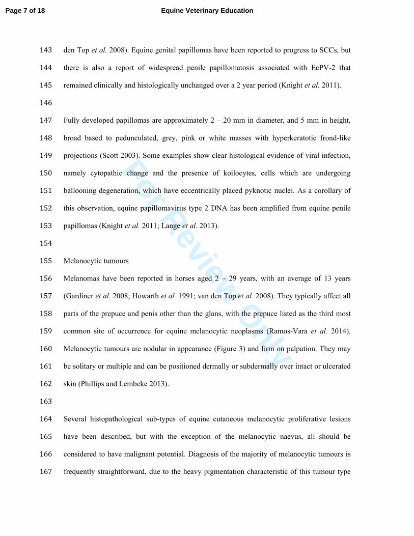
Histologically, squamous cell carcinomas, including those of the penis and prepuce, tend to 127
have a very characteristic appearance, with clusters of neoplastic cells exhibiting varying 128
degrees of keratinization, prominent nuclei often with conspicuous nucleoli, and frequently 129
prominent mitotic figures (Cramer et al. 2011) (Figure 2). Equine SCCs are frequently 130
infiltrated by CD3+ T lymphocytes, CD79+ B lymphocytes, IgG+ plasma cells and 131
macrophages (Perez et al. 1999). 132
133
Squamous papillomas 134
Squamous papillomas (warts) tend to occur on the nose, distal limbs and external genitalia. 135
They are the most common tumours in young horses, age 1 – 3 years (Scott 2003). 136
Papillomas on the external genitalia of male horses tend to affect older horses however, and 137
the published mean age range is 16.2 – 18 years (Gardiner et al. 2008; Howarth et al. 1991; 138
van den Top et al. 2008). Both congenital and acquired papillomas have been reported (Scott 139
2003; White et al. 2004). Papillomas begin as small, approximately 1 mm diameter, raised, 140
smooth, shiny grey to white papules (Van Den Top et al. 2010) and can be present over the 141
whole penis, although most appear over the glans, the urethral process and preputial fold (van 142
Page 6 of 18Equine Veterinary Education
For Review O
nly
den Top et al. 2008). Equine genital papillomas have been reported to progress to SCCs, but 143
there is also a report of widespread penile papillomatosis associated with EcPV-2 that 144
remained clinically and histologically unchanged over a 2 year period (Knight et al. 2011). 145
146
Fully developed papillomas are approximately 2 – 20 mm in diameter, and 5 mm in height, 147
broad based to pedunculated, grey, pink or white masses with hyperkeratotic frond-like 148
projections (Scott 2003). Some examples show clear histological evidence of viral infection, 149
namely cytopathic change and the presence of koilocytes, cells which are undergoing 150
ballooning degeneration, which have eccentrically placed pyknotic nuclei. As a corollary of 151
this observation, equine papillomavirus type 2 DNA has been amplified from equine penile 152
papillomas (Knight et al. 2011; Lange et al. 2013). 153
154
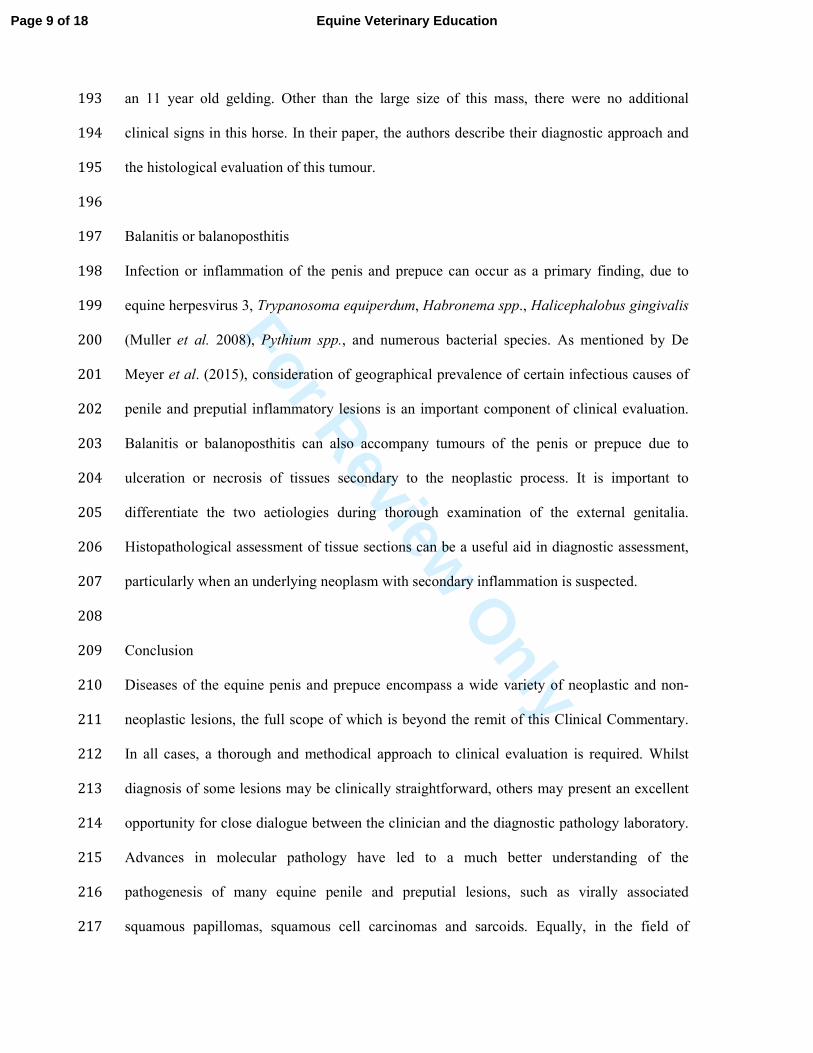
Melanocytic tumours 155
Melanomas have been reported in horses aged 2 – 29 years, with an average of 13 years 156
(Gardiner et al. 2008; Howarth et al. 1991; van den Top et al. 2008). They typically affect all 157
parts of the prepuce and penis other than the glans, with the prepuce listed as the third most 158
common site of occurrence for equine melanocytic neoplasms (Ramos-Vara et al. 2014). 159
Melanocytic tumours are nodular in appearance (Figure 3) and firm on palpation. They may 160
be solitary or multiple and can be positioned dermally or subdermally over intact or ulcerated 161
skin (Phillips and Lembcke 2013). 162
163
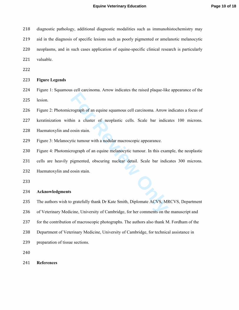
Several histopathological sub-types of equine cutaneous melanocytic proliferative lesions 164
have been described, but with the exception of the melanocytic naevus, all should be 165
considered to have malignant potential. Diagnosis of the majority of melanocytic tumours is 166
frequently straightforward, due to the heavy pigmentation characteristic of this tumour type 167
Page 7 of 18 Equine Veterinary Education

For Review O
nly
(Figure 4). However, diagnosis of poorly pigmented or amelanotic examples may be 168
challenging, particularly as these tumours may have a range of gross morphologies, which 169
historically led to human amelanotic melanoma being dubbed “the great masquerader” (Koch 170
and Lange 2000). These tumours may also exhibit a variable microscopic appearance. 171
Consequently immunohistochemical staining may be required for increased histopathological 172
diagnostic confidence, and in this regard it is notable that PNL2 has been suggested to be a 173
sensitive immunohistochemical marker of equine melanocytic neoplasms that is more 174
specific than S100 protein or PGP 9.5, both of which are used in the diagnosis of melanocytic 175
neoplasms in humans and other veterinary species (Ramos-Vara et al. 2014). 176
177
Other types of equine genital tumours 178
Sarcoids are tumours of fibroblastic origin, usually with an overlying hyperplastic epidermal 179
component. Bovine papillomaviruses (BPV) 1 and 2 have long been implicated in the 180
development of equine sarcoids and BPV nucleic acid has recently been visualised in these 181
tumours using in situ hybridization, adding to the weight of evidence suggesting a causative 182
association (Gaynor et al. 2015). Although sarcoids may occur at sites all over the body, 183
many different types are described in the preputial and paragenital regions. 184
185
Fibrosarcomas, a malignant proliferation of fibroblasts, may also arise in the penile and 186
preputial regions and are usually firm to fleshy infiltrative masses (Scott 2003). They are 187
invasive and capable of metastasis. Van den Top et al. (2008) have reported a fibrosarcoma 188
within the prepuce of a horse. Other tumour types are uncommon findings on the penis and 189
prepuce of horses, but reports include, lymphomas, lipomas, neurofibromas, 190
adenocarcinomas, basal cell carcinomas and haemangiosarcomas (Van Den Top et al. 2010). 191
The case report by De Meyer et al. (2015) describes the rare finding of a preputial fibroma in 192
Page 8 of 18Equine Veterinary Education
For Review O
nly
an 11 year old gelding. Other than the large size of this mass, there were no additional 193
clinical signs in this horse. In their paper, the authors describe their diagnostic approach and 194
the histological evaluation of this tumour. 195
196
Balanitis or balanoposthitis 197
Infection or inflammation of the penis and prepuce can occur as a primary finding, due to 198
equine herpesvirus 3, Trypanosoma equiperdum, Habronema spp., Halicephalobus gingivalis 199
(Muller et al. 2008), Pythium spp., and numerous bacterial species. As mentioned by De 200
Meyer et al. (2015), consideration of geographical prevalence of certain infectious causes of 201
penile and preputial inflammatory lesions is an important component of clinical evaluation. 202
Balanitis or balanoposthitis can also accompany tumours of the penis or prepuce due to 203
ulceration or necrosis of tissues secondary to the neoplastic process. It is important to 204
differentiate the two aetiologies during thorough examination of the external genitalia. 205
Histopathological assessment of tissue sections can be a useful aid in diagnostic assessment, 206
particularly when an underlying neoplasm with secondary inflammation is suspected. 207
208
Conclusion 209
Diseases of the equine penis and prepuce encompass a wide variety of neoplastic and non-210
neoplastic lesions, the full scope of which is beyond the remit of this Clinical Commentary. 211
In all cases, a thorough and methodical approach to clinical evaluation is required. Whilst 212
diagnosis of some lesions may be clinically straightforward, others may present an excellent 213
opportunity for close dialogue between the clinician and the diagnostic pathology laboratory. 214
Advances in molecular pathology have led to a much better understanding of the 215
pathogenesis of many equine penile and preputial lesions, such as virally associated 216
squamous papillomas, squamous cell carcinomas and sarcoids. Equally, in the field of 217
Page 9 of 18 Equine Veterinary Education
For Review O
nly
diagnostic pathology, additional diagnostic modalities such as immunohistochemistry may 218
aid in the diagnosis of specific lesions such as poorly pigmented or amelanotic melanocytic 219
neoplasms, and in such cases application of equine-specific clinical research is particularly 220
valuable. 221
222
Figure Legends 223
Figure 1: Squamous cell carcinoma. Arrow indicates the raised plaque-like appearance of the 224
lesion. 225
Figure 2: Photomicrograph of an equine squamous cell carcinoma. Arrow indicates a focus of 226
keratinization within a cluster of neoplastic cells. Scale bar indicates 100 microns. 227
Haematoxylin and eosin stain. 228
Figure 3: Melanocytic tumour with a nodular macroscopic appearance. 229
Figure 4: Photomicrograph of an equine melanocytic tumour. In this example, the neoplastic 230
cells are heavily pigmented, obscuring nuclear detail. Scale bar indicates 300 microns. 231
Haematoxylin and eosin stain. 232
233
Acknowledgments 234
The authors wish to gratefully thank Dr Kate Smith, Diplomate ACVS, MRCVS, Department 235
of Veterinary Medicine, University of Cambridge, for her comments on the manuscript and 236
for the contribution of macroscopic photographs. The authors also thank M. Fordham of the 237
Department of Veterinary Medicine, University of Cambridge, for technical assistance in 238
preparation of tissue sections. 239
240
References 241
Page 10 of 18Equine Veterinary Education

For Review O
nly
Bogaert, L., A. Willemsen, E. Vanderstraeten, M.A. Bracho, C. De Baere, I.G. Bravo, and A. 242
Martens. (2012). EcPV2 DNA in equine genital squamous cell carcinomas and 243
normal genital mucosa. Vet Microbiol 158, 33-41. 244
Brinsko, S.P. (1998). Neoplasia of the male reproductive tract. Vet Clin North Am Equine 245
Pract 14, 517-533. 246
Cramer, S.D., M.A. Breshears, and H.J. Qualls. (2011). Pathology in practice. Squamous cell 247
carcinoma of the penis with multifocal metastasis to the regional lymph nodes, lungs, 248
and heart. J Am Vet Med Assoc 238, 581-583. 249
De Meyer, A., S. Vandenabeele, C. Verbers, A. Martens, K. Roels, V. De Lange, M. 250
Hoogewijs, C. De Schauwer, and J. Govaere. (2015). Preputial fibroma in a gelding. 251
Equine Vet Educ. In press. 252
Ensink, J.M. (2015). Why clinicians should consider tumour staging and grading in horses. 253
Equine Vet J 47, 141. 254
Gardiner, D.W., J.P. Teifke, B.K. Podell, and D.A. Kamstock. (2008). Fibropapilloma of the 255
glans penis in a horse. J Vet Diagn Invest 20, 816-819. 256
Gaynor, A.M., K.W. Zhu, F.N. Cruz, Jr., V.K. Affolter, and P.A. Pesavento. (2015). 257
Localization of Bovine Papillomavirus Nucleic Acid in Equine Sarcoids. Vet Pathol. 258
In press. doi: 10.1177/0300985815594852. 259
Howarth, S., V.M. Lucke, and H. Pearson. (1991). Squamous cell carcinoma of the equine 260
external genitalia: a review and assessment of penile amputation and urethrostomy as 261
a surgical treatment. Equine Vet J 23, 53-58. 262
Hyland, J., and S. Church. (1995). The use of ultrasonography in the diagnosis and treatment 263
of a haematoma in the corpus cavernosum penis of a stallion. Aust Vet J 72, 468-469. 264
Page 11 of 18 Equine Veterinary Education

For Review O
nly
Knight, C.G., J.S. Munday, B.V. Rosa, and M. Kiupel. (2011). Persistent, widespread 265
papilloma formation on the penis of a horse: a novel presentation of equine 266
papillomavirus type 2 infection. Vet Dermatol 22, 570-574. 267
Koch, S.E., and J.R. Lange. (2000). Amelanotic melanoma: the great masquerader. J Am 268
Acad Dermatol 42, 731-734. 269
Lange, C.E., K. Tobler, A. Lehner, P. Grest, M.M. Welle, C.C. Schwarzwald, and C. Favrot. 270
(2013). EcPV2 DNA in equine papillomas and in situ and invasive squamous cell 271
carcinomas supports papillomavirus etiology. Vet Pathol 50, 686-692. 272
Mair, T.S., J.P. Walmsley, and T.J. Phillips. (2000). Surgical treatment of 45 horses affected 273
by squamous cell carcinoma of the penis and prepuce. Equine Vet J 32, 406-410. 274
Muller, S., M. Grzybowski, H. Sager, V. Bornand, and W. Brehm. (2008). A nodular 275
granulomatous posthitis caused by Halicephalobus sp. in a horse. Vet Dermatol 19, 276
44-48. 277
Nelson, B.B., E.F. Edmondson, J.M. Sonis, C.B. Frank, A. Valdes-Martinez, and B.S. Leise. 278
(2015). Multiple skeletal metastases from a penile squamous cell carcinoma in a 279
horse. Equine Vet Educ 27, 119-123. 280
Newkirk, K.M., D.V. Hendrix, E.A. Anis, B.W. Rohrbach, E.J. Ehrhart, J.A. Lyons, and S.A. 281
Kania. (2014). Detection of papillomavirus in equine periocular and penile squamous 282
cell carcinoma. J Vet Diagn Invest 26, 131-135. 283
Perez, J., E. Mozos, M.P. Martin, and M.J. Day. (1999). Immunohistochemical study of the 284
inflammatory infiltrate associated with equine squamous cell carcinoma. J Comp 285
Pathol 121, 385-397. 286
Phillips, J.C., and L.M. Lembcke. (2013). Equine melanocytic tumors. Vet Clin North Am 287
Equine Pract 29, 673-687. 288
Page 12 of 18Equine Veterinary Education

For Review O
nly
Ramos-Vara, J.A., C.B. Frank, D. DuSold, and M.A. Miller. (2014). Immunohistochemical 289
expression of melanocytic antigen PNL2, Melan A, S100, and PGP 9.5 in equine 290
melanocytic neoplasms. Vet Pathol 51, 161-166. 291
Scase, T., S. Brandt, C. Kainzbauer, S. Sykora, S. Bijmholt, K. Hughes, S. Sharpe, and A. 292
Foote. (2010). Equus caballus papillomavirus-2 (EcPV-2): an infectious cause for 293
equine genital cancer? Equine Vet J 42, 738-745. 294
Schumacher, J. (2006). Penis and prepuce. In: Equine Surgery, 3rd edition, Saunders Elsevier, 295
St Louis. pp 811-835. 296
Scott, D.W. and Miller, W. H. Jr. (2003). Equine Dermatology, 1st edition, Elsevier Science, 297
St Louis. p 707. 298
Strafuss, A.C. (1976). Squamous cell carcinoma in horses. J Am Vet Med Assoc 168, 61-62. 299
van den Top, J.G., N. de Heer, W.R. Klein, and J.M. Ensink. (2008). Penile and preputial 300
tumours in the horse: a retrospective study of 114 affected horses. Equine Vet J 40, 301
528-532. 302
Van den Top, J.G., J.M. Ensink, A. Grone, W.R. Klein, A. Barneveld, and P.R. Van Weeren. 303
(2010). Penile and preputial tumours in the horse: literature review and proposal of a 304
standardised approach. Equine Vet J 42, 746-757. 305
Van den Top, J.G., L. Harkema, C. Lange, J.M. Ensink, C.H. van de Lest, A. Barneveld, P.R. 306
van Weeren, A. Grone, and A. Martens. (2015). Expression of p53, Ki67, EcPV2- and 307
EcPV3 DNA, and viral genes in relation to metastasis and outcome in equine penile 308
and preputial squamous cell carcinoma. Equine Vet J 47, 188-195. 309
Van den Top, J.G.B., J.M. Ensink, A. Barneveld, and P.R. van Weeren. (2011). Penile and 310
preputial squamous cell carcinoma in the horse and proposal of a classification 311
system. Equine Vet Educ 23, 636-648. 312
Page 13 of 18 Equine Veterinary Education

For Review O
nly
Webster, J.D., V. Yuzbasiyan-Gurkan, R.A. Miller, J.B. Kaneene, and M. Kiupel. (2007). 313
Cellular proliferation in canine cutaneous mast cell tumors: associations with c-KIT 314
and its role in prognostication. Vet Pathol 44, 298-308. 315
White, K.S., R.N. Fuji, B.A. Valentine, and R.J. Bildfell. (2004). Equine congenital 316
papilloma: pathological findings and results of papillomavirus immunohistochemistry 317
in five cases. Vet Dermatol 15, 240-244. 318
Zappulli, V., R. Rasotto, D. Caliari, M. Mainenti, L. Pena, M.H. Goldschmidt, and M. 319
Kiupel. (2015). Prognostic evaluation of feline mammary carcinomas: a review of the 320
literature. Vet Pathol 52, 46-60. 321
Zhu, K.W., V.K. Affolter, A.M. Gaynor, F.N. Dela Cruz, Jr., and P.A. Pesavento. (2015). 322
Equine Genital Squamous Cell Carcinoma: In Situ Hybridization Identifies a Distinct 323
Subset Containing Equus caballus Papillomavirus 2. Vet Pathol. In press. 324
doi:10.1177/0300985815583095 325
326
Page 14 of 18Equine Veterinary Education

For Review O
nly
Figure 1: Squamous cell carcinoma. Arrow indicates raised plaque-like appearance of the lesion. 108x118mm (600 x 600 DPI)
Page 15 of 18 Equine Veterinary Education

For Review O
nly
Figure 2: Photomicrograph of an equine squamous cell carcinoma. Arrow indicates a focus of keratinization within a cluster of neoplastic cells. Scale bar indicates 100 microns. Haematoxylin and eosin stain.
75x56mm (300 x 300 DPI)
Page 16 of 18Equine Veterinary Education

For Review O
nly
Figure 3: Melanocytic tumour with a nodular macroscopic appearance.
98x96mm (600 x 600 DPI)
Page 17 of 18 Equine Veterinary Education

For Review O
nly
Figure 4: Photomicrograph of an equine melanocytic tumour. In this example, the neoplastic cells are heavily pigmented, obscuring nuclear detail. Scale bar indicates 300 microns. Haematoxylin and eosin stain.
75x56mm (300 x 300 DPI)
Page 18 of 18Equine Veterinary Education