Embed Size (px)
Citation preview

For producer information only. Not for use in sales situations.
Principal Financial Group®
Basics of the Echocardiogram:Diastolic Dysfunction & Left Ventricular
Hypertrophy (LVH)
Cindy MillerRN, Senior Underwriting Consultant

For producer information only. Not for use in sales situations.
Principal Financial Group®

For producer information only. Not for use in sales situations.
The Echo
History
• The technology of sonar or echosonography was originally developed during World War II to detect submarines
• The first “echo” was in the early 1950s. A Swedish physicist borrowed a sonar device from a local shipyard, modified it, and recorded echo’s from his own heart

For producer information only. Not for use in sales situations.
The Echo
Uses of the Ultrasound (US)
• Gynecology and obstetrics
• Vascular issues
• Musculoskeletal issues
• Detection of tumors in various areas in the body: prostate, colon, breast, heart, gastointestinal tract and other organs
• Cardiology

For producer information only. Not for use in sales situations.
The Echo
Cardiac Arena used for:
• Identification of structures of the heart (normal and abnormal)
• Allows for assessment of the motion and function of the heart and it’s various structures
• Follows blood flow through the heart and measures velocity of the blood flow
• Serves as a compliment to other diagnostic tests (eg. confirms EKG findings for LVH or chest x ray finding)
For producer information only. Not for use in sales situations.
The Echo
How is it completed?
• It involves a US machine, a wand which is known as a transducer that is connected to a US machine
• The patient lies on stretcher and the echosonographer who has been specifically trained in techniques of echosonography performs the scan
• The scan is completed in a very systematic format to obtain specific views of the heart that the cardiologists expects to see on any given echo

For producer information only. Not for use in sales situations.
The Echo
• 3 inter-related processes utilized during an ECHO
– M mode
– 2-D component
– Doppler (continuous wave/pulsed wave/ color flow)

For producer information only. Not for use in sales situations.
The Echo
M Mode
– Single dimension picture
– Enables us to look closely and measure the heart chambers and structures, the aorta (helpful with LVH, myxomatous valves, valvular stenosis, hypertrophic & dilated cardiomyopathies)

For producer information only. Not for use in sales situations.
The Echo
M Mode (LVID and Wall measurements / MV motion)

For producer information only. Not for use in sales situations.
The Echo
2D Mode
– Two dimensional picture of the heart is produced as result of sound waves going out of the transducer and bouncing off a structure and then returning back to the transducer
2D - 4 Chamber apical view
Diastole
MV open
Systole
MV closed

For producer information only. Not for use in sales situations.
The Echo
2D -parasternal

For producer information only. Not for use in sales situations.
The Echo
Doppler Mode
– Evaluates the path of the blood flow
– Evaluates the blood flow velocity as it moves through the heart and its structures
3 Types Doppler :
– Pulsed Wave
– Continuous Wave
– Color Flow

For producer information only. Not for use in sales situations.
The EchoPulsed Wave Doppler
Measures velocity of blood as it moves through the heart
• Provides specific information about blood flow through a specific site
• Helps asses the heart’s ability to contract and relax
E/A ratio
For producer information only. Not for use in sales situations.
The Echo
Continuous Wave Doppler
• Measures blood flow velocity as it moves through the valves
• Helps to quantify how severely a valve is leaking
• Measure degree of valvular regurgitation measuring the density, length and shape of the wave forms

For producer information only. Not for use in sales situations.
The Echo
• CW- Aortic Regurg
Mitral Regurgitation Aortic Insufficiency
Continuous WaveDoppler

For producer information only. Not for use in sales situations.
The Echo
Color Flow Doppler
• Measures the velocity and direction of blood flow via color patterns

For producer information only. Not for use in sales situations.
The Echo
Color Flow Doppler
MV Open MV Closed

For producer information only. Not for use in sales situations.
The Echo
Putting it all together
• During the test the images that have been obtained are recorded for later viewing by the MD
• The technician writes a report of their interpretation of the findings and prepares the report and a recording of the entire echo for the MD
• MD provides a final analysis from the recording

For producer information only. Not for use in sales situations.
The Echo
What Underwriters should note from the echo report:
• Why was it completed?
• Patient data (being aware of the persons age/any disease processes/body size )
• Quality of the image
• Assess the entire report and try not to focus on just one aspect *
• Do the findings make sense with the overall clinical picture? *

For producer information only. Not for use in sales situations.
The Echo
Limitations & Variables
• Interpretation of the echo can be somewhat subjective
• Body habitus and other physical deformities can alter the findings and ability to accurately obtain some images
• Equipment variables
• Sonographer technique and experience
• Some of the findings can’t be reproduced and are time specific

For producer information only. Not for use in sales situations.
The Echo
Additional Considerations
• Don’t take an isolated reading and automatically rate it, instead consider the following:
– Compare to prior echos
– Take age and body habits into consideration
– Note the BP and other impairment history
– Note why the test was done in the first place
– What if any clinical signs/symptoms are present
– What other clinical data is present that support the findings

For producer information only. Not for use in sales situations.
The Echo
Common Abnormal Echo Findings
• LVH
• Concentric: hypertension, valve diseases, aortic stenosis, cardiomyopathies, obesity
• Asymmetric Septal Hypertrophy (ASH): associated with valve disease
• Abnormal Wall Motion
• Global hypokensis (HK): cardiomyopathies
• Segmental HK or akinesia (AK): ischemic heart disease
• Dyskinesia: aneurysms, LBBB

For producer information only. Not for use in sales situations.
The Echo
Common Abnormal Echo Findings cont…
– Abnormal Valves: stenosis, insufficiency, bicuspid AV
– MAC (Mitral annular calcification): thickened cusps, calcium deposits
– Diastolic dysfunction
– PFO / ASD
– Atrial Enlargements: valve disease, diastolic dysfunction or atrial fib
– Aortic Root enlargements: CTD, valve disease
For producer information only. Not for use in sales situations.
The Echo
Less Common Findings…
• Tumors (malignant or benign)
• Pericardial effusions
• Congenital defects (great vessel anomalies)

For producer information only. Not for use in sales situations.
The Echo
Normal findings on a typical Elderly person ..
• LV wall thickness increases 15%;
• LV mass increases 1 gm/yr from ages 65-80
– senile septum (septum thickens slightly)
• LA dimensions increase ~ 16%

For producer information only. Not for use in sales situations.
The Echo
Normal findings on Elderly person cont…
• LV dimension unchanged
• Aortic Root diameter increases ~ 22%
• E/A velocity is often reversed ( diastolic dysfunction )
• Valvular disease and MAC

For producer information only. Not for use in sales situations.
The Echo
Normal Dimensions / Adult 2D-EchoRight ventricular dimension (RVD) 1.9 - 2.8
Left ventricular end diastolic dimension (LVEDD)
3.5 - 6.0
Left ventricular end systolic dimension (LVESD)
2.1 - 4.0
Posterior LV wall thickness (PW) 0.6 - 1.1
Interventricular septum wall thickness (IVS) 0.6 - 1.1Mild enlargement 1.2 – 1.3Mod 1.4 – 1.5 Severe 1.6 – 1.7
Left atrial dimension (LA) 1.9 - 4.0
Aortic root dimension (AR) 2.0 - 3.7
Cusp separation - aortic valve 1.5 - 2.6
Fractional shortening (FS) 25 – 42%
Ejection fraction (EF) 50 – 59%
Pulmonary Artery Pressure (RSVP) Up to 40

For producer information only. Not for use in sales situations.
Echocardiogram Reference Ranges- Left Atrium
Female
NormalRange
Mildly Abnormal Moderately Abnormal
Severely Abnormal
LA Diameter, cm 2.7 – 3.8 3.9 – 4.2 4.3 – 4.6 ≥ 4.7
LA Diameter BSA, cm 1.5 – 2.3 2.4 – 2.6 2.7 – 2.9 ≥ 3.0
LA Area ≤ 20 21 – 30 31 – 40 ≥ 40
LA Volume, ml 22 – 52 53 – 62 63 - 72 ≥ 73
Male
NormalRange
Mildly Abnormal Moderately Abnormal
Severely Abnormal
LA Diameter, cm 3.0 – 4.0 4.1 – 4.6 4.7 – 5.2 ≥ 5.3
LA Diameter BSA, cm 1.5 – 2.3 2.4 – 2.6 2.7 – 2.9 ≥ 3.0
LA Area ≤ 20 21 – 30 31 – 40 ≥ 40
LA Volume, ml 18 – 58 59 – 68 69 - 78 ≥ 79
LA Volume Index: Normal ≤ 28 ml/m²; Mild to Moderate- 29-39ml/m²; Severe- >40 ml/m²
Women and Men:

For producer information only. Not for use in sales situations.
Diastolic Dysfunction
LEFT VENTRICULAR DYSFUNCTION
• The Basics
– The heart is a pump: it has to be able to fill up (diastole) and then it has to be able pump the blood out (systole)
• Systolic dysfunction
– Pump failure equates to a low Ejection Fraction (EF) - Cardiomyopathy/CAD
– Heart muscle is damaged and is unable to pump the blood out to the body normally

For producer information only. Not for use in sales situations.
Diastolic Dysfuntion
Diastolic dysfunction
• LV can’t fill normally due to impaired relaxation/or restriction
• Ventricular systolic function is preserved
• Incidence increases with age and is seen in some degree in at least 50% of older patients
• More prevalent in women
• Signs and symptoms may be the same as in systolic failure

For producer information only. Not for use in sales situations.
Diastolic Dysfunction
Pathophysiology of Diastolic dysfunction:
• Normally the LV is passively filled, and then the atria contract and that provides additional “atrial packing.”
• In diastolic dysfunction the left ventricle cannot fill up with blood normally due to a hard stiff and non compliant LV and the blood has to be forced in

For producer information only. Not for use in sales situations.
Diastolic Dysfuntion
Causes of Diastolic Dysfunction
• Aging - lose general elasticity
• HTN - general wear and tear on the heart muscle causing it to hypertrophy and become stiff
• Aortic stenosis - LV becomes stiff because it’s overworked
• MI - scarring, damaged muscle
• Ischemic heart disease - damaged muscle
• Obesity - increases the workload and the muscle hypertrophies and becomes stiff and non compliant

For producer information only. Not for use in sales situations.
Diastolic Dysfuntion
Prognosis
• Depends on the degree of diastolic dysfunction
• If severe, can be as grim as systolic failure

For producer information only. Not for use in sales situations.
Diastolic Dysfunction
Signs & Symptoms
• Shortness of Breath / Dyspnea on Exertion
• SM and or S4 present
• Pedal edema
• Systolic Hypertension
• Increased proBNP (brain naturetic peptide - hormone made by the heart that increases when the heart is stressed)
For producer information only. Not for use in sales situations.
Diastolic Dysfuntion
Echo findings that support diagnosis of Diastolic Dysfunction:
• Abnormal E/A ratio –
– E/A ratio is the ratio between passive filling and active filling of the LV (normally the E wave is 80% process and A wave is 20%; in diastolic dysfunction this ratio is reversed)
For producer information only. Not for use in sales situations.
Diastolic Dysfunction
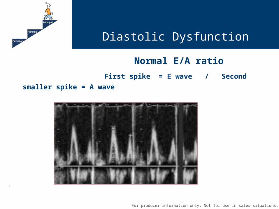
Normal E/A ratio
First spike = E wave / Second smaller spike = A wave
.

For producer information only. Not for use in sales situations.
Diastolic Dysfunction
Diastolic Dysfunction
– Equates to reversed E/A ratio (smaller E wave - taller A wave)

For producer information only. Not for use in sales situations.
Diastolic Dysfunction
Four Echocardiographic Patterns of Diastolic Dysfunction
• Grade I
– Abnormal relaxation
– Reversal of E/A ratio
– Some of this is normal with aging
– No significant clinical signs or symptoms

For producer information only. Not for use in sales situations.
Diastolic Dysfuntion
Four Echocardiographic Patterns of Diastolic Dysfunction
• Grade II
– Pseudo-normal filling (poorer prognosis at this stage)
– Moderate diastolic dysfunction
– Clinical symptoms apparent as well as have LAE and increased filling pressures
– Having more symptoms of SOB and possibly some edema
– Decreased exercise capacity

For producer information only. Not for use in sales situations.
Diastolic Dysfunction
• Grade III - IV Diastolic Dysfunction
– Restrictive filling
– Advanced diastolic dysfunction
– Left Atrial enlarged significantly
– May also have reduced EF
– This would be diastolic heart failure rather than systolic failure. (Often hard to differentiate whether its systolic or diastolic failure at this because of the complex issues at play and it’s probable they could be experiencing both at this point)

For producer information only. Not for use in sales situations.
Diastolic Dysfuntion
Treatment
Treat the cause / reduce the workload……
• Control hypertension
• Control the heart rate - maximize diastole/filling period (beta blockers)
• Improve LV relaxation (calcium channel blockers/ace inhibitors/angiotensin receptor blockers)
• Decrease the resistance the heart pumps against (afterload) and or decrease the filling pressure/pre-load by use of vasodilators
• Monitor build and salt intake
• Lose weight and exercise
• Regular follow up

For producer information only. Not for use in sales situations.
The Echo: Case Study #1
– 69 male NS
– 72 inches 260 #
– Impaired glucose tolerance & HTN
– Six months ago went to Emergency room complaining of SOB. No chest pain or palpitations. Last year had EBCT calcium score 10.
– Average BP 160/100
– Grade II/VI Systolic murmur; S4
– 1 + Pedal edema
– NT Pro BNP elevated
– Chest x-ray mild cardiomegaly

For producer information only. Not for use in sales situations.
Case Study
Echo
- Mild- Mod Aortic stenosis
- EF 55%
- Reversed E/A ratio
- IVS-1.2 ; PW-1.3
- LVID 5.6; LA 4.6
- Right sided chambers mildly dilated
- Mild Tricuspid regurg., and mild ^RSVP 39

For producer information only. Not for use in sales situations.
Case Study
RECAP - Indicators he may have significant diastolic dysfunction
– NL EF and still having unexplained symptoms not otherwise accounted for by another disease
– Age and Build
– Long standing Htn not optimally controlled
– Aortic stenosis
– S4 ( noncompliant ventricle)
– Elevated NT Pro BNP

For producer information only. Not for use in sales situations.
Contact information:
Cindy Miller: [email protected]: 515-235-9285
Contact for Questions

For producer information only. Not for use in sales situations.
Questions