Embed Size (px)
Citation preview

Follow-up for Metabolic Disorders:Fatty Acid Oxidation Disorders,
Galactosemia & Biotinidase Deficiency
Dr. Kathy Grange, MDDivision of Genetics and Genomic Medicine
Department of PediatricsWashington University School of Medicine

Fatty Acid Oxidation Disorders
• Blood spots should be collected between 24 and 48 hours of age
• Samples collected too late may result in a false negative result
• Metabolites in FAO disorders can decrease after the first few days of life as the baby is fed

Fatty Acid Oxidation Disorders• Follow-up testing
– Acylcarnitine profile– Total and free carnitine levels– Urine acylglycine profile– Urine organic acids– Comprehensive metabolic panel, uric acid, CK
• Counsel parents to feed infant frequently and seek medical care for any signs of illness
• For high risk cases (eg. high risk for VLCAD or LCHAD) may need immediate visit to geneticist and other evaluation such as echocardiogram
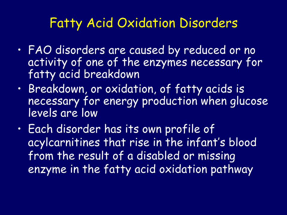
Fatty Acid Oxidation Disorders
• FAO disorders are caused by reduced or no activity of one of the enzymes necessary for fatty acid breakdown
• Breakdown, or oxidation, of fatty acids is necessary for energy production when glucose levels are low
• Each disorder has its own profile of acylcarnitines that rise in the infant’s blood from the result of a disabled or missing enzyme in the fatty acid oxidation pathway

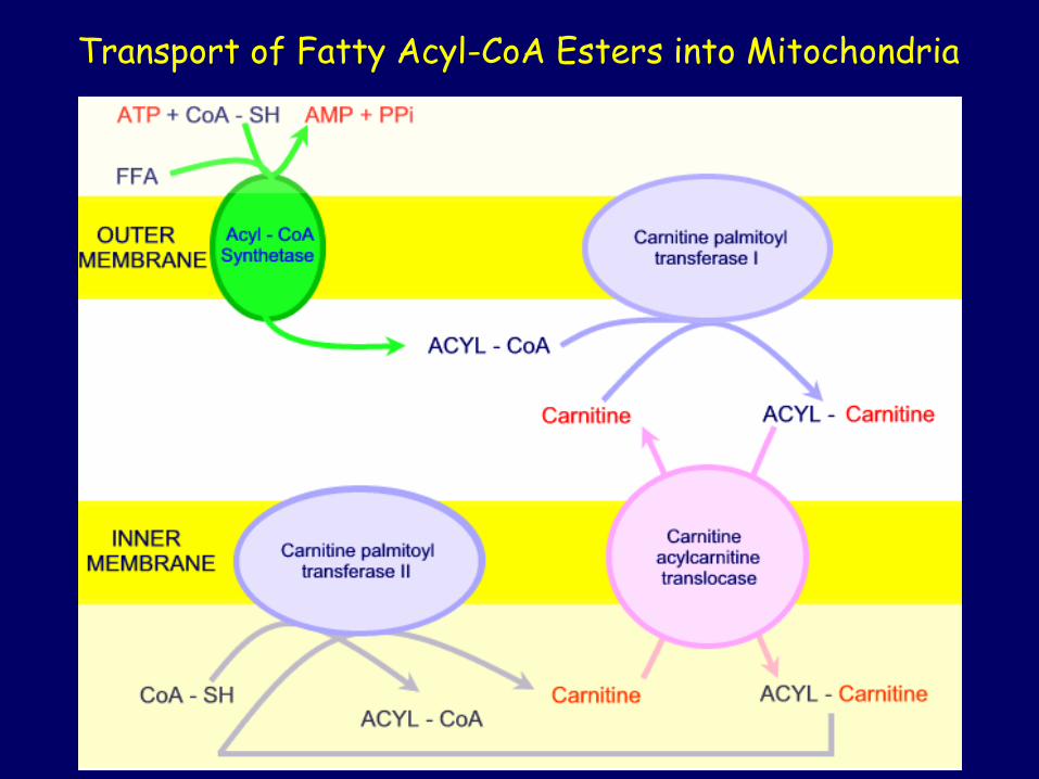
Transport of Fatty Acyl-CoA Esters into Mitochondria

Fatty AcidOxidationPathway
Each cycle removes 2 carbons &producesacetyl-CoA

Metabolic Needs• Brains must have glucose - no fat metabolism
in brain• Animals cannot make glucose from fats• If the body is low on glucose, oxaloacetate is
shunted away from the Krebs cycle to make glucose to feed the brain, and the Krebs cycle slows down, causing a loss of energy
• During severe starvation, the body makes ketone bodies from fatty acids (acetoacetic acid), this provides acetyl CoA for the brain– Glucose comes from protein degradation – Four carbon units are obtained from protein
degradation

During carbohydrate starvation, oxaloacetate in liver is depleted due to gluconeogenesis. This impedes acetyl-CoA entry to Krebs cycle. Acetyl-CoA in liver mitochondria is converted then to ketone bodies, acetoacetate & β-hydroxybutyrate.
Glucose-6-phosphatase glucose-6-P glucose Gluconeogenesis Glycolysis pyruvate fatty acids
acetyl CoA ketone bodies cholesterol oxaloacetate citrate
Krebs Cycle
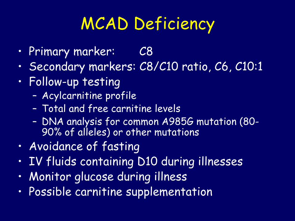
MCAD Deficiency• Primary marker: C8• Secondary markers: C8/C10 ratio, C6, C10:1• Follow-up testing
– Acylcarnitine profile– Total and free carnitine levels– DNA analysis for common A985G mutation (80-
90% of alleles) or other mutations• Avoidance of fasting• IV fluids containing D10 during illnesses• Monitor glucose during illness• Possible carnitine supplementation
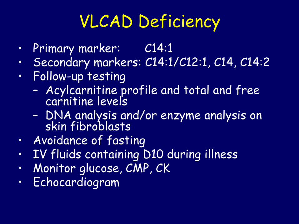
VLCAD Deficiency• Primary marker: C14:1• Secondary markers: C14:1/C12:1, C14, C14:2• Follow-up testing
– Acylcarnitine profile and total and free carnitine levels
– DNA analysis and/or enzyme analysis on skin fibroblasts
• Avoidance of fasting• IV fluids containing D10 during illness• Monitor glucose, CMP, CK• Echocardiogram

VLCAD Deficiency• Lipistart or Portagen formula in infancy • Low fat diet (30% of calories total with 10-
15% calories from long-chain fats)• MCT oil supplement to provide remainder of
fat calorie requirement• Essential fatty acid supplement with to
provide minimum of 3% kcal from linoleic acid and 0.5% kcal from linolenic acid
• Possible low dose carnitine supplementation if deficient

LCHAD or TFP Deficiency• Primary marker: C16-OH• Secondary markers: C18:1-OH, C18-OH, C14:1• Follow-up testing
– Acylcarnitine profile– Total and free carnitine levels– CMP, uric acid, CK– DNA analysis for the common mutation (G1528C)
and/or other mutations• Avoidance of fasting• IV fluids containing D10 during illnesses • Monitor echocardiograms at least yearly• Ophthalmology evaluations regularly for
pigmentary retinopathy

LCHAD or TFP Deficiency• Lipistart or Portagen formula in infancy • Low fat diet (30% of calories total with 10-
15% calories from long-chain fats)• MCT oil supplement to provide remainder of
fat calorie requirement• Essential fatty acid supplement with to
provide minimum of 3% kcal from linoleic acid and 0.5% kcal from linolenic acid
• Possible low dose carnitine supplementation if deficient

Multiple Acyl-CoA Dehydrogenase Deficiency (Glutaric Aciduria type II)
• Primary markers: C4, C5• Secondary markers: C18:1, C8, C12, C14, C16• Follow-up testing
– Acylcarnitine profile– Total and free carnitine levels– Urine organic acid analysis---multiple
compounds (ethylmalonic, adipic, glutaric, and lactic acids and others)• Distinguished from GA I by the presence of
2-OH-glutaric acid• GA II can be confirmed with ETF/ETF-QO
enzyme assay and gene sequencing

Multiple Acyl-CoA Dehydrogenase Deficiency (Glutaric Aciduria type II)
• Avoidance of fasting• IV fluids with glucose during illnesses• Low fat, low protein diet• Carnitine and riboflavin supplementation • Severe neonatal presentation can be
associated with renal and other congenital anomalies and may be fatal

SCAD Deficiency• Primary marker: C4• Follow-up testing
– Acylcarnitine profile– Total and free carnitine levels– Urine acylglycine profile– DNA analysis---common polymorphism
• Avoidance of fasting and IV fluids during illnesses
• Most people with SCAD deficiency are healthy without evidence of illness
• Severe forms with myopathy occur rarely

Carnitine Uptake Disorder• Primary marker: C0• Secondary marker: C2• Follow-up testing
– Blood total and free carnitine levels– Acylcarnitine profile– Urine carnitine levels– DNA analysis
• Test the mother as well – Many cases of maternal CUD have been diagnosed
through newborn screening of their infant• High dose carnitine supplementation• Echocardiogram

Carnitine palmitoyltransferase 1 deficiency (CPT 1)
• Primary markers: C0 (elevated)• Secondary markers: C0/C16, C0/C18• Follow-up testing:
– Acylcarnitine profile– Serum total and free carnitine levels---normal or
elevated in CPT1– CPT 1 DNA analysis
• Avoid fasting, heavy exercise and catabolic condition
• Watch for hypoketotic hypoglycemia, lethargy, hepatomegaly
• Monitor cardiac status with EKG and echocardiogram---risk for cardiomyopathy and arrhythmias

CPT2 and CACT Deficiencies• Primary marker: C16• Secondary markers: C18:1, C18:2, C14• Follow-up:
– Acylcarnitine profile– Serum total and free carnitine levels– DNA testing
• Avoid fasting, heavy exercise and catabolic condition
• Carnitine supplementation if levels low• MCT oil supplement and low long-chain fat
diet may help

CPT2 and CACT Deficiencies
• Monitor cardiac status with EKG and echocardiogram– High risk for cardiomyopathy and
arrhythmias• Perinatal form of CPT 2 deficiency is almost
invariably fatal• High mortality rate (24% survival) for CACT
deficiency

Galactosemia• Incidence is 1 in 45,000 newborns for classic
galactosemia• Autosomal recessive disorder• Deficiency of galactose-1-phosphate
uridyltransferase (GALT) enzyme • Inability to convert galactose-1-PO4 to
glucose-1-PO4

Metabolism of Galactose

Classic Galactosemia• Babies appear normal at birth• Symptoms begin in 1st week of life if
lactose is given:– Vomiting and diarrhea– Failure to thrive or weight loss– Hepatomegaly, jaundice, coagulopathy– Kidney dysfunction– Increased intracranial pressure/cerebral
edema– Cataracts (“oil droplet”)– E. coli sepsis

Classic Galactosemia
• Laboratory abnormalities include:– Galactosemia– Galactosuria (urine positive for reducing
substances)– Hyperbilirubinemia– Hypokalemia– Hyperchloremia– Hypoglycemia (+/-)– Hyperammonemia– Elevated PT/PTT and liver enzymes

Galactosemia
• Borderline Risk (3.1 – 4.0 U/gHb)– Variant galactosemia (Duarte-galactosemia
or others) – Carrier for galactosemia– Improperly handled sample (heat damage or
transit delay)• High Risk (≤ 3.0 U/gHb)
– Classical galactosemia (usually <1.5)– Variant galactosemia or carrier
(often 1.6-3.0)

Galactosemia
• Follow-up testing – Quantitative galactose-1-phosphate
uridyltransferase (GALT) enzyme assay– Red blood cell galactose-1-PO4 level– DNA analysis on the GALT gene
• Common mutations analyzed first• Sequencing of GALT gene if still unclear
• Change to a soy formula while awaiting test results

Biotinidase Deficiency
• Enzyme deficiency affects biotin availability• Lab test gives qualitative result (eg, 1+ to 4+)
– Reported as normal or abnormal to provider• Newborns are usually asymptomatic• Episodic hypoglycemia, lethargy, hypotonia, and
mild developmental delay can occur at any time from the neonatal period through childhood
• Untreated biotinidase deficiency leads to more severe developmental delay, seizures, alopecia, and hearing deficits
• Biotin treatment is highly effective

Biotin Cycle
Biotinidase Deficiency
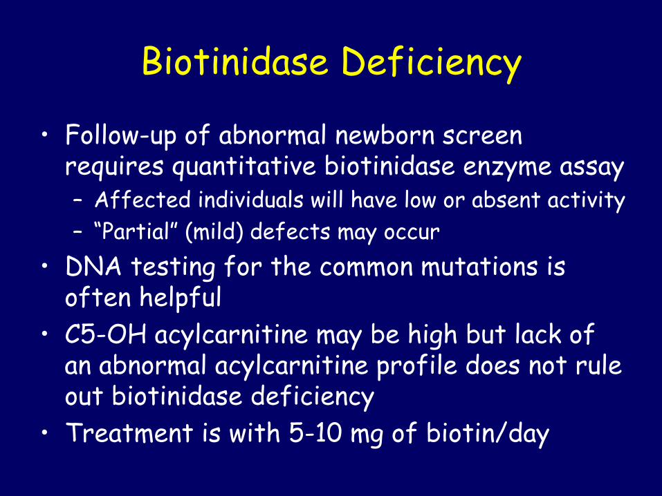
• Follow-up of abnormal newborn screen requires quantitative biotinidase enzyme assay – Affected individuals will have low or absent activity– “Partial” (mild) defects may occur
• DNA testing for the common mutations is often helpful
• C5-OH acylcarnitine may be high but lack of an abnormal acylcarnitine profile does not rule out biotinidase deficiency
• Treatment is with 5-10 mg of biotin/day

Who Should Get a Repeat NBS?
• Unsatisfactory sample on the first filter paper
• Premature infants• Infants who are ill and in the NICU• Borderline abnormal results on the first
screen• Suspicion for metabolic disease

Missouri Guidelines for NBS Specimens from Premature, Low Birth Weight, or Sick NICU Infants
• CORE CATEGORY = All NICU infants (unless transfused)– 1st specimen at 24-48 hours of age– 2nd specimen 10-14 days of age or at discharge
• RBC transfusion <24 hrs of age– 1st specimen collected before transfusion– 2nd specimen 24-48 hours after transfusion– 3rd specimen 30 days after transfusion
• NOT collected prior to RBC transfusion– 1st specimen 24-48 hours after transfusion– 2nd specimen 30 days after transfusion– 3rd specimen 90 days after transfusion
• Infants on TPN should follow Core Category unless transfused; may need another specimen after off TPN