Embed Size (px)
Citation preview

13 April 1968 CIRDICAL JOUMuAL
Papers and Originals
Folate Deficiency in Crohn's Disease: Incidence, Pathogenesis,and Treatment
A. V. HOFFBRAND,* B.M., M.R.C.P., D.C.P.; J. S. STEWARTJ M.B., M.R.C.P.; C. C. BOOTH4 M.D., F.R.C.P.D. L. MOLLIN,§ M.B., B.SC., M.R.C.P., M.C.PATH.
aBrt. mid.., 1968,2, 71-75
Megaloblastic anaemia due to folate deficiency is a well-recognized complication of idiopathic steatorrhoea and acutetropical sprue. These conditions are characterized by abnor-malities of mucosal structure and absorptive function of theproximal small intestine, the principal site of folate absorption.On the other hand, in patients with anatomical lesions of thesmall intestine such as blind loops, strictures, and ileal re-sections, megaloblastic anaemia, if it occurs, is almost invariablydue to vitamin B., deficiency (Mollin, 1960).
Nevertheless there are a few reports which suggest that severefolate deficiency, sometimes with megaloblastic anae mayoccur in patients with Crohn's disease. Klipstein (1963) founden extremely low serum folate level in one patient with thisdisease which he attributed to inadequate dietary intake of thevitamin. Rose (1965) found subnormal serum folate levelsin a fither three patients, two of whom showed " transitional "megaloblastic changes and one of the latter also excreted raisedamounts of formioglutamic acid (Figlu). The patient ofHall (1953) who developed megaloblastic anaemia which re-sponded to large doses of folic acid, and the four anaemicpatients of Knowles (1962) with positive Figlu tests, may alsohave been suffering from folate deficiency; but neither of thesestudies is conclusive, since B1, deficiency, which could haveequally well been present, was not excluded. Thompson andUngley (1955) describe the case of a patient who, though notnaemic, was suffering from diarrhoea and loss of weight while
an treatment with vitamin B12, which were promptly improvedwhen folic acid was given. Though no direct tests for folatedeficiency were available, it is likely that, this patient sufferedfrom such deficiency.In our experience folate deficiency is frequent in patients with
active Crohn's disease, and the purpose of this paper is to reportthe incidence and pathogenesis of the deficiency in a group of64 patients and describe the value of folic acid therapy in thiscondition.
Subjects Studied
Patients with Crohn's Disease
Observations were made on 64 patients (Table I) with Crohn'sdisease (27 males and 37 females), their ages ranging from 8
Lecturer in Haematology, St. Bartholomew's Hospital, London.Present address: Blood Research Laboratory, New England MedicalCenter Hospitals, 171 Harrison Avenue, Boston, Massachusetts,U.S.A.
t This work was done during the tenure of the T.K. Stubbins ResearchFellowship of the Royal College of Physicians, London. Present ap-pointment: Consultant Physician, West Middlesex Hospital,Isleworth, Middlesex.
t Professor of Medicine, Royal Postgraduate Medical School, LondonW.12.
Professor of Haemnatology, St. Bartholomew's Hospital, London E.C.1.
to 67 (mean 34.5) years. In 40 of the patients the diagnosishad been confirmed at laparotomy before the present studiowere made, and in a further seven the diagnosis was confirmedwhen they subsequently came to laparotomy. In the remaining17 the diagnosis was made because the patients suffered fromabdominal pain, diarrhoea, loss of weight, and fever, and showedcharacteristic narrowing with or without ulceration of the ileumon barium follow-through examination.The clinical histories of the patients ranged from a few
months to 25 years. Twenty-four were in hospital and 40were attending hospital as outpatients or were under the careof their general practitioner. Ten of the 64 patients werealready receiving regular parenteral B1, therapy (200 ,Agmonthly) for previously diagnosed megaloblastic anaemia dueto B,2 deficiency.TABLE I.-Peripheral Blood Findings in the 64 Patients, Showing the
Number of Patients in Each Clinical Group with Anaemia, Macro.cytes, and Hypersegmented Polymorphs
Group 1 Group 2 Group 3
Anaemic .. 2 0 0 8 5(62) 562 18 12(67) 9(50)Non-anaemic 19 1(5) 1 (5) 13 2 15) 3 23) 4 1 25) 2 50)Toa ..W 21 1 (5) 1 (5) 21 7 (33) 8 (38) 22 13 (59) 11 (50)
H-S. P. - Hypesegmented polymorphs.
The patients were divided into three groups according tothe severity of their symptoms, without foreknowledge of theresults of the study.Group 1.-Patients who were clinically well and symptomless
or who had no symptoms that could be attributed to Crohn'sdisease.Group 2.-Patients who were unwell with symptoms that could
be ascribed to Crohn's disease. These patients needed regularmedical attention, but their symptoms were not severe enough toprevent them from working or to warrant admission to hospital.Group 3.-Patients who were severely ill, the majority confined
to bed, and all in hospital or requiring admission to hospital.The mean ages of the patients from groups 1, 2, and 3 were.
38.6, 33.7, and 30.3 years respectively.
Control GroupThe control group was made up of 50 adult hospfra? or,-
patients. (25 males and 25 females) selected at random exceptthat antenatal patients and patients known to be aneic or tobe suffering from gastrointestinal or malignant disease wereexcluded. Their ages ranged from 17 to 83 (mean 49) years.
71

Methods
Serum folate levels were determined by microbiological assay
with Lactobacillus casei as test organism by the method ofWaters and Mollin (1961). The normal range is from 6 to 21m/Ag./ml.Red cell folate levels were measured by the modification of
the serum folate assay described by Hoffbrand et al. (1966b) in
which venous blood is haemolysed in distilled water containing1 g. of ascorbic acid per 100 ml. before assay. The normal
range is from 160 to 640 mj.tg./ml. of packed red cells.Figlu and urocanic acid were measured by the spectrophoto-
metric method of Chanarin and Bennett (1962a) in an eight-hour urine specimen collected after an oral dose of 15 g. of L-histidine monohydrochloride. The normal range for Figluexcretion is from 0 to 17 mg. in eight hours. Urocanic acidexcretion of 17 mg. or less in the eight-hour period is alsoregarded as normal (Hoffbrand et al., 1966a).Serum vitamin B1, levels were determined by microbio-
logical assay with the "z" strain of Euglena gracilis as testorganism (Anderson, 1964). The normal range is from 160 to925 juqug./ml.
Folic acid absorption was measured by microbiological assaywith Streptococcus faecalis as test organism as described byChanarin et al. (1958). In normal subjects the peak serumfolate level after the standard oral dose of folic acid (40 ,ug./kg. body weight) is more than 40 m~tg./ml.
Dietary folate intake was calculated by use of standard tables(McCance and Widdowson, 1960). So far as possible, allow-ance was made for loss of folate in cooking.Routine haematological methods were those described by
Dacie and Lewis (1963). Stained peripheral blood films wereexamined for the presence of macrocytes and hypersegmentedpolymorphs (one or more polymorphs with six or more nuclearlobes per 100 polymorphs). Bone marrows were classified asnormoblastic or megaloblastic. The megaloblastic marrowswere further subdivided into (1) those showing mild changes(rare intermediate megaloblasts and occasional giant meta-myelocytes, and (2) those showing intermediate changes (Dacieand White, 1949). Florid megaloblastic changes as occur insevere untreated Addisonian pernicious anaemia were not seen.
Rests
Serum B1, Concentrations.-The serum B., levels of 51of the 54 patients not already receiving regular B12 therapy were
E
t
E
0Ad,
numberrangemean
1 5
141312.1
12
10
9
8
6
5I
432
so
50
3-3-1867*1
0*
0
0
0
0
0
:
21
67
0
00
.I0
212-8-6-4
3.7
0
220-5-4-8
2-5
c----- ----- ----- - - --- >-- - -- -----
---------------- --- ---
WControls Group I Group 2 Group 3
FIG. 1.-Serum folate levels of 50 control patients and 64 patients withCrohn's disease. The latter are divided into three clinical groups (seWtext). The upper horizontal line is the lower limit of the range of semfolate levels in normal subjects. The lower horizontal line is the lower
limit of the control serum folate levels.
BemMVDICAL JOURNAL
normal, ranging from 160 to 825 (mean 373) ,Atg./ml. In the
other three patients the levels were subnormal (125, 130, and130 pjuqg./ml.) but not as low as usually occurs in untreatedpernicious anaemia.Serum Folate Concentrations.-Of the control subjects 21
(42%) had subnormal serum folate levels-that is, less than6 mjug./ml.-but all were above 3 mlug./ml. In contrast,as many as 52 (81%) of the patients with Crohn's disease hadsubnormal serum folate levels and 18 (28%) had levels less than3 mjAg./ml. (Fig. 1). Serum folate levels below the control range
occurred most frequently in the patients with the most activedisease. Thus 13 of the severely ill patients (group 3) hadserum folate levels less than 3 mndg./ml. compared with only 4of the less severely ill patients (group 2) (Fig. 1). Only one
of the symptom-free patients (group 1) had such a low level.On the other hand there was no relation between serum folatelevel and the age or sex of the patients, length of history, or
whether or not the patients had had an operation for thedisease.Red Cell Folate Concentrations.-Subnormal red cell folate
levels occurred in five of seven severely ill patients tested, in
two of eight mildly ill patients, but in none of four symptom-free patients.
Figlu Excretion.-This was measured in 24 patients, all ofwhom had normal serum B1. levels. Like subnormal serumand red cell folate levels, positive Figlu tests occurred mostoften in the patients with the most active disease (Fig. 2). ThusFiglu excretion was raised in all 10 severely ill patients tested,in only two of nine less severely ill patients, and in none of fivesymptom-free patients.
160
140
120
. 100
soe 80
60
40
20
0Groupi Group 2 Group 3
FIG. 2.-Excretion of Figlu and urocanic acid in 24 patients with Crohn'sdisease. The horizontal line denotes the upper limit of the normal rangeof Figlu excretion and the upper limit of the normal range of urocank
acid excretion.
Urocanic Acid Excretion.-This was raised in three of the10 severely ill patients with positive Figlu tests, but was normalin the other 21 patients tested (Fig. 2).
Haematological Findings
Peripheral Blood.-Anaemia was present in 28 of the patients(Table I). The anaemic patients were generally severely ill withmultiple causes of anaemia-for example, active inflammato"disease, iron deficiency, etc. They usually required urgenttherapy with steroids, antibiotics, iron, transfusions, etc., so itwas difficult to observe the effect of folic acid therapy alone,and therefore impossible to estimate in how many of the 28patients folate deficiency contributed to anaemia. There was,however, an obvious association between the presence ofanaemia and presence of haematological changes due to folatedeficiency in the peripheral blood. Thus macrocytosis and/or
72 13 April 1968 Crohn's Disease-Hoffbrand et al.

hypersegented polymorphs, like anaemia, occurred far moreoften in the severely ill patients than in those without symptoms(Table I). Moreover, macrocytosis was seen in the films of17 of the anaemic patients but in the films of only 4 of thenon-anaemic patients; and hypersegmented polymorphsoccurred in 14 of the anaemic and only 6 of the non-anaemicpatients. Macrocytosis and hypersegmented polymorphs couldalmost certainly be ascribed to folate deficiency, since all but3ne of the patients showing these features had subnormal serumfolate and normal serum B1, levels. The remaining patient hadsubnormal serum folate and serum B1, levels (2.7 m~tg./ml. and'30 pjsg./mL repectvely).
Bone Marrow.-Bone marrow examinations were performed
;n 17 anaensc and six non-anaemic patients (all with normalierum B, levels (Table II). The marrow showed megalo-,lastic changes im 12 anaemic and three non-anaemic patients.Like other evidence of folate deficiency, megaloblastic changeswere particularly frequent in the patients with the most activeIisease (Table II).
All 15 patients with megaloblastic changes had subnormalserum folate levels, and in 11 these were less than 3 m~tg./ml.Of the patients with normoblastic erythropoiesis four had sub-aormal serum folate levels, but these were invariably above3 mjtg./ml. Surprisingly, two patients with normoblastichaemopoiesis had positive Figlu tests, usually indicating severefolate deficiecy.
Results of Folic Acid Therapy
It was possible to study the effect of folic acid therapy n
Heven patients (four with intermediate megaloblastic changesand three with mild megaloblastic changes). None of themshowed a complete haematological remission on folic acid treat-ment alone. The best response occurred in one of the severely
Ill patient (Case 13, Table II).This patient had suffered from Crohn's disease for four years
and was admitted to the hospital with a four-month history ofIncreasing anorexia and diarrhoea. He was analmc (haemo-globin 6.6 g./100 ml., P.C.V. 20%, red cell count 1,700,000/,.u. mm., reticulocytosis 0.5%, white cell count 5,000/cu. mm.,platelets 150,000/cu. mm.). The stained peripheral blood filmshowed obvious macrocytosis and hypersegmented polymorphs,and the bone marrow showed intermediate megaloblasticchanges with adequate iron stores but siderotic granules almost
TABLE II.-Deaied Haematological and Biochemical FindingsPerfo
BDrrIsaMEDICAL JOURNAL 73
completely absent from the developing erythroblasts, indicatingdefective iron utilization.He was transfused with the packed cells from 2 pints (1,140
ml.) of blood and then given folic acid (15 mg. intramuscularlydaily). There was a reticulocytosis to a peak of 13% with aslow rise of haemoglobin to 12 g./100 ml. and a rise in leuco-cytes from normal to supranormal levels (Fig. 3). Both the,
B.T.,+ Folic acid 15mg.daily
30-
,. 20-
0
'S 10-
43.
OJ
3a
8920.-080 9.0,
0
or
x._1
° 3-0
oM
I-10 0 laDays of, therapy
FIG. 3.-Haematological response of one patient (Case 1) to folkc acid
haemoglobin and leucocyte count became normal when thepatient was subsequently treated with A.C.T.H. (40 units daily)and sulphasalazine (1 g. t.d.s.).Two further patients (Cases 2 and 12, Table II) with inter-
mediate megaloblastic change due to folate deficiency andinitial haemoglobin concentrations of 8.9 and 11.9 g./100 mlshowed significant reticulocyte responses and increases in
haemoglobin of 3.1 and 2.0 g./100 ml. respectively with folicacid therapy (15 mg. daily by mouth). On the other hand fourmildly anaemic patients (Cases 3, 9, 15, and 16, haemoglobinrange from 10.4 to 13.0 g./100 ml.) showed no definite haema-tological response to folic acid in large doses. However, allseven patients treated with folic acid showed striking subjectiveimprovement, increased appetite, and gain in weight.
Pathogenesis of the Folate DeficiencyThere are three possible causes for folate deficiency in Crohn's
disease: malnutrition, malabsorption, and excess folate utiliza-
in the 23 Patints on whom Bone Marrow xnations oere
Blood___ Bone____Marrowgi Urocanic Calculated Folic Acid AbsorptionCase SeS I I PerihmBood BoneMarowBi| Serum Red Cell Acid Daily (mpg./ml.)NO. (S.1100 ~~~~~~~~~~Folate Folate (m./ Folate(m .I3)No. ml.) ~~~~~ H-S. P. M-G.-G.0 rns r.
M| I H | Iron I (PPg.Im. |(mpg.jnl.) mp.jlrl.). 8 br..) 8 hrs.) hr. hr. 2|hr.
Group 31 66 + + Int.meg. Present 255 0.5 39 110 25 24 0 64 432 F 849 + + Int.meg. Present Rec. Bis 1.1 53 _ - - 0 34 363 F 10-7 + + Int.meg. Present 165 4-8 122 28 0 33 0 97 924 M 12-2 + + Int.meg. Absent 330 3-8 - 515 8 0 9 155 M 10 8 + _ Mildmeg. Present 260 3-6 88 158 0 - - - -6 M 12 6 + - Mildmeg. Present 465 2-0 103 85 0 _ _ _7 M 1340 + + Midmeg. Present 190 1-6 - 121 9 - _8 F 7-4 - Mild meg. Absent 640 2-3 - 22 0 24 0 9 789 M 10-4 + Mildmeg. Absent 235 2-6 - 74 0 - - - -10 F 9-6 - + Norno Absent Rec. Bs 3-5 250 94 49 38 0 39 4011 F 100 - - Normo Absent 700 7-8 240 131 65 49 0 57 73
Group 212 M 11-9 + + Int. meg. Present Rec. Bis 3-2 111 18 0 200 0 16 12713 F 14-7 + + Int.me& Present 175 4-8 126 30 0 56 0 30 4914 bS 13-3 + + Mildmeg. Present Rec. Bis 3*3 237 12 0 86 0 > 100 > 10015 M 13-0 + Mildmeg. Present 265 3-8 313 8 0 72 0 42 4516 M 12-0 + - Mildmeg. Absent 825 3-2 198 0 0 - -_ _17 F 12-1 _ + Mildmeg. Present 300 4-6 185 9 0 77 0 9 818 M 15-5 - Normo Present Rec. Bis 3-6 230 6 0 53 0 10 3019 P 124 - - Normo Absent 320 2-9 307 8 0 86 0 32 43
Group 120 M 13-9 - - Normo Present Rec. B1I 7-1 369 11 0 - 0 67 5421 F 12-3 - - Normo Present 305 5-6 457 - - - 0 78 8522 F 112 1 - Normo Absent 215 8-7 601 0 0 - -23 F 11-3 1 - Normo Absent 370 8 0 533 0 0 - - I I° MaY-ruenwald-Gemsa stain. Mild meg.=Mild megaloblastic. Int. Meg. -Intermediate megaloblastic. H-S.P- Hypersegmental polymorh.
13 April 1968 Crohn's Disease-Hoffbrand et al.
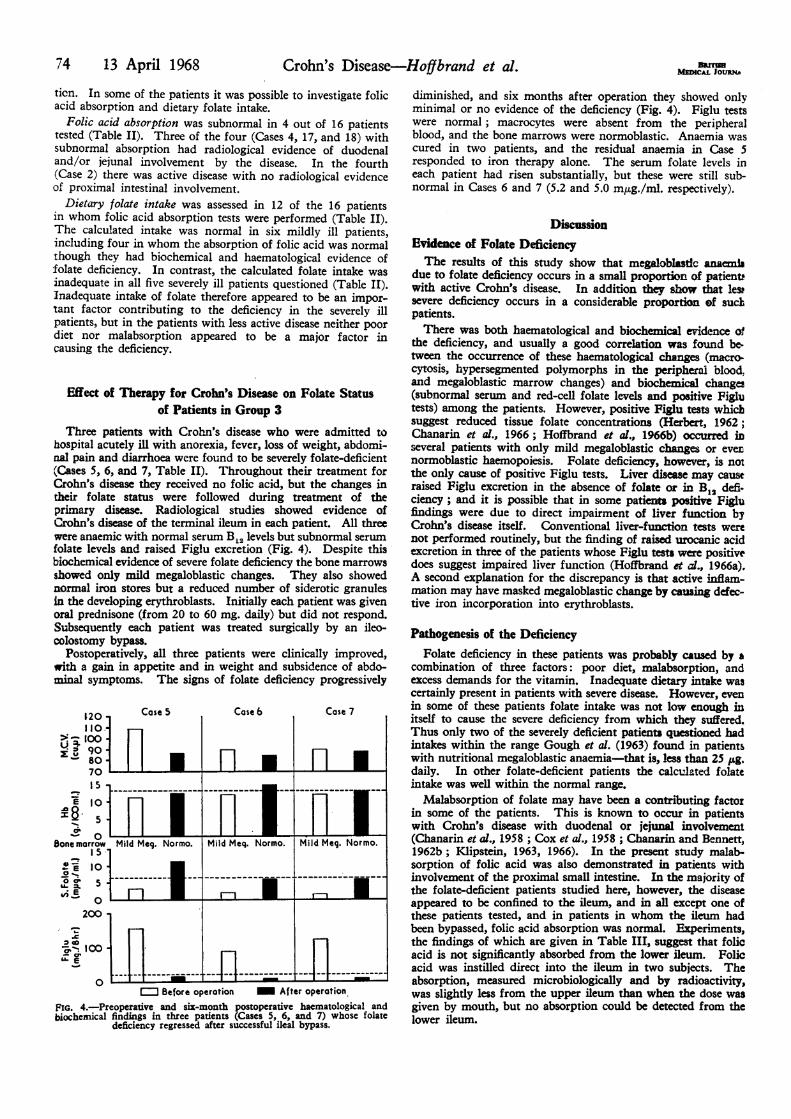
74 13 April 1968 Crohn's Disease-Hoffbrand et al.ticn. In some of the patients it was possible to investigate folicacid absorption and dietary folate intake.
Folic acid absorption was subnormal in 4 out of 16 patientstested (Table II). Three of the four (Cases 4, 17, and 18) withsubnormal absorption had radiological evidence of duodenaland/or jejunal involvement by the disease. In the fourth(Case 2) there was active disease with no radiological evidenceof proximal intestinal involvement.
Dietary folate intake was assessed in 12 of the 16 patientsin whom folic acid absorption tests were performed (Table II).The calculated intake was normal in six mildly ill patients,including four in whom the absorption of folic acid was normalthough they had biochemical and haematological evidence offolate deficiency. In contrast, the calculated folate intake wasinadequate in all five severely ill patients questioned (Table II).Inadequate intake of folate therefore appeared to be an impor-tant factor contributing to the deficiency in the severely illpatients, but in the patients with less active disease neither poordiet nor malabsorption appeared to be a major factor incausing the deficiency.
Effect of Therapy for Crohn's Disease on Folate Status
of Patients in Group 3
Three patients with Crohn's disease who were admitted tohospital acutely ill with anorexia, fever, loss of weight, abdomi-
aal pain and diarrhoea were found to be severely folate-deficient
(Cases 5, 6, and 7, Table II). Throughout their treatment forCrohn's disease they received no folic acid, but the changes intheir folate status were followed during treatment of the
primary disease. Radiological studies showed evidence ofCrohn's disease of the terminal ileum in each patient. All three
were anaemic with normal serum B12 levels but subnormal serumfolate levels and raised Figlu excretion (Fig. 4). Despite thisbiochemical evidence of severe folate deficiency the bone marrowsshowed only mild megaloblastic changes. They also showednormal iron stores but a reduced number of siderotic granulesin the developing erythroblasts. Initially each patient was givenoral prednisone (from 20 to 60 mg. daily) but did not respond.Subsequently each patient was treated surgically by an ileo-
colostomy bypass.Postoperatively, all three patients were clinically improved,
with a gain in appetite and in weight and subsidence of abdo-
minal symptoms. The signs of folate deficiency progressively
120
110.j
, 100a90180
70
15
e 10
5g5
Cy% C0Bone marrow
-0'.
5
.E
0
20
Case 5
LlI1I
Case6 Case 7
.r. . . .
Mild Meg. Normo. Mild Meq. Normo. Mild Meg. Normo.
* 1-1iL .= Before operation = After operation
FIG. 4.-Preoperative and six-month postoperative haematological andbiochemical findings in three patients (Cases 5, 6, and 7) whose folate
deficiency regressed after successful ileal bypass.
diminished, and six months after operation they showed onlyminimal or no evidence of the deficiency (Fig. 4). Figlu testswere normal; macrocytes were absent from the peripheralblood, and the bone marrows were normoblastic. Anaemia wascured in two patients, and the residual anaemia in Case 5responded to iron therapy alone. The serum folate levels ineach patient had risen substantially, but these were still sub-normal in Cases 6 and 7 (5.2 and 5.0 mjug./ml. respectively).
Discussion
Evidence of Folate DeficiencyThe results of this study show that megaloblastic anaemd
due to folate deficiency occurs in a small proportion of patientswith active Crohn's disease. In addition they show that lewsevere deficiency occurs in a considerable proportion of suckpatients.There was both haematological and biochemical evidence of
the deficiency, and usually a good correlation was found be-
tween the occurrence of these haematological changes (macro.cytosis, hypersegmented polymorphs in the peripheral blood,and megaloblastic marrow changes) and biochemical changes(subnormal serum and red-cell folate levels and positive Figlutests) among the patients. However, positive Figlu tests whichsuggest reduced tissue folate concentrations (Herbert, 1962;Chanarin et d., 1966; Hoffbrand et at., 1966b) occurred inseveral patients with only mild megaloblastic changes or evernormoblastic haemopoiesis. Folate deficiency, however, is notthe only cause of positive Figlu tests. Liver disease may causeraised Figlu excretion in the absence of folate or in B1, defi-ciency; and it is possible that in some patients positive Figlufindings were due to direct impairment of liver function byCrohn's disease itself. Conventional liver-function tests werenot performed routinely, but the finding of raised urocanic acidexcretion in three of the patients whose Figlu tests were positivedoes suggest impaired liver function (Hoffbrand et al., 1966a).A second explanation for the discrepancy is that active inflam-mation may have masked megaloblastic change by causing defec-tive iron incorporation into erythroblasts.
Pathogenesis of the DeficiencyFolate deficiency in these patients was probably caused by a
combination of three factors: poor diet, malabsorption, andexcess demands for the vitamin. Inadequate dietary intake wascertainly present in patients with severe disease. However, evenin some of these patients folate intake was not low enough initself to cause the severe deficiency from which they suffered.Thus only two of the severely deficient patients questioned hadintakes within the range Gough et al. (1963) found in patientswith nutritional megaloblastic anaemia-that is, less than 25 jig.daily. In other folate-deficient patients the calculated folateintake was well within the normal range.
Malabsorption of folate may have been a contributing factorin some of the patients. This is known to occur in patientswith Crohn's disease with duodenal or jejunal involvement(Chanarin et al., 1958 ; Cox et al., 1958 ; Chanarin and Bennett,1962b; Klipstein, 1963, 1966). In the present study malab-sorption of folic acid was also demonstrated in patients withinvolvement of the proximal small intestine. In the majority ofthe folate-deficient patients studied here, however, the diseaseappeared to be confined to the ileum, and in all except one ofthese patients tested, and in patients in whom the ileum hadbeen bypassed, folic acid absorption was normal. Experiments,the findings of which are given in Table III, suggest that folicacid is not significantly absorbed from the lower ileum. Folicacid was instilled direct into the ileum in two subjects. Theabsorption, measured microbiologically and by radioactivity,was slightly less from the upper ileum than when the dose wasgiven by mouth, but no absorption could be detected from thelower ileum.
BarrMEDCAL JoutA

13 April 1968 Crohn's Disease-Hoffbrand et al. mf 75
TABLE III.-Comparison of the Absorption of Folic Acid in Two NormalSubjects (A and B) and One Patient with Untreated IdiopathicSteatorrhoea (C) When the Dose was Given (a) by Mouth and (b)by Direct Instillation Into the Small Intestine. Each Dose was 40ug. Folic Acid/kg. Body Weight Labelled with 20 pCi Tritium, andthe Absorption was Determined by the Rise in Serum Folic AcidMeasured by Str. faccalis Microbiological Assay and by the 24-hourUrinary Excretion of Radioactivity After a 15-mg. IntramuscularFlushing Dose of Non-radioactive Folic Acid Administered ThreeHours After the Oral or Intestinal Dose
UrinaryDistance of Radiological SerumPolate ExcretionTip of Small Pition of Site of Levels (Hours) Radio.
Case Intestinal Tip of Adminis- activityTube From Small tration of Iin 24 hours
Incisor Intestinal Polk Acid %Teeth (cm.) Tube 0 1 2 3 (Normal
>30%)A - _ Oral 0 105 82 48 350
170. Lower Lower 0 66 63 40 302jejunum jejunum
B _ -e j.Oral 0 98 77 56 45-6250 Mid-lower Mid-lower 0 3 1 0 0.0
ileum ileumC -_ Oral 1 23 34 31 -
200 Upper pper 018S 124 -ileum ilreu
One severely ill patient (Case 2) did malabsorb folic acid eventhough her disease was apparently confined to the terminalileum. This suggests that extremely active disease of the ileummight affect jejunal function. However, in other patients withextremely active disease of the ileum folic acid absorption wasnormal (Table II).The third factor of possible importance in the pathogenesis
of the deficiency is increased folate utilization due to the activeinflammatory disease. Inflammation may provoke folate defi-Ciency in the experimental animal (May et al., 1952) and inpatients with rheumatoid arthritis (Gough et al., 1964) andtuberculosis (Roberts et al., 1966). In these conditions thedeficiency is presumably caused by increased folate requirementsdue to increased production of granulocytes and other inflam-matory cells. In active Crohn's disease there is likely to beIncreased folate utilization from this cause. Since dietary folateintake and folic acid absorption were both well within thenormal range in some patients with folate deficiency, in thesepatients, at least, increased utilization was likely to be a majorfEactor in causing the deficiency.
Value of Folic Acid TherapyIt was impossible to estimate to what extent folate deficiency
contributed to anaemia among these 64 patients. There was asignificant though incomplete haematological response to folicacid in three anaemic patients with intermediate megaloblasticchanges. All seven deficient patients given folic acid showedmarked subjective improvement, with a prompt gain in appetiteand weight. It would therefore seem advisable to treat withfolic acid all patients with active Crohn's disease and haemato-logical and biochemical changes due to the deficiency. Theincidence of patients needing folic acid therapy is likely to behigh among those with severely active disease.Vitamin B1 deficiency, which causes haematological changes
identical to folate deficiency, also occurs frequently in patientswith Crohn's disease, particularly after ileal resection. Beforegiving folic acid therapy to patients with Crohn's disease it istherefore essential to exclude B12 deficiency-for instance, bymeasuring the serum B12 level-in order to avoid precipitatingneurological damage.As shown in the three patients studied here, before and after
surgical intervention, folate deficiency may also be corrected bysuccessful treatment of the Crohn's disease without folic acidtherapy. In these three patients the diet improved postopera-tively, and presumably the intake of folate increased and thedemands for the vitamin diminished. Spontaneous remissionof a macrocytic anaemia in Crohn's disease after successfuloperation was recorded by Butt and Watkins (1936). Theydid not establish the cause of the macrecytic anaemia in their
patients, but the phenomenon they observed is probably the onerecorded in these three patients here.
SummaryIn the present study of 64 patients with Croha's disease .
high incidence of folate deficiency was found among patients frthe active stage of the condition. Haematological changes' duto the deficiency included macrocytosis and hypsegmentedpolymorphs in the peripheral blood and megaloblastic changetin the bone marrow. Biochemical changes included low serumand red cell folate levels and positive Figlu tests. Vitamin B1,deficiency was excluded as a cause of these changes in all buione patient.
Folate deficiency appeared to be due to a combination oJfactors-inadequate dietary intake, excess utilization, andmalabsorption of the vitamin. The-re was a marked sub-jective improvement and gain in weight in all seven folate-deficient patients given folic acid therapy and definite haemato-logical improvement in the three with the most severe deficiency.Folate deficiency in three patients with active Crohn' diseaseremitted spontaneously after successful surgical therapy of theCrohn's disease itself. It is concluded that severe folatedeficiency is a frequent complication of active Croin's diseaseand that folic acid therapy may prove beneficial in a highproportion of such patients.
Because of the high frequency of B1, deficiency in Crohn'sdisease, however, it is always essential to exclude this beforegiving folic acid. Folate deficiency in Crohn's dCse may becured slowly without folic acid therapy, by successful treatmentof the Crohn's disease itself.
We wish to thank the physicians and surgeons of HsmmersmitbHospital and Dr. N. F. Cogill and the other physicians andsurgeons of the West Middlesex Hospital for permission to studypatients under their care, and Dr. A. M. Dawson, St. Bartholo-mew's Hospital, for permission to study and publish details oiCase X, which was under his care. In addition we are grateful toDr. A. H. Waters for helpful discussions during this study and toMr. J. Morgan, Miss B. F. A. Newcombe, Miss J. D. Cowan, andMiss J. Mercy for valuable technical assistance. D. L. M. andA. V. H. are members of the Medical Research Council Group forInvestigation of Megaloblastic and Sideroblastic Anaemis.
Requests for reprints should be addressed to Dr. A. V. Hoffbrand.St. Bartholomew's Hospital, London E.C.I.
REFERENCESAnderson, B. B. (1964). 7. clin. Path., 17, 14.Butt, H. R., and Watkins, C. H. (1936). Ann. nsuern. Med. 10 222.Chanarin, I., Anderson, B. B., and Mollin, D. L. (1958). Bria.k.aema.,
4, 156.Chanarin, I., and Bennett, MA C. (1962a). Brit. med. 7., 1, 27.Chanarin, I., and Bennett, M. C. (1962b). Bait. med. 7., 1, 985.Chanarin, I., Hutchinson, M., McLean, A., and Moulc, M. (1966). Brm
ned. 7., 1, 396.Cox, B. V., Meynel, M. J., Cooke, W. T., sad Gaddie, R. (1958).
Gastroenterology, 35, 390.Dade, J. V., and Lewis, S. M. (1963). Practical Haematlogy, 3rd Cd.
London.Dacie, J. V., and White, J. C. (1949). 7. clan. Path., 2, 1.Gough, K. R., McCarthy, C., Read, A. B., Mollin, D. L., and Waters,
A. H. (1964). Brit. med. 7., 1, 212.Gough, K. R., Read, A. E., McCarthy, C. F., and Waters, A. H. (1963).
Quart. 7. Med., 32, 243.Herbert, V. (1962). Trans. Ass. Amer. Phycns, 7S, 307.Hoffbrand, A. V., Neale, G., Hines, J. D., and Mollin,.D. L. (1966a).
Lancet, 1, 1231.Hoffbrand, A. V., Newcombe, B. F. A., and Mollin, D. L. (1966b). I
clin. Path., 19, 17.Klipstein, F. A. (1963). Blood, 21, 626.Klipstein, F. A. (1966). Bull. N.Y. Aced. Mod., 42, 638.Knowles, J. P. (1962). Gut, 3, 42.McCance, R. A., and Widdowson, E. M. (1960). Spec. Rep. Ser. med
Res. Coun. (Lond.), No. 297.May, C. D., Stewart C T., Hamilton, A., and Salmon, R. J. (1952).
Amer. 7. Dis. Child., 84, 718.Mollin, D. L. (1960). Ann. Rev. Med., 11, 333.Roberts, P. D., Hoffbrand, A. V., and Mollin, D. L. (1966). Brit. meui
7., 2, 198.Rose, D. P. (1965). Brit. med. 7., 1, 1031.Thompson, R. B., and Ungley, C. C. (1955). Blood, 10, 771.Waters, A. H., and Mollin, D. L. (1961). 7. elin. Path., 14, 335.


![Late Maternal Folate Supplementation Rescues …...and thus, their deficiency results in decreased methylation capacities [9, 10]. Moreover, B vitamins participate to the regulation](https://img.dokumen.tips/doc/110x75/5fc46692503c6337c6596a71/late-maternal-folate-supplementation-rescues-and-thus-their-deficiency-results.jpg)