Embed Size (px)
Citation preview

fMRI/EEG integration Ruth M Krebs ‐ 18.04.2014
fMRI/EEG integrationSimultaneous recording of fMRI and EEG data in one session
fMRI EEG

Outline
1. Data Acquisition
2. Data Preprocessing
3. Data Integration
4. Benefits and Limitations
fMRI/EEG integration Ruth M Krebs ‐ 18.04.2014

EEG Recording
EEG‐Amplifier
Sync preAmp
Sync Box
MR chamber
StimulationPC
ResponseButtons
Projector
MR volume trigger
Adapted from R. Huster
USB Adapter
fMRIScannerElectronic
1. Data Acquisition
fMRI/EEG integration Ruth M Krebs ‐ 18.04.2014
1. Data Acquisition
fMRI/EEG integration Ruth M Krebs ‐ 18.04.2014
Things to consider upfront
MR and pulse (cardio) artifact!
• Synchronization of fMRI and EEG is essential for MR artifact correction! (TR = multiple of EEG clock period of 200 μsec)
• Attenuate noise sources to reduce artifacts:‐ fMRI sequences may have to be optimized‐ Some head coils are better than others ‐ Straight cable routing, isolate from MR table, use tape and sandbags to reduce vibrations‐ Turn off cryo pumps during scanning ‐ Help participant to keep head still because motion amplifies artifacts!
• Deliver task like in a regular fMRI experiment, but:• Include event codes in EEG data for later conditional averaging • Stimulus timing should not be locked to the TR (fMRI volume) or slice repetition frequency
Paradigm adjustments
• Requires extra time and space• Requires MR‐compatible EEG system• Extra electrode on the back for cardiac signal (but real ECG is better)• The lower the EEG impedances, the smaller the MR artifact!
EEG preparation at the scanner
EEG recording in the scanner

! SAFETY FIRST !(in addition to the rules for regular fMRI experiments)
‐ Only use MR‐certified non‐magnetic equipment (caps, amplifiers, cables) !‐ Only use “safe” MR sequences, check with local MR physicist (risk of heating) !‐ Be extra careful when multiple helpers are involved (assign responsibilities) !
1. Data Acquisition
fMRI/EEG integration Ruth M Krebs ‐ 18.04.2014
Things to consider upfront

fMRI/EEG integration Ruth M Krebs ‐ 18.04.2014
1. Data AcquisitionThe procedure in brief
Mullinger et al. 2013, University of Nottingham
Prepare: MR scanner, stimulus computer, EEG system for capping
Participant arrival: informed consent, MR checklist, instruction and practice, metal check
Placing EEG and ECG: correct cap size, electrode‐scalp connection, impedance and signal check
Positioning: noise protection, ensure “comfortable position”, padding, place head coil, scanner table positioning, connect amplifiers, fixate cables

Connect hardware: amplifiers (via fiber optic cables), EEG acquisition laptop, MR scanner, and stimulus computer (via sync box) Run experiment: anatomical MR, check EEG signal, check synchronization, turn off cryo pumps, start EEG recording, start paradigm and fMRI acquisition, check MR and event markers in EEG
fMRI/EEG integration Ruth M Krebs ‐ 18.04.2014
1. Data AcquisitionThe procedure in brief

Outline
1. Data Acquisition
2. Data Preprocessing
3. Data Integration
4. Benefits and Limitations
fMRI/EEG integration Ruth M Krebs ‐ 18.04.2014

EEG as acquired
fMRI/EEG integration Ruth M Krebs ‐ 18.04.2014
2. Data PreprocessingRegular for fMRI, special for EEG
after scanner artifact removal after pulse artifact removal
Mullinger et al. 2013
The problem

EEG preprocessing single subject
2. Data Preprocessing
fMRI/EEG integration Ruth M Krebs ‐ 18.04.2014

Scanner artifact correction
2. Data Preprocessing
Looks bad, but hurray, it’s a technical artifact! (stable)
template
Different methods for marker‐based template generation:‐ Average across all volumes (OK if there’s no movement)‐ Sliding average (better in case the participant moved)‐ Average across selected volumes (if there are known bad volumes)
MR markers: volume‐based vs. slice‐based correction (depends on scanner, here volume‐based), check timing! (should be stable)
fMRI/EEG integration Ruth M Krebs ‐ 18.04.2014

2. Data PreprocessingPulse (cardio) artifact correction
Problem: this is a non‐technical artifact (not stable)!
Different approaches ‐ can be used in combination:‐Marker‐based template generation and subtraction‐ Optimal Basis Set (EEGlab): finds and removes principle components in the EEG signal that carry the signature of the ECG‐ ICA and inverse ICA (Analyzer): decomposition into ICs, suggesting ECG‐related ICs, removal of those ICs (after visual inspection!)
Place ECG markers for template generation (semi‐automatic mode!)
fMRI/EEG integration Ruth M Krebs ‐ 18.04.2014

2. Data Preprocessing
‐ Re‐referencing ‐ Segmentation (based on trial event markers)‐ Regular artifact rejection (blinks etc.) ‐ Baseline correction‐ Condition‐based averaging (ERPs)
After artifact correction: From continuous EEG to ERPs
fMRI/EEG integration Ruth M Krebs ‐ 18.04.2014

2. Data Preprocessing
(Hopefully) clean ERPs!
After artifact correction: From continuous EEG to ERPs
fMRI/EEG integration Ruth M Krebs ‐ 18.04.2014
Outline
1. Data Acquisition
2. Data Preprocessing
3. Data Integration
4. Benefits and Limitations
fMRI/EEG integration Ruth M Krebs ‐ 18.04.2014

fMRI/EEG integration Ruth M Krebs ‐ 18.04.2014
fMRI
3. Data Integration
EEGCoregistrationSlice‐time correctionRealignmentNormalizationSmoothing
MR and pulse artifact correctionRe‐referencing SegmentationRegular artefact rejection (blinks etc.)Baseline correction
Across‐subject averaging and stats (voxel‐wise analysis, ROI, ICA, functional connectivity)
Across‐subject averaging and stats (ERPs, amplitude and latency, topography, ICA, frequencies)
Condition‐based BOLD averaging (General Linear Model, GLM)ICAFunctional connectivity
Condition‐based averaging(amplitude or oscillatory power)ICASource reconstruction
Two data sets per participant
Pre‐processing
1st level (subject)
2nd level(group)
EEG
fMRI/EEG integration Ruth M Krebs ‐ 18.04.2014
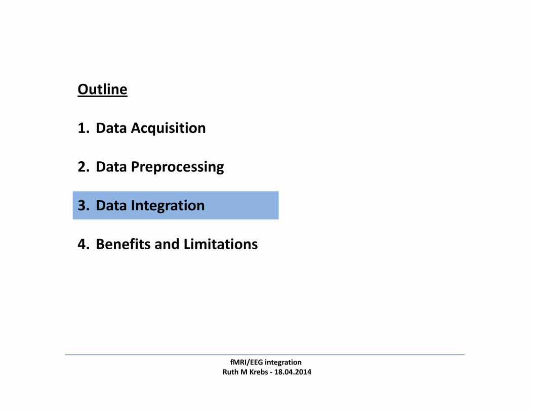
3. Data Integration3.1 Treat as separate data sets. Get two experiments for the price of one.
CoregistrationSlice‐time correctionRealignmentNormalizationSmoothing
MR and pulse artifact correctionRe‐referencing SegmentationRegular artefact rejection (blinks etc.)Baseline correction
Across‐subject averaging and stats (ERPs, amplitude and latency, topography, ICA, frequencies)
Condition‐based BOLD averaging (General Linear Model, GLM)ICAFunctional connectivity
Condition‐based averaging(amplitude or oscillatory power)ICASource reconstruction
Across‐subject averaging and stats (voxel‐wise analysis, ROI, ICA, functional connectivity)
fMRI
Pre‐processing
1st level (subject)
2nd level(group)

Pre‐processing
1st level (subject)
2nd level(group)
fMRI/EEG integration Ruth M Krebs ‐ 18.04.2014
3. Data Integration3.2 Covariation of EEG and fMRI activity. Use averaged EEG measure (amplitude or oscillatorypower) as covariate in 2nd level fMRI analysis or correlate directly with BOLD signal.
CoregistrationSlice‐time correctionRealignmentNormalizationSmoothing
MR and pulse artifact correctionRe‐referencing SegmentationRegular artefact rejection (blinks etc.)Baseline correction
Across‐subject averaging and stats (voxel‐wise analysis, ROI, ICA, functional connectivity)
Across‐subject averaging and stats (ERPs, amplitude and latency, topography, ICA, frequencies)
Condition‐based BOLD averaging(General Linear Model, GLM)ICAFunctional connectivity
Condition‐based averaging(amplitude or oscillatory power)ICASource reconstruction
fMRI EEG
correlation
include covariate
Plichta et al. 2013 (JNeurosci) average CNV during reward anticipation is correlated with averaged BOLD activity in the supplementary motor area, striatum, and thalamus (across trials)
Example:One across‐subject covariate (CNV):
(Examples: Liebenthal et al. 2003; Plichta et al. 2013)

fMRI/EEG integration Ruth M Krebs ‐ 18.04.2014
3. Data Integration
Pre‐processing
3.3 fMRI‐informed EEG source localization. Especially useful in clinical contexts. In fact, theorigin of simultaneous fMRI/EEG lies in epilepsy treatment.
1st level (subject)
2nd level(group)
CoregistrationSlice‐time correctionRealignmentNormalizationSmoothing
MR and pulse artifact correctionRe‐referencing SegmentationRegular artefact rejection (blinks etc.)Baseline correction
Condition‐based BOLD averaging (General Linear Model, GLM)ICAFunctional connectivity
Condition‐based averaging(amplitude or oscillatory power)ICASource reconstruction
Across‐subject averaging and stats (ERPs, amplitude and latency, topography, ICA, frequencies)
Across‐subject averaging and stats (voxel‐wise analysis, ROI, ICA, functional connectivity)
fMRI EEG
(Examples: Lemieux et al. 2004; Vanni et al. 2004; Grouiller et al. 2011)

fMRI/EEG integration Ruth M Krebs ‐ 18.04.2014
3. Data Integration3.4 EEG‐informed fMRI analysis. Use single‐trial EEG measure (amplitude or oscillatory power)as parametric modulator in 1st‐level fMRI model and test at 2nd level (additional variance?).
CoregistrationSlice‐time correctionRealignmentNormalizationSmoothing
MR and pulse artifact correctionRe‐referencing SegmentationRegular artefact rejection (blinks etc.)Baseline correction
Across‐subject averaging and stats (ERPs, amplitude and latency, topography, ICA, frequencies)
Condition‐based BOLD averaging (General Linear Model, GLM)ICAFunctional connectivity
Condition‐based averaging(amplitude or oscillatory power)ICASource reconstruction
Single‐trial data Single‐trial data
Across‐subject averaging and stats (voxel‐wise analysis, ROI, ICA, functional connectivity)
fMRI EEG
parametric modulation
Pre‐processing
1st level (subject)
2nd level(group)
Example:
Baumeister et al. 2014 (NeuroImage) N2 and P3 amplitudes during response inhibition are anti‐correlated and correlated with the BOLD signal in distinct regions on a trial‐to‐trial basis
Two single‐trial parametric modulators (N2, P3):
(Examples: Debener et al. 2005; Benar et al. 2007; Scheeringa et al. 2009; Nguyen et al. 2014; Baumeister et al. 2014)

fMRI/EEG integration Ruth M Krebs ‐ 18.04.2014
3. Data Integration3.5 Symmetrical integration of fMRI/EEG data. e.g. joint ICA (data‐driven, integratedspatiotemporal ICs) and complex neural models (model‐based, integration of multiple levels).
CoregistrationSlice‐time correctionRealignmentNormalizationSmoothing
MR and pulse artifact correctionRe‐referencing SegmentationRegular artefact rejection (blinks etc.)Baseline correction
Across‐subject averaging and stats (ERPs, amplitude and latency, topography, ICA, frequencies)
Condition‐based BOLD averaging (General Linear Model, GLM)ICAFunctional connectivity
Condition‐based averaging(amplitude or oscillatory power)ICASource reconstruction
Across‐subject averaging and stats (voxel‐wise analysis, ROI, ICA, functional connectivity)
neural model
fMRI EEG
joint ICA
Pre‐processing
1st level (subject)
2nd level(group)
(Examples: Valdes‐Sosa et al. 2009; Mijovic et al. 2014)

Outline
1. Data Acquisition
2. Data Preprocessing
3. Data Integration
4. Benefits and Limitations
fMRI/EEG integration Ruth M Krebs ‐ 18.04.2014

fMRI/EEG integration Ruth M Krebs ‐ 18.04.2014
4. Benefits and LimitationsBenefits (presuming good data quality and sufficient power!)
Simultaneous fMRI/EEG is favorable compared to separate data sets, even if those were acquired in the same participant:
‐ no between‐subject variance (obvious)‐ no order and practice effects‐ identical situation with respect to task performance, stimulus perception, body position, noise level, instruction /experimenter effects
these aspects increase statistical power and ensure that differences between conditions in one measure are not due to differences between fMRI and EEG session
Simultaneous fMRI/EEG allows trial‐by‐trial covariation of spatial and temporal signatures of condition‐specific brain states, exceeding across‐participant approaches the same logic applies to covariation analyses between neural activity and task performance
time
EEG Amplitude
trials
fMRI GLM
regressors

fMRI/EEG integration Ruth M Krebs ‐ 18.04.2014
4. Benefits and Limitations
Experimental limitations in both acquisition modalities due to compatibility issues(e.g., sub‐optimal stimulus timing; special sequences may not be allowed)
Limitations (beyond practical and technical issues)
Amplitudes
trials
fMRI GLMFrequencies
X
The high number of degrees of freedom require good a priori hypotheses and adequatecorrections for multiple comparisons
well, let’s consider this a luxury problem!
Voxels / ROIs
Channels
X

fMRI/EEG integration Ruth M Krebs ‐ 18.04.2014
4. Benefits and Limitations
Take home:Simultaneous fMRI/EEG is more complicated to set up,
but you can get the best out of two worlds with just a little more effort.
In best case, the data can be related to one another (and to performance) to gain insights into both the WHERE and the WHEN of a specific cognitive process.
But: good hypotheses are all the more important here as degrees of freedom are very high! (if you correlate stuff wildly, you may find something by accident)

fMRI/EEG integration Ruth M Krebs ‐ 18.04.2014
That’s all!
Useful references: ‐ Huster et al. 2012, JNeuroscience (review article data integration)‐Mullinger et al. 2013, JOVE (best practice data acquisition, incl. movie)‐ Jorge et al. 2014, NeuroImage (review article data integration) ‐ Debener et al. 2006, TICS (opinion article single‐trial analysis)