Embed Size (px)
Citation preview
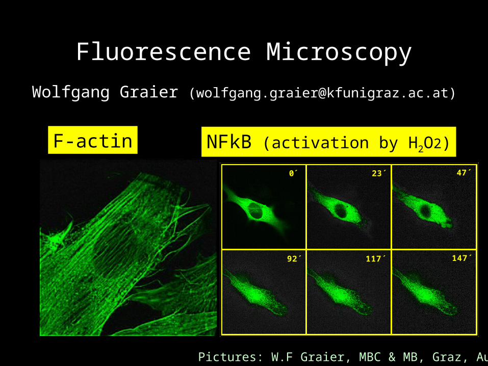
Fluorescence Microscopy
Wolfgang Graier ([email protected])
0́ 23´ 47´
92´ 117´ 147´
F-actin NFkB (activation by H2O2)
Pictures: W.F Graier, MBC & MB, Graz, Austria
NOTE:This Powerpoint presentation also includes so far not
published pictures and results. It has been released only for teaching the principles and possibilities of high
resolution micrsocopy to graduate and post-graduate students. - Thank you very much for your fairness.
If any other use is planed please contact:Prof. Wolfgang F. Graier
Department of Medical Biochemistry and Medical Molecular BiologyKarl-Franzens University of Graz
Harrachgasse 21/IIIA-8010 Graz
Tel. +43-316-380-7560Fax. +43-316-380-9615
E- mail: [email protected]

Basics and IntroductionFluorescence/TransmissionmicroscopyAdvantage/Drawback of light microscopyFluorescence DyesGFPs
Instrumental DevicesConfocal laser scan microscopy (CLSM) Imaging in living cellsDeconvolution microscopy
Comparison of techniques available
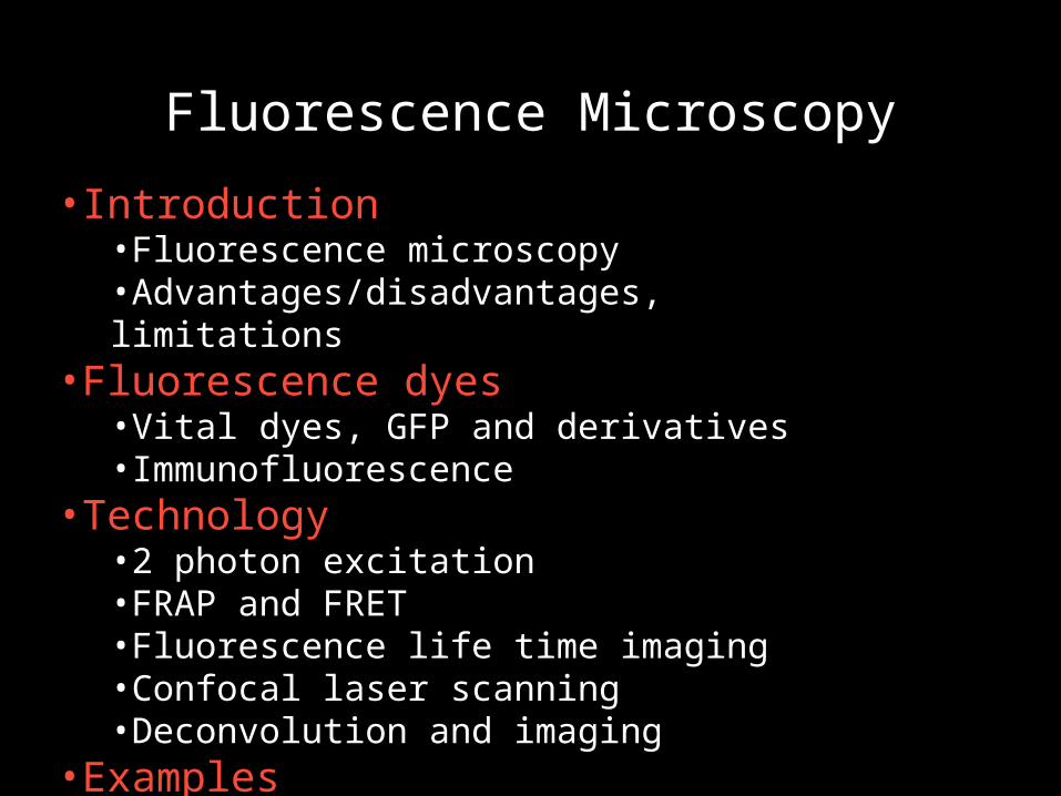
Fluorescence Microscopy
•Introduction•Fluorescence microscopy•Advantages/disadvantages, limitations
•Fluorescence dyes•Vital dyes, GFP and derivatives•Immunofluorescence
•Technology•2 photon excitation•FRAP and FRET•Fluorescence life time imaging•Confocal laser scanning•Deconvolution and imaging
•Examples
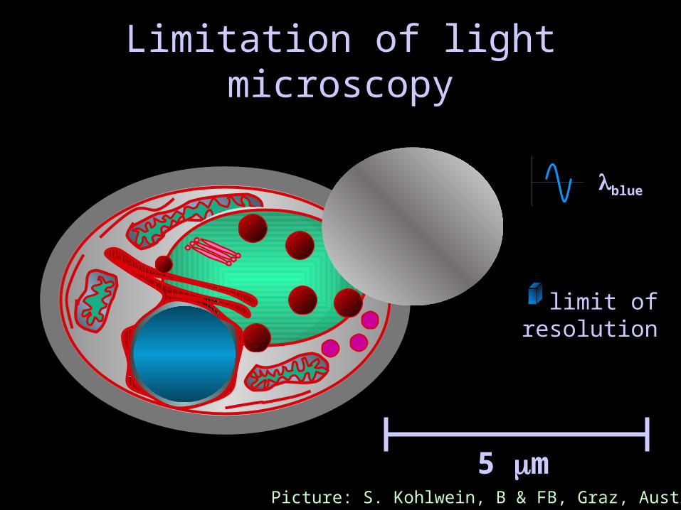
Limitation of light microscopy
5 m
limit ofresolution
blue
Picture: S. Kohlwein, B & FB, Graz, Austria
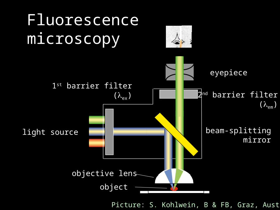
Fluorescencemicroscopy
light source
object
objective lens
1st barrier filter(ex) 2nd barrier filter
(em)
beam-splittingmirror
eyepiece
Picture: S. Kohlwein, B & FB, Graz, Austria

Fluorescence Microscopy
• Life Cell and Immuno Fluorescence
• Applications - dyesOrganelle-specific, pH, membrane potential, ionConcentration
• Caged compounds
• GFP, BFP, RFP, YFP; Aequorin; GFP and FRET
• Sample Preparation
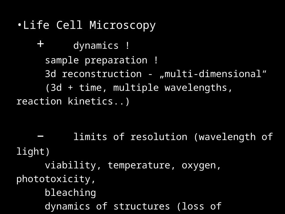
•Life Cell Microscopy
+ dynamics !
sample preparation !
3d reconstruction - „multi-dimensional“
(3d + time, multiple wavelengths, reaction kinetics..)
– limits of resolution (wavelength of light)
viability, temperature, oxygen, phototoxicity,
bleaching
dynamics of structures (loss of resolution)
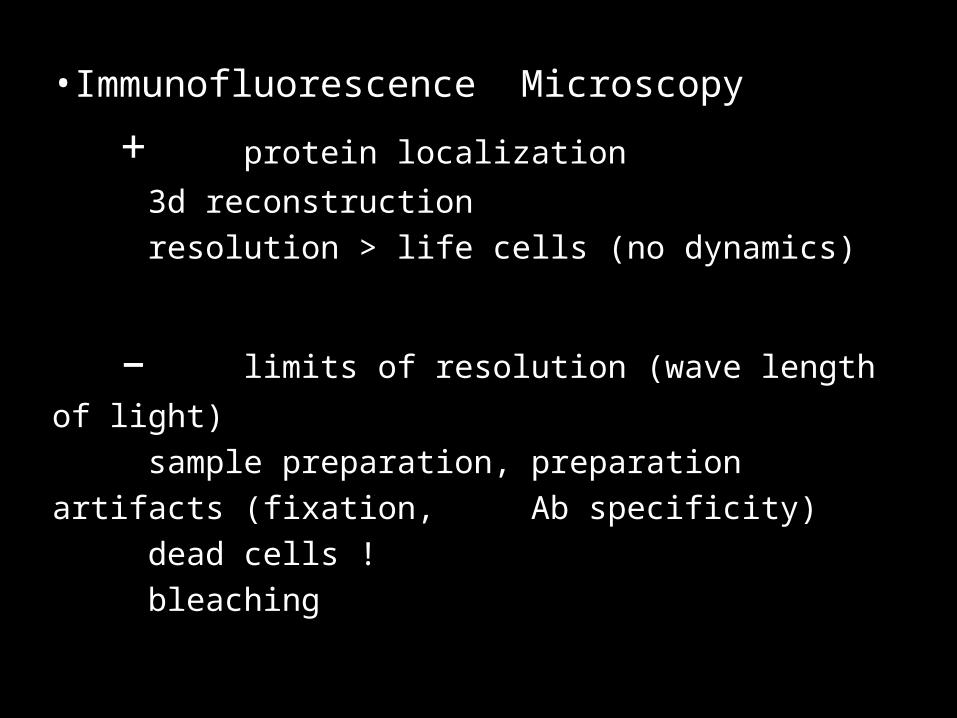
•Immunofluorescence Microscopy
+ protein localization
3d reconstruction
resolution > life cells (no dynamics)
– limits of resolution (wave length of light)
sample preparation, preparation artifacts (fixation,
Ab specificity)
dead cells !
bleaching
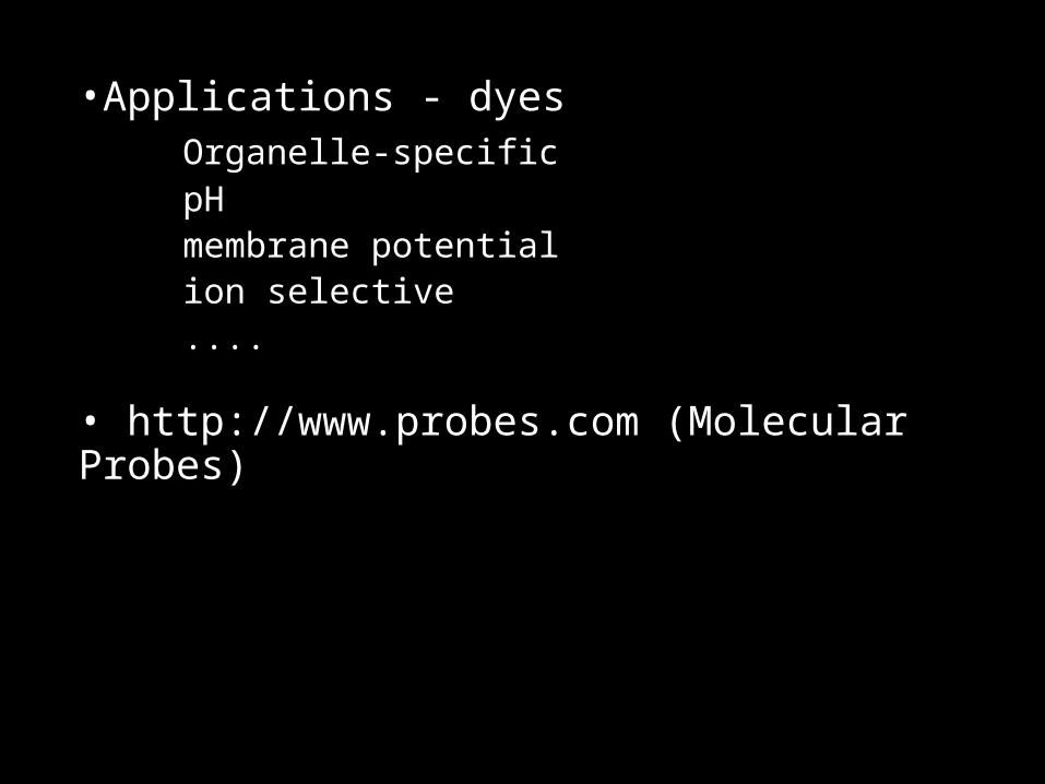
•Applications - dyesOrganelle-specificpHmembrane potentialion selective....
• http://www.probes.com (Molecular Probes)
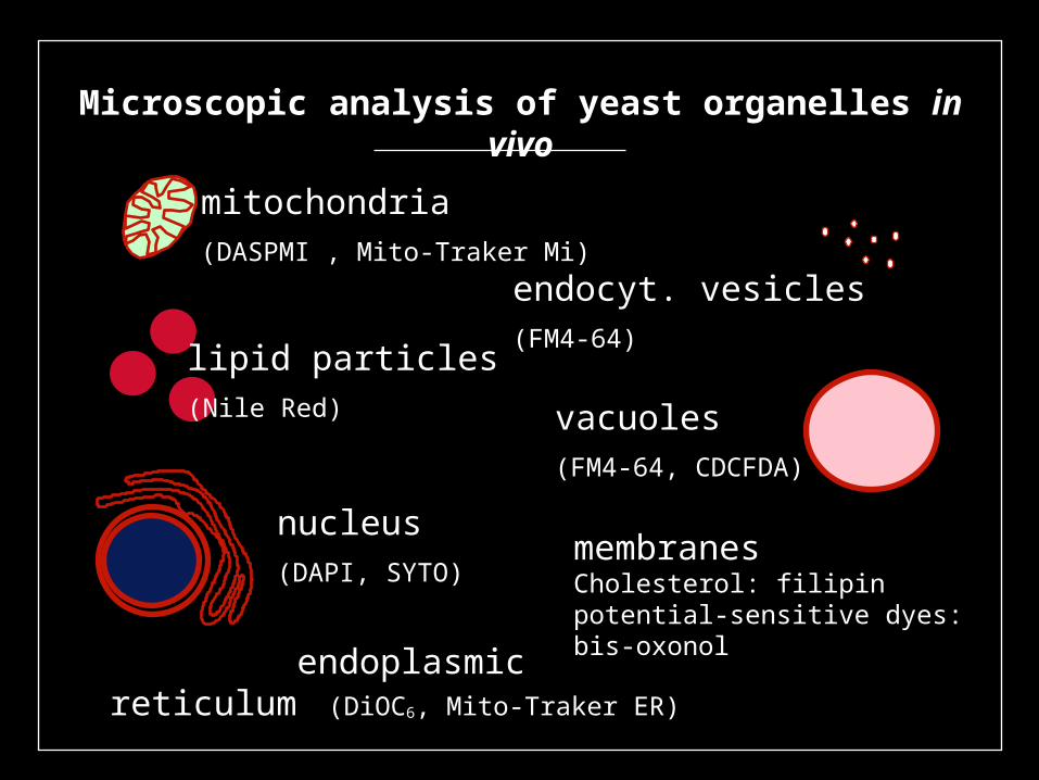
Microscopic analysis of yeast organelles in vivo
mitochondria(DASPMI , Mito-Traker Mi)
lipid particles(Nile Red)
nucleus(DAPI, SYTO)
endoplasmic reticulum (DiOC6, Mito-Traker ER)
vacuoles(FM4-64, CDCFDA)
endocyt. vesicles(FM4-64)
membranesCholesterol: filipinpotential-sensitive dyes: bis-oxonol
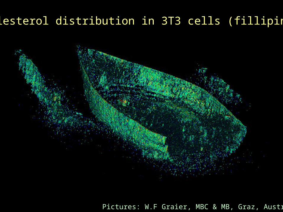
Cholesterol distribution in 3T3 cells (fillipin)
Pictures: W.F Graier, MBC & MB, Graz, Austria

Pictures: W.F Graier, MBC & MB, Graz, Austria
DiOC6
deconvoluted
deconvoluted
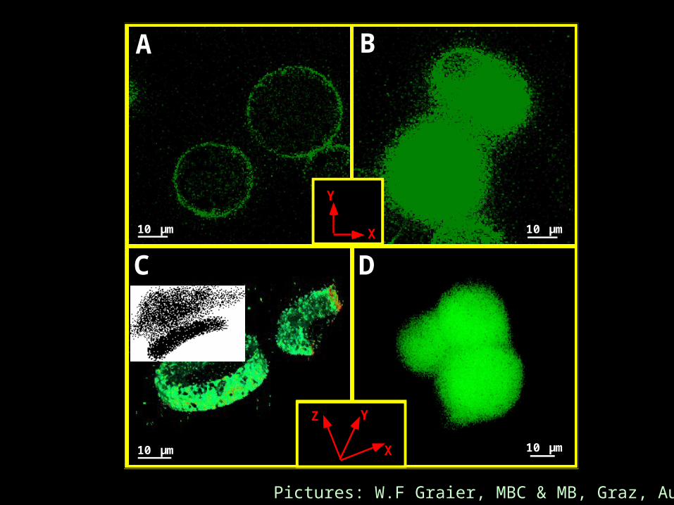
C D
X
YZ
A B
10 µm
10 µm
10 µm
10 µm
X
Y
Pictures: W.F Graier, MBC & MB, Graz, Austria
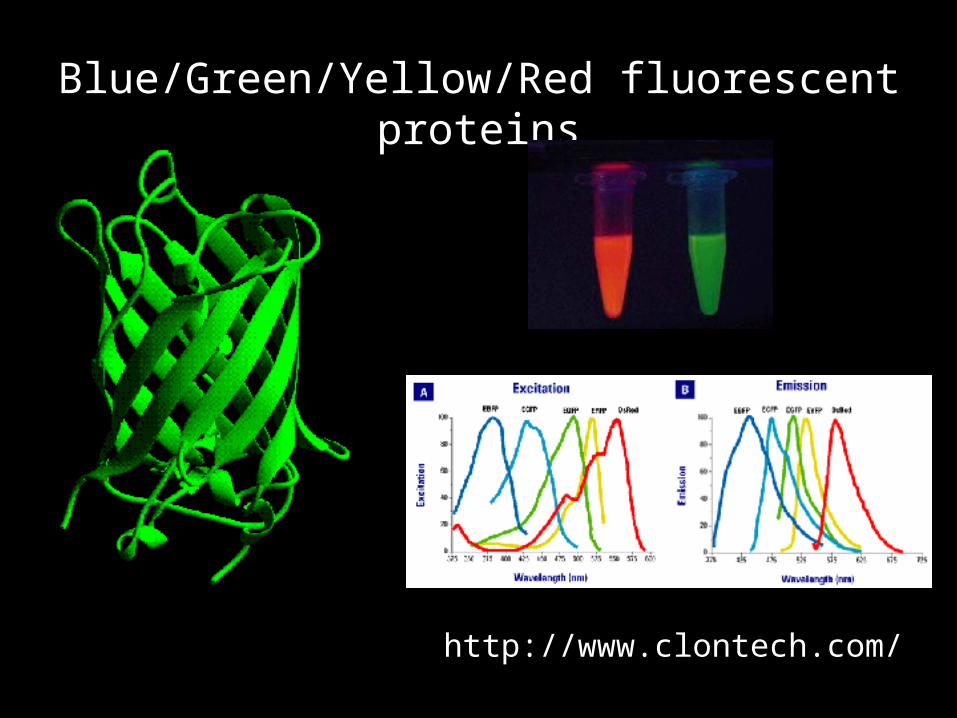
Blue/Green/Yellow/Red fluorescent proteins
http://www.clontech.com/
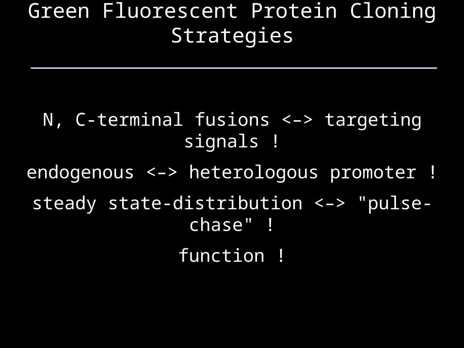
Green Fluorescent Protein Cloning Strategies
N, C-terminal fusions <–> targeting signals !
endogenous <–> heterologous promoter !
steady state-distribution <–> "pulse-chase" !
function !
GFP kanMX6ERG
Chromosomal fusionvia homologous recombination
GFP kanMX6
Chromosome
GFP
kanMX6
Plasmid
ERG Chromosome
PCR
GFP kanMX6ERG
Chromosomal fusionvia homologous recombination
GFP kanMX6
Chromosome
GFP
kanMX6
Plasmid
ERG Chromosome
PCR
GFP kanMX6ERG
Chromosomal fusionvia homologous recombination
GFP kanMX6
Chromosome
GFP
kanMX6
Plasmid
ERG Chromosome
PCR
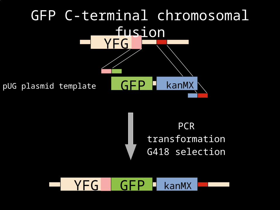
YFG
GFP kanMX
YFG GFP kanMX
PCRtransformationG418 selection
YFG
GFP kanMX
YFG GFP kanMX
PCRtransformationG418 selection
GFP C-terminal chromosomal fusion
pUG plasmid template
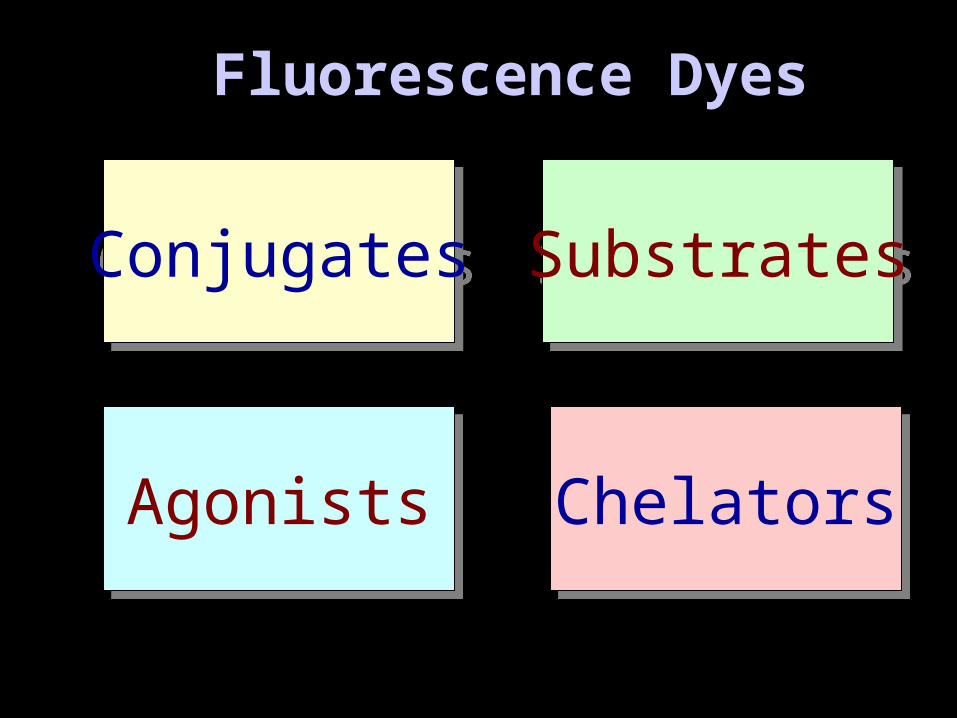
Fluorescence DyesFluorescence Dyes
ConjugatesConjugates SubstratesSubstrates
AgonistsAgonists ChelatorsChelators
ConjugatesConjugates
Principles:primary antibodysecondary antibody
(dye coupled)
Samples:Alexa, Cy-X
Immunfluorescence
Pictures: Molecular Probes
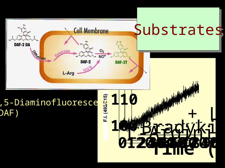
0120240360480600720840960100
110
120
L-ArginineBradykinin+ L-NA
Time (min)
4,5-Diaminofluorescein(DAF)
SubstratesSubstrates
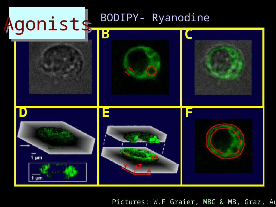
A B
D E F
CAgonistsAgonists BODIPY- Ryanodine
Pictures: W.F Graier, MBC & MB, Graz, Austria

ChelatorsChelators
Targeting of chelators by specific groups (e.g. fatty acids)
F.I.
Wavelenghts (nm)
Ca2+
340 360 380 EX
EM510
Ca2+
Na+
H+
K+
Cl-
..
..
..
Fura-2

Fluorescence Microscopy
•Technology
Deconvolution Microscopy
Confocal Laser Scanning Microscopy
2 Photon Microscopy; time-resolved FM
FRAP fluorescence recovery after photo bleaching
FRET fluorescence resonance energy transfer
Fluorescence Microscopy
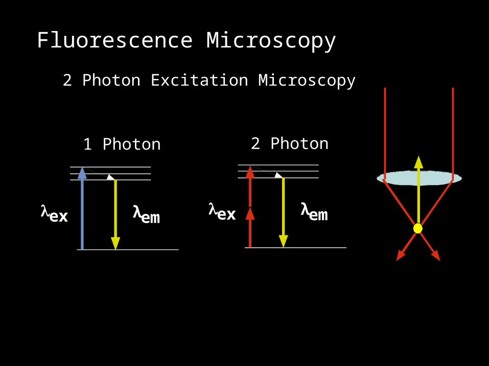
2 Photon Excitation Microscopy
ex λem
1 Photon
ex λem
2 Photon
Fluorescence Microscopy
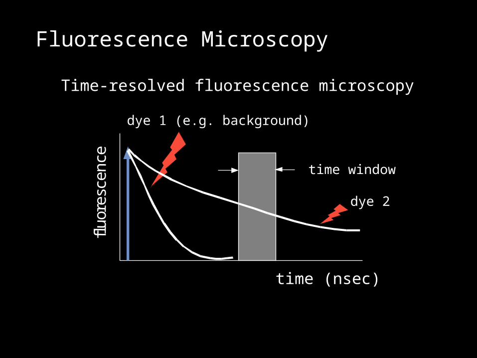
Time-resolved fluorescence microscopyflu
ore
scen
ce
time (nsec)
dye 1 (e.g. background)
dye 2
time window
Fluorescence Microscopy
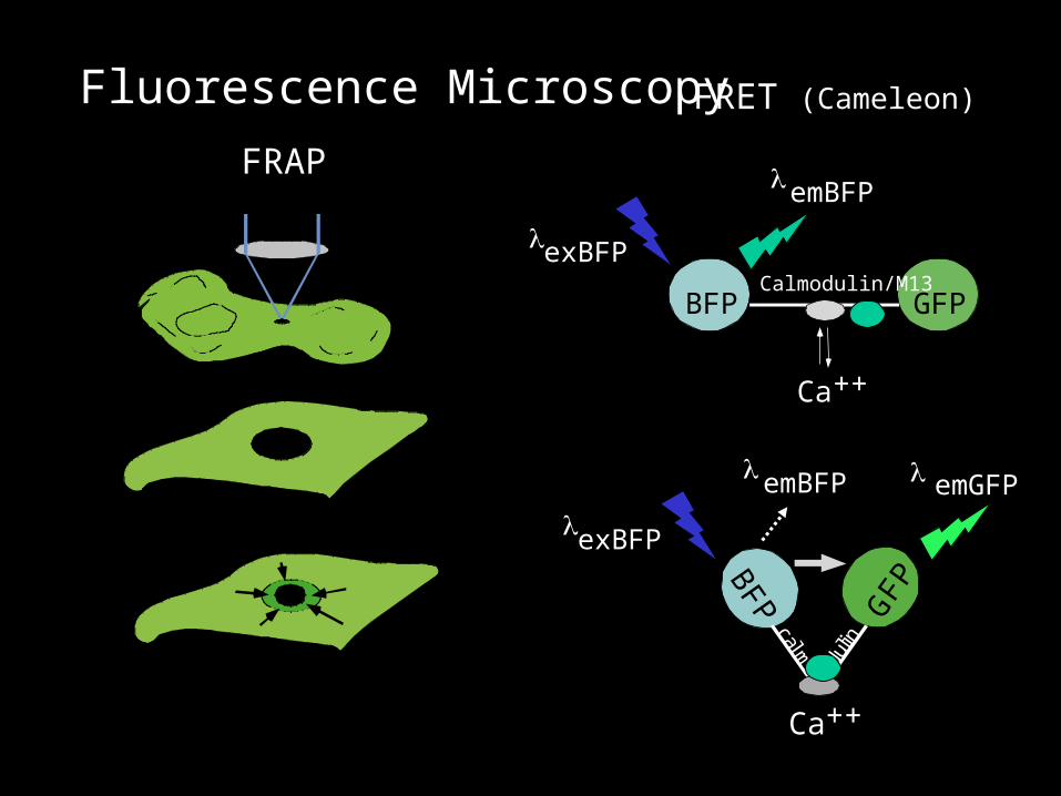
FRAP
FRET (Cameleon)
BFP GFP
Ca++
o
calm
dulin
exBFP
emBFP emGFP
BFP GFPCalmodulin/M13
Ca++
emBFP
exBFP
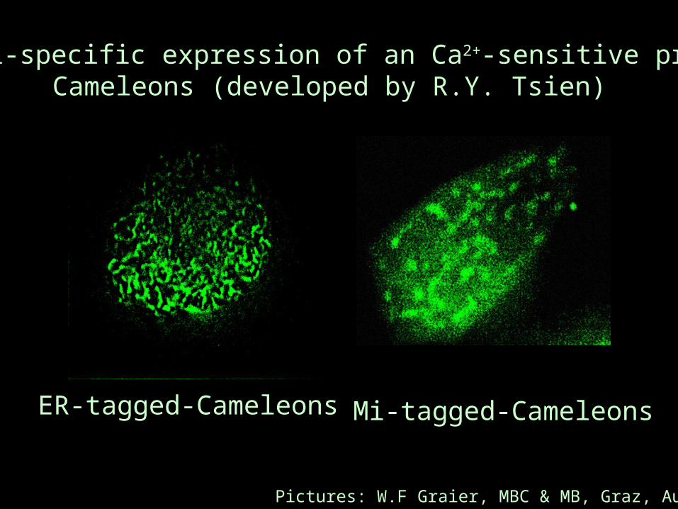
ER-tagged-Cameleons Mi-tagged-Cameleons
Pictures: W.F Graier, MBC & MB, Graz, Austria
Organell-specific expression of an Ca2+-sensitive proteineCameleons (developed by R.Y. Tsien)
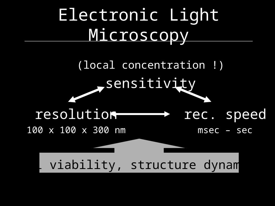
(local concentration !)
sensitivity
resolution rec. speed100 x 100 x 300 nm msec – sec
Electronic Light Microscopy
cell viability, structure dynamics
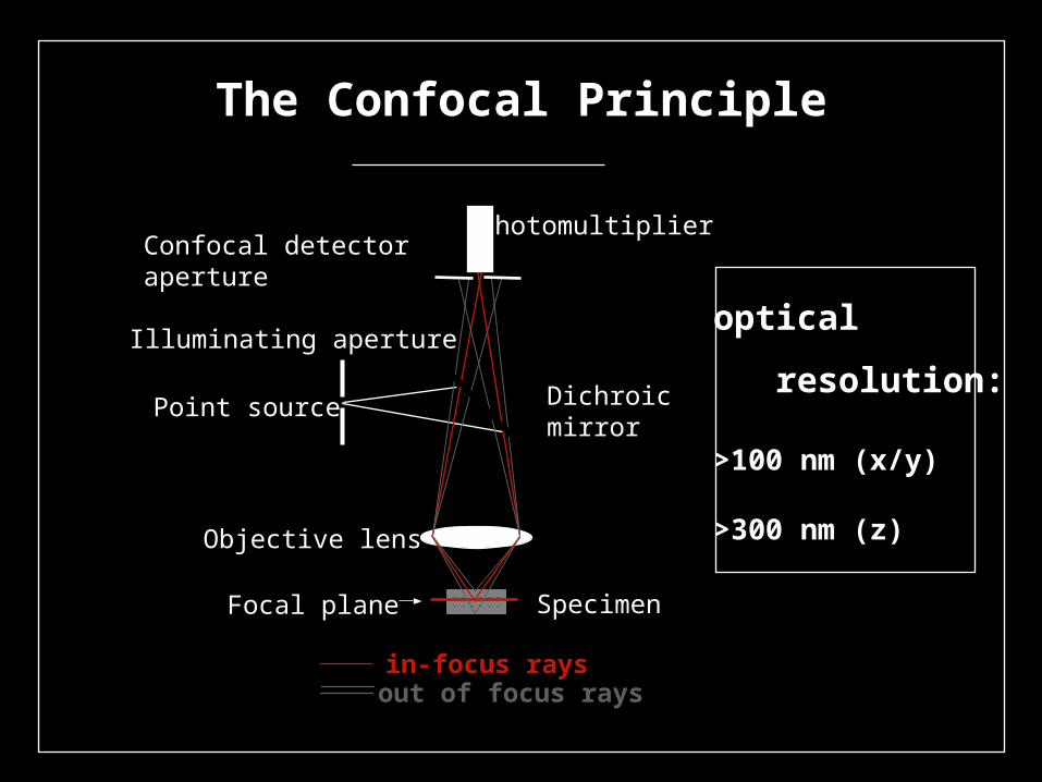
The Confocal Principle
optical
resolution:
>100 nm (x/y)
>300 nm (z)
Point source
Objective lens
Focal plane Specimen
Dichroicmirror
Illuminating aperture
Confocal detectoraperture
Photomultiplier
in-focus raysout of focus rays
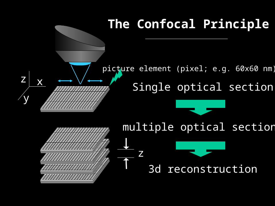
The Confocal Principle
Single optical section
multiple optical sections
3d reconstruction
z
z x
y
picture element (pixel; e.g. 60x60 nm)
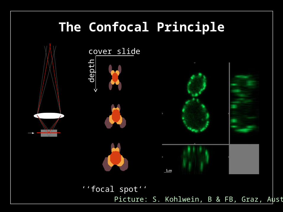
The Confocal Principle
cover slide
dep
th
‘‘focal spot‘‘Picture: S. Kohlwein, B & FB, Graz, Austria
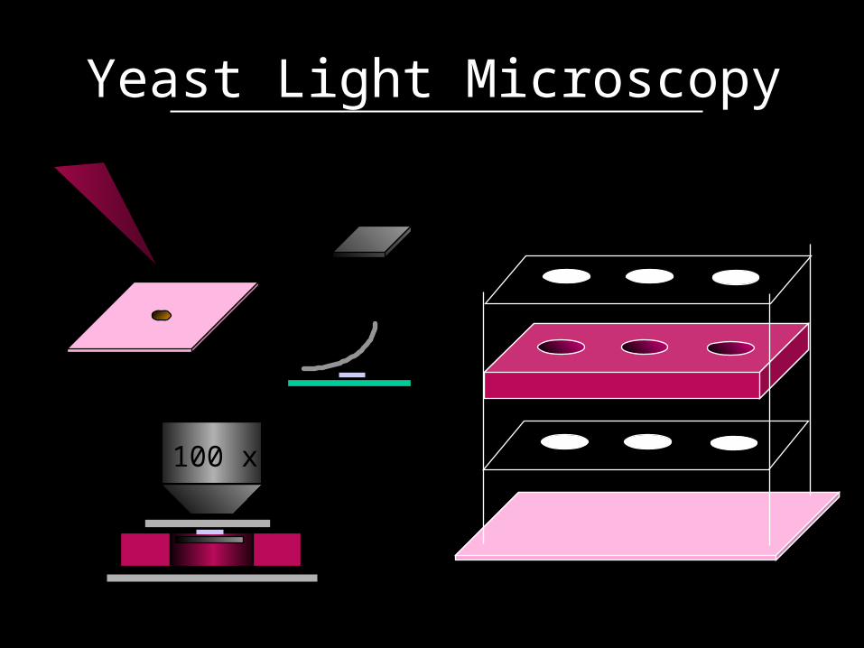
Yeast Light Microscopy
100 x
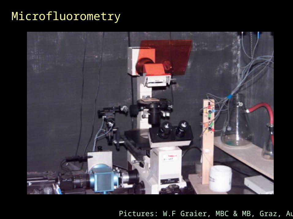
Microfluorometry
Pictures: W.F Graier, MBC & MB, Graz, Austria
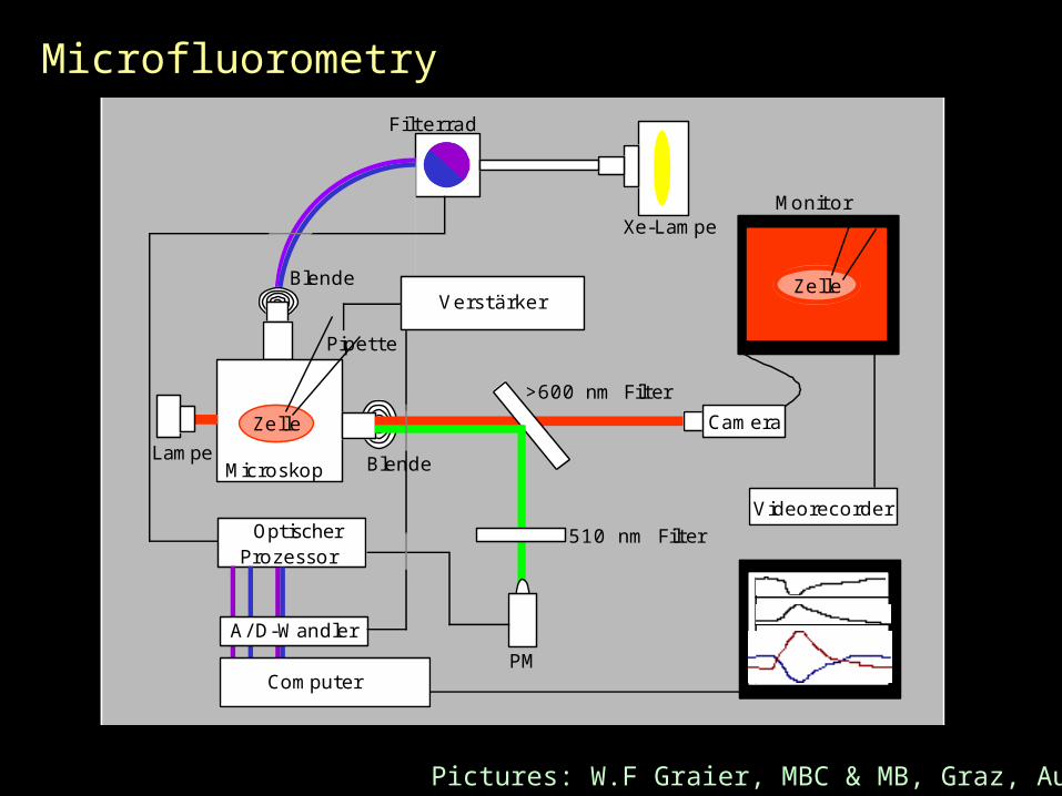
Computer
A/ D-Wandler
OptischerProzessor
Videorecorder
Camera
Xe-LampeMonitor
Filterrad
PM
Microskop
Zelle
Zelle
Blende
BlendeLampe
510 nm Filter
>600 nm Filter
Pipette
Verstärker
Microfluorometry
Pictures: W.F Graier, MBC & MB, Graz, Austria
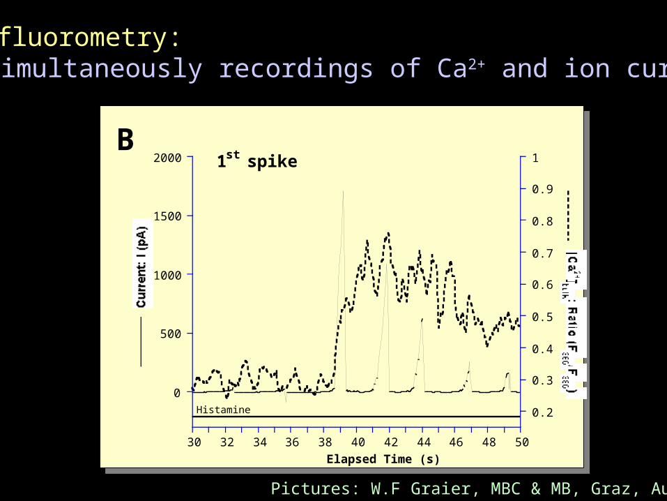
Microfluorometry:Simultaneously recordings of Ca2+ and ion currents
0
500
1000
1500
2000
0.2
0.3
0.4
0.5
0.6
0.7
0.8
0.9
1
30 32 34 36 38 40 42 44 46 48 50
Current: I (pA) [Ca2+]
bulk
: Ratio (F
360
/F380
)
Elapsed Time (s)
1st spikeB
Histamine
Pictures: W.F Graier, MBC & MB, Graz, Austria
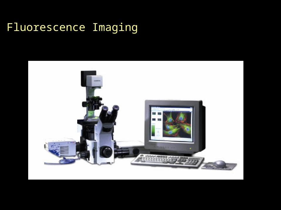
Fluorescence Imaging
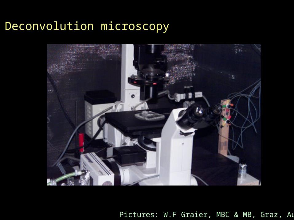
Deconvolution microscopy
Pictures: W.F Graier, MBC & MB, Graz, Austria

Point spread function

Focus
Out-of-focus fluorescence
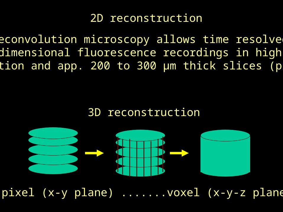
3D reconstruction
pixel (x-y plane) .......voxel (x-y-z plane)
2D reconstruction
Deconvolution microscopy allows time resolvedtwo dimensional fluorescence recordings in high x-y
resolution and app. 200 to 300 µm thick slices (pixel)

Confocal vs Deconvolution
Out-of-focus light
Signal to noise ratio
Serial lines (time scan)
Image acquisition
Imaging quality
Thick samples
Excitation
Costs
pinhole
low (10 p/px)
√
slow
== f(object) >
100 µm
# laser lines
PSF & comput.
high (104 p/px)
n.a.
fast
== f(object)
<< 100 µm
Spectral lamp
![Anti-Inflammatory Constituents of Plants: A Revie · saffron) Colchicaceae Colchicine 13 (Alkaloid) Inhibition of NFkB, COX-2 and AP-1 [20,21] 13 Curcuma longa (Tumeric) Zingiberaceae](https://img.dokumen.tips/doc/110x75/5eb4511d062c011da64136d0/anti-inflammatory-constituents-of-plants-a-saffron-colchicaceae-colchicine-13.jpg)