Embed Size (px)
Citation preview
Hindawi Publishing Corporatione Scienti�c �orld JournalVolume 2013, Article ID 920595, 11 pageshttp://dx.doi.org/10.1155/2013/920595
Review ArticleFluctuating Roles of Matrix Metalloproteinase-9 inOral Squamous Cell Carcinoma
Suvi-Tuuli Vilen,1 Tuula Salo,1, 2, 3 Timo Sorsa,1, 4 and Pia Nyberg2, 3
1 Biomedicum Helsinki, Institute of Dentistry, Research Laboratory, University of Helsinki, P.O. Box 63,Haartmaninkatu 8, 00014 Helsinki, Finland
2Department of Diagnostics and Oral Medicine, Institute of Dentistry and the Oulu Center for Cell-Matrix Research,University of Oulu, P.O. Box 5281, 90014 Oulu, Finland
3Oulu University Hospital, Oulu, Finland4Department of Oral and Maxillofacial Diseases, Helsinki University Hospital, Helsinki, Finland
Correspondence should be addressed to Suvi-Tuuli Vilen; suvi-tuuli.vilen�helsinki.�
Received 5 November 2012; Accepted 10 December 2012
Academic Editors: S. Bhan, A. Chuang, and J. E. J. Lee
Copyright © 2013 Suvi-Tuuli Vilen et al. is is an open access article distributed under the Creative Commons AttributionLicense, which permits unrestricted use, distribution, and reproduction in any medium, provided the original work is properlycited.
One hallmark of cancer is the degradation of the extracellular matrix (ECM), which is caused by proteinases. In oral cancers, matrixmetalloproteinases (MMPs), especially MMP-9, are associated with this degradation. MMPs break down the ECM allowing cancerto spread; they also release various factors from their cryptic sites, including cytokines. ese factors modulate cell behavior andenhance cancer progression by regulating angiogenesis, migration, proliferation, and invasion.e development of earlymetastasesis typical for oral cancer, and increasedMMP-9 expression is associated with a poor disease prognosis. However, many studies fail torelateMMP-9 expression with metastasis formation. Contrary to earlier models, recent studies show thatMMP-9 plays a protectiverole in oral cancers. erefore, the role of MMP-9 is complicated and may �uctuate throughout the different types and stages oforal cancers.
1. Introduction
Oral cancer is one of the ten most common cancers world-wide. Nearly 3% of all cancer cases are oral cancers; they aremore common in men than in women; and two-thirds oforal cancer cases occur in developing countries [1, 2]. Oneimportant hallmark of cancer progression is the degradationof the extracellular matrix (ECM), which allows cancer cellsto invade the surrounding tissue. Matrix metalloproteinases(MMPs) are zinc-dependent endopeptidases that efficientlydegrade the components of the ECM and basement mem-branes (BM). MMPs also release cytokines, chemokines, andgrowth factors from their proforms or their cryptic sites[3–6]. To date, at least 24 distinct MMP genes have beenidenti�ed in humans. MMPs are classi�ed according to their
substrate speci�cities: gelatinases, collagenases, matrilysins,and stromelysins. e structures of all MMPs include an N-terminal signal peptide that directs the protein to either theplasma membrane insertion or to the secretory pathway; itsprodomain confers its latency, and its catalytic domain hasa zinc atom in its active site. MMPs are either anchoredin the membrane or secreted, primarily as latent proformsthat require activation before becoming catalytically com-petent [7–9]. Two different soluble gelatinases have beenidenti�ed: gelatinase A, 72 kDa (MMP-2), and gelatinase B,92 kDa (MMP-9). Both contain a collagen-binding domainwithin their catalytic domain, distinguishing them fromother MMPs. A more detailed structure of these enzymes isdescribed in a review by Björklund and Koivunen [10].
2 e Scienti�c World �ournal
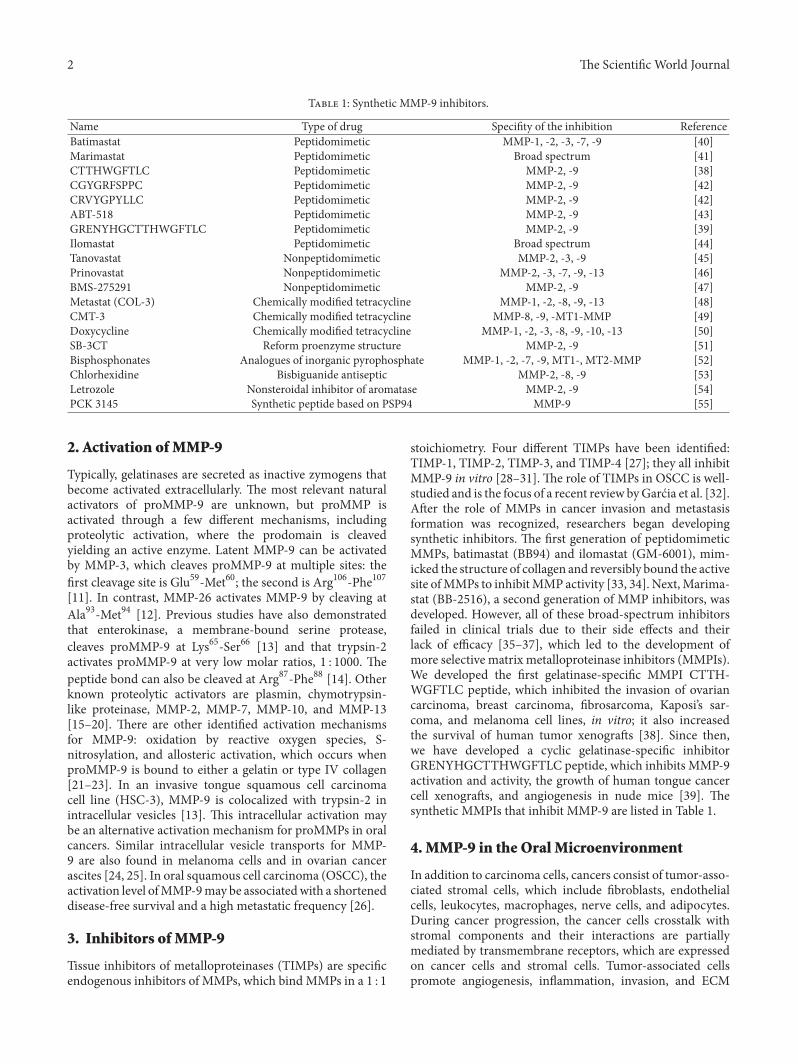
T 1: Synthetic MMP-9 inhibitors.
Name Type of drug Speci�ty of the inhibition ReferenceBatimastat Peptidomimetic MMP-1, -2, -3, -7, -9 [40]Marimastat Peptidomimetic Broad spectrum [41]CTTHWGFTLC Peptidomimetic MMP-2, -9 [38]CGYGRFSPPC Peptidomimetic MMP-2, -9 [42]CRVYGPYLLC Peptidomimetic MMP-2, -9 [42]ABT-518 Peptidomimetic MMP-2, -9 [43]GRENYHGCTTHWGFTLC Peptidomimetic MMP-2, -9 [39]Ilomastat Peptidomimetic Broad spectrum [44]Tanovastat Nonpeptidomimetic MMP-2, -3, -9 [45]Prinovastat Nonpeptidomimetic MMP-2, -3, -7, -9, -13 [46]BMS-275291 Nonpeptidomimetic MMP-2, -9 [47]Metastat (COL-3) Chemically modi�ed tetracycline MMP-1, -2, -8, -9, -13 [48]CMT-3 Chemically modi�ed tetracycline MMP-8, -9, -MT1-MMP [49]Doxycycline Chemically modi�ed tetracycline MMP-1, -2, -3, -8, -9, -10, -13 [50]SB-3CT Reform proenzyme structure MMP-2, -9 [51]Bisphosphonates Analogues of inorganic pyrophosphate MMP-1, -2, -7, -9, MT1-, MT2-MMP [52]Chlorhexidine Bisbiguanide antiseptic MMP-2, -8, -9 [53]Letrozole Nonsteroidal inhibitor of aromatase MMP-2, -9 [54]PCK 3145 Synthetic peptide based on PSP94 MMP-9 [55]
2. Activation of MMP-9
Typically, gelatinases are secreted as inactive zymogens thatbecome activated extracellularly. e most relevant naturalactivators of proMMP-9 are unknown, but proMMP isactivated through a few different mechanisms, includingproteolytic activation, where the prodomain is cleavedyielding an active enzyme. Latent MMP-9 can be activatedby MMP-3, which cleaves proMMP-9 at multiple sites: the�rst cleavage site is Glu59-Met60; the second is Arg106-Phe107[11]. In contrast, MMP-26 activates MMP-9 by cleaving atAla93-Met94 [12]. Previous studies have also demonstratedthat enterokinase, a membrane-bound serine protease,cleaves proMMP-9 at Lys65-Ser66 [13] and that trypsin-2activates proMMP-9 at very low molar ratios, 1 : 1000. epeptide bond can also be cleaved at Arg87-Phe88 [14]. Otherknown proteolytic activators are plasmin, chymotrypsin-like proteinase, MMP-2, MMP-7, MMP-10, and MMP-13[15–20]. ere are other identi�ed activation mechanismsfor MMP-9: oxidation by reactive oxygen species, S-nitrosylation, and allosteric activation, which occurs whenproMMP-9 is bound to either a gelatin or type IV collagen[21–23]. In an invasive tongue squamous cell carcinomacell line (HSC-3), MMP-9 is colocalized with trypsin-2 inintracellular vesicles [13]. is intracellular activation maybe an alternative activation mechanism for proMMPs in oralcancers. Similar intracellular vesicle transports for MMP-9 are also found in melanoma cells and in ovarian cancerascites [24, 25]. In oral squamous cell carcinoma (OSCC), theactivation level ofMMP-9may be associatedwith a shorteneddisease-free survival and a high metastatic frequency [26].
3. Inhibitors of MMP-9
Tissue inhibitors of metalloproteinases (TIMPs) are speci�cendogenous inhibitors of MMPs, which bind MMPs in a 1 : 1
stoichiometry. Four different TIMPs have been identi�ed:TIMP-1, TIMP-2, TIMP-3, and TIMP-4 [27]; they all inhibitMMP-9 in vitro [28–31]. e role of TIMPs in OSCC is well-studied and is the focus of a recent review byGarćia et al. [32].Aer the role of MMPs in cancer invasion and metastasisformation was recognized, researchers began developingsynthetic inhibitors. e �rst generation of peptidomimeticMMPs, batimastat (BB94) and ilomastat (GM-6001), mim-icked the structure of collagen and reversibly bound the activesite ofMMPs to inhibitMMP activity [33, 34]. Next,Marima-stat (BB-2516), a second generation of MMP inhibitors, wasdeveloped. However, all of these broad-spectrum inhibitorsfailed in clinical trials due to their side effects and theirlack of efficacy [35–37], which led to the development ofmore selective matrix metalloproteinase inhibitors (MMPIs).We developed the �rst gelatinase-speci�c MMPI CTTH-WGFTLC peptide, which inhibited the invasion of ovariancarcinoma, breast carcinoma, �brosarcoma, Kaposi�s sar-coma, and melanoma cell lines, in vitro; it also increasedthe survival of human tumor xenogras [38]. Since then,we have developed a cyclic gelatinase-speci�c inhibitorGRENYHGCTTHWGFTLC peptide, which inhibits MMP-9activation and activity, the growth of human tongue cancercell xenogras, and angiogenesis in nude mice [39]. esynthetic MMPIs that inhibit MMP-9 are listed in Table 1.
4. MMP-9 in the Oral Microenvironment
In addition to carcinoma cells, cancers consist of tumor-asso-ciated stromal cells, which include �broblasts, endothelialcells, leukocytes, macrophages, nerve cells, and adipocytes.During cancer progression, the cancer cells crosstalk withstromal components and their interactions are partiallymediated by transmembrane receptors, which are expressedon cancer cells and stromal cells. Tumor-associated cellspromote angiogenesis, in�ammation, invasion, and ECM

e Scienti�c World �ournal 3
modeling through cell-cell contact and the production ofgrowth factors, hormones, cytokines, and proteinases suchas MMPs [56–58]. In OSCC tumors, MMP-9 is expressed incarcinoma and in�ammatory cells around carcinoma islands.Meanwhile, MMP-2 is mainly found in carcinoma-associated�broblasts (CAFs) [59, 60]. In an oral squamous cell car-cinoma cell line SCC-25, CAFs increase the expression ofMMP-9, in vitro, which is thought to occur via a �bronectin-integrin 𝛼𝛼v𝛽𝛽6 pathway [61]. In the aggressive human tonguesquamous cell carcinoma cell line HSC-3, MMP-2 was onlyfound in its latent form, whereas MMP-9 was found in itsactive form [13]. MMP-9
′s effect during HSC-3 cell line
invasion was studied in a human organotypic model basedon myoma tissue [62], which contains �broblasts, smoothmuscle cells, lymphocytes, macrophages, endothelial cells,and MMP-2, but not MMP-9 [62]. erefore, this model isa better predictor of the in vivo tumor microenvironmentcompared with the commonly used rat tail-derived type Icollagen and/or the mouse EHS sarcoma-derived Matrigelinvasion assay. In the myoma organotypic invasion assay,aer inhibiting gelatinase activity in HSC-3 cells using aspeci�c gelatinase inhibitor CTTHWGFTLC [38], the tumorcells were surprisingly more invasive than in the controlgroup (unpublished data). Mice bearing HSC-3 xenogratumors treated with the gelatinase inhibitor CTTHWGFTLChad smaller primary tumors in vivo than the control group[39], but the inhibition of gelatinases did not affect localinvasion or metastasis formation [63]. e ability of cancercells to change their migration under certain circumstancesfrom proteolytic to non-proteolytic, amoeboid type duringprotease-inhibitor treatment helps to explain the OSCCbehaviors we observed. us, these cells change their shapeand adapt to squeeze through tissue gaps without degradingthe ECM [64]. MMP-9 may not be the only, or even themost important, proteolytic enzyme in the OSCC invasionprocess, but it may be important for indirect cell signalingby controlling the bioavailability and bioactivity of moleculesthat target speci�c receptors, which regulate cell growth,migration, in�ammation, and angiogenesis [65–69].
5. The Role of MMP-9 in OSCC InvasionandMetastasis
MMP-9 is associated with the aggressive nature of many can-cers, including OSCC [81–84], and this aggressive nature wasthought to cause type IV collagen degradation, a main com-ponent of basement membranes [85]. To date, the spectrumof MMP-9 matrix substrates has signi�cantly increased, andaside from substrates, which originate in the matrix, MMP-9 has other bioactive substrates that independently modulatecarcinogenesis, such as the pro-transforming growth factor-𝛽𝛽1 (TGF-𝛽𝛽1) and the pro-tumor necrosis factor-𝛼𝛼 (TNF-𝛼𝛼) [10, 86, 87]. MMP-9 has traditionally been associatedwith the aggressive nature of OSCC. However, in spiteof increased MMP-9 expression levels, many researchershave presented contradictory results [70–72] (Table 2). Forexample, Guttman et al. [73] did not �nd a correlationbetween MMP-9 expression and the size of the primary
tumor or the neck metastasis in tongue SCC patients. Mean-while, another study reported that high levels of MMP-9expression in OSCC patients were correlated with regionallymph node and/or distant metastases and a poor prognosis[74]. In addition, De Vicente et al. [78] showed that MMP-9 expression was not associated with clinical variables, suchas tumor stage or recurrence rate. In a study conducted byIkebe et al., gelatinolytic activity and increased expressionof both MMP-2 and MMP-9 in OSCC tumors were relatedto the invasiveness, but not to the metastatic potential ofOSCC tumors [75]. Finally, Kato and co-workers [76] showedthat, although MMP-9 expression was high in OSCCs, theactivated form:proform ratio was very low, while activatedMMP-2s were elevated and associated with advanced stagesof disease. ese �ndings suggest that MMP-9 may not bea universal cancer progression promotion factor in OSCCs;instead, it may have �uctuating roles.
6. MMP-9 in theModulation ofCan�e�-Related In�a��ation
Chronic in�ammation is associated with epithelial cancers,and it differs from normal in�ammation because it is notself-limiting. Cancer cells produce different cytokines thatattract innate immune cells, such as mast cells, granulo-cytes, and macrophages. ese innate immune cells thensecrete interleukins, chemokines, reactive oxygen species,and MMPs that modulate angiogenesis, cell proliferation,tumor growth, and invasion [88–90]. In OSCCs, the levelof a multifunctional cytokine, transforming growth factor-𝛽𝛽1, is upregulated, which leads to the enhanced expressionof snail. Snail is a transcription factor that increases MMP-9expression and triggers an epithelial-mesenchymal transition(EMT); then, carcinoma cells change their morphology,reduce their intercellular and cell-matrix adhesions, andincrease their motility [91, 92]. Interestingly, the inactiveform of TGF-𝛽𝛽1 is activated by MMP-9 [93]. Many othercytokines are also substrates for MMP-9, including TNF-𝛼𝛼,CXCL1, CXCL4, CXCL7, CXCL8, and interleukin-1𝛽𝛽 [65–67]. Interleukin-1𝛽𝛽 is secreted by tumor cells and inducesthe expression of lipocalin 2 [94, 95]. e plasma levels oflipocalin 2, MMP-9 and the lipocalin 2/MMP-9 complex areassociated with more advanced clinical stages and/or tumorsizes in OSCC patients. Interestingly, MMP-9 levels are notcorrelated with either lymph nodes or distant metastases[77]. Chemokine CXCL8 can induce the release of MMP-9 from tertiary neutrophil granules, and increased CXCL8expression is associated with OSCC. e CXCL8 expressedin tumor cells is also secreted by OSCC cell lines, and CXCL8mRNA expression is enhanced by the addition of TNF-𝛼𝛼 andIL- 1𝛽𝛽. CXCL8 fromOSCC cell lines increases cell migration,induces invasion, and increases the expression of MMP-7.However, it does not have an effect on MMP-9 expression.erefore, CXCL8-induced expression of MMP-9 may becell-type speci�c [96–98]. e chemokine receptor, CXCR4,modulates the invasion of OSCCs by regulating MMP-9expression. In patients, this expression correlates with lymphnode metastasis and MMP-9 expression [99, 100]. Cyclo-oxygenase (COX)-2 is an enzyme that converts arachidonic
4 e Scienti�c World �ournal
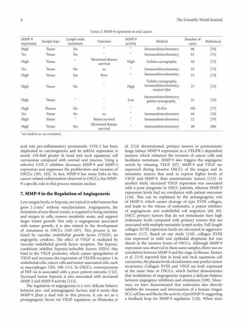
T 2: MMP-9 expression in oral cancer.
MMP-9expression Sample type Lymph node
metastasis Outcome MMP-9activity Method Number of
cases References
High Tissue Yes ∗ ∗ Immunohistochemistry 96 [70]Yes Tissue Yes ∗ ∗ Immunohistochemistry 61 [71]
High Tissue ∗ Shortened diseasesurvival High Gelatin zymography, 44 [72]
Yes Tissue No nc ∗ Immunohistochemistry 23 [73]High Tissue Yes Poor ∗ Immunohistochemistry 53 [74]
High Tissue No ∗ HighGelatin zymography,
immunohistochemistry,western blot
57 [75]
High Tissue ∗ ∗ LowImmunohistochemistry,gelatin zymography, 31 [76]
High Plasma No ∗ ∗ ELISA 195 [77]Yes Tissue No nc ∗ Immunohistochemistry 68 [78]High Tissue ∗ Better survival ∗ Immunohistochemistry 12 [79]
High Tissue Yes Shortened diseasesurvival
∗ Immunohistochemistry 48 [80]∗not studied, nc: no correlation.
acid into pro-in�ammatory prostanoids. COX-2 has beenimplicated in carcinogenesis and its mRNA expression isnearly 150-fold greater in head and neck squamous cellcarcinomas compared with normal oral mucosa. Using aselective COX-2 inhibitor decreases MMP-9 and MMP-2expression and suppresses the proliferation and invasion ofOSCCs [101, 102]. In fact, MMP-9 has many links to thecancer-related in�ammation observed in OSCCs, but MMP-9′s speci�c role in this process remains unclear.
7. MMP-9 in the Regulation of Angiogenesis
Low oxygen levels, or hypoxia, are typical in solid tumors thatgrow 1-2mm3 without vascularization. Angiogenesis, theformation of new blood vessels, is required to bring nutritionand oxygen to cells, remove metabolic waste, and supportlarger tumor growth. Not only is angiogenesis associatedwith tumor growth, it is also related to the developmentof metastases in OSCCs [103–107]. is process is ini-tiated by vascular endothelial growth factor (VEGF), anangiogenic cytokine. e effect of VEGF is mediated byvascular endothelial growth factor receptors. e hypoxicconditions stabilize hypoxia-inducible factors (HIFs) thatbind to the VEGF promoter, which causes upregulation ofVEGF and increases the expression of VEGFR-receptor-1 inendothelial cells, cancer cells and tumor-associated cells, suchas macrophages [105, 108–111]. In OSCCs, overexpressionof HIF-1𝛼𝛼 is associated with a poor patient outcome [112].Increased tumor hypoxia is also associated with increasedMMP-2 and MMP-9 activity [113].
e regulation of angiogenesis is a very delicate balancebetween pro- and antiangiogenic factors, and it seems thatMMP-9 plays a dual role in this process. It can act as aproangiogenic factor via VEGF regulation; as Hiratsuka et
al. [114] demonstrated, primary tumors in premetastaticlungs induce MMP-9 expression in a VEGFR-1-dependentmanner, which enhances the invasion of cancer cells andfacilitates metastasis. MMP-9 also triggers the angiogenicswitch by releasing VEGF [87]. MMP-9 and VEGF areexpressed during invasive OSCCs of the tongue and inmetastatic tumors that tend to express higher levels ofVEGF and MMP-9, than nonmetastatic tumors [115]. Inanother study, increased VEGF expression was associatedwith a poor prognosis in OSCC patients, whereas MMP-9expression levels had no correlation with patient outcomes[116]. is can be explained by the antiangiogenic roleof MMP-9, which causes cleavage of type XVIII collagen,and leads to the release of endostatin, a potent inhibitorof angiogenesis and endothelial cell migration [68, 69].OSCC primary tumors that do not metastasize have highendostatin levels compared with primary tumors that areassociated with multiple metastatic lymph nodes. Full-lengthcollagen XVIII expression levels are decreased in aggressivetumors [117]. Based on our study [118], collagen XVIIIwas expressed in mild oral epithelial dysplasias but wasabsent in the invasive fronts of OSCCs. Although MMP-9expression was observed in these same samples, there was nocorrelation between MMP-9 and the stage of disease. Homeret al. [119] reported that in head and neck squamous cellcarcinoma, the plasma levels of endostatinmay predict tumorrecurrence. Collagen XVIII and VEGF are both expressedat the same time in OSCCs, which further demonstratesthat modulation of angiogenesis requires a delicate balancebetween angiogenic inhibitors and stimulators [109]. More-over, we have demonstrated that endostatin also directlyinhibits the invasion and intravasation of a human tongueSCC cell line and blocks the activity of proMMP-9, suggestinga feedback loop for MMP-9 regulation [120]. When mice

e Scienti�c �orld �ournal 5
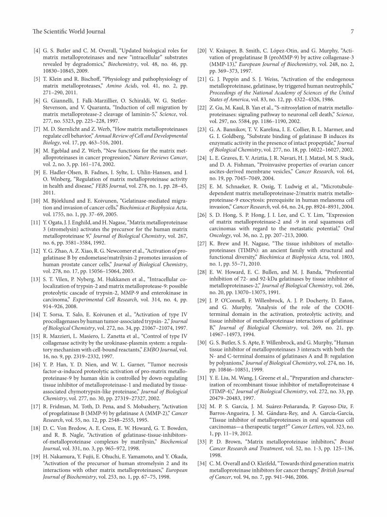
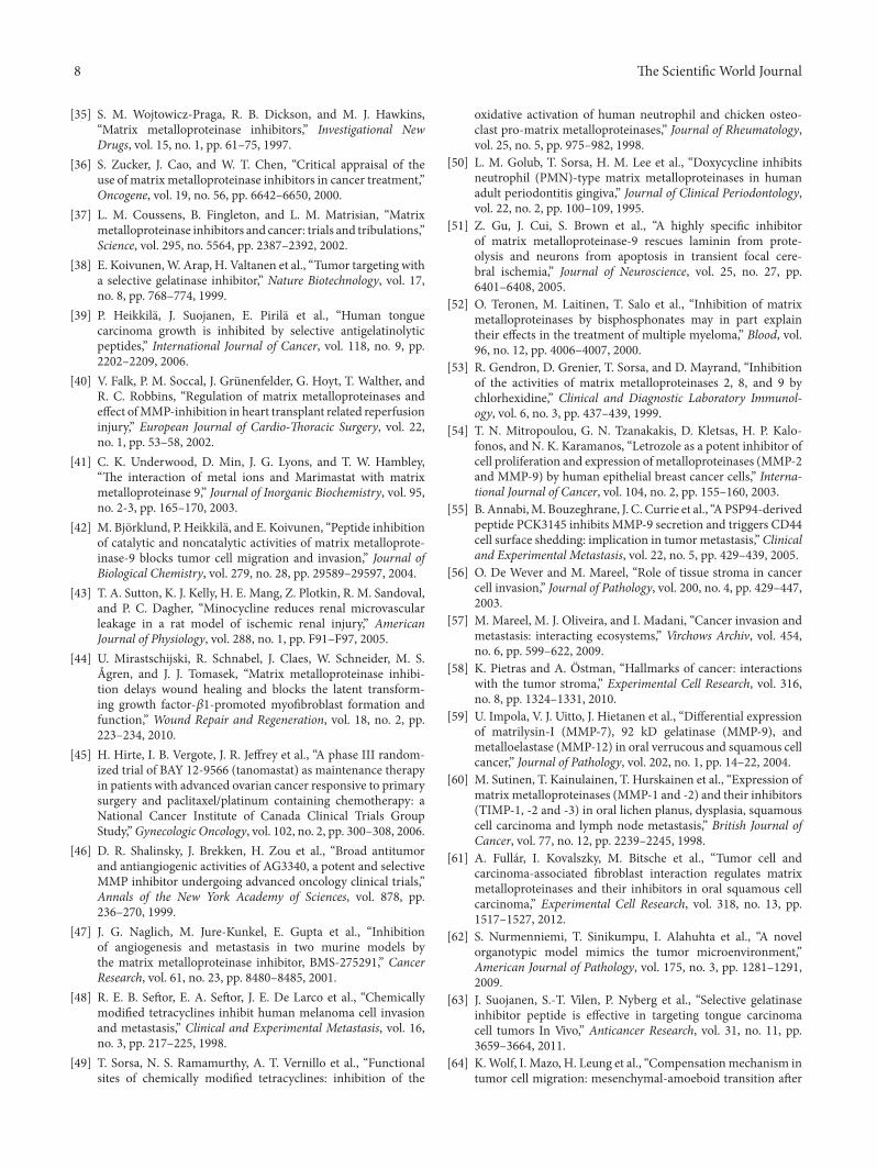
MMP-9
VEGFTumstatinangiostatin
endostatin
Pro-angiogenicAnti-angiogenic
F 1: Effect of MMP-9 on angiogenesis in oral cancer. MMP-9 inhibits angiogenesis by releasing antiangiogenic factors from theirprecursors. MMP-9 enhances angiogenesis by releasing and activating VEGF from extracellular proteoglycans.
bearing nasopharyngeal carcinoma tumors were treated withendostar (recombinant endostatin), the treatment led to asigni�cant decrease in MMP-9 and VEGF expression levelsand normalized the tumor vasculature [121]. Endostatinwas not the only cryptic angiogenesis inhibitor liberatedby MMP-9. Angiostatin, a fragment of plasminogen, alsoinhibits endothelial cell proliferation [86, 122]. Pozzi etal. [123] proved that decreased MMP-9 plasma levels intumor-bearing mice also decreases angiostatin levels, whichincreases tumor vascularization and growth.Matsumoto et al.[124] observed that angiostatin-overexpressing SCC tumorsin mice grew slower than the control tumors. Aside fromendostatin and angiostatin production, MMP-9 is also asso-ciated with the proteolytic degradation of type IV collagen,which produces an angiogenesis inhibitor, tumstatin [125,126]. ese �ndings suggest that in cancer angiogenesisthe variations in spatial and temporal MMP-9 expressionmay switch between two roles: from a proangiogenic to anantiangiogenic molecule (Figure 1).
8. Effect of Genetic and Environmental Factorson the Expression of MMP-9
e development and progression of OSCCs are a resultof interactions between accumulating genetic alterationsand environmental factors, such as alcohol, tobacco, viralinfection, or chronic in�ammation [127]. Despite newercancer treatments, approximately 50% of patients die within5 years of diagnosis [128]. is can be partially explainedby the theory of oral �eld cancerization: an oral mucosaexposed to carcinogens, such as alcohol, causes multiplegenetic abnormalities in the entire epithelium and increasesthe risk of developing several dysplastic lesions [129]. Poly-morphisms in the MMP9 gene allele are associated with anincreased risk of developing the initial stages of oral canceramong patients without a family history of cancer and highsmoking and/or alcohol use [130].MMP-9 expression did notcorrelate with age, gender, tumor location, or smoking habits,whereas an association with tumor grade differentiation andalcohol consumption was observed [78]. MMP-9 was notexpressed in the normal oral mucosa or dysplasia, whereas
in situ carcinomas were weakly detectable. In OSCC, it wasexpressed in the same areas where collagen (IV) chain losswas observed at the invasive fronts, whereas in other studies,its overexpression was detected in 85% of oral dysplasias andin all of the oral cancer samples. e mRNA levels of MMP-9 were higher in oral dysplasia that progressed to oral SCC[131, 132]. Ogbureke et al. 2012 [133] proposed that MMP-9expression at histologically negative surgical margins couldpredict OSCC recurrence. Interestingly, MMP-9 was absentfrom the margins of tumors ≤4 cm and only 10% of tumorswithout later node metastasis expressed it. MMP-9 expres-sion alternates between different stages of malignant trans-formations. Different clinicopathological variables in OSCCscan partially be explained by viral infections in epithelial cells.e human papilloma virus (HPV) is associated withOSCCs,especially type 16. HPV16 transgenic mice, HPV16/MMP-9 +/+ and HPV16/MMP-9 +/− develop a similar incidenceof SCC aer 12 months of age, whereas HPV16/MMP-9 −/−mice have fewer tumors, but these tumors were more poorlydifferentiated than those in the other groups. e expressionof HPV16 oncoproteins in human keratinocytes inducedthe upregulation of MMP-9 activity. At the same time,two natural MMP inhibitors were downregulated. One ofthem was the reversion-inducing cysteine-rich protein withKazal motifs (RECK), which affects transcription, synthesis,activation, and the activity of MMPs; the other was TIMP-2[134–136]. HPV16 infection may be one mechanism behindthe contradictory expression of MMP-9 in OSCC patients;however, further studies are necessary to understand itssigni�cance.
9. Methods for MMP-9 Investigation inOral Cancer
e most commonly used immunohistological analyses ofMMP-9 expression can be misleading because most antibod-ies do not distinguish between the pro- and active formsof the protein. e total amount of protein expression doesnot necessarily mean that the enzyme is in an active form[137]. Gelatin zymography or in situ zymography [138, 139]are better methods to evaluate the level of gelatinase activity

6 e Scienti�c World Journal
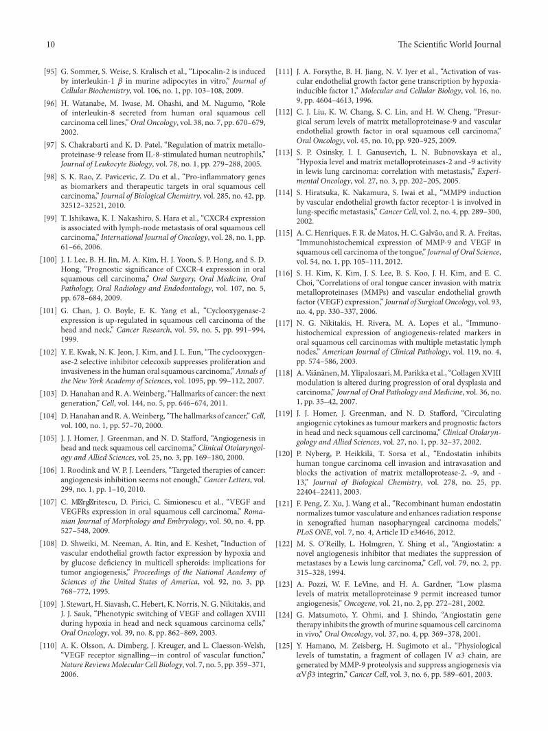
MMP-9
Processing of cytokines, cell surface and
cell adhesion proteins, growth factorsProteolytic processing of ECM
Anti-angiogenesis Angiogenesis Invasion,tumor growth,
metastasis
Inflammation EMT
F 2: Schematic picture of the events that are modulated by MMP-9 in oral cancer. Proteolytic degradation of ECM components(including types III, IV, and V collagens, as well as gelatin) by MMP-9 facilitates carcinoma cell invasion and leads to the release of growthfactors, such as VEGF, that enhance angiogenesis and tumor progression. At the same time anti-antiangiogenic endostatin, angiostatin, andtumstatin are released. Processing of proin�ammatory chemokines (C�CL1, -4, -8, -9, -11, -12), proforms of cytokines (proTNF-𝛼𝛼, proIL-1𝛽𝛽), cell adhesion proteins such as intercellular adhesion molecule-1 triggers an in�ammatory reaction and modulates transcription factorsleading to an epithelial-to-mesenchymal transition and enhanced carcinogenesis.
and would have provided more information in the studiesreferred to here (Table 2) [139]. Many other techniques caninvestigate the presence, amount and function ofMMP-9. Forexample, in situ hybridization could de�ne the location andnumber of cells that expressMMP-9mRNA in tissue sections,whereas the polymerase chain reaction (PCR) could detectthe presence and amount of mRNA in tissues or cell extracts[140, 141]. More speci�c intracellular localization of MMP-9 could easily be achieved using confocal laser scanningmicroscopy, which enables a 3-dimensional de�nition of pro-tein localization [13]. Enzyme-linked immunosorbent assays(ELISAs) could be used to determine the concentration ofMMP-9 protein in serum and plasma samples; based on thesestudies, MMP-9 is a systemic biomarker that monitors theeffectiveness of OSCC treatment [112, 141, 142]. Differentblotting methods, Western, Northern, and Southern, revealthe expression of protein, RNA or DNA, respectively, invarious samples [75, 143]. MMPIs, transfected cell lines, andknock-out animal models give more functional data aboutMMP-9 [144, 145]. Most likely, the variety of methodologiesand the low sample sizes explain the high variation observedamong the previous OSCC results. erefore, more studieswith larger well-documented clinicalmaterials, using delicatemethods, are necessary to determine the impact ofMMP-9 inOSCC.
10. Conclusions
Generally MMP-9 has been associated with aggressive headand neck cancers, but novel studies have shown that it acts asa protective molecule during carcinogenesis and metastasis.For example, in salivary gland myoepithelial carcinoma,MMP-9 expression predicts a better overall survival, and inregional metastases of head and neck cancers, MMP9 geneexpression was decreased [79, 146]. Similarly, the expressionof MMP-9 is associated with a better outcome in breast-and colitis-associated carcinomas [147, 148]. Additionally, in
oral cancer the role of MMP-9 was purely associated withthe degradation of the ECM, which led to the enhancementof carcinoma cell invasion. However, the philosophy oforal carcinoma progression has become signi�cantly morecomplicated; now, MMP-9 is known as a multifunctionalmodulator that is involved in very complex cell-signalingcascades (Figure 2). erefore, in the case of MMP-9, onereason for the obvious failures of broad-spectrumand speci�cMMPIs in cancer treatment might be due to its �uctuatingrole in cancer, which not only affects carcinoma cells but alsoother cell populations. e tumor microenvironment matrixexpresses and sequestersMMP-9. Taken together, our currentknowledge of MMP-9 has been extended; it can act as eithera carcinoma protector or promoter depending on the speci�csituation, which is related to patient characteristics, includingthe stage, grade, and location of the tumor.
Acknowledgments
is study was supported by the Academy of Finland, FinnishDental Society Apollonia, Finnish Cultural Foundation,Sigrid Juselius Foundation, Funds from the Helsinki andOuluUniversityHospital state support for research (EVOandKEVO), and Helsinki Biomedical Graduate School.
References
[1] A. Jemal, F. Bray, M. M. Center, J. Ferlay, E. Ward, and D.Forman, “Global cancer statistics,” CA Cancer Journal forClinicians, vol. 61, no. 2, pp. 69–90, 2011.
[2] P. E. Petersen, “Oral cancer prevention and control—theapproach of the World Health Organization,” Oral Oncology,vol. 45, no. 4-5, pp. 454–460, 2009.
[3] H. Nagase, R. Visse, and G. Murphy, “Structure and func-tion of matrix metalloproteinases and TIMPs,” CardiovascularResearch, vol. 69, no. 3, pp. 562–573, 2006.
e Scienti�c World Journal 7
[4] G. S. Butler and C. M. Overall, “Updated biological roles formatrix metalloproteinases and new “intracellular” substratesrevealed by degradomics,” Biochemistry, vol. 48, no. 46, pp.10830–10845, 2009.
[5] T. Klein and R. Bischoff, “Physiology and pathophysiology ofmatrix metalloproteases,” Amino Acids, vol. 41, no. 2, pp.271–290, 2011.
[6] G. Giannelli, J. Falk-Marzillier, O. Schiraldi, W. G. Stetler-Stevenson, and V. Quaranta, “Induction of cell migration bymatrix metalloprotease-2 cleavage of laminin-5,” Science, vol.277, no. 5323, pp. 225–228, 1997.
[7] M. D. Sternlicht and Z. Werb, “How matrix metalloproteinasesregulate cell behavior,”Annual Review ofCell andDevelopmentalBiology, vol. 17, pp. 463–516, 2001.
[8] M. Egeblad and Z. Werb, “New functions for the matrix met-alloproteinases in cancer progression,” Nature Reviews Cancer,vol. 2, no. 3, pp. 161–174, 2002.
[9] E. Hadler-Olsen, B. Fadnes, I. Sylte, L. Uhlin-Hansen, and J.O. Winberg, “Regulation of matrix metalloproteinase activityin health and disease,” FEBS Journal, vol. 278, no. 1, pp. 28–45,2011.
[10] M. Björklund and E. Koivunen, “Gelatinase-mediated migra-tion and invasion of cancer cells,” Biochimica et Biophysica Acta,vol. 1755, no. 1, pp. 37–69, 2005.
[11] Y.Ogata, J. J. Enghild, andH.Nagase, “Matrixmetalloproteinase3 (stromelysin) activates the precursor for the human matrixmetalloproteinase 9,” Journal of Biological Chemistry, vol. 267,no. 6, pp. 3581–3584, 1992.
[12] Y. G. Zhao, A. Z. Xiao, R. G. Newcomer et al., “Activation of pro-gelatinase B by endometase/matrilysin-2 promotes invasion ofhuman prostate cancer cells,” Journal of Biological Chemistry,vol. 278, no. 17, pp. 15056–15064, 2003.
[13] S. T. Vilen, P. Nyberg, M. Hukkanen et al., “Intracellular co-localization of trypsin-2 andmatrixmetalloprotease-9: possibleproteolytic cascade of trypsin-2, MMP-9 and enterokinase incarcinoma,” Experimental Cell Research, vol. 314, no. 4, pp.914–926, 2008.
[14] T. Sorsa, T. Salo, E. Koivunen et al., “Activation of type IVprocollagenases by human tumor-associated trypsin- 2,” Journalof Biological Chemistry, vol. 272, no. 34, pp. 21067–21074, 1997.
[15] R. Mazzieri, L. Masiero, L. Zanetta et al., “Control of type IVcollagenase activity by the urokinase-plasmin system: a regula-torymechanismwith cell-bound reactants,” EMBO Journal, vol.16, no. 9, pp. 2319–2332, 1997.
[16] Y. P. Han, Y. D. Nien, and W. L. Garner, “Tumor necrosisfactor-𝛼𝛼-induced proteolytic activation of pro-matrix metallo-proteinase-9 by human skin is controlled by down-regulatingtissue inhibitor of metalloproteinase-1 and mediated by tissue-associated chymotrypsin-like proteinase,” Journal of BiologicalChemistry, vol. 277, no. 30, pp. 27319–27327, 2002.
[17] R. Fridman, M. Toth, D. Pena, and S. Mobashery, “Activationof progelatinase B (MMP-9) by gelatinase A (MMP-2),” CancerResearch, vol. 55, no. 12, pp. 2548–2555, 1995.
[18] D. C. Von Bredow, A. E. Cress, E. W. Howard, G. T. Bowden,and R. B. Nagle, “Activation of gelatinase-tissue-inhibitors-of-metalloproteinase complexes by matrilysin,” BiochemicalJournal, vol. 331, no. 3, pp. 965–972, 1998.
[19] H. Nakamura, Y. Fujii, E. Ohuchi, E. Yamamoto, and Y. Okada,“Activation of the precursor of human stromelysin 2 and itsinteractions with other matrix metalloproteinases,” EuropeanJournal of Biochemistry, vol. 253, no. 1, pp. 67–75, 1998.
[20] V. Knäuper, B. Smith, C. López-Otin, and G. Murphy, “Acti-vation of progelatinase B (proMMP-9) by active collagenase-3(MMP-13),” European Journal of Biochemistry, vol. 248, no. 2,pp. 369–373, 1997.
[21] G. J. Peppin and S. J. Weiss, “Activation of the endogenousmetalloproteinase, gelatinase, by triggered human neutrophils,”Proceedings of the National Academy of Sciences of the UnitedStates of America, vol. 83, no. 12, pp. 4322–4326, 1986.
[22] Z. Gu, M. Kaul, B. Yan et al., “S-nitrosylation of matrix metallo-proteinases: signaling pathway to neuronal cell death,” Science,vol. 297, no. 5584, pp. 1186–1190, 2002.
[23] G. A. Bannikov, T. V. Karelina, I. E. Collier, B. L. Marmer, andG. I. Goldberg, “Substrate binding of gelatinase B induces itsenzymatic activity in the presence of intact propeptide,” Journalof Biological Chemistry, vol. 277, no. 18, pp. 16022–16027, 2002.
[24] L. E. Graves, E. V. Ariztia, J. R. Navari, H. J. Matzel, M. S. Stack,and D. A. Fishman, “Proinvasive properties of ovarian cancerascites-derived membrane vesicles,” Cancer Research, vol. 64,no. 19, pp. 7045–7049, 2004.
[25] E. M. Schnaeker, R. Ossig, T. Ludwig et al., “Microtubule-dependent matrix metalloproteinase-2/matrix matrix metallo-proteinase-9 exocytosis: prerequisite in human melanoma cellinvasion,”Cancer Research, vol. 64, no. 24, pp. 8924–8931, 2004.
[26] S. D. Hong, S. P. Hong, J. I. Lee, and C. Y. Lim, “Expressionof matrix metalloproteinase-2 and -9 in oral squamous cellcarcinomas with regard to the metastatic potential,” OralOncology, vol. 36, no. 2, pp. 207–213, 2000.
[27] K. Brew and H. Nagase, “e tissue inhibitors of metallo-proteinases (TIMPs): an ancient family with structural andfunctional diversity,” Biochimica et Biophysica Acta, vol. 1803,no. 1, pp. 55–71, 2010.
[28] E. W. Howard, E. C. Bullen, and M. J. Banda, “Preferentialinhibition of 72- and 92-kDa gelatinases by tissue inhibitor ofmetalloproteinases-2,” Journal of Biological Chemistry, vol. 266,no. 20, pp. 13070–13075, 1991.
[29] J. P. O’Connell, F. Willenbrock, A. J. P. Docherty, D. Eaton,and G. Murphy, “Analysis of the role of the COOH-terminal domain in the activation, proteolytic activity, andtissue inhibitor of metalloproteinase interactions of gelatinaseB,” Journal of Biological Chemistry, vol. 269, no. 21, pp.14967–14973, 1994.
[30] G. S. Butler, S. S. Apte, F. Willenbrock, and G. Murphy, “Humantissue inhibitor of metalloproteinases 3 interacts with both theN- and C-terminal domains of gelatinases A and B: regulationby polyanions,” Journal of Biological Chemistry, vol. 274, no. 16,pp. 10846–10851, 1999.
[31] Y. E. Liu, M. Wang, J. Greene et al., “Preparation and character-ization of recombinant tissue inhibitor of metalloproteinase 4(TIMP-4),” Journal of Biological Chemistry, vol. 272, no. 33, pp.20479–20483, 1997.
[32] M. P. S. García, J. M. Suárez-Peñaranda, P. Gayoso-Diz, F.Barros-Angueira, J. M. Gándara-Rey, and A. García-García,“Tissue inhibitor of metalloproteinases in oral squamous cellcarcinomas—a therapeutic target?” Cancer Letters, vol. 323, no.1, pp. 11–19, 2012.
[33] P. D. Brown, “Matrix metalloproteinase inhibitors,” BreastCancer Research and Treatment, vol. 52, no. 1-3, pp. 125–136,1998.
[34] C.M.Overall andO. Kleifeld, “Towards third generationmatrixmetalloproteinase inhibitors for cancer therapy,” British Journalof Cancer, vol. 94, no. 7, pp. 941–946, 2006.
8 e Scienti�c World Journal
[35] S. M. Wojtowicz-Praga, R. B. Dickson, and M. J. Hawkins,“Matrix metalloproteinase inhibitors,” Investigational NewDrugs, vol. 15, no. 1, pp. 61–75, 1997.
[36] S. Zucker, J. Cao, and W. T. Chen, “Critical appraisal of theuse of matrix metalloproteinase inhibitors in cancer treatment,”Oncogene, vol. 19, no. 56, pp. 6642–6650, 2000.
[37] L. M. Coussens, B. Fingleton, and L. M. Matrisian, “Matrixmetalloproteinase inhibitors and cancer: trials and tribulations,”Science, vol. 295, no. 5564, pp. 2387–2392, 2002.
[38] E. Koivunen,W. Arap, H. Valtanen et al., “Tumor targeting witha selective gelatinase inhibitor,” Nature Biotechnology, vol. 17,no. 8, pp. 768–774, 1999.
[39] P. Heikkilä, J. Suojanen, E. Pirilä et al., “Human tonguecarcinoma growth is inhibited by selective antigelatinolyticpeptides,” International Journal of Cancer, vol. 118, no. 9, pp.2202–2209, 2006.
[40] V. Falk, P. M. Soccal, J. Grünenfelder, G. Hoyt, T. Walther, andR. C. Robbins, “Regulation of matrix metalloproteinases andeffect ofMMP-inhibition in heart transplant related reperfusioninjury,” European Journal of Cardio-oracic Surgery, vol. 22,no. 1, pp. 53–58, 2002.
[41] C. K. Underwood, D. Min, J. G. Lyons, and T. W. Hambley,“e interaction of metal ions and Marimastat with matrixmetalloproteinase 9,” Journal of Inorganic Biochemistry, vol. 95,no. 2-3, pp. 165–170, 2003.
[42] M. Björklund, P. Heikkilä, and E. Koivunen, “Peptide inhibitionof catalytic and noncatalytic activities of matrix metalloprote-inase-9 blocks tumor cell migration and invasion,” Journal ofBiological Chemistry, vol. 279, no. 28, pp. 29589–29597, 2004.
[43] T. A. Sutton, K. J. Kelly, H. E. Mang, Z. Plotkin, R. M. Sandoval,and P. C. Dagher, “Minocycline reduces renal microvascularleakage in a rat model of ischemic renal injury,” AmericanJournal of Physiology, vol. 288, no. 1, pp. F91–F97, 2005.
[44] U. Mirastschijski, R. Schnabel, J. Claes, W. Schneider, M. S.Ågren, and J. J. Tomasek, “Matrix metalloproteinase inhibi-tion delays wound healing and blocks the latent transform-ing growth factor-𝛽𝛽1-promoted myo�broblast formation andfunction,” Wound Repair and Regeneration, vol. 18, no. 2, pp.223–234, 2010.
[45] H. Hirte, I. B. Vergote, J. R. Jeffrey et al., “A phase III random-ized trial of BAY 12-9566 (tanomastat) as maintenance therapyin patients with advanced ovarian cancer responsive to primarysurgery and paclitaxel/platinum containing chemotherapy: aNational Cancer Institute of Canada Clinical Trials GroupStudy,”Gynecologic Oncology, vol. 102, no. 2, pp. 300–308, 2006.
[46] D. R. Shalinsky, J. Brekken, H. Zou et al., “Broad antitumorand antiangiogenic activities of AG3340, a potent and selectiveMMP inhibitor undergoing advanced oncology clinical trials,”Annals of the New York Academy of Sciences, vol. 878, pp.236–270, 1999.
[47] J. G. Naglich, M. Jure-Kunkel, E. Gupta et al., “Inhibitionof angiogenesis and metastasis in two murine models bythe matrix metalloproteinase inhibitor, BMS-275291,” CancerResearch, vol. 61, no. 23, pp. 8480–8485, 2001.
[48] R. E. B. Seor, E. A. Seor, J. E. De Larco et al., “Chemicallymodi�ed tetracyclines inhibit human melanoma cell invasionand metastasis,” Clinical and Experimental Metastasis, vol. 16,no. 3, pp. 217–225, 1998.
[49] T. Sorsa, N. S. Ramamurthy, A. T. Vernillo et al., “Functionalsites of chemically modi�ed tetracyclines: inhibition of the
oxidative activation of human neutrophil and chicken osteo-clast pro-matrix metalloproteinases,” Journal of Rheumatology,vol. 25, no. 5, pp. 975–982, 1998.
[50] L. M. Golub, T. Sorsa, H. M. Lee et al., “Doxycycline inhibitsneutrophil (PMN)-type matrix metalloproteinases in humanadult periodontitis gingiva,” Journal of Clinical Periodontology,vol. 22, no. 2, pp. 100–109, 1995.
[51] Z. Gu, J. Cui, S. Brown et al., “A highly speci�c inhibitorof matrix metalloproteinase-9 rescues laminin from prote-olysis and neurons from apoptosis in transient focal cere-bral ischemia,” Journal of Neuroscience, vol. 25, no. 27, pp.6401–6408, 2005.
[52] O. Teronen, M. Laitinen, T. Salo et al., “Inhibition of matrixmetalloproteinases by bisphosphonates may in part explaintheir effects in the treatment of multiple myeloma,” Blood, vol.96, no. 12, pp. 4006–4007, 2000.
[53] R. Gendron, D. Grenier, T. Sorsa, and D. Mayrand, “Inhibitionof the activities of matrix metalloproteinases 2, 8, and 9 bychlorhexidine,” Clinical and Diagnostic Laboratory Immunol-ogy, vol. 6, no. 3, pp. 437–439, 1999.
[54] T. N. Mitropoulou, G. N. Tzanakakis, D. Kletsas, H. P. Kalo-fonos, and N. K. Karamanos, “Letrozole as a potent inhibitor ofcell proliferation and expression of metalloproteinases (MMP-2and MMP-9) by human epithelial breast cancer cells,” Interna-tional Journal of Cancer, vol. 104, no. 2, pp. 155–160, 2003.
[55] B. Annabi,M. Bouzeghrane, J. C. Currie et al., “A PSP94-derivedpeptide PCK3145 inhibits MMP-9 secretion and triggers CD44cell surface shedding: implication in tumor metastasis,” Clinicaland Experimental Metastasis, vol. 22, no. 5, pp. 429–439, 2005.
[56] O. De Wever and M. Mareel, “Role of tissue stroma in cancercell invasion,” Journal of Pathology, vol. 200, no. 4, pp. 429–447,2003.
[57] M. Mareel, M. J. Oliveira, and I. Madani, “Cancer invasion andmetastasis: interacting ecosystems,” Virchows Archiv, vol. 454,no. 6, pp. 599–622, 2009.
[58] K. Pietras and A. Östman, “Hallmarks of cancer: interactionswith the tumor stroma,” Experimental Cell Research, vol. 316,no. 8, pp. 1324–1331, 2010.
[59] U. Impola, V. J. Uitto, J. Hietanen et al., “Differential expressionof matrilysin-I (MMP-7), 92 kD gelatinase (MMP-9), andmetalloelastase (MMP-12) in oral verrucous and squamous cellcancer,” Journal of Pathology, vol. 202, no. 1, pp. 14–22, 2004.
[60] M. Sutinen, T. Kainulainen, T. Hurskainen et al., “Expression ofmatrix metalloproteinases (MMP-1 and -2) and their inhibitors(TIMP-1, -2 and -3) in oral lichen planus, dysplasia, squamouscell carcinoma and lymph node metastasis,” British Journal ofCancer, vol. 77, no. 12, pp. 2239–2245, 1998.
[61] A. Fullár, I. Kovalszky, M. Bitsche et al., “Tumor cell andcarcinoma-associated �broblast interaction regulates matrixmetalloproteinases and their inhibitors in oral squamous cellcarcinoma,” Experimental Cell Research, vol. 318, no. 13, pp.1517–1527, 2012.
[62] S. Nurmenniemi, T. Sinikumpu, I. Alahuhta et al., “A novelorganotypic model mimics the tumor microenvironment,”American Journal of Pathology, vol. 175, no. 3, pp. 1281–1291,2009.
[63] J. Suojanen, S.-T. Vilen, P. Nyberg et al., “Selective gelatinaseinhibitor peptide is effective in targeting tongue carcinomacell tumors In Vivo,” Anticancer Research, vol. 31, no. 11, pp.3659–3664, 2011.
[64] K.Wolf, I. Mazo, H. Leung et al., “Compensationmechanism intumor cell migration: mesenchymal-amoeboid transition aer

e Scienti�c World Journal 9
blocking of pericellular proteolysis,” Journal of Cell Biology, vol.160, no. 2, pp. 267–277, 2003.
[65] A. J. H. Gearing, P. Beckett, M. Christodoulou et al., “Matrixmetalloproteinases and processing of pro-TNF-𝛼𝛼,” Journal ofLeukocyte Biology, vol. 57, no. 5, pp. 774–777, 1995.
[66] A. Ito, A. Mukaiyama, Y. Itoh et al., “Degradation of interleukin1𝛽𝛽 by matrix metalloproteinases,” Journal of Biological Chem-istry, vol. 271, no. 25, pp. 14657–14660, 1996.
[67] P. E. Van Den Steen, P. Proost, A. Wuyts, J. Van Damme, and G.Opdenakker, “Neutrophil gelatinase B potentiates interleukin-8 tenfold by aminoterminal processing, whereas it degradesCTAP-III, PF-4, and GRO-𝛼𝛼 and leaves RANTES and MCP-2intact,” Blood, vol. 96, no. 8, pp. 2673–2681, 2000.
[68] M. Ferreras, U. Felbor, T. Lenhard, B. R. Olsen, and J. M.Delaissé, “Generation and degradation of human endostatinproteins by various proteinases,” FEBS Letters, vol. 486, no. 3,pp. 247–251, 2000.
[69] R. Heljasvaara, P. Nyberg, J. Luostarinen et al., “Generation ofbiologically active endostatin fragments from human collagenXVIII by distinct matrix metalloproteases,” Experimental CellResearch, vol. 307, no. 2, pp. 292–304, 2005.
[70] S.-I. Kurahara, M. Shinohara, T. Ikebe et al., “Expression ofMMPs, MT-MMP, and TIMPs in squamous cell carcinoma ofthe oral cavity: correlations with tumor invasion and metasta-sis,” Head and Neck, vol. 21, no. 7, pp. 627–638, 1999.
[71] C. X. Zhou, Y. Gao, N. W. Johnson, and J. Gao, “Immunoex-pression of matrix metalloproteinase-2 and matrix metallopro-teinase-9 in the metastasis of squamous cell carcinoma of thehuman tongue,” Australian Dental Journal, vol. 55, no. 4, pp.385–389, 2010.
[72] C. W. Yorioka, R. D. Coletta, F. Alves, I. N. Nishimoto, L.P. Kowalski, and E. Graner, “Matrix metalloproteinase-2 and-9 activities correlate with the disease-free survival of oralsquamous cell carcinoma patients,” International Journal ofOncology, vol. 20, no. 1, pp. 189–194, 2002.
[73] D. Guttman, Y. Stern, T. Shpitzer, D. Ulanovski, T. Druzd, andR. Feinmesser, “Expression of MMP-9, TIMP-1, CD-34 andfactor-8 as prognostic markers for squamous cell carcinoma ofthe tongue,” Oral Oncology, vol. 40, no. 8, pp. 798–803, 2004.
[74] A. Katayama, N. Bandoh, K. Kishibe et al., “Expressions ofmatrix metalloproteinases in early-stage oral squamous cellcarcinoma as predictive indicators for tumor metastases andprognosis,”Clinical Cancer Research, vol. 10, no. 2, pp. 634–640,2004.
[75] T. Ikebe,M. Shinohara,H. Takcuchi et al., “Gelatinolytic activityof matrix metalloproteinase in tumor tissues correlates withthe invasiveness of oral cancer,” Clinical and ExperimentalMetastasis, vol. 17, no. 4, pp. 315–323, 1999.
[76] K. Kato, A. Hara, T. Kuno et al., “Matrix metalloproteinases2 and 9 in oral squamous cell carcinomas: manifestation andlocalization of their activity,” Journal of Cancer Research andClinical Oncology, vol. 131, no. 6, pp. 340–346, 2005.
[77] C.-W. Lin, S.-W. Tseng, S.-F. Yang et al., “Role of lipocalin 2and its complexwithmatrixmetalloproteinase-9 in oral cancer,”Oral Diseases, vol. 18, no. 8, pp. 734–740, 2012.
[78] J. C. De Vicente, M. F. Fresno, L. Villalain, J. A. Vega, andG. Hern�ndez Vallejo, “Expression and clinical signi�cance ofmatrix metalloproteinase-2 and matrix metalloproteinase-9 inoral squamous cell carcinoma,”Oral Oncology, vol. 41, no. 3, pp.283–293, 2005.
[79] A. Stokes, J. Joutsa, R. Ala-aho et al., “Expression pro�les andclinical correlations of degradome components in the tumor
microenvironment of head and neck squamous cell carcinoma,”Clinical Cancer Research, vol. 16, no. 7, pp. 2022–2035, 2010.
[80] H.-X. Fan, H.-X. Li, Z.-X. Gao, and J.-H. Zheng, “Changesin the expression of MMP2, MMP9, and ColIV in stromalcells in oral squamous tongue cell carcinoma: relationships andprognostic implications,” Journal of Experimental and ClinicalCancer Research, vol. 31, no. 1, article 90, 2012.
[81] M. Siewko, B. Mroczko, and M. Szmitkowski, “e roleof matrix metalloproteinases (MMPs) and their inhibitors(TIMPs) in the development of esophageal cancer,” Folia His-tochemica et Cytobiologica, vol. 50, no. 1, pp. 12–19, 2012.
[82] X. Hu, D. Li, W. Zhang, J. Zhou, and B. Tang, “Matrixmetalloproteinase-9 expression correlates with prognosis andinvolved in ovarian cancer cell invasion,”Archives of Gynecologyand Obstetrics, vol. 26, 2012.
[83] Q. W. Zhang, L. Liu, and R. Chen, “Matrix metalloproteinase-9 as a prognostic factor in gastric cancer: a meta-analysis,”Asian Paci�c Journal of Cancer Prevention, vol. 13, no. 6, pp.2903–2908, 2012.
[84] H. Ruokolainen, P. Pääkkö, and T. Turpeenniemi-Hujanen,“Serummatrixmetalloproteinase-9 in head and neck squamouscell carcinoma is a prognostic marker,” International Journal ofCancer, vol. 116, no. 3, pp. 422–427, 2005.
[85] W. G. Stetler-Stevenson, S. Aznavoorian, and L. A. Liotta,“Tumor cell interactions with the extracellular matrix duringinvasion and metastasis,” Annual Review of Cell Biology, vol. 9,pp. 541–573, 1993.
[86] B. C. Patterson andQ. A. Sang, “Angiostatin-converting enzymeactivities of human matrilysin (MMP-7) and gelatinase B/typeIV collagenase (MMP.9),” Journal of Biological Chemistry, vol.272, no. 46, pp. 28823–28825, 1997.
[87] G. Bergers, R. Brekken, G.McMahon et al., “Matrixmetallopro-teinase-9 triggers the angiogenic switch during carcinogenesis,”Nature Cell Biology, vol. 2, no. 10, pp. 737–744, 2000.
[88] A. Mantovani, “Cancer: in�ammation by remote control,”Nature, vol. 435, no. 7043, pp. 752–753, 2005.
[89] B. B. Aggarwal, S. Shishodia, S. K. Sandur, M. K. Pandey,and G. Sethi, “In�ammation and cancer: how hot is the link?”Biochemical Pharmacology, vol. 72, no. 11, pp. 1605–1621, 2006.
[90] D. Ribatti and E. Crivellato, “Mast cells, angiogenesis, andtumour growth,” Biochimica et Biophysica Acta, vol. 1822, no.1, pp. 2–8, 2012.
[91] L. Sun, M. E. Diamond, A. J. Ottaviano, M. J. Joseph, V.Ananthanarayan, and H. G. Munshi, “Transforming growthfactor-𝛽𝛽1 promotes matrix metalloproteinase-9- mediated oralcancer invasion through snail expression,” Molecular CancerResearch, vol. 6, no. 1, pp. 10–20, 2008.
[92] S. Takayama, M. Hatori, Y. Kurihara, Y. Kinugasa, T. Shirota,and S. Shintai, “Inhibition of TGF-𝛽𝛽1 suppresses motilityand invasiveness of oral squamous cell carcinoma cell linesvia modulation of integrins and down-regulation of matrix-metalloproteinases,” Oncology Reports, vol. 21, no. 1, pp.205–210, 2009.
[93] Q. Yu and I. Stamenkovic, “Cell surface-localized matrixmetalloproteinase-9 proteolytically activates TGF-𝛽𝛽 and pro-motes tumor invasion and angiogenesis,” Genes and Develop-ment, vol. 14, no. 2, pp. 163–176, 2000.
[94] K. Shchors and G. Evan, “Tumor angiogenesis: cause or con-sequence of cancer?” Cancer Research, vol. 67, no. 15, pp.7059–7061, 2007.
10 e Scienti�c World Journal
[95] G. Sommer, S. Weise, S. Kralisch et al., “Lipocalin-2 is inducedby interleukin-1 𝛽𝛽 in murine adipocytes in vitro,” Journal ofCellular Biochemistry, vol. 106, no. 1, pp. 103–108, 2009.
[96] H. Watanabe, M. Iwase, M. Ohashi, and M. Nagumo, “Roleof interleukin-8 secreted from human oral squamous cellcarcinoma cell lines,”Oral Oncology, vol. 38, no. 7, pp. 670–679,2002.
[97] S. Chakrabarti and K. D. Patel, “Regulation of matrix metallo-proteinase-9 release from IL-8-stimulated human neutrophils,”Journal of Leukocyte Biology, vol. 78, no. 1, pp. 279–288, 2005.
[98] S. K. Rao, Z. Pavicevic, Z. Du et al., “Pro-in�ammatory genesas biomarkers and therapeutic targets in oral squamous cellcarcinoma,” Journal of Biological Chemistry, vol. 285, no. 42, pp.32512–32521, 2010.
[99] T. Ishikawa, K. I. Nakashiro, S. Hara et al., “CXCR4 expressionis associated with lymph-node metastasis of oral squamous cellcarcinoma,” International Journal of Oncology, vol. 28, no. 1, pp.61–66, 2006.
[100] J. I. Lee, B. H. Jin, M. A. Kim, H. J. Yoon, S. P. Hong, and S. D.Hong, “Prognostic signi�cance of CXCR-4 expression in oralsquamous cell carcinoma,” Oral Surgery, Oral Medicine, OralPathology, Oral Radiology and Endodontology, vol. 107, no. 5,pp. 678–684, 2009.
[101] G. Chan, J. O. Boyle, E. K. Yang et al., “Cyclooxygenase-2expression is up-regulated in squamous cell carcinoma of thehead and neck,” Cancer Research, vol. 59, no. 5, pp. 991–994,1999.
[102] Y. E. Kwak, N. K. Jeon, J. Kim, and J. L. Eun, “e cyclooxygen-ase-2 selective inhibitor celecoxib suppresses proliferation andinvasiveness in the human oral squamous carcinoma,”Annals ofthe New York Academy of Sciences, vol. 1095, pp. 99–112, 2007.
[103] D. Hanahan and R. A.Weinberg, “Hallmarks of cancer: the nextgeneration,” Cell, vol. 144, no. 5, pp. 646–674, 2011.
[104] D.Hanahan andR.A.Weinberg, “ehallmarks of cancer,”Cell,vol. 100, no. 1, pp. 57–70, 2000.
[105] J. J. Homer, J. Greenman, and N. D. Stafford, “Angiogenesis inhead and neck squamous cell carcinoma,” Clinical Otolaryngol-ogy and Allied Sciences, vol. 25, no. 3, pp. 169–180, 2000.
[106] I. Roodink andW. P. J. Leenders, “Targeted therapies of cancer:angiogenesis inhibition seems not enough,” Cancer Letters, vol.299, no. 1, pp. 1–10, 2010.
[107] C. M�rg�ritescu, D. Pirici, C. Simionescu et al., “VEGF andVEGFRs expression in oral squamous cell carcinoma,” Roma-nian Journal of Morphology and Embryology, vol. 50, no. 4, pp.527–548, 2009.
[108] D. Shweiki, M. Neeman, A. Itin, and E. Keshet, “Induction ofvascular endothelial growth factor expression by hypoxia andby glucose de�ciency in multicell spheroids: implications fortumor angiogenesis,” Proceedings of the National Academy ofSciences of the United States of America, vol. 92, no. 3, pp.768–772, 1995.
[109] J. Stewart, H. Siavash, C. Hebert, K. Norris, N. G. Nikitakis, andJ. J. Sauk, “Phenotypic switching of VEGF and collagen XVIIIduring hypoxia in head and neck squamous carcinoma cells,”Oral Oncology, vol. 39, no. 8, pp. 862–869, 2003.
[110] A. K. Olsson, A. Dimberg, J. Kreuger, and L. Claesson-Welsh,“VEGF receptor signalling—in control of vascular function,”Nature ReviewsMolecular Cell Biology, vol. 7, no. 5, pp. 359–371,2006.
[111] J. A. Forsythe, B. H. Jiang, N. V. Iyer et al., “Activation of vas-cular endothelial growth factor gene transcription by hypoxia-inducible factor 1,” Molecular and Cellular Biology, vol. 16, no.9, pp. 4604–4613, 1996.
[112] C. J. Liu, K. W. Chang, S. C. Lin, and H. W. Cheng, “Presur-gical serum levels of matrix metalloproteinase-9 and vascularendothelial growth factor in oral squamous cell carcinoma,”Oral Oncology, vol. 45, no. 10, pp. 920–925, 2009.
[113] S. P. Osinsky, I. I. Ganusevich, L. N. Bubnovskaya et al.,“Hypoxia level and matrix metalloproteinases-2 and -9 activityin lewis lung carcinoma: correlation with metastasis,” Experi-mental Oncology, vol. 27, no. 3, pp. 202–205, 2005.
[114] S. Hiratsuka, K. Nakamura, S. Iwai et al., “MMP9 inductionby vascular endothelial growth factor receptor-1 is involved inlung-speci�c metastasis,” Cancer Cell, vol. 2, no. 4, pp. 289–300,2002.
[115] A. C. Henriques, F. R. deMatos, H. C. Galvão, and R. A. Freitas,“Immunohistochemical expression of MMP-9 and VEGF insquamous cell carcinoma of the tongue,” Journal of Oral Science,vol. 54, no. 1, pp. 105–111, 2012.
[116] S. H. Kim, K. Kim, J. S. Lee, B. S. Koo, J. H. Kim, and E. C.Choi, “Correlations of oral tongue cancer invasion with matrixmetalloproteinases (MMPs) and vascular endothelial growthfactor (VEGF) expression,” Journal of Surgical Oncology, vol. 93,no. 4, pp. 330–337, 2006.
[117] N. G. Nikitakis, H. Rivera, M. A. Lopes et al., “Immuno-histochemical expression of angiogenesis-related markers inoral squamous cell carcinomas with multiple metastatic lymphnodes,” American Journal of Clinical Pathology, vol. 119, no. 4,pp. 574–586, 2003.
[118] A. Väänänen,M. Ylipalosaari,M. Parikka et al., “CollagenXVIIImodulation is altered during progression of oral dysplasia andcarcinoma,” Journal of Oral Pathology andMedicine, vol. 36, no.1, pp. 35–42, 2007.
[119] J. J. Homer, J. Greenman, and N. D. Stafford, “Circulatingangiogenic cytokines as tumourmarkers and prognostic factorsin head and neck squamous cell carcinoma,” Clinical Otolaryn-gology and Allied Sciences, vol. 27, no. 1, pp. 32–37, 2002.
[120] P. Nyberg, P. Heikkilä, T. Sorsa et al., “Endostatin inhibitshuman tongue carcinoma cell invasion and intravasation andblocks the activation of matrix metalloprotease-2, -9, and -13,” Journal of Biological Chemistry, vol. 278, no. 25, pp.22404–22411, 2003.
[121] F. Peng, Z. Xu, J. Wang et al., “Recombinant human endostatinnormalizes tumor vasculature and enhances radiation responsein xenograed human nasopharyngeal carcinoma models,”PLoS ONE, vol. 7, no. 4, Article ID e34646, 2012.
[122] M. S. O’Reilly, L. Holmgren, Y. Shing et al., “Angiostatin: anovel angiogenesis inhibitor that mediates the suppression ofmetastases by a Lewis lung carcinoma,” Cell, vol. 79, no. 2, pp.315–328, 1994.
[123] A. Pozzi, W. F. LeVine, and H. A. Gardner, “Low plasmalevels of matrix metalloproteinase 9 permit increased tumorangiogenesis,” Oncogene, vol. 21, no. 2, pp. 272–281, 2002.
[124] G. Matsumoto, Y. Ohmi, and J. Shindo, “Angiostatin genetherapy inhibits the growth of murine squamous cell carcinomain vivo,” Oral Oncology, vol. 37, no. 4, pp. 369–378, 2001.
[125] Y. Hamano, M. Zeisberg, H. Sugimoto et al., “Physiologicallevels of tumstatin, a fragment of collagen IV 𝛼𝛼3 chain, aregenerated by MMP-9 proteolysis and suppress angiogenesis via𝛼𝛼V𝛽𝛽3 integrin,” Cancer Cell, vol. 3, no. 6, pp. 589–601, 2003.

e Scienti�c World Journal 11
[126] Y. Maeshima, M. Manfredi, C. Reimerli et al., “Identi�cation ofthe anti-angiogenic site within vascular basement membrane-derived tumstatin,” Journal of Biological Chemistry, vol. 276, no.18, pp. 15240–15248, 2001.
[127] S. Choi and J. N. Myers, “Molecular pathogenesis of oralsquamous cell carcinoma: implications for therapy,” Journal ofDental Research, vol. 87, no. 1, pp. 14–32, 2008.
[128] D. M. Walker, G. Boey, and L. A. McDonald, “e pathology oforal cancer,” Pathology, vol. 35, no. 5, pp. 376–383, 2003.
[129] M. G. C. T. Van Oijen and P. J. Slootweg, “Oral �eld canceri-zation: carcinogen-induced independent events or micrometa-static deposits?” Cancer Epidemiology Biomarkers and Preven-tion, vol. 9, no. 3, pp. 249–256, 2000.
[130] E. Vairaktaris, S. Vassiliou, E. Nkenke et al., “A metalloprote-inase-9 polymorphism which affects its expression is associatedwith increased risk for oral squamous cell carcinoma,” EuropeanJournal of Surgical Oncology, vol. 34, no. 4, pp. 450–455, 2008.
[131] R. Tamamura, H. Nagatsuka, C. H. Siar et al., “Comparativeanalysis of basal lamina type IV collagenalpha chains, matrixmetalloproteinases-2 and -9 expressions in oral dysplasia andinvasive carcinoma,” Acta Histochemica. In press.
[132] R. C. K. Jordan, M. Macabeo-Ong, C. H. Shiboski et al.,“Overexpression ofmatrixmetalloproteinase-1 and -9mRNA isassociated with progression of oral dysplasia to cancer,” ClinicalCancer Research, vol. 10, no. 19, pp. 6460–6465, 2004.
[133] K. U. Ogbureke, P. M. Weinberger, S. W. Looney et al.,“Expressions of matrix metalloproteinase-9 (MMP-9), dentinsialophosphoprotein (DSPP), and osteopontin (OPN) at histo-logically negative surgical margins may predict recurrence oforal squamous cell carcinoma,” Oncotarget, vol. 3, no. 3, pp.286–298, 2012.
[134] L. M. Coussens, C. L. Tinkle, D. Hanahan, and Z. Werb, “MMP-9 supplied by bone marrow-derived cells contributes to skincarcinogenesis,” Cell, vol. 103, no. 3, pp. 481–490, 2000.
[135] T. Ramqvist and T. Dalianis, “Oropharyngeal cancer epidemicand human papillomavirus,” Emerging Infectious Diseases, vol.16, no. 11, pp. 1671–1677, 2010.
[136] M. A. Andreoli, C. di Loreto, A. L. Filho, L. L. Villa, and S.S. Maria-Engler, “HPV16 oncoproteins induce MMPs/RECK-TIMP-2 imbalance in primary keratinocytes: possible implica-tions in cervical carcinogenesis,” PLoSONE, vol. 7, no. 3, ArticleID e33585, 2012.
[137] S.-T. Vilen, J. Suojanen, F. Salas et al., “Trypsin-2 enhancescarcinoma invasion by processing tight junctions and activatingpromt1-mmp,” Cancer Investigation, vol. 30, no. 8, pp. 583–592,2012.
[138] E. Pirilä, P. Maisi, T. Salo, E. Koivunen, and T. Sorsa, “Invivo localization of gelatinases (MMP-2 and -9) by in situzymography with a selective gelatinase inhibitor,” Biochemicaland Biophysical Research Communications, vol. 287, no. 3, pp.766–774, 2001.
[139] P. A. M. Snoek-van Beurden and J. W. Von Den Hoff, “Zymo-graphic techniques for the analysis ofmatrixmetalloproteinasesand their inhibitors,” BioTechniques, vol. 38, no. 1, pp. 73–83,2005.
[140] L. Jin and R. V. Lloyd, “In situ hybridization: methods andapplications,” Journal of Clinical Laboratory Analysis, vol. 11, no.1, pp. 2–9, 1997.
[141] R. D. Singh, N. Haridas, J. B. Patel et al., “Matrix metallo-proteinases and their inhibitors: correlation with invasion andmetastasis in oral cancer,” Indian Journal of Clinical Biochem-istry, vol. 25, no. 3, pp. 250–259, 2010.
[142] B. P. Patel, S. V. Shah, S. N. Shukla, P. M. Shah, and P. S. Patel,“Clinical signi�cance of MMP-2 and MMP-9 in patients withoral cancer,” Head and Neck, vol. 29, no. 6, pp. 564–572, 2007.
[143] J. M. Berg, J. L. Tymoczko, and L. Stryer, Biochemistry, W.H.Freeman, New York, NY, USA, 5th edition, 2002.
[144] J. Suojanen, T. Salo, E. Koivunen, T. Sorsa, and E. Pirilä, “Anovel and selective membrane type-1 matrix metalloproteinase(MT1-MMp) inhibitor reduces cancer cell motility and tumorgrowth,” Cancer Biology and erapy, vol. 8, no. 24, pp.2362–2370, 2009.
[145] K. Hotary, X. Y. Li, E. Allen, S. L. Stevens, and S. J. Weiss,“A cancer cell metalloprotease triad regulates the basementmembrane transmigration program,” Genes and Development,vol. 20, no. 19, pp. 2673–2686, 2006.
[146] H. Luukkaa, P. Klemi, I. Leivo et al., “Expression of matrixmetalloproteinase-1, -7, -9, -13, Ki-67, and HER-2 in epithelial-myoepithelial salivary gland cancer,”Head andNeck, vol. 32, no.8, pp. 1019–1027, 2010.
[147] C. Bendrik, J. Robertson, J. Gauldie, and C. Dabrosin, “Genetransfer of matrix metalloproteinase-9 induces tumor regres-sion of breast cancer in vivo,” Cancer Research, vol. 68, no. 9,pp. 3405–3412, 2008.
[148] P. Garg, D. Sarma, S. Jeppsson et al., “Matrix metalloproteinase-9 functions as a tumor suppressor in colitis-associated cancer,”Cancer Research, vol. 70, no. 2, pp. 792–801, 2010.

Submit your manuscripts athttp://www.hindawi.com
Stem CellsInternational
Hindawi Publishing Corporationhttp://www.hindawi.com Volume 2014
Hindawi Publishing Corporationhttp://www.hindawi.com Volume 2014
MEDIATORSINFLAMMATION
of
Hindawi Publishing Corporationhttp://www.hindawi.com Volume 2014
Behavioural Neurology
EndocrinologyInternational Journal of
Hindawi Publishing Corporationhttp://www.hindawi.com Volume 2014
Hindawi Publishing Corporationhttp://www.hindawi.com Volume 2014
Disease Markers
Hindawi Publishing Corporationhttp://www.hindawi.com Volume 2014
BioMed Research International
OncologyJournal of
Hindawi Publishing Corporationhttp://www.hindawi.com Volume 2014
Hindawi Publishing Corporationhttp://www.hindawi.com Volume 2014
Oxidative Medicine and Cellular Longevity
Hindawi Publishing Corporationhttp://www.hindawi.com Volume 2014
PPAR Research
The Scientific World JournalHindawi Publishing Corporation http://www.hindawi.com Volume 2014
Immunology ResearchHindawi Publishing Corporationhttp://www.hindawi.com Volume 2014
Journal of
ObesityJournal of
Hindawi Publishing Corporationhttp://www.hindawi.com Volume 2014
Hindawi Publishing Corporationhttp://www.hindawi.com Volume 2014
Computational and Mathematical Methods in Medicine
OphthalmologyJournal of
Hindawi Publishing Corporationhttp://www.hindawi.com Volume 2014
Diabetes ResearchJournal of
Hindawi Publishing Corporationhttp://www.hindawi.com Volume 2014
Hindawi Publishing Corporationhttp://www.hindawi.com Volume 2014
Research and TreatmentAIDS
Hindawi Publishing Corporationhttp://www.hindawi.com Volume 2014
Gastroenterology Research and Practice
Hindawi Publishing Corporationhttp://www.hindawi.com Volume 2014
Parkinson’s Disease
Evidence-Based Complementary and Alternative Medicine
Volume 2014Hindawi Publishing Corporationhttp://www.hindawi.com