Embed Size (px)
Citation preview

Flow-induced arterial enlargement is inhibited by suppression of nitric oxide synthase activity in vivo F&ml J. Guzman, MD, Keith Abe, BA, and Christopher K. Zarins, MD, Stanford, Calif:
Background. Acute flow-induced arterial dilation is mediated by nitric oxide (NO). The role of NO in chronic flaw-induced adaptive enlargement is unknown. We assessed the role of NO in arterial adapta- tion to increased blood flow (BF). Methods. Iliac artery BF was increased in adult male rats by creating a left femoral arteriovenousfis- tula. Left iliac BF and diameter were measured, and wall shear stress was calculated. The effect of the NO synthase inhibitor IV”-nitroL-arginine-methyl ester (L-NAME) was studied in arta’ovenous fistula rats divided into three groups (group 1, vehicle; group 2, 0.5 mg/ml; group 3, 2 mg/ml) in drinking water Arterial diameter; blood pressure, and medial cell density were assessed after 21 days. Left iliac cyclic guanosine monophosphate content was measured in an additional group of animals. Results. BF and wall shear stress in the left iliac artery increased fourfold immediately after arteriove- nous @ula. Arterial enlargement was evident after 7 days, and wall shear stress normalized aftter 42 days. Flow-induced arterial enlargement was inhibited by both low- and high-dose L-NAME compared with control (analysis of variance p ~0.05). Blood pressure was elevated only in animals treated with high-dose L-NAME. Left iliac cyclic guanosine monophosphate content was lower in rats treated with L- NAME than in the control group (p ~0.05). Conclusions. NO suppression by L-NAME inhibits flow-induced iliac artery enlargement in rats. This
finding suggests that NO plays a role inflow-induced arterial remodeling. (Surgery 1997;122:273-80.)
From the Department of Surgery, Division of Vascular Surgery, Stanford University Medical Center; Stanfmd, Calif:
ARTERIES RESPOND TO CHRONIC INCREASES in blood flow by increasing luminal diameter, a process that results in reduction of wall shear stress (WSS) toward normal.‘, 2 Flow-induced arterial remodel- ing is endothelium dependent and is thought to involve structural modification of the vessel wa11.3 Microscopic and ultrastructural studies of the arte- rial wall proximal to an arteriovenous fistula (AVF) have shown extensive tears and fragmentation of the internal elastic lamina,4 and recent workers have demonstrated enlarged fenestrae in the inter- nal elastic lamina of flow-loaded rabbit carotid arteries with laser scanning confocal microscopy.5
Investigations have previously demonstrated multiple changes in endothelial cells exposed to shear stress in culture. These changes include tran- sient increase in cytosolic calcium,6 activation of G
Presented at the Fifty-eighth Annual Meeting of the Society of University Surgeons, Tampa, Fla., Feb. 13-15, 1997.
Reprint requests: Raul J. Guzman, MD, Division of Vascular
Surgery, H-3600, Stanford University Medical Center, Stanford, CA 94305.
Copyright 0 1997 by Mosby-Year Book, Inc.
0039-6060/97/$5.00 + 0 11/6/82034
proteins,’ increased phosphorylation of endothe- lial nitric oxide synthase (NOS),’ and alterations in the expression of multiple genes including tis- sue plasminogen activatort (tPA),g endothelin,‘O,ll platelet-derived growth factor (PDGF) A and B chain,12*13 thrombomodulin, t4 adhesion mole- cules,15 and c-fos. l6 In addition, increased in vivo production of PDGF-A in prosthetic grafts subject- ed to increased flow has been demonstrated,l’ as has a sustained increase in aortic endothelial NOS expression in a rat model of chronic high blood flow. l8
Acute changes in arterial diameter are mediat- ed by endothelial production of nitric oxide (NO) .lg In this arterial response to increased flow NO is synthesized from the conversion of L-argi- nine to citrulline by the enzyme NOS. The effects of NO on smooth muscle tone are thought to be mediated by the second messenger cyclic guano- sine monophosphate (cGMP), which induces im- mediate changes in intracellular calcium concen- trations. The synthesis of NO from L-arginine can be inhibited by the L-arginine analog w-nitro-t- arginine-methyl ester (L-NAME).
SURGERY 273

274 Guzman, Abe, Zarins surgery
August 1997
Fii. 1. A, In viva photograph of aortoiliac segment with dilated left common iliac artery caused by left femoral AVE. Left iliac vein has red blood discoloration as result of patent AVF. B, Doppler velocimetry mea- surements in control rat used for blood flow determinations. Left common iliac is normal size.
The purpose of this series of experiments was to test the hypothesis that NO mediates chronic adap- tive arterial enlargement in response to increased blood flow. We have developed a reproducible model of flow-induced arterial enlargement in rats. Suppression of NO was accomplished with L- NAME, and the effect on flow-induced arterial enlargement was determined.
MATERIAL AND METHODS
Adult Sprague-Dawley rats (n = 98) weighing 350 to 450 gm were used for these experiments. All pro- cedures were performed with the rats under gen- eral anesthesia and with sterile techniques. General anesthesia was administered intraperitoneally with 40 mg/kg Nembutal (Abbott Laboratories). Beef lung heparin (Upjohn) in a dose of 100 units/kg was routinely given intravenously before the vessels were cross-clamped for fistula construction. On completion of each procedure animals were allowed to recover in a warmer and were then allowed free access to food and water. All animals
were studied under protocols approved by the Animal Care and Use Committee of Stanford University and in accordance to Guide for the Care and Use of Laboratory Animals, Department of Health and Human Services publication no. [NIH] 8fS23, revised 1985.
AVF creation. An AVF was constructed between the left femoral artery and vein in 60 rats. After anesthesia was induced, the left femoral region was prepped, and the femoral vessels were exposed. Heparin was then infused into the femoral vein, and 3 minutes were allowed to elapse for the heparin to circulate. The vessels were then cross- clamped with microvascular clamps (Roboz Scientific), and a 2 mm fistula was constructed between the end of the femoral vein and the side of the femoral artery with a continuous 8-O nylon suture (Microsurgical Instruments Co., Belair, Texas) under an operating microscope (D.F. Vasconcellas, Brazil). At the completion of the pro- cedure the clamps were removed, and the patency of the fistula was assessed by noting pulsatile red

surgmy Volume 122, Number 2
Guzman, Abe, &wins 275
A
B
1.8
1.6
1
O-J,,,,,,,,,,,,,, 0 7 14 21 28 35 42 49 56 63 70 77 84 91
Days
60
0.8- , , , , , , , , , , , , , , 0 0 7 14 21 28 35 42 49 56 63 70 77 84 91
Days
C
*
I I I I I I I , , , , ( , (
0 7 14 21 28 35 42 49 56 63 70 77 84 91
Days
Fig. 2. A, Time course of iliac artery blood flow changes proximal to AVF. B, Time course of iliac artery diameter changes after AVE. C, Time course of left iliac WSS changes after AVE. (*p < 0.05 compared with baseline control group).
blood flow in the vein. The incision was then closed with 40 Vicryl (Ethicon Inc.) suture, and the ani- mals were allowed to recover from anesthesia in a
warm environment. Arterial diameter measurement. At various time
points after fistula creation ranging from 1 hour to 90 days, animals were reanesthetized, the femoral incision was reopened, and the fistula was assessed for patency. Of the 60 rats used for this initial series
of experiments, nine had nonfunctional fistulas and did not undergo further evaluation. An abdominal incision was then made, the small intes- tine was eviscerated to the right, and the infrarenal aorta and iliac vessels were carefully exposed. In vivo assessment of arterial diameter was then made
with microcalipers (L.S. Starrett), and in vivo pho- tography was undertaken with an internal scale for subsequent measurement by projecting the slides
276 Chzman, Abe, Zarins surgery August 1997
A 1.6
- 1.4
2
f 1.2 .e n a ‘S
2 1.0
0.8
C 15.0
3 3 10.0
g
‘: 3
1 5.0
0
0.0
conirol 1‘7 6’7 L-NAME (average mg/day)
control 17 67
L-NAME (average mg/day)
I-
S-
control 17 6-7
L-NAME (average mg/day)
control 17 6-7
L-NAME (average mg/day)
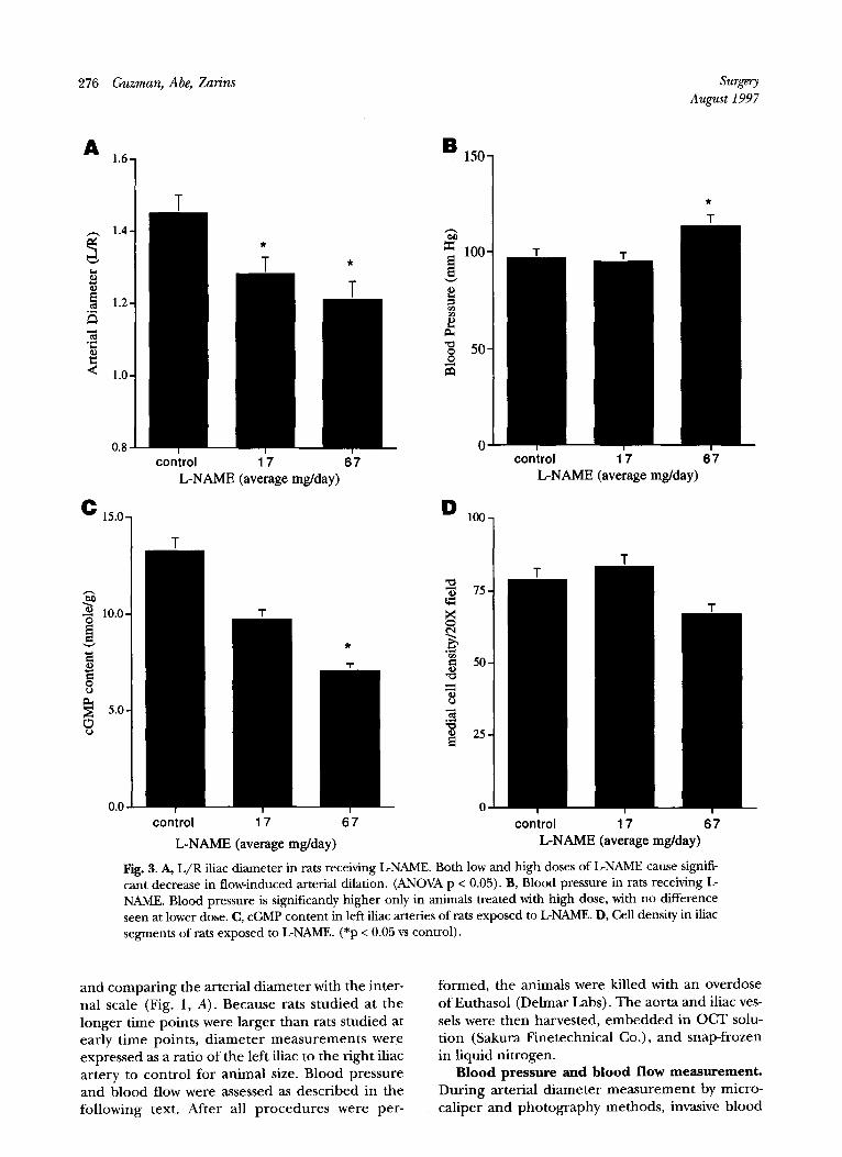
Fig. 3. A, L/R iliac diameter in rats receiving LNAME. Both low and high doses of L-NAME cause signifi- cant decrease in flow-induced arterial dilation. (ANOVA p < 0.05). B, Blood pressure in rats receiving L NAME. Blood pressure is significantly higher only in animals treated with high dose, with no difference seen at lower dose. C, cGMP content in left iliac arteries of rats exposed to LNAME. D, Cell density in iliac segments of rats exposed to LNAME. (*p < 0.05 vs control).
and comparing the arterial diameter with the inter- nal scale (Fig. 1, A). Because rats studied at the longer time points were larger than rats studied at early time points, diameter measurements were expressed as a ratio of the left iliac to the right iliac artery to control for animal size. Blood pressure and blood flow were assessed as described in the following text. After all procedures were per-
formed, the animals were killed with an overdose of Eutbasol (Delmar Labs). The aorta and iliac ves- sels were then harvested, embedded in OCT solu- tion (Sakura Finetechnical Co.), and snap-frozen in liquid nitrogen.
Blood pressure and blood flow measurement. During arterial diameter measurement by micro- caliper and photography methods, invasive blood

surgery ?6lume 122, Number 2
Guzman, Abe, Zarins 277
pressure monitoring was continuously performed by way of the left carotid artery with an intraarteri- al Millar Mikro-tip catheter transducer (Millar Instruments, Inc.) connected to a dual pressure transducer amplifier module (Crystal Biotech, Hopkinton, Mass.). Immediately after in vivo assess- ment of arterial diameter and blood pressure was performed, blood flow measurements were under- taken with a miniature Doppler blood flow trans- ducer connected to a 20 MHz high velocity pulsed Doppler velocimeter (Crystal Biotech). Measure- ments were taken in each iliac vessel and in the infrarenal aorta (Fig. 1, B) . Flow in the proximal iliac vessel is expressed as the ratio of the left iliac artery flow to the right iliac artery flow (L/R) to control for differences in animal size.
To assess blood pressure in animals exposed to L-NAME, animals were anesthetized in the usual manner, and arterial diameter was assessed. No extra doses of anesthesia were given. Blood pres- sure and heart rate were then assessed every minute for a total of 5 minutes during a time when no other interventions were being performed. Blood pressure was calculated as the average of the last three measurements taken while the animal was anesthetized and not undergoing any proce- dure.
WSS calculation. WSS, expressed as dynes per centimeter squared, was calculated with the Hagen- Poiseuille formula WSS = 4 @J7cr3, where 1-1 is the viscosity of blood (taken to be 0.035 poise), Qis the blood flow (ml/set), and ris one half the external- ly measured diameter without correction for wall thickness.
NOS inhibition with L-NAME. The effect of NO inhibition on arterial diameter was studied in 23 animals with a left femoral AVF. Animals were divid- ed into three groups that received (group 1, n = 10) vehicle (H*O), (group 2, n = 6) 0.5 mg/ml, or (group 3, n = 7) 2 mg/ml of L-NAME (Sigma Chemical Co.) in drinking water with the pH adjusted to 7.4. L-NAME treatment was started 2 days before AVF construction and was continued for 21 days after AVF, at which time the animals were killed and measurements were taken of blood pressure, blood flow, and arterial diameter as pre- viously described.
Assessment of cGMP content. Analysis of cGMP content was undertaken to assess suppression of NO in a separate group of 15 rats with a left femoral AVF. Animals were divided into three groups that received (group 1, n = 6) vehicle (H20), (group 2, n = 6) 0.5 mg/ml or (group 3, n = 3) 2 mg/ml L-NAME in drinking water. Five days after fistula construction (7 days after initiation of
treatment), rats were anesthetized in the usual manner. The aorta and right and left iliac vessels were then quickly dissected and excised. The adventitia was removed from the vessels, and they were snap-frozen in liquid nitrogen and stored at -80” C until ready for further use.
Arterial samples were then crushed to a fine powder under liquid nitrogen, and extraction of cGMP was performed in 6% trichloroacetic acid. Measurement of cGMP levels was undertaken with the cGMP enzyme-immunoassay system according to the nonacetylation protocol (Amersham Life Sciences). cGMP levels were expressed as the read- ing performed at 650 nm divided by the wet weight of the tissue sample.
Medial cell density assessment. Frozen tissues embedded in OCT were cut on a cryostat and mounted on coated slides (Superfrost brand; Fisher Scientific). Sections were then stained with hematoxylin-eosin. Medial cell density was deter- mined by counting the number of medial nuclei under x20 magnification. Two independent blind- ed observers evaluated five different sections for each arterial segment. Cell density is expressed as the average of the 10 different determinations for each sample.
Statistical analysis. Results are expressed as mean f SEM. For comparison of arterial diameter, blood flow, and WSS in the initial series of experi- ments, all groups were compared with control unoperated animals. For comparison of arterial diameter in the L-NAME experiments, analysis of variance (ANOVA) was performed with p value less than 0.05 considered significant, and groups were subsequently compared by unpaired t tests, with p value less than 0.05 considered significant. Un- paired t tests among low-dose, high-dose, and con- trol groups were used for comparison of blood pressure, cGMP, and medial cell density.
RESULTS
Effect of femoral AVF on blood blow and iliac artery size. Immediately after left femoral AVF con- struction was performed, L/R blood flow increased fourfold (baseline, 1.00 _+ 0.03 vs 4.47 + 1.28; p < 0.05 ) (Fig. 2, A). The L/R flow ratio continued to increase for 2 weeks after AVF construction and then remained stable for the remainder of the 3- month study period.
The L/R diameter did not change significantly during the first 7 days after AVF construction (base- line, 0.95 + 0.02, vs 7 days, 1.02 f 0.05; p = not sig- nificant). However, after 7 days a significant increase in L/R diameter was seen compared with baseline at all time points, and this difference

278 Guzman, Abe, Zarins surgery August 1997
increased during the observed 90 days (14 days, 1.25 f 0.08, p< 0.05; 21 days, 1.44 rt 0.09, p< 0.05; 42 days, 1.41 * 0.07, p < 0.05; and 90 days, 1.55 + O.O8,p< 0.05) (Fig. 2, B).
WSS and arterial enlargement. Calculated left iliac WSS increased immediately after AVF con- struction (control, 16.86 f 1.68 dynes/cm* to 43.11 f 13.99; p <: 0.05). However, after 2 weeks calculat- ed WSS began to drop as a result of arterial enlargement. At 42 days after AVF, WSS was not sig- nificantly different from control (20.00 ? 5.33, p = not significant) (Fig. 2, C).
Effect of L-NAME on flow-induced arterial enlargement. LNAME was administered in drink- ing water to two groups of rats, which were com- pared with the control group (group 1, control; group 2, 0.5 mg/ml; group 3, 2 mg/ml). Average daily H,O intake was 35 ml/day and did not differ significantly among groups. Average L-NAME intake was 16.27 * 3.22 mg/day for rats in the low- dose group and 67.71 f 7.73 mg/day for rats in the high-dose group.
Both groups of L-NAME-treated rats had a sig- nificantly lower L/R diameter from the control group, suggesting that inhibition of NOS caused inhibition of flow-induced arterial enlargement (group 1, 1.45 ? 0.05, versus group 2, 1.28 * 0.05, p < 0.05, and versus group 3, 1.21 k 0.05, p < 0.01, ANOVA p < 0.05) (Fig. 3, A).
Three rats from each group underwent blood pressure measurement at the time of tissue harvest. No significant difference was seen between the control and the low-dose groups (group 1,96.44 + 6.27 mm Hg, versus group 2, 94.5 * 8.15 mm Hg); however, a significant difference in blood pressure was seen between the control group and the high- dose group (117.11 + 3.13 mm Hg, p < 0.04) (Fig.
3, B). cGIvIP is lower in vessels treated with CNAME.
Effectiveness of NO suppression was determined by measurement of arterial cGMP content. High-dose LNAME treatment resulted in a significant reduc- tion in left iliac cGMP content compared with the control group, whereas low-dose L-NAME treat- ment did not result in a significant decrease (group 1, 13.27 + 1.48 nmol/gm, versus group 2, 9.74 * 2.10 nmol/gm, p = not significant, versus group 3, 7.05 k 2.30 nmol/gm, p= 0.02) (Fig. 3, C).
Effect of GNAME on cell density. The effect of LNAME administration on medial cell density was undertaken to determine whether we could identi- fy any changes in medial composition. Cell density was evaluated on histologic sections obtained at the time of harvest. No significant difference was noted among the three groups, suggesting that the influ-
ence of NOS inhibition was not mediated by an alteration in medial cell number (group 1, 78.73 ? 9.83, versus group 2, 83.27 + 29.07, versus 67.00 k 10.10) (Fig. 3, C).
DISCUSSION
Arterial remodeling in response to increased blood flow involves a complex interaction between the endothelium, which acts as the signal sensor, and medial smooth muscle cells, which must effect a structural reorganization. A specific signal trans- ducing system that regulates the remodeling phe- nomenon has yet to be identified. Investigators have previously identified a number of genes that are specifically up-regulated by shear stress in cul- ture. These genes code for proteins with a diverse set of functions including vasoconstrictors, nuclear signal transducers, thrombolysins, and NOS, the gene responsible for the production of NO.
A number of recent reports have demonstrated the up-regulation of specific gene products by shear stress in vivo. Kraiss et al.” described the induction of PDGF-A gene expression in a baboon prosthetic graft model caused by an acute reduc- tion in blood flow. They localized the increased PDGF-A mRNA and protein expression to the lumi- nal endothelium and subjacent smooth muscle cells. They also showed that smooth muscle cell proliferation in low-flow grafts exceeded that in high-flow grafts. Nadaud et a1.18 recently used a rat aortocaval fistula model of chronic high blood flow to study aortic endothelial cell NO synthase gene expression. They found that NOS3 mRNA levels were increased by quantitative polymerase chain reaction in fistula rats compared with a control group. They also showed increased immunoreac- tivity to NOS3 levels by Western blots, increased NOS activity with the L-arginine to citrulline assay, and increased cGMP levels at 6 weeks. In addition, Tronc et a1.20 recently demonstrated a role for NO in flow-induced remodeling of the rabbit common carotid artery. In that study they showed that arte- rial enlargement in response to increased blood flow is reduced by nonhypertensive doses of L- NAME. Vascular reactivity studies were performed to demonstrate an effect of L-NAME on NO syn- thesis.
In this study we demonstrate that inhibiting NO synthesis with the synthetic analog L-NAME results in a dose-dependent reduction in flow-induced arterial remodeling. We show that this effect is independent of L-NAME’s effect on blood pressure and that medial cell density is not affected by L- NAME treatment. cGMP content was reduced in high-dose L-NAME-treated vessels demonstrating

surgery Volume 122, Number 2
Guzman, Abe, Zarins 279
suppression of NO. Blood pressure did not differ significantly between control and low-dose L-
NAME-treated groups; however, a significant influ- ence of L-NAME treatment on blood pressure was
seen at the higher dose. As such, it is possible that the effect of L-NAME treatment on arterial diame-
ter at the higher dose was related to an effect on blood pressure; however, the finding of a signifi-
cant effect of L-NAME treatment of arterial
enlargement at a nonhypertensive dose argues against this possibility.
We chose to evaluate the effect of L-NAME treat-
ment on arterial diameter at 21 days after fistula construction. This time point was chosen on the basis of our time course data, which showed a sig-
nificant increase in diameter beginning 14 days after fistula construction and a leveling off of arte-
rial enlargement after 21 days. However, it is impor- tant to note that WSS was not normalized at 21
days, and therefore we cannot make any conclu-
sions relating to the effect of NO suppression on normalization of WSS. Our time course study
demonstrated that WSS returned to normal at 42
days in this model. Therefore a longer term study of L-NAME treatment extended past 42 days would
be necessary to determine whether NO suppres- sion alters an artery’s ability to normalize WSS with- in the usual time frame.
Finally, we have demonstrated that inhibition of NO synthesis resulted in only a partial blockage of
arterial enlargement, and thus it is likely that other signaling mediators are involved in this process.
This may involve systems such as the angiotensin or
endothelin cascades. In addition, alterations in enzymes responsible for matrix degradation and
resynthesis may play an important role in the long-
term regulation of arterial adaptation. Further studies will be needed to identify which factors are involved in this complicated process.
REFERENCES 1. Zarins CK, Zatina MA, Giddens DP, Ku DN, Glagov S. Shear
stress regulation of artery lumen diameter in experimental
atherogenesis.J Vast Surg 1987;5:413-20.
2. Kimaya A, Togawa T. Adaptive regulation of wall shear
stress to flow change in the canine carotid artery. Am J
Physiol 1980;239:H14-21.
3. Langille BL, O’Donnell F. Reductions in arterial diameter produced by chronic decreases in blood flow are endothe-
lium dependent. Science 3986;231:405-7.
4. Jones GT, Stethbrns WE. The ultrastructure of arteries proximal to chronic experimental carotidjugular fistulae
in rabbit?. Pathology 1995;27:3&42.
5. Wang LCY, Langille BL. Developmental remodeling of the internal elastic lamina of rabbit arteries: effect of blood
flow. Circ Res 1996;78:799-805. 6. Shen J, Luscinakas FW, Connolly A, Dewey Jr CF, Gimbrone
MA. Fluid shear stress modulates sytosolic free calcium in
vascular endothelial cells. Am J Physiol 1992;262:C38490. 7. Gudi SRI’, Clark CB, Frangos JA. Fluid flow rapidly activates
G proteins in human endothelial cells: involvement of G proteins in mechanochemical signal transduction. Circ Res 1996;79:8349.
8. Carson MA, James NL, Latta SE, Nerem RM, Berk BC, Harrison DC. Phosphorylation of endothelial nitric oxide
synthase in response to fluid shear stress. Circ Res 1996;79:98491.
9. Diamond SL, Sharefkin JB, Dieffenbach CW, Fraiser-Scott
KF, Mclntire LV, Eskin SG. Tissue plasminogen activator messenger RNA levels increase in cultured human
endothelial cells exposed to laminar shear stress. J Cell
Physiol 1990;143:36471. 10. Kuchan MJ, Frangos JA. Shear stress regulated endothelin-
1 release via protein kinase C and cGMP in cultured endothehal cells. Am J Physiol 1993;264:H150-6.
11. Sharefkin JB, Diamond SL. Eskin SG, Dieffenbach CW,
McInire LV. Fluid flow decreases endothelin mRNA levels and suppresses endothelin peptide release in cultured
human endothelial cells. J Vast Surg 1991;14:1-9. 12. Hsieh HJ, Li NQ, Frangos JA. Shear stress increases
endothelial platelet-derived growth factor mRNA levels.
Am J Physiol 1991;260:H642-6. 13. Hsieh HJ, Li NQ, Frangos JA. Shear-induced platelet-
derived growth factor gene expression in human endothe-
lial cells is mediated by protein kinase C. J Cell Physiol 1992;150:552-8.
14. Malek AM, Jackman R, Rosenberg RD, Izumo S. Endothelial expression of thrombomodulins reversibly reg-
ulated by fluid shear stress. Circ Res 1994;74:852-60. 15. Nagel T, Resnick N, Atkinson WJ, Dewey DF, Gimbrone
MA. Shear stress selectively upregulates intracellular adhe-
sion molecule-l expression in cultured human vascular endothelial cells. J Clin Invest 1994;94:885-91.
16. Hsieh HJ, Li NQ Frangos JA. Pulsatile and steady flow
induces c-fos expression in human endothelial cells. J Cell Physiol 1993;154:143-51,
17. Kraiss LW, Geary RL, Mattsson EJR, Verge1 S, Au YPT, Clowes AW. Acute reductions in blood flow and shear stress
induce platelet-derived growth factor-A expression in
baboon prosthetic grafts. Circ Res 1996;79:45-53. 18. Nadaud S, Philippe M, Arnal JF, Michel JB, Soubrier F.
Sustained increase in aortic endothelial nitric oxide syn- thase expression in vivo in a model of chronic high blood
flow. Circ Res 1996;79:857-63. 19. Pohl U, Herlan K, Huang A, Bassenge E. EDRF-mediated
shear-induced dilatation opposes myogenic vasconstriction
in small rabbit arteries. Am J Physiol 1991;261:H201&23. 20. Tronc F, Wassef M, Esposito B, Henrion D, Glagov S,
Tedgui A. Role of NO in flow-induced remodeling of the
rabbit common carotid artery. Arterioscler Thromb Vast Biol 1996:16:125&62.
DISCUSSION
Dr. J. Jeffrey Alexander (Cleveland, Ohio). In the control animals there was a progressive arterial dilatation that continued for a period of 90 days. This occurred
despite a calculated reduction in shear stress that was noted at 14 days and that normalized at 42 days. If shear activates NOS and this is the mechanism for arterial dilatation, how do you explain this discrepancy in the
arterial enlargement despite normalization of shear? In addition, your measurements of the treated animals were

280 Guzman, Abe, Zarins surgery August 1997
taken at a time when shear was at its maximal. Were sim- ilar measurements performed later in the time course to demonstrate that chronic arterial dilatation was also due to this mechanism?
Second, have you studied the effects of altering shear by using this model? This could be accomplished by changing the hemodynamics of the fistula to determine whether the size of the shunt is related to NO activity.
Finally, it has been shown by other investigators that prostacyclin is produced in a flow-dependent manner by the vascular endothelium and that it may contribute to vasodilatation. Have you adequately excluded the contri- butions of prostacyclin to this effect?
Dr. Guzman. With regard to the right iliac artery enlargement in the control animal group, one of the problems that we experienced was that the rats grew dur- ing the 9Oday time point of the study. This caused all the vessels in the body to increase in size. For that reason we decided to express our data in terms of the L/R iliac ratio, so that we had an internal standard that would take growth into account. As to the effect of L-NAME treat- ment on calculated WSS in the treated group, we do not have later time points, although we did measure WSS at 21 days. This is the time at which shear is maximum, but there is a lot of variability in our measurements. There was a tendency for the L-NAME-treated rats to have a higher WSS at that time, but that was not statistically sig- nificant. Measurements at later time points would be important in determining what effect L-NAME treat- ment had on normalization of shear stress in those groups.
With regard to altering shear stress to understand the overall effect, we do not have those data right now. Your point about the role of prostacyclin is also very impor- tant. We are continuing to evaluate this model with emphasis on evaluating that issue.
Dr. John B. Pracyk (Bethesda, Md.). Could you elabo- rate on the rationale for selecting a L/R ratio instead of just measuring the left iliac diameter? Second, was there any effect of NOS inhibition on the right iliac vessel or
perhaps an unoperated vessel? Third, would you specu- late on some potential mechanisms for NO in arterial remodeling? Last, you measured blood pressure changes in animals that were anesthetized. Were measurements made on sedated or nonanesthetized animals?
Dr. Gunman. The choice of using the L/R iliac ratio was primarily based on the variability in animal size. However, if you look at the LNAME data, you do not need to use that ratio, and comparison to the diameter of the left iliac artery diameter is sufficient. However, we decided to present our data in a consistent manner.
As to the effect of NOS inhibition on the right iliac diameter, we did carefully measure right iliac diameters and there was no significant alteration in the right iliac artery diameter by L-NAME treatment. That is somewhat different from results obtained by other groups. I am not able to explain the difference, except to suggest that pos- sibly there is a species difference.
With regard to mechanism, there are some recent data suggesting that in cultured endothelial cells NO may be involved in the up-regulation of matrix metallo proteinase genes. This may be one of the initial steps involved in the process, such that the degradation of the matrix wall begins with the expression of these enzymes.
Finally, it is very difficult to measure blood pressure in rats. We did use intraarterial invasive methods in anes- thetized rats, and we tried to be as careful and consistent as possible to exclude the effect of blood pressure in that low-dose treated group. But clearly blood pressure assess- ment in awake rats would be a much more realistic assessment of the effect of LNAME on blood pressure.
Dr. Timothy R. Billiar (Pittsburgh, Pa.) This study sug- gests that it is just as likely that inducible NOS (iNOS) is induced by the trauma. Have you checked for that? I would suggest that you confirm or carry out these stud- ies in knockout mice. That should give you a much more definitive answer.
Dr. Guzman. We are currently carrying out some of these experiments.