Embed Size (px)
Citation preview

Flecainide increases Kir2.1 currents by interactingwith cysteine 311, decreasing the polyamine-induced rectificationRicardo Caballeroa,1, Pablo Dolz-Gaitóna,1, Ricardo Gómeza,2, Irene Amorósa, Adriana Baranaa, Marta González de laFuentea, Lourdes Osunaa, Juan Duarteb, Angelica López–Izquierdoc, Ignacio Moraledad, Enrique Gálvezd,José Antonio Sánchez–Chapulac, Juan Tamargoa, and Eva Delpóna
aDepartment of Pharmacology, School of Medicine, Universidad Complutense, 28040 Madrid, Spain; bDepartment of Pharmacology, School of Pharmacy,Universidad de Granada, 18071 Granada, Spain; cUnidad de Investigación Carlos Méndez, Centro Universitario de Investigaciones Biomédicas, Universidad deColima, 28045 Colima, Mexico; and dDepartment of Organic Chemistry, School of Pharmacy, Universidad de Alcalá, 28871 Alcalá de Henares, Spain
Edited by Lily Yeh Jan, University of California, San Francisco, CA, and approved July 23, 2010 (received for review March 25, 2010)
Both increase and decrease of cardiac inward rectifier current (IK1) areassociated with severe cardiac arrhythmias. Flecainide, a widely usedantiarrhythmic drug, exhibits ventricular proarrhythmic effects whileeffectively controlling ventricular arrhythmias associated with muta-tions in the geneencodingKir2.1 channels that decrease IK1 (Andersensyndrome). Here we characterize the electrophysiological and molec-ular basis of the flecainide-induced increase of the current generatedby Kir2.1 channels (IKir2.1) and IK1 recorded in ventricular myocytes.Flecainide increases outward IKir2.1 generated by homotetramericKir2.1 channels by decreasing their affinity for intracellular poly-amines, which reduces the inward rectification of the current. Flecai-nide interacts with the HI loop of the cytoplasmic domain of thechannel, Cys311 being critical for the effect. This explains why flecai-nide does not increase IKir2.2 and IKir2.3, because Kir2.2 and Kir2.3 chan-nels do not exhibit a Cys residue at the equivalent position. Wefurther show that incubation with flecainide increases expression offunctional Kir2.1 channels in themembrane, aneffect alsodeterminedby Cys311. Indeed, flecainide pharmacologically rescues R67W, butnot R218W, channelmutations found inAndersen syndromepatients.Moreover, ourfindings provide noteworthy clues about the structuraldeterminants of the C terminus cytoplasmic domain of Kir2.1 channelsinvolved in the control of gating and rectification.
cardiac IK1 | Kir2.2 channel | Kir2.3 channel | Andersen mutations | inwardrectifying channel
The cardiac inwardly rectifying K+ current (IK1) stabilizes restingmembrane potential (RMP) close to the reversal potential of K+
(EK) and shapes the final repolarization phase of the action po-tential (AP) (1). Three inwardly rectifying channels (Kir2.1, Kir2.2,and Kir2.3) contribute to IK1 in the human heart assembled ashomo- and/or heterotetramers (2). Experimental data suggest thatin humans, Kir2.1 is the major isoform underlying ventricular IK1,whereas its relative contribution to atrial IK1 seems to be lower (3).The strong inward rectification of Kir2.x channels, i.e., the prefer-ential conduction of inward compared with outward current, de-pends on the binding of intracellular Mg2+ and polyamines to thecytoplasmic pore and to the inner vestibule of the channel (4).Gain- and loss-of-function mutations in the gene that encodes
Kir2.1 (KCNJ2) have been reported, and both the IK1 increase anddecrease produced by these mutations are associated with severeventricular arrhythmias (1). Furthermore, experimental data showedthat as the amplitude of the outward component of the IK1 increases,the frequency of the fast and stable reentry of spiral waves (rotors)increases. Indeed, the importance of IK1 in the establishment ofrotors and ventricular fibrillation dynamics has been shown (5).Flecainide is a class I antiarrhythmic drug that, besides its Na+
channel-blocking properties, exhibits class III antiarrhythmic effects[i.e., prolongs AP duration (APD) and refractoriness] at the atrialbut not at the ventricular level (6, 7). Flecainide is widely used tosuppress recent onset atrial fibrillation (AF) (8), though it is asso-ciated with an increased risk of ventricular proarrhythmia, especially
in patients with coronary artery disease and/or heart failure. Con-versely, in a limited number of patients, it has been reported thatflecainide is effective in controlling ventricular tachycardia associ-ated with the autosomal dominant trait Andersen syndrome (AS),which is produced by loss-of-function mutations in KCNJ2 (9, 10).We hypothesized that a putative differential flecainide effect on
atrial and ventricular IK1 (blockade of atrial and increase of ven-tricular IK1) can account for the selective prolongation of the atrialAPD, the ventricular proarrhythmic effects, and the antiarrhyth-mic effects in the AS patients. Therefore, we have analyzed theflecainide effects on the currents generated by human WT andmutated Kir2.1, Kir2.2, and Kir2.3 channels (IKir2.x) and comparedits effects on atrial and ventricular IK1. Acutely applied flecainideselectively increased ventricular IK1 and IKir2.1, an effect deter-mined by the presence of Cys311 at the cytoplasmic domain ofKir2.1 channels. Binding of flecainide reduced the Kir2.1 poly-amine blockade, thus reducing the inward rectification. Addition-ally, incubation with flecainide increased functional ion channeldensity, an effect also determined by Cys311. Overall, the findingsshow that Kir2.1 channels can be affected through the pharma-cological modulation of their polyamine blockade by a therapeu-tically used drug.
ResultsFlecainide Increases Kir2.1 Currents. Figure 1A shows IKir2.1 tracesrecorded in transiently transfected Chinese hamster ovary (CHO)cells by applying 250-ms pulses from −60 mV to potentials ranging−120 and +20 mV in the absence and presence of 1 μM flecainide.Flecainide increased IKir2.1 at voltages negative (16.5 ± 2.4% at−120mV) and, more importantly, at voltages positive to EK (45.8±9.8% at −50 mV, n = 8, P < 0.05; Fig. 1 A and B). These effectswere completely reversible upon washout (Fig. S1). The concen-tration that produces the half-maximum effect (EC50) and themaximum effect (Emax) were calculated by fitting the Hill equationto the increase produced by different flecainide concentrations andaveraged 0.8± 0.01 μM(nH=1.6± 0.4) and 22± 1.9%at−120mV,respectively (Fig. S1). At −50 mV, the EC50 and the Emax averaged0.4 ± 0.01 μM (nH = 2.2 ± 0.2) and 53.9 ± 3.6%, respectively (Fig.S1). Therefore, flecainide preferentially increased the outwardIKir2.1 generated at physiological potentials.
Author contributions: R.C., J.T., and E.D. designed research; R.C., P.D.-G., R.G., I.A., A.B.,M.G.d.l.F., L.O., J.D., A.L.-I., I.M., E.G., and J.A.S.-C. performed research; R.C., P.D.-G., R.G.,I.A., A.B., M.G.d.l.F., and J.A.S.-C. analyzed data; and J.T. and E.D. wrote the paper.
The authors declare no conflict of interest.
This article is a PNAS Direct Submission.
Freely available online through the PNAS open access option.1R.C. and P.D.-G. contributed equally to this work.2To whom correspondence should be addressed. E-mail: [email protected].
This article contains supporting information online at www.pnas.org/lookup/suppl/doi:10.1073/pnas.1004021107/-/DCSupplemental.
www.pnas.org/cgi/doi/10.1073/pnas.1004021107 PNAS | August 31, 2010 | vol. 107 | no. 35 | 15631–15636
PHARM
ACO
LOGY

To determine the effect of flecainide in a physiologically relevantsetting, we used an epicardial AP voltage-clamp protocol (Fig. S2).Under these conditions, flecainide increased the charge crossing themembrane estimated from the integral of the current traces,reaching 137 ± 28% at 1 μM (n = 8; Fig. S2).
Flecainide Does Not Modify Kir2.2 and Kir2.3 Currents. BecauseKir2.2 and Kir2.3 proteins also contribute to cardiac IK1, the effectsof flecainide on these channels were studied. The findings showedthat flecainide failed tomodify IKir2.2 and IKir2.3 (P> 0.05 vs. control,n = 10; Fig. 1C and Fig. S3). We also studied the flecainide effectson cells cotransfected (1:1 ratio) with bothKir2.1 andKir2.2 (Kir2.1/Kir2.2) andwithKir2.1 andKir2.3 (Kir2.1/Kir2.3). Kir2.1/Kir2.2 andKir2.1/Kir2.3 currents displayed activation kinetics significantlydifferent from those of the respective homotetrameric channels(Fig. S4), demonstrating the heterotetrameric nature of the chan-nels. Under these conditions, flecainide failed to increase inwardand outward currents (P > 0.05 vs. control, n= 10; Fig. 1C and Fig.S4), demonstrating that its effects were only apparent in channelscomposed of four Kir2.1 subunits.
Flecainide Increases IK1 in Ventricular but Not in Atrial Myocytes.Considering that flecainide selectively increases IKir2.1, we sur-mised that flecainide will differentially affect atrial and ventricularIK1. Figure 2 A and B show original recordings and mean I–Vrelationships for IK1 obtained in isolated human atrial myocytesby applying a voltage ramp from −100 to −10 mV in the absenceandpresence offlecainide (1μM).Flecainide did notmodify eitherthe inward (−5.5 ± 1.2 vs. −5.6 ± 1.2 pA/pF at −100 mV; P > 0.05,n = 5) or outward (0.5 ± 0.1 vs. 0.6 ± 0.1 pA/pF at −10 mV; P >0.05) current.
Because human ventricular myocytes were not available, wecompared the effects of flecainide on the IK1 recorded in guinea-pigatrial and ventricular myocytes (Fig. 2 C–F). As in humans, inguinea-pig ventricles, Kir2.1 expression is higher than in the atria(1, 11, 12). In ventricular myocytes, 1 μM flecainide significantlyincreased both the inward (19.5 ± 3.2% at −120 mV) and outward(38.0 ± 9.5% at −40 mV) current (P < 0.05, n = 4) without modi-fying atrial IK1 (P > 0.05, n= 4; Fig. 2 C–F). All these experimentswere performed in the presence of atropine (1 μM) and glibencla-mide (10 μM) to block the acetylcholine-activated component(IKACh) and the ATP-sensitive (IKATP) inward rectifier currents,
-140 -60 20
-2.5
-2
-1.5
-1
Control
I1.
2ri
K)
An (
Membrane potential (mV)
Fleca 1 μM
**
**
**
*
** *
-0.5
50 ms
An
1
ControlFlecainide 1 μM
-60 mV
+20 mV
-120 mV
-80 -60 -40 -20
0.2
0.1
-0.2
-0.1
**
* 0.3
A
B
C
-10
0
10
20
30
40
50
60
70de
cu
dni-
ed i
n ia
cel
F)
%(e
gn
ah
c
Vm= -50 mV
* ** *
Kir2.1
Kir2.2
Kir2.3
Kir2.1
+2.2
Kir2.1
+2.3
Fig. 1. Flecainide increases IKir2.1. (A) IKir2.1 traces recorded by applying theprotocol shown in the absence and presence of flecainide. (B) I–V curves forcurrents measured at the end of the pulses. (Inset) Data at potentials positiveto EK in an expanded scale. *P < 0.05 and **P < 0.01 vs. control. (C) Flecainide-induced change on the current recorded at −50 mV in cells expressing homo-tetramers of Kir2.1, Kir2.2, or Kir2.3 channels or heterotetramers of Kir2.1 +Kir2.2 and Kir2.1 + Kir2.3. *P < 0.05 vs. Kir2.1. Each point/bar represents themean ± SEM of five or more experiments.
50 ms Control50 ms
Fp/
Ap
02
50 ms Flecainide 1 μM
-130 -110 -90 -70 -50 -30
-30
-20
-10
10
Control
Fleca 1 μM
Il
airt
agi
p-a
eni
uG
1K
)F
p/A
p (
Membrane potential (mV)
-130 -110 -90 -70 -50 -30
-50
-40
-30
-20
-10
10
Control
Fleca 1 μM
Ir
alu
cirt
ne
vgi
p-a
en i
uG
1K
)F
p /A
p (
Membrane potential (mV)
*
*
*
*
*
B A
DC
FE
-110 -90 -70 -50 -30
-8
-6
-4
-2
2
Control
Fleca 1 μM
Il
airt
an
am
uH
1K
)F
p /A
p (
Membrane potential (mV)
-40 mV
-40 mV
-120 mV
Control
Fleca 1 μM
Fp/
Ap
2
20 mV
0 mV
50 ms Control50 ms
Fp/
Ap
01
50 msFlecainide 1 μM
-130 -110 -90 -70 -30
-50
-40
-30
-20
-10
10
Ba2+ sens Control
Ba2+ sens Fleca
Ir
alu
cirt
ne
vgi
p -a
en i
uG
1K
)F
p /A
p (
Membrane potential (mV)
*
*
*
*
*
Ba2+ insens Control
Ba2+ insens Fleca
HG
600
300
1000
bp
Atria
Kir2.1
Kir2.2
Kir2.3
Mar
ker
Ventricles
Kir2.1
Kir2.2
Kir2.3
GAPDH
Kir2.x
1 2 43 65 7
Fig. 2. Flecainide increases ventricular but not atrial IK1. Voltage ramp (800ms)from −100 to −10 mV (A) and I–V curves (B) for human atrial IK1 in the absenceandpresenceofflecainide.Representative IK1 traces recorded inguinea-pig atrial(C) or ventricular (E) myocytes by applying the protocol shown. I–V curves forguinea-pig atrial (D) or ventricular (F) IK1. (G) I–V of the Ba2+-sensitive and Ba2+-insensitive currents recorded in ventricular myocytes before and after appli-cation of flecainide. (H) mRNA expression level of Kir2.x channels in guinea-pigatrial and ventricular samples. First lane shows molecular weight marker (1,000–100 bp). Lanes 2–4 and 5–7 show Kir2.1 (325 bp), Kir2.2 (291 bp), and Kir2.3 (303bp) mRNA expression in atrial and ventricular tissue, respectively. GAPDH genewas used as internal standard. Each point represents the mean ± SEM of fourexperiments in each group. *P < 0.05 vs. control.
15632 | www.pnas.org/cgi/doi/10.1073/pnas.1004021107 Caballero et al.

respectively. Identical findings were obtained when the ventricularIK1 was measured as the Ba2+-sensitive current (Fig. 2G).A previous report proposed that Kir2.1 is the only Kir2.x channel
expressed in guinea-pig atria (11). Conversely, our findings sug-gested that guinea-pig ventricular IK1 is mainly carried by Kir2.1homotetramers, whereas Kir2.x heterotetramers generate atrial IK1.For testing whether Kir2.1 is the only Kir2.x channel present in theatria, we analyzed the mRNA expression of Kir2.x in guinea-pigatria and ventricles. In agreement with another report (12), we coulddetect Kir2.1, Kir2.2, and Kir2.3 mRNA in both the atria and theventricles (Fig. 2H).
Flecainide Increases Open Probability of Kir2.1 Channels. Figure 3Ashows single-channel recordings in a CHO cell expressing Kir2.1channels by applying 10-s pulses to −80 mV from a holding po-tential of 0 mV in the absence and presence of 1 μM flecainide. Incontrol conditions, Kir2.1 channel activity was characterized by fewand long events, leading to an opening frequency (fo) of 2.0 ± 0.1Hz and a mean open probability (Po) of 0.27 ± 0.01 (n= 6; Fig. 3Dand E). Flecainide did not modify unitary current amplitude (Fig.3B) but changed channel gating by significantly increasing the meanopen time and fo (Fig. 3 C and D), which eventually resulted in
a significant increase in the Po (Fig. 3E). Figure 3F compares thevoltage dependence of the Po (Po–V curves) in control conditionsand in the presence of flecainide. As described, Po decreased as themembrane potential became more negative (1, 13). Flecainideshifted the midpoint of the curve to more negative potentials (from−55.3 ± 1.8 to −71.1 ± 2.4 mV; P < 0.05) without modifying theslope. In Fig. 3G, the voltage dependence of the single-channelcurrent amplitude is depicted. Flecainide did not modify the slopeconductance values (γ = 34.7 ± 1.7 pS) yielded by the fit of a linearfunction to the data. All of these effects resemble those producedby PIP2 (14), suggesting that flecainide potentiates the activatingPIP2 effects on the channel. To test this hypothesis, we studied theeffects of flecainide on L222I Kir2.1 channels, a mutation thatdecreases the channel affinity for PIP2 but renders functionalchannels (14). Fig. S5 shows that the decrease of the channel af-finity for PIP2 suppressed the flecainide IKir2.1- increasing effects atpotentials negative to the EK, whereas increasing effects at positivepotentials were still apparent.
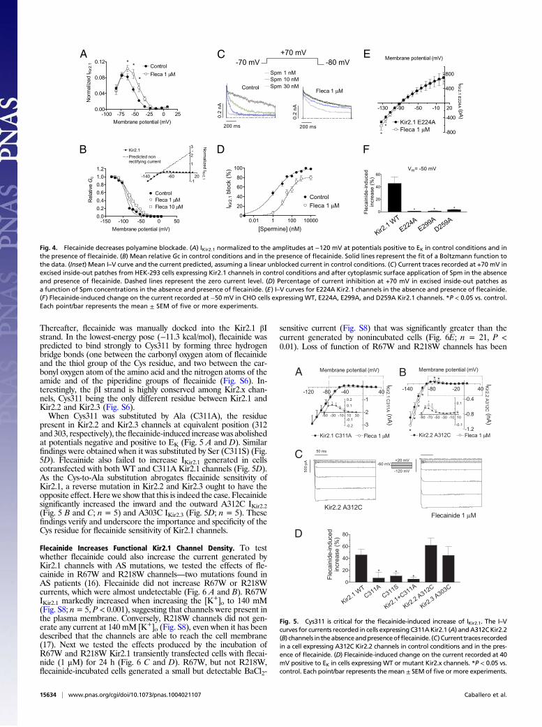
Flecainide Increases Kir2.1 Currents by Decreasing the PolyamineBlockade. Figure 4A illustrates the I–V curve normalized to theamplitudes at −120 mV in control conditions and in the presenceof 1 μM flecainide at potentials positive to EK. As can be observed,flecainide induced a marked increase of the outward IKir2.1 andshifted the potential at which the outward current peaks, sug-gesting that it decreased the channel rectification.The degree of rectification of Kir2.1 channels was estimated as
the relative chord conductance (Gc; Fig. 4B) in control conditionsand in the presence of flecainide. Flecainide shifted the midpoint ofthe curves to more positive potentials (from −91.9 ± 1.8 to −87.1 ±1.8 and −80.5 ± 1.4 mV at 1 and 10 μM flecainide, respectively; P <0.05) and decreased the steepness of rectification, reflected bya lower z value in the presence of flecainide (2.4 ± 0.3 and 1.6 ± 0.6for 1 and 10 μM flecainide, respectively) than in control conditions(3.0 ± 0.5; Fig. 4B).Because the strong rectification of Kir2.1 channels is explained by
the voltage-dependent block produced by intracellular polyamines(4), ourfindings suggested thatflecainidedecreases theaffinity of thechannel for polyamines. Therefore, flecainide effects were furthertested in excised inside-out macropatches in the presence of in-creasing concentrations of spermine (Spm, 1 nM–10 μM). Figure 4Cshows that in control conditions, Spm produced a concentration-dependent block of the outward current at +70 mV. The concen-tration that produces the half-maximum inhibition (IC50) was 1.5 ±0.1 nM (Fig. 4D; n = 8). In the presence of flecainide (1 μM),blockade produced by all of the Spm concentrations tested was de-creased, an effect that shifted rightward (29.0 ± 2.1 nM, n= 8) theconcentration-effect curve of Spm (Fig. 4D). Importantly, in thepresence of flecainide, Emax produced by Spm reached saturation at82.1 ± 5.5%.Finally, we analyzed the effects of flecainide on E224A, D259A,
and E299A Kir2.1 channels, three cytoplasmic residues that areimportant in determining the extent of Kir2.1 inward rectification(15). Figure 4E shows the E224A I–V relationship in the absenceand presence of flecainide. The mutation disrupted the rectifica-tion mechanism of the channel and completely abolished the fle-cainide increase at potentials positive to the EK. Identical findingswere obtained when analyzing the effects of flecainide on D259Aand E299A Kir2.1 channels (Fig. 4F).
Cys311 Is Critical for the Flecainide-Induced Increase of IKir2.1. Toexplore the putative binding site of flecainide within Kir2.1 chan-nels, a molecular model was developed. A blind docking for fle-cainide with a full-length channel composed of the crystal structureof one cytoplasmic domain of Kir2.1 (15) and a modeled trans-membrane domain based on KirBac1.1 crystal structure was per-formed. The findings showed that the most practical binding site onKir2.1 channels was located on the βI strand within the C terminusof the cytoplasmic domain (Fig. S6 and Table S1). In contrast,when blind docking was performed on Kir2.2 and Kir2.3, none ofthe conformations obtained were located on the βI (Fig. S7).
C
O
C
O
C
O
Control
C
O
C
O
C
O
C
O
Fleca 1 μM
C
O
Ap
5
0.5 s
Control Fleca0
1
2
3
4
)A
p(e
dutil
pm
A
Control Fleca0
100
200
300
400
)s
m(e
mitn
ep
on
ae
M
**
Control Fleca0
1
2
3
4
f o)
zH(
**
Control Fleca0.0
0.1
0.2
0.3
0.4
0.5
Po
*
CB
A
ED
GF
-120 -100 -80 -60 -40 -200.0
0.2
0.4
0.6
0.8
1.0Control
Fleca
Membrane potential (mV)
Po
**
**
* * -100 -80 -60 -40 -20 0
-4
-3
-2
-1
Control
Fleca
Membrane potential (mV)
i1.
2ri
K)
Ap(
Fig. 3. Flecainide 1 μM increases mean open time, fo, and Po, of Kir2.1channels. (A) Single-channel recordings under control conditions and afterperfusion with flecainide. Closed- and open-channel levels are indicated by Cand O, respectively. Unitary current amplitude (B), mean open time (C), fo(D), and Po (E) in the absence and presence of flecainide. (F) Po–V in controlconditions and in the presence of flecainide. Solid lines represent the fit ofa Boltzmann function to the data. (G) Single-channel current-voltage rela-tionships in the absence and presence of flecainide. Solid lines represent thefit of a linear function to the data. Each point/bar represents the mean ±SEM of six experiments. *P < 0.05 and **P < 0.01 vs. control.
Caballero et al. PNAS | August 31, 2010 | vol. 107 | no. 35 | 15633
PHARM
ACO
LOGY
Thereafter, flecainide was manually docked into the Kir2.1 βIstrand. In the lowest-energy pose (−11.3 kcal/mol), flecainide waspredicted to bind strongly to Cys311 by forming three hydrogenbridge bonds (one between the carbonyl oxygen atom of flecainideand the thiol group of the Cys residue, and two between the car-bonyl oxygen atom of the amino acid and the nitrogen atoms of theamide and of the piperidine groups of flecainide (Fig. S6). In-terestingly, the βI strand is highly conserved among Kir2.x chan-nels, Cys311 being the only different residue between Kir2.1 andKir2.2 and Kir2.3 (Fig. S6).When Cys311 was substituted by Ala (C311A), the residue
present in Kir2.2 and Kir2.3 channels at equivalent position (312and 303, respectively), theflecainide-induced increase was abolishedat potentials negative and positive to EK (Fig. 5 A and D). Similarfindings were obtained when it was substituted by Ser (C311S) (Fig.5D). Flecainide also failed to increase IKir2.1 generated in cellscotransfected with both WT and C311A Kir2.1 channels (Fig. 5D).As the Cys-to-Ala substitution abrogates flecainide sensitivity ofKir2.1, a reverse mutation in Kir2.2 and Kir2.3 ought to have theopposite effect. Here we show that this is indeed the case. Flecainidesignificantly increased the inward and the outward A312C IKir2.2(Fig. 5 B and C; n = 5) and A303C IKir2.3 (Fig. 5D; n = 5). Thesefindings verify and underscore the importance and specificity of theCys residue for flecainide sensitivity of Kir2.1 channels.
Flecainide Increases Functional Kir2.1 Channel Density. To testwhether flecainide could also increase the current generated byKir2.1 channels with AS mutations, we tested the effects of fle-cainide in R67W and R218W channels—two mutations found inAS patients (16). Flecainide did not increase R67W or R218Wcurrents, which were almost undetectable (Fig. 6 A and B). R67WIKir2.1 markedly increased when increasing the [K+]o to 140 mM(Fig. S8; n= 5, P < 0.001), suggesting that channels were present inthe plasma membrane. Conversely, R218W channels did not gen-erate any current at 140 mM [K+]o (Fig. S8), even when it has beendescribed that the channels are able to reach the cell membrane(17). Next we tested the effects produced by the incubation ofR67W and R218W Kir2.1 transiently transfected cells with flecai-nide (1 μM) for 24 h (Fig. 6 C and D). R67W, but not R218W,flecainide-incubated cells generated a small but detectable BaCl2-
sensitive current (Fig. S8) that was significantly greater than thecurrent generated by nonincubated cells (Fig. 6E; n = 21, P <0.01). Loss of function of R67W and R218W channels has been
001- 57- 05- 52- 0 5200.0
40.0
80.0
21.0lortnoC
1acelF μM
**
*
)Vm(laitnetopenarbmeM
Norm
aliz
ed I
Kir
2.1
051- 001- 05- 0 050.0
2.0
4.0
6.0
8.0
0.1
2.1
lortnoC
1acelF μM
01acelF μM
)Vm(laitnetopenarbmeM
Rela
tive
Gc 041- 06- 02
1-
1
2
31.2riK
Norm
aliz
ed
IKir2
.1
nondetciderPtnerrucgniyfitcer
B
A
F
E
D
C
Mn1mpS
Mn01mpS
Mn03mpS
sm002
0.2
nA
lortnoC
sm002
0.2
nA
1acelF μM
Vm08-Vm07-
Vm07+
10.0 1 001 000010
02
04
06
08
001
1acelF μM
lortnoC
)Mn(]enimrepS[I K
ir2
.1 b
lock (
%)
0
02
04
06
Fle
ca
inid
e-i
nd
uce
din
cre
ase
(%
)
Vm Vm05-=
* * *
Kir2.1
WT
E224A
E299A
D259A
031- 09- 05- 01- 02
008-
004-
004
008
A422E1.2riK
1acelF μM
IKir2
.1 E
224A (p
A)
)Vm(laitnetopenarbmeM
*
*
Fig. 4. Flecainide decreases polyamine blockade. (A) IKir2.1 normalized to the amplitudes at −120 mV at potentials positive to EK in control conditions and inthe presence of flecainide. (B) Mean relative Gc in control conditions and in the presence of flecainide. Solid lines represent the fit of a Boltzmann function tothe data. (Inset) Mean I–V curve and the current predicted, assuming a linear unblocked current in control conditions. (C) Current traces recorded at +70 mV inexcised inside-out patches from HEK-293 cells expressing Kir2.1 channels in control conditions and after cytoplasmic surface application of Spm in the absenceand presence of flecainide. Dashed lines represent the zero current level. (D) Percentage of current inhibition at +70 mV in excised inside-out patches asa function of Spm concentrations in the absence and presence of flecainide. (E) I–V curves for E224A Kir2.1 channels in the absence and presence of flecainide.(F) Flecainide-induced change on the current recorded at −50 mV in CHO cells expressing WT, E224A, E299A, and D259A Kir2.1 channels. *P < 0.05 vs. control.Each point/bar represents the mean ± SEM of five or more experiments.
Kir2.2 A312C
50 ms
Ap
00
5
-120 -80 -40 40
-3
-2
-1
Kir2.1 C311A Fleca 1 μM
IA
11
3C
1.2r
iK
)A
n(
Membrane potential (mV)
-50 -30 -10 10 30
0.2
0.1
-0.2
-0.1
C
BA
D
0
20
40
60
80
de
cu
dni-
edi
nia
cel
F)
%(e
sa
erc
n i * **
iKr2
.1W
T
C311A
C311S
iKr2
.1+C311A
iKr2
.3A303C
-140 -80 -20 40
-1.2
-0.8
-0.4
Kir2.2 A312C Fleca 1 μM
IC
21
3A
2.2r
iK
)A
n(
Membrane potential (mV)
*
*
*
* *
*
-90 -70 -50 -30 -10 10
0.1
-0.1
**
iKr2
.2A312C
-60 mV+20 mV
-120 mV
Flecainide 1 μM
Fig. 5. Cys311 is critical for the flecainide-induced increase of IKir2.1. The I–Vcurves for currents recorded in cells expressing C311AKir2.1 (A) andA312CKir2.2(B) channels in theabsenceandpresenceofflecainide. (C) Current tracesrecordedin a cell expressing A312C Kir2.2 channels in control conditions and in the pres-ence of flecainide. (D) Flecainide-induced change on the current recorded at 40mV positive to EK in cells expressing WT or mutant Kir2.x channels. *P < 0.05 vs.control. Each point/bar represents the mean ± SEMof five or more experiments.
15634 | www.pnas.org/cgi/doi/10.1073/pnas.1004021107 Caballero et al.

attributed to a decreased affinity of the channels for PIP2 (17);therefore, we also tested the effects of the 24-h incubation withflecainide on L222I channels. Figure 6F shows that flecainide alsosignificantly increased L222I IKir2.1 density.These findings suggested that flecainide increased the functional
ion channel density. To test this hypothesis, we incubatedKir2.1WTtransfected cells with flecainide, which significantly increased IKir2.1density (Fig. 6G). Importantly, acute addition of flecainide in-creased IKir2.1 generated by the incubated cells, producing the sameincrease as in nonincubated cells (42.7± 9.5%at−50mV, n=4,P<0.05). This suggests that acute effects of flecainide are additive to itseffects on channel density. Conversely, incubation with flecainidedid not increase C311A Kir2.1 channel density (Fig. 6H), indicatingthat the presence of Cys311 is also critical for this effect. Indeed,incubation with flecainide failed to increase density of Kir2.2 andKir2.3 channels, whereas it increased those of A312C Kir2.2 and
A303C Kir2.3 (Fig. S9). Kir2.1/Kir2.2 and Kir2.1/Kir2.3 densitieswere also not modified with the flecainide incubation (Fig. S9).
DiscussionOur findings show that flecainide acutely increased human IKir2.1mainly by reducing the polyamine blockade of the channel. Addi-tionally, incubation with flecainide increased functional Kir2.1channel density. Flecainide did notmodify either IKir2.2 and IKir2.3 orKir2.2 and Kir2.3 channel density. This selectivity could be attrib-uted to the specific presence of Cys311 on the C terminus of thecytoplasmic region of Kir2.1 channels.
Flecainide Increases IKir2.1 but Not IKir2.2 and IKir2.3. Acutely appliedflecainide increased IKir2.1, and this effect was only apparent inKir2.1 homotetrameric channels. Therefore, flecainide-increasingeffects would be apparent only in those species and tissues in whichIK1 is mainly generated by homotetrameric Kir2.1 channels. Thus,our pharmacological findings suggested that guinea-pig ventricularIK1 is mainly generated by homotetrameric Kir2.1 channels. Recentdata showed that Kir2.1 is the major isoform underlying humanventricular IK1, whereas its relative contribution to atrial IK1 seemsto be lower (3). This could explain why flecainide did not increasehuman atrial IK1.Flecainide preferentially increased the outward IKir2.1 generated
at potentials positive to the EK, which influences cardiac RMP,excitability, and APD. Our findings suggested that flecainide de-creased polyamines’ affinity for the channel in a dose-dependentmanner, thus reducing the strength of rectification. Furthermore,the effects of flecainide on the concentration dependence of theSpm-induced block suggested that it decreases the Spm block bya “noncompetitive” mechanism. Indeed, in the presence of flecai-nide, Emax produced by Spm reached saturation at ≈82%. Thisfinding suggests that flecainide does not compete with Spm for thesame binding site at the channel level, but that interaction of fle-cainide to its own receptor site allosterically reduces the binding ofpolyamines. Previous studies demonstrated that the Glu224,Asp259, and Glu299 cytoplasmic residues help to maximize the rateof polyamine channel block (4, 15). Thus, we tested whether mu-tation of these residues decreased the outward IKir2.1 flecainide in-crease. The findings show that in E224A, D259A, and E299Anonrectifying channels, flecainide did not increase the IKir2.1. Itcould be possible that flecainide interaction to the Cys311 at the βIstrandmodifies the position of the rings of negatively charged aminoacids that create a complimentary electrostatic match for the bind-ing of positively charged polyamines (4, 18). Therefore, this showsthat a therapeutically used drug can affect Kir2.1 channels bymodulating their interaction with polyamines.Our findings strongly suggest that Cys311 located in the βI strand
of the C terminus cytoplasmic domain of the Kir2.1 channeldetermines the flecainide binding. Furthermore, the presence of thisCys is critical for the increasing effects absent in Kir2.2 and Kir2.3channels that do not exhibit a Cys residue at the equivalent position.The βI and βH strands form the HI loop or G loop (residues 300–315) (15). Mutations in the HI loop disrupted gating and affectedinward rectification of Kir2.1 channels (15), and thus it seems rea-sonable to assume that binding of flecainide to this region canproduce the observed effects. Moreover, the HI loop is structurallydistinct from but functionally coupled to the PIP2 binding site (13–15). Indeed, the adjacent residue to Cys311 within the βI strand(Arg312 in Kir2.1 channels) modulates the PIP2-channel inter-actions (14, 18). Furthermore, it has been previously shown thatsubstitution of Cys311 by polar residues strongly modifies the Kir2.1channel kinetic properties, producing long-lasting closed-timeintervals that decreased the channelPo—effects that were attributedto a destabilization of PIP2-Kir2.1 interaction (13). Therefore, wesurmised that flecainide interaction with Cys311 could increase thechannel-activating PIP2 effects. Our findings demonstrated thatflecainide increased the Po of the channel by shifting the Po–V re-lationship to more negative potentials. Moreover, it did not modifythe unitary current amplitude or the single-channel conductance,but augmented the mean open time and the fo of the channel. All
A
E
Incubated with
Fleca 1 μM
100 ms
Ap
02
100 ms
Ap
00
2
Control
Fleca 1 μM
C
R67W -60 mV
+20 mV
-120 mV
-60 20
-5
-3
1
3
5
Kir2.1 R67W
Kir2.1 R67W (inc)
IW
76
R1.
2ri
K)
Fp/
Ap (
yti
sn
ed
Membrane potential (mV)
* * *
**
*
*
*
*
-140
-120 -40 40
-80
-60
-40
-20
IA
11
3C
1.2 r
iK
)F
p /A
p (y t
is
ne
d
Kir2.1 C311A
Kir2.1 C311A (inc)
Membrane potential (mV)
-140 -60 -20
-140
-100
-60
-20
II
22
2L
1.2 r
iK
)F
p/A
p(yt
is
ne
d
Kir2.1 L222I
Kir2.1 L222I (inc)*
*
*
*
* **
Membrane potential (mV)
H
-140 -60 -20 20
-150
-100
-50
50
Kir2.1 WT
Kir2.1 WT (inc)
*
*
**
* * *
I1.
2ri
K)
Fp /
Ap (
y ti
sn
ed
Kir2.1 WT (inc) + Fleca 1 μM
φφφφ
φ φ φ
Membrane potential (mV)
-100 -60 -20-20
20
40
60φφ
φ*
**
-80 -40
-10
-5
5
10
-100 -60 -20
-15
-10
-5
5
10**
*
F
G
B
100 ms
Ap
00
2
Control
Fleca 1 μM
R218W
Incubated withFleca 1 μM
100 ms
Ap
02
D
Fig. 6. Effects of flecainide on Kir2.1 channel density. Current traces recordedin a cell expressing R67W (A) and R218W (B) Kir2.1 in the absence and presenceof flecainide. Current traces recorded in cells expressing R67W (C) and R218W(D) Kir2.1 channels incubated with flecainide for 24 h. The I–V curves for cur-rents recorded in cells expressing R67W (E), L222I (F), WT (G), and C311A (H)Kir2.1 channels in control conditions and after incubation with flecainide. In G,squares represent acute effects of flecainide produced in cells incubated withflecainide. *P < 0.01 vs. control. ΦP < 0.05 vs. incubated cells. Each point rep-resents the mean ± SEM of eight experiments.
Caballero et al. PNAS | August 31, 2010 | vol. 107 | no. 35 | 15635
PHARM
ACO
LOGY

these effects resemble those produced by PIP2 (14). Finally, theL222I mutation, which decreased Kir2.1 affinity for PIP2, sup-pressed the flecainide-increasing effects at potentials negative to theEK, leaving increasing effects at positive potentials unaltered.Overall, these findings suggest that flecainide also potentiates theactivating PIP2 actions on the channel, which contributes to its in-creasing effects—particularly at potentials negative to EK.
Flecainide Increases Kir2.1 Channel Density. Incubation for 24 h withflecainide increased functionalKir2.1 channeldensity.Thiseffectwasalso dependent on the presence of Cys311 and, importantly, wasadditive to the acute flecainide-increasing effects. Therefore, flecai-nide would substantially increase homotetrameric Kir2.1 IK1 by twomechanisms. To test whetherflecainide increases the current and thefunctional density of AS-mutated channels, we tested its effects onR67WandR218W channels. As expected, acutely applied flecainidedid not increase either R67W or R218W IKir2.1, because both chan-nels do not exhibit affinity for PIP2. However, incubation with fle-cainide for 24 h significantly increased R67W and L222I but notR218W IKir2.1 densities. Therefore, these preliminary findings sug-gest that in AS patients who carry mutations that produce functionalchannels, flecainide would increase IKir2.1 generated by WT andmutated channels by increasing their membrane density (i.e., flecai-nide would pharmacologically rescue some AS mutations).Growing evidence suggests that ion-channel density can be mod-
ified pharmacologically. It has been described that two therapeuti-cally used drugs that acutely block Kir2.1 also modify the expressionof the channels in the membrane, i.e., incubation with chloroquineincreases, and with pentamidine decreases, the IKir2.1 density oftransfected cells (19). Further studies are needed to elucidatewhetherflecainide increases the anterogradedelivery toordecreasesthe endocytosis of channel protein from the membrane.
Therapeutic Implications. Flecainide is a potent Na+ channelblocker that also blocks several voltage-dependent K+ channels(Kv). Indeed, flecainide inhibits the Ca2+-independent transientoutward K+ current (Ito1) and the rapid component of the delayedrectifier current (IKr) at concentrations close to those needed forblocking Na+ channels (20, 21). Flecainide frequency-dependentlyincreases the human atrial APD and refractoriness (6) withoutmodifying human ventricular APD, as measured from the QT in-terval of the electrocardiogram (7). The selective increase of theventricular IK1 could account for this differential effect on atrialand ventricular APD. Indeed, because IK1 plays a critical role in thefinal phase of repolarization, it could be speculated that the ven-tricular IK1 increase overcomes the putative APD prolongation
produced by the simultaneous blockade of other Kv channels.However, in the atrial tissue, in which flecainide did not increasethe IK1, the APD prolongation produced by the blockade of Kvchannels will be apparent. Furthermore, the ventricular IK1 in-crease could contribute to the ventricular proarrhythmic effects offlecainide. The importance of IK1 in the establishment and main-tenance of the stability of rotors and ventricular fibrillation dy-namics has been demonstrated (5). The prediction is that an IK1increase accelerates and stabilizes the reentry (5).Finally, it is noteworthy that the flecainide effects here de-
scribed were produced at concentrations that are therapeuticallyrelevant, because peak plasma concentrations after administra-tion of therapeutic doses are between 0.4 and 2.2 μM (22).
ConclusionsOverall, our findings show that Kir2.1 channels can be positivelymodulated through the decrease of their polyamine blockade bya therapeutically used drug. Moreover, flecainide increases thedensity of Kir2.1 channels, an effect that could produce the phar-macological rescue of functional AS-mutated channels. Botheffects were based on the presence of a Cys residue at position 311within the βI strand, which further stresses the role of the cyto-plasmic domain and Cys residues of Kir2.x channels in controllinggating and rectification.
MethodsThis study was approved by the Investigation Committee of the Hospital Uni-versitarioGregorioMarañón (CNIC-13) and conforms to theprinciples outlined inthe Declaration of Helsinki. Human atrial myocytes were enzimatically isolatedfrom right atrial appendages obtained from patients that underwent cardiacsurgery (Table S2) (12). Guinea-pig cardiomyocytes were also enzimatically iso-lated (23). Reverse transcription-PCR analysis of Kir2.x channels was developedwith theprimers depicted in Table S3.WTandmutatedhumanKir2.1, Kir2.2, andKir2.3 channels were transiently transfected in CHO cells (24). Macroscopic andsingle-channel currents were recorded using the whole-cell and cell-attachedpatch-clamp configurations, respectively. Inside-out recordings of IKir2.1 weredeveloped in transiently transfected HEK-293 cells. All I–V curves were correctedaccording to the calculated liquid junction potential. Blind andmanual dockingsof flecainide on a modeled Kir2.1 channel were performed with AutoDock 4.0and QUANTA/CHARMm software, respectively (23). Detailed SI Methods areavailable online.
ACKNOWLEDGMENTS. This workwas supported byMinisterio de Educación yCiencia Grant SAF2008-04903; Ministerio de Sanidad y Consumo, Instituto deSalud Carlos III Grants Red HERACLES RD06/0009 and PI080665; UniversidadComplutense de Madrid (4195); Fundación LILLY; and Centro Nacional deInvestigaciones Cardiovasculares (CNIC-13).
1. Anumonwo JM, Lopatin AN (2010) Cardiac strong inward rectifier potassiumchannels. J Mol Cell Cardiol 48:45–54.
2. Wang Z, Yue L, White M, Pelletier G, Nattel S (1998) Differential distribution of inwardrectifier potassium channel transcripts in human atrium versus ventricle. Circulation 98:2422–2428.
3. Gaborit N, et al. (2007) Regional and tissue specific transcript signatures of ionchannel genes in the non-diseased human heart. J Physiol 582:675–693.
4. Lu Z (2004) Mechanism of rectification in inward-rectifier K+ channels. Annu RevPhysiol 66:103–129.
5. Jalife J (2009) Inward rectifier potassium channels control rotor frequency in ventricularfibrillation. Heart Rhythm 6(11, Suppl):S44–S48.
6. Wang ZG, Pelletier LC, Talajic M, Nattel S (1990) Effects of flecainide and quinidine onhuman atrial action potentials. Role of rate-dependence and comparison with guineapig, rabbit, and dog tissues. Circulation 82:274–283.
7. Katritsis D, et al. (1995) Effect of flecainide on atrial and ventricular refractoriness andconduction in patients with normal left ventricle. Implications for possible antiarrhythmicand proarrhythmic mechanisms. Eur Heart J 16:1930–1935.
8. Crijns HJ, et al. (1988) Acute conversion of atrial fibrillation to sinus rhythm: Clinicalefficacy of flecainide acetate. Comparison of two regimens. Eur Heart J 9:634–638.
9. Pellizzón OA, Kalaizich L, Ptácek LJ, Tristani-Firouzi M, Gonzalez MD (2008) Flecainidesuppresses bidirectional ventricular tachycardia and reverses tachycardia-inducedcardiomyopathy in Andersen–Tawil syndrome. J Cardiovasc Electrophysiol 19:95–97.
10. Bökenkamp R, Wilde AA, Schalij MJ, Blom NA (2007) Flecainide for recurrentmalignant ventricular arrhythmias in two siblings with Andersen–Tawil syndrome.Heart Rhythm 4:508–511.
11. Dhamoon AS, et al. (2004) Unique Kir2.x properties determine regional and speciesdifferences in the cardiac inward rectifier K+ current. Circ Res 94:1332–1339.
12. Liu GX, et al. (2001) Comparison of cloned Kir2 channels with native inward rectifierK+ channels from guinea-pig cardiomyocytes. J Physiol 532:115–126.
13. Garneau L, Klein H, Parent L, Sauvé R (2003) Contribution of cytosolic cysteine residues tothe gating properties of the Kir2.1 inward rectifier. Biophys J 84:3717–3729.
14. Logothetis DE, Jin T, Lupyan D, Rosenhouse-Dantsker A (2007) Phosphoinositide-mediated gating of inwardly rectifying K(+) channels. Pflugers Arch 455:83–95.
15. Pegan S, et al. (2005) Cytoplasmic domain structures of Kir2.1 and Kir3.1 show sites formodulating gating and rectification. Nat Neurosci 8:279–287.
16. Plaster NM, et al. (2001) Mutations in Kir2.1 cause the developmental and episodicelectrical phenotypes of Andersen’s syndrome. Cell 105:511–519.
17. Lopes CM, et al. (2002) Alterations in conserved Kir channel-PIP2 interactions underliechannelopathies. Neuron 34:933–944.
18. Bichet D, Haass FA, Jan LY (2003) Merging functional studies with structures ofinward-rectifier K(+) channels. Nat Rev Neurosci 4:957–967.
19. de Boer TP, Houtman MJ, Compier M, van der Heyden MA (2010) The mammalianKIR2.x inward rectifier ion channel family: Expression pattern and pathophysiology.Acta Physiol (Oxf) 199:243–256.
20. Wang Z, Fermini B, Nattel S (1995) Effects of flecainide, quinidine, and 4-aminopyridine on transient outward and ultrarapid delayed rectifier currents inhuman atrial myocytes. J Pharmacol Exp Ther 272:184–196.
21. Follmer CH, Colatsky TJ (1990) Block of delayed rectifier potassium current IK byflecainide and E-4031 in cat ventricular myocytes. Circulation 82:289–293.
22. Slama R, LeClercq JF (1985) The clinical use of oral flecainide. Drugs 29(Suppl 4):28–29.23. Núñez L, et al. (2006) Nitric oxide blocks hKv1.5 channels by S-nitrosylation and by
a cyclic GMP-dependent mechanism. Cardiovasc Res 72:80–89.24. Gómez R, et al. (2009) Nitric oxide increases cardiac IK1 by nitrosylation of cysteine 76
of Kir2.1 channels. Circ Res 105:383–392.
15636 | www.pnas.org/cgi/doi/10.1073/pnas.1004021107 Caballero et al.


![Phytochromes and Phytochrome Interacting Factors1[OPEN] · Update on Phytochromes and Phytochrome Interacting Factors Phytochromes and Phytochrome Interacting Factors1[OPEN] Vinh](https://img.dokumen.tips/doc/110x75/5e9224c5cbd0a85457462c45/phytochromes-and-phytochrome-interacting-factors1open-update-on-phytochromes-and.jpg)


