Embed Size (px)
Citation preview

Tokai J Exp Clin Med., Vol. 39, No. 3, pp. 116-121, 2014
―116―
INTRODUCTION
Flavonoids are widely distributed in plants as pig-mentary compounds, and are routinely consumed by humans from vegetables, fruits and grain. They have a variety of bioactivities, thus constituting one of the most important medically applicable natural substanc-es [1-5]. Since the various bioactivities include anti-allergic and anti-viral actions, they are now attracting considerable attention as a potential medicinal drug [6, 7]. We have previously shown that quercetin, a rep-resentative �avonoid, is a powerful promoter of mel-anogenesis in the HMV-II cell line (Human Melanoma of Vagina II) [8-10]. Addition of quercetin to cell culture medium markedly increased melanin content, tyrosinase activity, and expression of tyrosinase pro-tein. Tyrosinase activation by quercetin was blocked by actinomycin-D or cycloheximide suggesting that both transcriptional and translational events may have been involved in the stimulation of melanogenesis. These findings were also confirmed in a 3D dermal model and murine buccal follicular tissues.
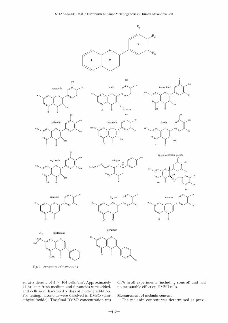
Flavonoid is a generic name for a chemical com-pound possessing 2 phenyl groups (rings A and B) bound to one another with 3 carbon atoms (Fig. 1). Flavonoids are classified into 5 classes: flavonols, fla-vones, isoflavones, flavanols and flavones. Currently, around 8000 �avonoids have been identi�ed. The side chains and locations of hydroxyl groups in the phenyl group may differ among the �avonoids, characterizing them with different chemical properties. It is therefore assumed that �avonoids with different chemical struc-
tures have different biological actions that include a stimulation of melanogenesis. The present study was aimed at determining the melanogenesis-promoting actions of 14 chemically different �avonoids to explore relationships between their chemical structures and melanogenesis-promoting actions.
MATERIALS AND METHODS
Chemicals and reagentsMushroom tyrosinase, melanin, L-dopa, quercetin,
rutin, kaempferol, robinetin, rhamnetin, fisetin, epigallocatechin gallate (EGCg), myricetin, chrysin, ipriflavone, genistein and naringin were obtained from Sigma Chemical Co. (St. Louis, MO, USA). Apigenin was purchased from Indofine (Somerville, NJ). Luteolin was purchased from Extrasynthese (Lyon, France). Ham's F12 medium and trypsin/EDTA were obtained from GIBCO (Carlsbad, CA, USA). Dimethylsulfoxide (DMSO) was purchased from Merck & Company (Darmstadt, Germany).
Cell cultureHMVII is a human melanoma cell line that was
established from a black-brown malignant melanoma in the vaginal wall of woman [11]. Human melanoma HMVII cells were kindly provided by the RIKEN Cell Bank. HMVII cells were cultured in Ham's F12 me-dium containing 10% fetal bovine serum (FBS) in a humidi�ed 37ºC atmosphere consisting of 5% CO2 and 95% air. Cell plating densities were arranged so that cells were in the log phase of growth for the duration of incubation with drug. Subcultures of cells were plat-
Flavonoids Enhance Melanogenesis in Human Melanoma Cells
Susumu TAKEKOSHI, Hidetaka NAGATA and Kanae KITATANI
Department of Cell Biology, Division of Host Defense Mechanism, Tokai University School of Medicine
(Received June 9, 2014; Accepted June 14, 2014)
Flavonoids are pigmentary compounds existing widely in plants. We have reported that quercetin (3, 3’, 4’, 5, 7-pentahydroxylflavone), one of the typical flavonoids, strongly promotes melanogenesis by melanocytes. Meanwhile, there are 8000 or more flavonoids having a chemical structure different from each other in the natu-ral world. Their distinctive chemical properties suggest that they may be different in melanogenic actions. In the present study, the melanogenic actions of 14 flavonoids were analyzed to correlate their chemical structures with melanogenic actions. To evaluate the effects of flavonoids on melanogenesis, the HMV II cell line derived from human malignant melanoma was used. Flavonols including quercetin, kaempferol, rhamnetin and fisetin, flavones including apigenin, luteolin and chrysin, and isoflavones including genestein showed melanogenesis-promoting actions but rutin, robinetin, myricetin, ipriflavone, epigalocatechin gallate (EGCg) and naringin did not. From analyses of the relationships between the chemical structures of flavonoids and their melanogenesis-promoting actions, it was inferred that a hydroxyl group bound to the phenyl group plays an important role in stimulating melanogenesis. From the above results, 8 flavonoids were identified as melanogenesis promoters. Also, correla-tions were established between the melanogenesis-promoting actions of flavonoids and their chemical structures.
Key words: Flavonoid, tyrosinase, melanin, melanogenesis, HMVII cell
Susumu TAKEKOSHI, Department of Cell Biology, Division of Host Defense Mechanism, Tokai University School of Medicine, 143 Shimokasuya, Isehara, Kanagawa 259-1193, Japan Tel: +81-463-93-1121 ext. 2578 Fax: +81-463-94-2976 E-mail: [email protected]
S. TAKEKOSHI et al. / Flavonoids Enhance Melanogenesis in Human Melanoma Cell
―117―
ed at a density of 4 × 10⁴ cells/cm2. Approximately 24 hr later, fresh medium and �avonoids were added, and cells were harvested 7 days after drug addition. For testing, �avonoids were dissolved in DMSO (dim-ethylsulfoxide). The final DMSO concentration was
0.1% in all experiments (including control) and had no measurable effect on HMVII cells.
Measurement of melanin contentThe melanin content was determined as previ-
Fig. 1 Structure of �avonoids

S. TAKEKOSHI et al. / Flavonoids Enhance Melanogenesis in Human Melanoma Cell
―118―
ously described [12]. After washes in PBS, cells were detached by short incubation in trypsin/EDTA (0.05%/0.02% in PBS). An aliquot was used for cell counts. The remaining cells were sonicated and incu-bated overnight in 500 nl 1 M NaOH. Melanin concen-tration was calculated by comparing the OD at 475 nm of unknown samples with a standard curve obtained with synthetic melanin.
Tyrosinase activityCellular tyrosinase activity using L-DOPA as the
substrate was assayed by the method of Maeda and Fukuda [13]. 1 × 10⁶ cells were washed with 10 mM phosphate-buffered saline (PBS) and lysed with 45 nl of 1% Triton X-100-PBS. After sonication, 5 nl of 20 mM L-DOPA were added to the wells. The 96 well plates were incubated at 37ºC for 1 hr, and the absorbance was measured at 475 nm in the model SPECTRAmax 250 microplate reader (Molecular Device Co). The absorbance values were compared with a standard curve obtained with purified mush-room tyrosinase. The standard curve was linear within the range of experimental values.
RESULTS
Effects of individual flavonoids on melanogenesisThe effects on melanogenesis by 14 �avonoids dif-
fering from each other in chemical structure were test-ed in HMV II cells (Fig. 1). The 5 classes of �avonoids and compounds tested were 1) flavonol: quercetin, rutin, kaempferol, robinetin, rhamnetin, fisetin, and myrietin; 2) �avone: apigenin, luteolin, and chrysin; 3) isoflavone; ipriflavone, genestein, (4) flavanol: epiga-locatechin gallate (EGCg); and 5) naringin: naringin. The concentration of the tested �avonoids was 20 nM DEMSO, which was a solvent for the tested �avonoids, was added to the control dish. Quercetin, kaempferol, rhamnetin and fisetin of the flavonol class exhibited a strong melanogenesis-promoting action but was not observed in rutin, robinetin, or myricetin. Rutin has a glycoside attached on ring A as well as 3 hydroxyl groups bound to ring B (�g. 1). Robinetin and myrice-tin have hydroxyl groups on R2 of ring B. Apigenin, luteolin, and chrysin of the �avone class and genestein of the iso�avone class signi�cantly increased melano-genesis. However, ipri�avone, another iso�avone with a side chain bound to ring A lacked stimulatory activ-ity. Such stimulatory action was also not shown by epi-galocatechin gallate (EGCg) of the �avanol class or by naringin of the naringin class. Thus, it was determined that the chemical structure of flavonoids influences the stimulation of melanogenesis.
Flavonoid effects on tyrosinase activity in HMVII cellsTyrosinase is a key enzyme in melanin synthesis by
melanocytes. Using HMVII cells, the effects of 14 dif-ferent flavonoids on tyrosinase activity were studied (Fig. 3). The concentration of each flavonoid in the medium was set at 20 nM. Quercetin, kaempferol, rhamnetin and fisetin of the flavonol class, when added to culture medium, resulted in a remarkable increase of tyrosinase activity of HMVII cells compared to the control medium containing only DEMSO. In contrast, rutin, robinetin or myricetin lacked a
tyrosinase-stimulating action. These results were not inconsistent with melanogenesis-stimulating actions of flavonoids. Apigenin, luteolin and chrysin, which belong to the flavone class, significantly increased tyrosinase activity of HMVII cells. In the isoflavones, genestein showed tyrosinase-stimulation, but was not observed in ipri�avone, which has a side chain in ring A. Epigalocatechin gallate (EGCg) of the flavanol class and naringin of the naringin class did not show stimulatory activity. From the above results, it was clear that some �avonoids were able to stimulate the activity of tyrosinase, a rate-limiting enzyme of the melanin synthesis pathway in the cell. It was clarified that degrees of tyrosinase stimulation were positively cor-related with amounts of melanin production and that promotion of melanogenesis by �avonoids depended on increased intracellular tyrosinase activity.
Flavonoid concentration-dependent stimulation of melanogenesis
Melanogenesis stimulation was exerted by querce-tin, kaempferol, �setin, rhamnetin, apigenin, luteolin, chrysin, or genestein. We had previously reported that quercetin had a concentration-dependent stimulatory action on melanogenesis [8]. Therefore, the 7 remain-ing flavonoids were examined for concentration-dependent production of melanin (Fig. 4). On treat-ing HMVII cells with 1, 5, 10 and 20 nM �avonoids for 7 days, melanin content was signi�cantly increased, as shown below: (results are expressed as multiples of the values for DMSO-treated control cells, at each of the �avonoid concentrations, respectively):
a) kaempferol to 1.21-, 1.44-, 1.54- and 3.30-fold; b) apigenin to 1.20-, 1.75-, 2.08-, 5.53-fold; c) luteolin to 1.49-, 2.76-, 4.64-, 8.90-fold; d) chrysin to 1.35-, 1.89-, 2.27-, 4.17-fold; e) �setin to 1.11-, 1.36-, 1.39-, 2.94-fold; f) rhamnetin to 0.84-, 0.87-, 1.43-, 3.50-fold; g) genestein to 1.43-, 1.87-, 2.89-, 4.46-fold
As a result, concentration-dependent promotion of melanogenesis was demonstrated in all of the 7 fla-vonoids.
DISCUSSION
It has now been clari�ed that 8 �avonoids, including quercetin, kaempferol, rhamnetin, fisetin, apigenin, luteolin, chrysin, and genestein, are promoters of melanin production in HMVII cells whose tyrosinase activity was stimulated by the flavonoids. Tyrosinase is a key enzyme for the melanin synthesis pathway of melanin-producing cells [14]. With these results, we established that these 8 �avonoids promoted melanin production via enhancing intracellular tyrosinase activ-ity. We reported previously that quercetin, which was among the �avonoids in the present study, enhanced tyrosinase activity in HMVII cells and murine buccal follicular tissues through induction of tyrosinase pro-tein expression [8-10]. It strongly suggested that the other 7 flavonoids might share the same mechanism of tyrosinase activity stimulation with quercetin.
Quercetin, kaempferol and rhamnetin, which are melanogenesis-stimulating members of the flavonol class, are characterized by the flavonoid structure where R2 of ring B binds a hydroxyl group (Fig. 1). It

S. TAKEKOSHI et al. / Flavonoids Enhance Melanogenesis in Human Melanoma Cell
―119―
is therefore inferred that a hydroxyl group at position R2 of ring B of the �avonoid skeleton is essential for the melanogenic action. Robinetin, myricetin and epigal-locatechin gallate, although having a hydroxyl group at position R2 of ring B, did not show a melanogenesis-promoting action. These �avonoids are characterized by the presence of additional hydroxyl groups at posi-tions R1 and R3 near R2 of ring B. Thus, the structure
where hydroxyl groups are bound to R1, R2, and R3 of ring B may suppress a melanogenesis-stimulating ac-tion. Other �avonoids are not melanogenesis promot-ers, such as rutin and naringin which have a glycoside bound to ring A, and ipri�avone which has a similarly bound isopropoxy side chain. These side chains may suppress melanogenesis-stimulating actions.
Seven flavonoids from the present study have
Fig. 2 Flavonoids increased the melanin content in HMVII cells. Melanin content was determined by measuring the absorbance at 475 nm of HMVII cells treated with 20 nM �avonoids for 7 days, as described in Materials and Methods. Each value of melanin content is the mean ± SD of �ve determinations. Signi�cant differences were determined by Student's t-test; *P < 0.01, **P < 0.001.
control (none)
DMSO
quercetin
rutin
kaempferol
robinetin
rhamnetin
�setin
myricetin
apigenin
luteolin
chrysin
ipri�avone
genestein
EGCg
naringin
0 20 40 60 80 100 120 140
�avonol
�avone
iso�avone
�avanol
naringin
melanin content (μg/105 cells)
Fig. 3 Flavonoids enhanced tyrosinase activity in HMVII cells. Tyrosinase activity was measured using L-DOPA (1 mM) as the substrate. Cells were treated with 20 nM �avonoids for 7 days. Tyrosinase activity is the mean ± SD of �ve determinations (n = 5). Signi�cant differences were determined by Student's t-test; *P < 0.01, **P < 0.001.
control (none)DMSO
quercetinrutin
kaempferolrobinetin
rhamnetin�setin
myricetinapigenin
luteolinchrysin
ipri�avonegenestein
EGCgnaringin
0 20 40 60 80 100
Tyrosinase activity (units/105 cells)
�avonol
�avone
iso�avone
�avanol naringin

S. TAKEKOSHI et al. / Flavonoids Enhance Melanogenesis in Human Melanoma Cell
―120―
been added to the list of melanogenesis-promoting flavonoids that hitherto included only quercetin. It was also shown that �avonoid chemical structures have correlations with melanogenesis-stimulating actions.
Flavonoids are expected to improve acquired depig-mentation of skin and hair like vitiligo and white hair, respectively. Hopefully, the results of the present study will contribute to the development of more powerful
Fig. 4 Various concentrations of �avonoids stimulated melanogenesis in dose dependent manner. Melanin content was determined by measuring the absorbance at 475 nm of HMVII cells treated with various concentrations (1, 5, 10, 20 nM). of �avonoids for 7 days, as described in Materials and Methods. Each value of melanin content is the mean ± SD of �ve determinations. Signi�cant differences were deter-mined by Student's t-test; *P < 0.01, **P < 0.001.

S. TAKEKOSHI et al. / Flavonoids Enhance Melanogenesis in Human Melanoma Cell
―121―
melanogenesis-stimulating agents.
ACKNOWLEDGMENTS
The authors thank Takashi Kasuga for provid-ing HMVII cell lines and Johbu Itoh and Hiroshi Kamiguchi for invaluable technical advice. This work was supported by a Grant-in-Aid for Scienti�c Research (C) (no. 20590385) from the Japanese Society for the Promotion of Science.
REFERENCES1) Nagata, H., Takekoshi, S., Takagi, T., Honma, T., and Watanabe,
K. (1999). Antioxidative action of flavonoids, quercetin and catechin, mediated by the activation of glutathione peroxidase. Tokai J Exp Clin Med 24, 1-11.
2) Russo, M., Palumbo, R., Mupo, A., Tosto, M., Iacomino, G., Scognamiglio, A., Tedesco, I., Galano, G., and Russo, G. L. (2003). Flavonoid quercetin sensitizes a CD95-resistant cell line to apop-tosis by activating protein kinase Calpha. Oncogene 22, 3330-3342.
3) Shalini, V., Bhaskar, S., Kumar, K. S., Mohanlal, S., Jayalekshmy, A., and Helen, A. (2012). Molecular mechanisms of anti-in�ammato-ry action of the �avonoid, tricin from Njavara rice (Oryza sativa L.) in human peripheral blood mononuclear cells: possible role in the in�ammatory signaling. Int Immunopharmacol 14, 32-38.
4) Shi, R., Huang, Q., Zhu, X., Ong, Y. B., Zhao, B., Lu, J., Ong, C. N., and Shen, H. M. (2007). Luteolin sensitizes the anticancer effect of cisplatin via c-Jun NH2-terminal kinase-mediated p53 phos-phorylation and stabilization. Mol Cancer Ther 6, 1338-1347.
5) Kawai, Y. (2014). beta-Glucuronidase activity and mitochondrial dysfunction: the sites where �avonoid glucuronides act as anti-in�ammatory agents. J Clin Biochem Nutr 54, 145-150.
6) dos Santos, A. E., Kuster, R. M., Yamamoto, K. A., Salles, T. S.,
Campos, R., de Meneses, M. D., Soares, M. R., and Ferreira, D. (2014). Quercetin and quercetin 3-O-glycosides from Bauhinia longifolia (Bong.) Steud. show anti-Mayaro virus activity. Parasit Vectors 7, 130.
7) Quan, G. H., Chae, H. S., Song, H. H., Ahn, K. S., Lee, H. K., Kim, Y. H., Oh, S. R., and Chin, Y. W. (2013). Anti-allergic flavones from Arthraxon hispidus. Chem Pharm Bull (Tokyo) 61, 920-926.
8) Nagata, H., Takekoshi, S., Takeyama, R., Homma, T., and Yoshiyuki Osamura, R. (2004). Quercetin enhances melanogene-sis by increasing the activity and synthesis of tyrosinase in human melanoma cells and in normal human melanocytes. Pigment Cell Res 17, 66-73.
9) Takekoshi, S., Matsuzaki, K., and Kitatani, K. (2013). Quercetin stimulates melanogenesis in hair follicle melanocyte of the mouse. Tokai J Exp Clin Med 38, 129-134.
10) Takeyama, R., Takekoshi, S., Nagata, H., Osamura, R. Y., and Kawana, S. (2004). Quercetin-induced melanogenesis in a recon-stituted three-dimensional human epidermal model. J Mol Histol 35, 157-165.
11) Hasumi, K., Sakamoto, G., Sugano, H., Kasuga, T., and Masubuchi, K. (1978). Primary malignant melanoma of the vagina: study of four autopsy cases with ultrastructural �ndings. Cancer 42, 2675-2686.
12) Rosenthal, M. H., Kreider, J. W., and Shiman, R. (1973). Quantitative assay of melanin in melanoma cells in culture and in tumors. Anal Biochem 56, 91-99.
13) Maeda, K., and Fukuda, M. (1996). Arbutin: mechanism of its depigmenting action in human melanocyte culture. J Pharmacol Exp Ther 276, 765-769.
14) Hearing, V. J., and Jimenez, M. (1987). Mammalian tyrosinase--the critical regulatory control point in melanocyte pigmentation. Int J Biochem 19, 1141-1147.


![EVALUATION OF MELANOGENESIS IN A-375 MELANOMA …malignant melanoma cells. In addition, DMC induces processes associated with melanogenesis in these cells [21, 22]. Fig. 1. The chemical](https://img.dokumen.tips/doc/110x75/60fb51d6e32fcb33e065fcc0/evaluation-of-melanogenesis-in-a-375-melanoma-malignant-melanoma-cells-in-addition.jpg)