Embed Size (px)
Citation preview
Flavin-Induced Oligomerization in Escherichia coli AdaptiveResponse Protein AidBMichael J. Hamill,†,‡ Marco Jost,‡ Cintyu Wong,‡,@ Sean J. Elliott,*,† and Catherine L. Drennan*,‡,§,∥,⊥
†Department of Chemistry, Boston University, 590 Commonwealth Avenue, Boston, Massachusetts 02215, United States‡Department of Chemistry, §Department of Biology, ∥Howard Hughes Medical Institute, and ⊥Center for Environmental Health,Massachusetts Institute of Technology, 77 Massachusetts Avenue, Cambridge, Massachusetts 02139, United States
*S Supporting Information
ABSTRACT: The process known as “adaptive response” allowsEscherichia coli to respond to small doses of DNA-methylatingagents by upregulating the expression of four proteins. While therole of three of these proteins in mitigating DNA damage is wellunderstood, the function of AidB is less clear. Although AidB is aflavoprotein, no catalytic role has been established for the boundcofactor. Here we investigate the possibility that flavin plays astructural role in the assembly of the AidB tetramer. We report thegeneration and biophysical characterization of deflavinated AidBand of an AidB mutant that has greatly reduced affinity for flavinadenine dinucleotide (FAD). Using fluorescence quenching and analytical ultracentrifugation, we find that apo AidB has a highaffinity for FAD, as indicated by an apparent dissociation constant of 402.1 ± 35.1 nM, and that binding of substoichiometricamounts of FAD triggers a transition in the AidB oligomeric state. In particular, deflavinated AidB is dimeric, whereas theaddition of FAD yields a tetramer. We further investigate the dimerization and tetramerization interfaces of AidB by determininga 2.8 Å resolution crystal structure in space group P32 that contains three intact tetramers in the asymmetric unit. Taken together,our findings provide strong evidence that FAD plays a structural role in the formation of tetrameric AidB.
Exposure of Escherichia coli cells to small doses of DNA-methylating agents initiates a response that mitigates the
mutagenic and cytotoxic effects of DNA methylation.1−3 Thisprocess, known as the adaptive response, involves theupregulation of four proteins: Ada, AlkA, AlkB, and AidB.1,4
Ada is a DNA methyltransferase that irreversibly transfers amethyl group from the DNA phosphodiester backbone to itsCys38 side chain or from O 4-methyl-T and O 6-methyl-Glesions to its Cys321 side chain.5,6 Upon methylation at Cys38,Ada becomes a transcription factor and activates the tran-scription of its own encoding gene and the other adaptiveresponse genes.7,8 AlkA is a DNA glycosylase that repairs avariety of lesions, including 3-methyl-A, through a base excisionmechanism.9,10 The third member of the adaptive response,AlkB, is an α-ketoglutarate- and Fe(II)-dependent dioxygenasethat repairs 1-methyl-A and 3-methyl-C lesions by an oxidativedemethylation mechanism.11−15 While the roles of Ada, AlkA,and AlkB in the adaptive response are well-established, AidB isstill enigmatic.Although reported to diminish the mutagenic effect of the
methylating agent N-methyl-N′-nitro-N-nitrosoguanidine(MNNG),16 an exact AidB phenotype has been difficult toestablish. While in vitro studies clearly show protection of DNAfrom methylating agents such as MNNG,17 cells with aninactivated aidB gene do not show the expected increase inmethylation sensitivity.2,18 Recently, it has been suggested thatlocalization of AidB and its protective function to specific
regions of the genome could obscure the AidB phenotype,although more work is needed to fully resolve this issue.17 Ourunderstanding of how AidB exerts its protective function is alsoin its infancy. AidB is known to be a flavin adenine dinucleotide(FAD)-containing protein19 that shares sequence homologywith members of the acyl-coenzyme A dehydrogenase (ACAD)flavoenzyme family and exhibits low levels of isovaleryl-coenzyme A dehydrogenase (IVD) activity.16 However, thisactivity is 1000-fold lower than that of other known ACADs,indicating that it does not represent the biological function ofAidB.19 The concomitant discovery that AidB can bind double-stranded DNA nonspecifically with low micromolar dissocia-tion constants19,20 led to a functional model in which AidBscreens DNA and directly repairs methylated bases by adehydrogenation reaction.19 While there is structural andbiochemical support for the presence of DNA-binding domainson AidB,19,20 the AidB structure also shows that the four FADbinding sites per tetramer are far from these DNA-bindingregions, raising doubt that AidB directly repairs DNA.20
In a previous crystallographic study, AidB was crystallized inthe presence of DNA oligonucleotides, and although theseDNA molecules were not observed in the crystal structure, theassembly of AidB molecules in the crystal lattice created 25 Å
Received: August 24, 2011Revised: October 11, 2011Published: October 17, 2011
Article
pubs.acs.org/biochemistry
© 2011 American Chemical Society 10159 dx.doi.org/10.1021/bi201340t |Biochemistry 2011, 50, 10159−10169
pores lined by the DNA-binding regions of AidB.20 On thebasis of this structure, a model was put forward in which AidBsheaths DNA from destructive modification by completelysequestering it in these pores, with a potential secondary rolefor the FAD cofactor in detoxifying damaging agents via anunknown mechanism.20 It was later shown that AidB can bindto certain DNA sequences with enhanced affinity duringnormal cell growth, including the upstream sequence of its ownpromoter, and that AidB plays a protective role even when itsDNA-binding regions are deleted, suggesting that the purposeof the DNA binding ability of AidB is to localize detoxificationactivity rather than to protect DNA by providing a sheath.21
Again, the exact nature of this putative detoxification reaction isunknown, with a recent study ruling out MNNG as an obvioussubstrate candidate.22
As outlined above, numerous models have been proposed forthe role of AidB in the adaptive response. At the heart of thismystery is the role of the bound FAD. Is AidB an enzyme thatuses the FAD as a cofactor for detoxification or DNA repair? IsAidB a DNA-binding protein for which the FAD plays astructural role, or could FAD play both a catalytic and astructural role in a multifaceted AidB protein? To address thesequestions, we investigated for the first time the properties ofbinding of FAD to AidB. Using a combination of techniques,we find that FAD binds tightly and cooperatively to AidB,inducing a change in the oligomeric state of the protein, fromdimer to tetramer.
■ MATERIALS AND METHODSCloning and Site-Directed Mutagenesis. The AidB
gene was amplified from E. coli strain AB1157 using a protocolreported previously.19 The amplified gene was subcloned into apET28a vector (Novagen) that had been digested with NcoIand HindIII. The resulting tag-free construct, pET28a-wtAidB,was transformed into E. coli BL21(DE3) cells for expression.An AidB triple mutant (T185V/S191R/R324D, mtAidB) wasgenerated from the wild-type AidB-pET28a clone using theQuikChange Site-Directed Mutagenesis Kit (Stratagene) andthe primers listed in Table S1 (Supporting Information).Overexpression and Purification. Luria-Bertani medium
(4 × 1 L) containing 50 μg/mL kanamycin was inoculated witha starter culture of E. coli BL21(DE3) transformed withpET28a-wtAidB. Cells were grown at 37 °C to an opticaldensity of 0.5 at 600 nm, at which point the cells were inducedwith 1 mM isopropyl β-D-1-thiogalactopyranoside. The cultureswere then transferred to 21 °C for overnight growth. Cells wereharvested by centrifugation at 10000g for 10 min, and theresulting cell pellets were resuspended in AidB buffer [50 mMTris (pH 7.8), 1 mM ethylenediaminetetraacetic acid (EDTA),300 mM NaCl, 10% (v/v) glycerol, and 5 mM β-mercaptoethanol (β-ME)] and lysed by sonication. Cell debriswas separated from the soluble supernatant by centrifugation at35000g for 30 min at 4 °C. The cell lysate was treated withammonium sulfate at final concentrations of 30 and 45% in astepwise fashion. After each addition, the solution was stirredgently for 1 h at room temperature to reach equilibrium.Precipitated protein was removed by centrifugation at 6000g for30 min at 4 °C. The protein that precipitated after the additionof 45% ammonium sulfate was separated from the supernatantand dissolved in 10 mL of AidB buffer. This solution,containing crude wtAidB, was loaded onto a low-substitutionphenyl Sepharose column (GE Healthcare) pre-equilibratedwith AidB buffer, and the column was washed with 10 column
volumes of AidB buffer. wtAidB was eluted with AidB buffersupplemented with 0.4% (w/v) deoxycholate. Fractionscontaining wtAidB, as judged by color and sodium dodecylsulfate−polyacrylamide gel electrophoresis (SDS−PAGE),were merged, concentrated to 5 mL, and loaded onto a HiPrep26/60 Sephacryl S200 size exclusion column (AmershamBioscience) pre-equilibrated with AidB buffer. Protein waseluted with 1.5 column volumes of AidB buffer. Fractionscontaining pure wtAidB, as judged by SDS−PAGE, weremerged and used for experiments within 2 days (here, “fresh”AidB is protein that is <2 days old). mtAidB was overexpressedand purified in the same fashion as wtAidB. Reduced wtAidBwas generated by incubation of a solution of 1 mg/mL wtAidBin AidB buffer with a 2-fold molar excess of sodium dithionitein an oxygen-free environment (Coy Scientific chamber under a95% Ar/5% H2 atmosphere).Deflavination. wtAidB (10 mL) that eluted from the
Sephacryl S200 size exclusion column was diluted to a finalvolume of 50 mL with deflavination buffer [250 mM sodiumphosphate (pH 7.5), 3 M KBr, 1 mM EDTA, 10% (v/v)glycerol, and 5 mM β-ME]. The mixture was incubated at 4 °Cfor 4 days, concentrated to 5 mL, and applied to a PD-10desalting column (GE Healthcare) pre-equilibrated withdeflavination buffer to remove free FAD. The amount ofresidual FAD was determined photometrically by theabsorbance at 450 nm (ε 450 = 11300 M−1 cm−1 19), and thedeflavination process was repeated if necessary. The final apowtAidB sample was buffer exchanged with AidB buffer using aPD-10 column. UV−vis spectra of apo wtAidB were recordedon a Cary 50 Bio spectrophotometer (Varian).Circular Dichroism Spectroscopy. Circular dichroism
spectra were recorded on an Aviv 62 DS circular dichroismspectrometer at 25 °C using a 0.1 cm optical path length cell.The protein concentration was 1.2 mg/mL in a buffercontaining 200 mM potassium phosphate (pH 7.5), 1 mMEDTA, 10% (v/v) glycerol, and 5 mM β-ME. Ellipticity wasrecorded from 200 to 240 nm in 1 nm steps with a 20 saveraging time and a 1.5 nm bandwidth. At least three scanswere averaged for each sample. Mean residue ellipticity, θ indegrees square centimeters per decimole, was calculated fromthe equation θ = MRW × θ obs/(10dc), where θ obs is theobserved ellipticity measured in degrees, MRW is the meanresidue molecular mass (112.0 Da), c is the proteinconcentration in grams per milliliter, and d is the optical pathlength of the cell in centimeters.23
Fluorescence Quenching. The affinity of apo wtAidB forfree FAD was determined by monitoring the extent offluorescence quenching24 at 20 °C, using a SpectraMax M2microplate reader (Molecular Devices). Fluorescence emissionof FAD was detected at 520 nm with excitation at 350 nm.Fresh apo wtAidB (200 nM to 5 μM) and 1 μM FAD, each inAidB buffer, were mixed by being shaken for 2 min.Fluorescence was recorded over time until equilibrium wasreached. The change in fluorescence intensity as a function ofbinding of apo wtAidB to FAD was fit to the Hill equation(eq 1), which accounts for cooperative binding:
(1)
where ΔF is the change in FAD fluorescence at 520 nm at eachAidB concentration, ΔFmax is the change in fluorescence at 520nm at a saturating wtAidB concentration, Kd is the dissociation
Biochemistry Article
dx.doi.org/10.1021/bi201340t |Biochemistry 2011, 50, 10159−1016910160

constant, h is the Hill coefficient, and [AidB] is theconcentration of wtAidB.Analytical Ultracentrifugation. Sedimentation velocity
experiments were performed using a Beckman Coulter OptimaXL-I analytical ultracentrifuge equipped with a BeckmanAn60Ti rotor and an XL-A monochromator. Absorbance datawere collected at 280, 350, or 385 nm, 20 °C, and 30000 rpmuntil sedimentation was complete. All experiments wereperformed with at least two different protein samples. Theprotein concentrations for the different samples were 9.0 and9.6 μM for holo wtAidB, 6.1 and 16.7 μM for apo wtAidB, 7.0and 8.5 μM for apo mtAidB, 6.8 and 16.7 μM for reconstitutedwtAidB, and 7.4 and 7.4 μM for reduced wtAidB. All proteinsamples were freshly prepared in AidB buffer. For reducedwtAidB, the protein solution and the analytical ultracentrifuga-tion cells were handled in an oxygen-free environment (CoyScientific chamber under a 95% Ar/5% H2 atmosphere) untilthe cells were sealed from air. Stoichiometric reconstitutionexperiments were performed by incubation of apo wtAidB withfree FAD at 4 °C for 16 h. For the following sedimentationvelocity experiments, the FAD absorbance at 385 nm wassimultaneously monitored along with the absorbance at 280nm. The density and viscosity of the buffer solution at 20 °Cwere calculated with Sednterp, which uses formulae based on adatabase of known values.25 Hydropro was used to calculatetheoretical AidB hydrodynamic properties,26 based on ahydrodynamic model created from our AidB crystal structure.The distribution of sedimentation coefficients was calculated byfitting sedimentation velocity data using Sedfit.27 For thisanalysis, the continuous distribution c(s) Lamm equation modelwas used, which accounts for protein diffusion.27 All Sedfitsedimentation coefficient results were confirmed by additionalg*(s) analysis using DCDT+.28
Fluorescence Anisotropy. Fluorescence anisotropy ex-periments were performed using a SpectraMax M5 microplatereader (Molecular Devices) as described previously.20 Theoligonucleotide consisted of a 28-mer of DNA that is known tobind to AidB (UP element with the −35 box of the rrnB P1promoter) and contained a fluorescein label at the 5′-end: 5′-fluorescein-GAAAATTATTTTAAATTTCCTCTTGTCA-3′and 5′-TGACAAGAGGAAATTTAAAATAATTTTC-3′.17
Polarized fluorescence was monitored using excitation andemission wavelengths of 495 and 538 nm, respectively. Samplesof 50 nM fluorescein-labeled DNA in AidB buffer were mixedwith 0−20 μM holo or apo wtAidB or apo mtAidB, inagreement with conditions used previously.20 Anisotropy wasmeasured after equilibrium was reached. Binding curves were fitto a two-state binding model to determine Kd as follows:
(2)
where r0 is the anisotropy of labeled DNA, rmax is the anisotropyat saturating concentrations of AidB, Kd is the dissociationconstant, and [AidB] is the concentration of AidB.Crystallization of AidB and Data Collection in the P32
Crystal Form. wtAidB was crystallized at 25 °C by thehanging drop vapor diffusion technique; 1 μL of a proteinsolution [10 mg/mL wtAidB in 10 mM Tris (pH 7.8), 100 mMNaCl, 10% (v/v) glycerol, and 2 mM β-ME] was mixed with 1μL of a precipitant solution [100 mM HEPES (pH 7.5), 20%(v/v) ethanol, and 200 mM MgCl2] on a coverslip and sealedover 0.5 mL of precipitant solution. Trigonal crystals with
dimensions of ∼0.3 mm × 0.2 mm × 0.1 mm appeared within 2weeks. Crystals were flash-frozen in liquid nitrogen without theuse of additional cryoprotectants.All crystals belonged to space group P32. Data were collected
to 2.8 Å resolution at the Advanced Photon Source (Argonne,IL) on beamline 24ID-C using an ADSC Q315 detector. Alldata were collected at 100 K. Data were reduced in Denzo andscaled using Scalepack.29 Data collection statistics aresummarized in Table S2 of the Supporting Information.Determination and Refinement of the P32 Crystal
Structure of AidB. The structure of wtAidB was determinedby molecular replacement in Phaser30 using data to 2.8 Åresolution. The search model was the published structure ofwtAidB (Protein Data Bank entry 3DJL20) without any cofactoror water atoms. The best rotational and translational solutionhad a correlation coefficient of 26.9 with 12 wtAidB protomersper asymmetric unit, corresponding to three tetramer unitsrelated by noncrystallographic symmetry (NCS). The resultingmodel was subjected to rigid body refinement followed bysimulated annealing refinement in CNS.31,32 After the firstround of refinement, Rcryst and Rfree were 31.9 and 31.5%,respectively. Cofactors, ions, and a modest number of watermolecules were added to the model at 2.8 Å resolution followedby iterative rounds of model building in Coot33 and refinementin PHENIX.34 NCS restraints were applied across the sixwtAidB dimers in the asymmetric unit to reduce the number ofvariables in the refinement as opposed to applying NCSrestraints across wtAidB tetramers. Residues involved in crystallattice contacts were excluded from NCS restraints. The finalcycles of refinement included TLS parametrization using oneTLS group per tetramer.35 In all chains, either residues 2−540or residues 1−540 were observed out of a total of 541 residues.In addition, each chain contained one molecule of FAD, onechloride ion, and a disulfide bridge between Cys28 and Cys540.Simulated annealing composite omit maps calculated in CNSwere used to validate the model.31,32 The final structure wasrefined to 2.8 Å resolution with Rcryst and Rfree values of 20.4and 22.9%, respectively. The resulting model exhibitedexcellent stereochemistry and small root-mean-square devia-tions from ideal values for bond lengths and bond angles; 0.1%of the residues are in disallowed regions of the Ramachandranplot. For most of the outliers, the backbone is involved in FADbinding, possibly providing stabilization for less favorablebackbone conformations. Refinement statistics for the finalmodel are summarized in Table S3 of the SupportingInformation. The geometry of the final model was analyzedusing MolProbity.36 Figures were generated in PyMOL.37
■ RESULTSPreparation and Spectroscopic Analysis of Deflavi-
nated AidB. To investigate the role of the bound flavincofactor, we generated deflavinated AidB (apo AidB) both by achemical method using KBr (apo wtAidB) and by creating aflavin-binding deficient triple mutant (T185V/S191R/R324D)of AidB (apo mtAidB). Whereas deflavination by typicaltreatments, such as dialysis and chromatography,38 wasunsuccessful with AidB, dilution of fresh wtAidB into a buffercontaining KBr, a chaotropic agent that competes with theflavin for the phosphate binding site, quantitatively yielded apowtAidB. To generate a mutant form of AidB with greatlyreduced FAD affinity, we inserted the T185V, S191R, andR324D mutations into the sequence of wtAidB. All three ofthese residues are directly involved in FAD binding (Figure S1
Biochemistry Article
dx.doi.org/10.1021/bi201340t |Biochemistry 2011, 50, 10159−1016910161
of the Supporting Information). During the size exclusionchromatography step of the protein purification procedure, theresulting triple mutant mtAidB eluted as two species. A minorspecies with a shorter retention time exhibited a bright yellowcolor, indicative of bound FAD, while the major species wascolorless and appeared to be deflavinated. Because we did notoverexpress our mutant protein in an aidB knockout strain, theminor species most likely represents endogenous AidB whilethe major species is our overexpressed mutant protein.To ensure deflavination of all samples used in these
experiments, we analyzed both apo wtAidB and apo mtAidBby UV−vis spectroscopy. The characteristic FAD absorptionfeatures were not observed in either apo wtAidB or apo mtAidB,indicating that no or very little FAD was present in eithersample (Figure S2A of the Supporting Information). We thenperformed circular dichroism (CD) spectroscopy on holowtAidB, apo wtAidB, and apo mtAidB to assess whether thedeflavinated protein was properly folded. The CD spectra ofapo wtAidB and apo mtAidB were almost identical to that ofholo wtAidB, with no deviations above background (Figure S2Bof the Supporting Information). Upon stoichiometric recon-stitution of apo wtAidB with FAD, the absorption features wererestored and the CD spectrum remained unchanged. Therefore,we concluded that we achieved deflavination without majorstructural perturbations and used these deflavinated species forfurther analyses.
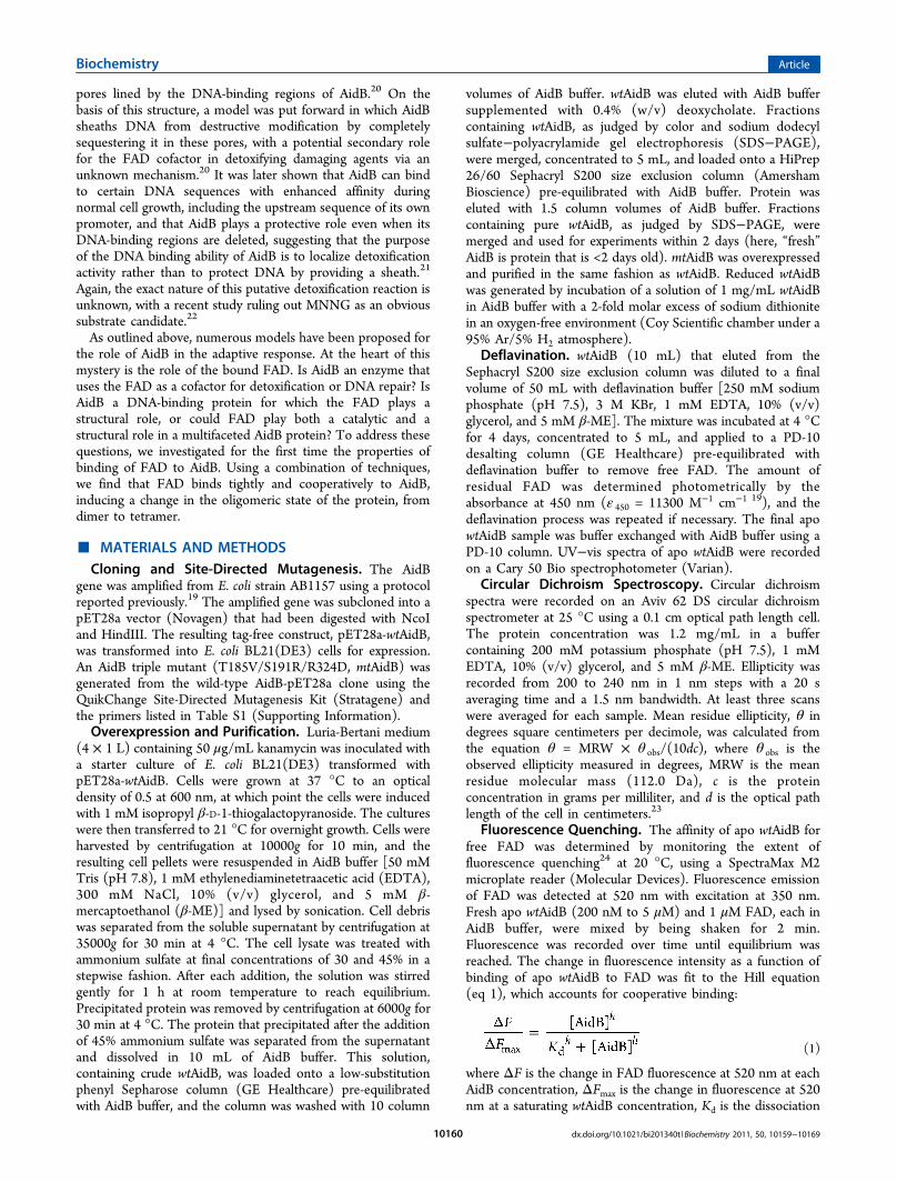
wtAidB Binds FAD Tightly and Cooperatively. Toinvestigate the interaction of FAD with wtAidB, we measuredthe dissociation constant (Kd) by fluorescence quenching(Figure 1). FAD has a fluorescence emission band centered at
520 nm when it is excited at 350 nm. When FAD binds towtAidB, the fluorescence emission of FAD at 520 nm isquenched by a factor of 3.1. Binding data were fit to a Hillequation (eq 1) to account for cooperative binding. Becausecooperativity implies multiple and unequal binding equilibriumstates, the resulting Kd is a rough estimation of the averagedissociation constant. We calculate an apparent Kd of 402.1 ±35.1 nM with a Hill coefficient (h) that is greater than 1 (1.73± 0.21), consistent with tight and cooperative binding of FADto wtAidB.The Oligomeric State of AidB Is Flavin-Dependent.We
performed sedimentation velocity analytical ultracentrifugation
(AUC) experiments to determine the oligomeric state ofAidB under different conditions. Sedimentation velocity datawere fit to a continuous distribution c(s) Lamm equation modelusing Sedfit,27 yielding the experimental sedimentationcoefficient distribution. All sedimentation coefficients, whichare dependent on the mass and shape of the protein, werenormalized to 20 °C in water (s20,w). We also used Hydropro28 todetermine theoretical s20,w values for different oligomers of AidBbased on our crystal structure (see below). The calculated s20,wvalue for the putative AidB tetramer was 11.5 S. In contrast, thes20,w values for possible dimer combinations ranged between 6.4and 7.0 S, and the s20,w value for an AidB monomer was 4.2 S.These values were compared to experimentally determined s20,wvalues, allowing us to assign oligomeric states to the differentAidB samples.First, we collected AUC data on holo wtAidB, apo wtAidB,
and apo mtAidB to assess the influence of the FAD cofactor onthe oligomeric state. For holo wtAidB, we obtained anexperimental s20,w value of 11.5 S (Figure 2A), indicative of atetramer structure and in agreement with previously reportedresults.19,20 Trace amounts of smaller species (asterisks in Figure2A) likely represent a small amount of misfolded, denatured, ormonomeric protein. In contrast, the major species of apo wtAidBexhibited an s20,w value of 7.0 S, indicating that apo wtAidB is adimer (Figure 2B). Small amounts of a tetrameric species areobserved in the sample, but the ratio of dimer to tetramer did notchange with a 2.7-fold increase in protein concentration. For apomtAidB, we obtained an s20,w value of 7.1 S for the dominantspecies (Figure 2C), in close agreement with the value obtainedfor apo wtAidB. Thus, both deflavinated AidB species are dimericin solution, indicating that AidB undergoes a change in theoligomerization state, from a tetramer to a dimer, upon removalof the FAD cofactor.Using a stepwise reconstitution protocol of apo wtAidB with
free FAD, we then tested whether the observed changes inoligomerization state are reversible. FAD was added to apowtAidB at molar ratios of 0.25, 0.5, and 1.0 with respect to theconcentration of the AidB monomer. The samples were thensubjected to AUC with simultaneous monitoring of theabsorbance at both 280 and 385 nm (Figure 2D−F).Sedimentation coefficients of the various forms of AidB werecalculated from the absorbance at 280 nm (Figure 2D-F, toptraces), while the absorbance at 385 nm was used to identifyFAD-containing AidB (Figure 2D-F, bottom traces). Uponreconstitution of apo wtAidB with 0.25 equiv of FAD, weobserved a species with an s20,w value of 11.5 S in the AUCexperiment, accounting for 47% of the total AidB (Figure 2D).A second species exhibited an s20,w value of 7.0 S, accounting forthe remaining AidB. The absorbance at 385 nm was observedonly in the species with an s20,w value of 11.5 S. Thus, 47% ofthe AidB in solution had formed a tetramer, and all FAD wasbound to this tetrameric species; no FAD was bound to adimeric species that was also present in solution. When weincreased the amount of FAD to 0.5 equiv relative to theconcentration of AidB monomer, 77% of AidB was present in atetrameric state. Again, FAD was only observed in thosetetramers (Figure 2E). Finally, reconstitution using 1 equiv ofFAD relative to AidB monomer resulted in 95% tetramerformation, a value similar to that observed for holo wtAidB(Figure 2F). All in all, these results suggest the presence of anFAD-dependent equilibrium between the dimeric and tetra-meric states of AidB in which binding of FAD induces atransition to the tetramer.
Figure 1. Quenching of FAD fluorescence upon binding to apowtAidB. Plotted is the ratio between the change in FAD fluorescenceat each wtAidB concentration (ΔF) and the change in fluorescence atsaturating wtAidB concentrations (ΔFmax) vs the concentration ofwtAidB. The data were fit to the Hill equation (eq 1). Data points anderror bars represent the average of three experiments.
Biochemistry Article
dx.doi.org/10.1021/bi201340t |Biochemistry 2011, 50, 10159−1016910162
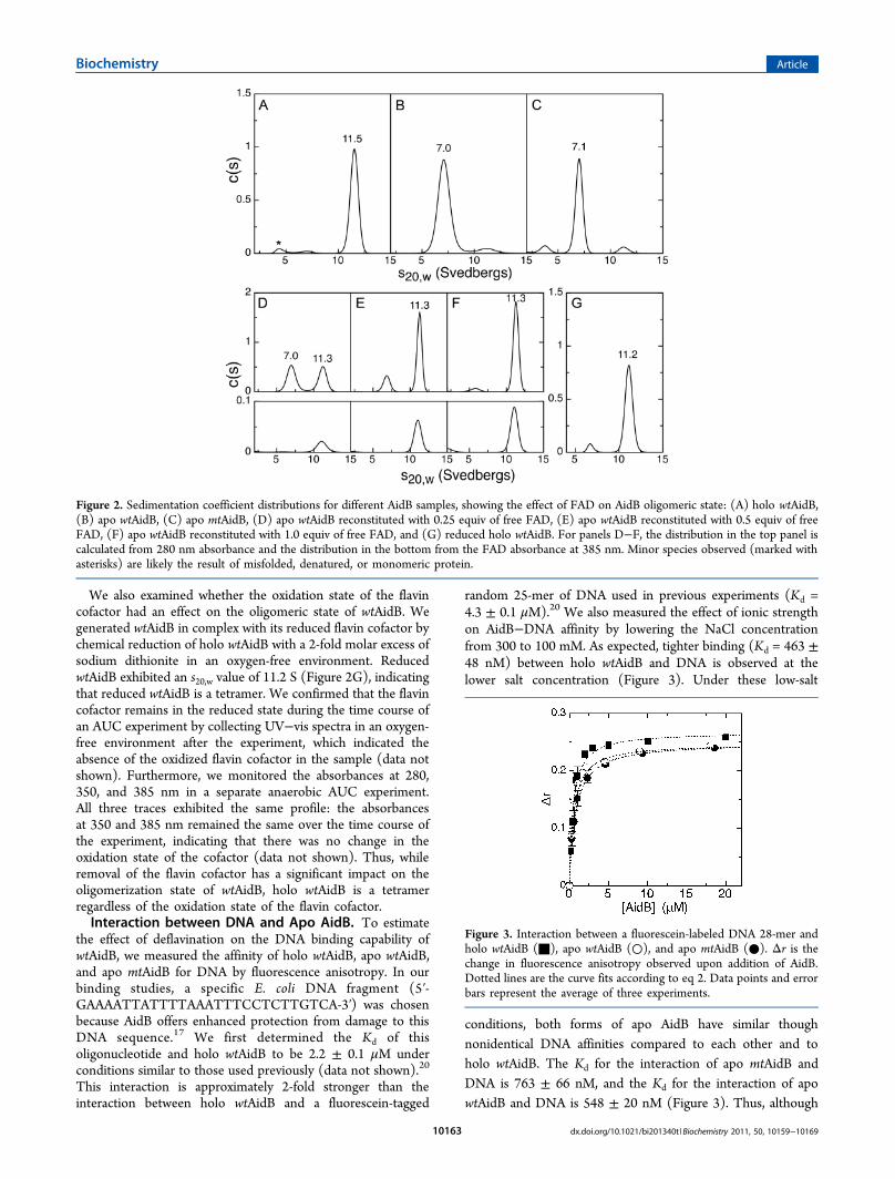
We also examined whether the oxidation state of the flavincofactor had an effect on the oligomeric state of wtAidB. Wegenerated wtAidB in complex with its reduced flavin cofactor bychemical reduction of holo wtAidB with a 2-fold molar excess ofsodium dithionite in an oxygen-free environment. ReducedwtAidB exhibited an s20,w value of 11.2 S (Figure 2G), indicatingthat reduced wtAidB is a tetramer. We confirmed that the flavincofactor remains in the reduced state during the time course ofan AUC experiment by collecting UV−vis spectra in an oxygen-free environment after the experiment, which indicated theabsence of the oxidized flavin cofactor in the sample (data notshown). Furthermore, we monitored the absorbances at 280,350, and 385 nm in a separate anaerobic AUC experiment.All three traces exhibited the same profile: the absorbancesat 350 and 385 nm remained the same over the time course ofthe experiment, indicating that there was no change in theoxidation state of the cofactor (data not shown). Thus, whileremoval of the flavin cofactor has a significant impact on theoligomerization state of wtAidB, holo wtAidB is a tetramerregardless of the oxidation state of the flavin cofactor.Interaction between DNA and Apo AidB. To estimate
the effect of deflavination on the DNA binding capability ofwtAidB, we measured the affinity of holo wtAidB, apo wtAidB,and apo mtAidB for DNA by fluorescence anisotropy. In ourbinding studies, a specific E. coli DNA fragment (5′-GAAAATTATTTTAAATTTCCTCTTGTCA-3′) was chosenbecause AidB offers enhanced protection from damage to thisDNA sequence.17 We first determined the Kd of thisoligonucleotide and holo wtAidB to be 2.2 ± 0.1 μM underconditions similar to those used previously (data not shown).20
This interaction is approximately 2-fold stronger than theinteraction between holo wtAidB and a fluorescein-tagged
random 25-mer of DNA used in previous experiments (Kd =4.3 ± 0.1 μM).20 We also measured the effect of ionic strengthon AidB−DNA affinity by lowering the NaCl concentrationfrom 300 to 100 mM. As expected, tighter binding (Kd = 463 ±48 nM) between holo wtAidB and DNA is observed at thelower salt concentration (Figure 3). Under these low-salt
conditions, both forms of apo AidB have similar thoughnonidentical DNA affinities compared to each other and toholo wtAidB. The Kd for the interaction of apo mtAidB andDNA is 763 ± 66 nM, and the Kd for the interaction of apowtAidB and DNA is 548 ± 20 nM (Figure 3). Thus, although
Figure 2. Sedimentation coefficient distributions for different AidB samples, showing the effect of FAD on AidB oligomeric state: (A) holo wtAidB,(B) apo wtAidB, (C) apo mtAidB, (D) apo wtAidB reconstituted with 0.25 equiv of free FAD, (E) apo wtAidB reconstituted with 0.5 equiv of freeFAD, (F) apo wtAidB reconstituted with 1.0 equiv of free FAD, and (G) reduced holo wtAidB. For panels D−F, the distribution in the top panel iscalculated from 280 nm absorbance and the distribution in the bottom from the FAD absorbance at 385 nm. Minor species observed (marked withasterisks) are likely the result of misfolded, denatured, or monomeric protein.
Figure 3. Interaction between a fluorescein-labeled DNA 28-mer andholo wtAidB (■), apo wtAidB (○), and apo mtAidB (●). Δr is thechange in fluorescence anisotropy observed upon addition of AidB.Dotted lines are the curve fits according to eq 2. Data points and errorbars represent the average of three experiments.
Biochemistry Article
dx.doi.org/10.1021/bi201340t |Biochemistry 2011, 50, 10159−1016910163

there is a significant change to the oligomerization state upondeflavination of AidB, we find the effect on DNA binding to benegligible.
P32 Crystal Structure of the AidB Tetramer. Toinvestigate the molecular basis of the FAD-dependent changesin the AidB oligomerization state, we determined the crystalstructure of holo wtAidB in space group P32 to 2.8 Å resolution(Figure 4B−D). Unlike the previous I222 structure,20 thiscrystal form has three tetramers of AidB in the asymmetric unitas opposed to a single monomer. The P32 structure wasdetermined by molecular replacement using the I222 crystalstructure.20 Protomers from the two crystal structures super-impose with a root-mean-square deviation (rmsd) of 0.34 Åover 538 Cα atoms, indicating that the structures of wtAidBprotomers are unchanged. Also, the wtAidB tetramer from theP32 structure and the AidB tetramer generated by symmetryfrom the I222 structure superpose well, with an rmsd of 0.40 Åover 2152 Cα atoms. However, there are profound differencesin the crystal packing arrangement of these tetramers (Figure4A,B). The previous structure contained a crystal lattice inwhich four AidB tetramers form another higher-order oligomer,with the AidB DNA-binding regions lining a central pore with a25 Å diameter (Figure 4A). In the P32 crystal structure, thethree tetramers in the asymmetric unit are arranged in atriangular shape with the DNA-binding domains pointing
outward and no obvious pore (Figure 4B). Even whenconsidering symmetry-related molecules in the P32 crystallattice, the resulting arrangement does not reveal a central porewith which AidB could sequester DNA (Figure S3 of theSupporting Information). Thus, although the tetramer of AidBis conserved in both crystal forms, the assembly of thosetetramers into higher-order oligomers is not.Structural Basis for Flavin-Dependent Oligomeriza-
tion. In the P32 crystal structure, each protomer of an AidBtetramer interacts with each of the other protomers. Thus, thereare three different types of dimers that could be formed upondeflavination: “up and down” (AB or CD dimers), “side byside” (AC or BD dimers), and “diagonal” (AD or BC dimers)(Figure 5). To investigate which of the three possible dimers ofAidB is present after deflavination, we calculated theoretical s20,wvalues for each of the dimers from our crystal structure andcompared these values to the results from our AUC experiments.We obtained theoretical s20,w values of 7.0, 6.6, and 6.4 S for theAB dimer, the AC dimer, and the AD dimer, respectively. Thesevalues reflect their differences in shape, as the AB dimer would bemore globular while the AC and the AD dimers would be moreelongated (Figure 5). Our observed s20,w values for apo wtAidBand apo mtAidB are 7.1 and 7.0 S, respectively, consistent withthe value calculated for the AB dimer of 7.0 S.
Figure 4. Crystal structures of holo wtAidB. (A) Higher-order oligomer of AidB observed in the I222 crystal structure,20 with the central AidBtetramer colored by protomer as described below. (B) Three AidB tetramers in the asymmetric unit of the P32 crystal structure, with the bottomAidB tetramer colored by protomer. (C) Tetramer of holo wtAidB with protomers labeled A−D. (D) Tetramer of holo wtAidB, rotated by 45° withrespect to panel C. Ribbons are transparent to emphasize the bound FAD molecules and the L1′2′ loops of each protomer (thicker ribbons, markedby arrows). The general coloring scheme was as follows: protomers in a tetramer colored yellow (A), orange (B), cyan (C), and blue (D). Additionaltetramers are colored pink. The putative DNA-binding regions of AidB are highlighted in purple and magenta. Bound FAD molecules are shown inball-and-stick representation with carbon atoms colored green.
Biochemistry Article
dx.doi.org/10.1021/bi201340t |Biochemistry 2011, 50, 10159−1016910164

In the P32 crystal structure, the AB dimer is held together byan extensive interface, burying a combined area of more than6500 Å2 from solvent (Figure 5A). The dimer interface features33 hydrogen bonds and 24 salt bridges. In contrast, the AC andAD dimer interfaces have solvent-buried areas of only 1782 and1319 Å2, respectively (Figure 5B,C), providing an additionalrationale for the presence of an AB dimer in solution. The FADof each protomer is bound at the AB dimer interface, with bothprotomers contributing to the binding of FAD (Figure 6A).FAD is bound by 12 hydrogen bonds from one protomer, whilethe second protomer adds another two hydrogen bonds andtwo salt bridges to binding of the pyrophosphate moiety andthe adenine ring (Figure 6A). In contrast, FAD makes no directcontacts across the AC or AD interfaces.The AidB tetramer, as observed in both crystal structures, is a
dimer of dimers with three 2-fold symmetry axes (Figure4C,D). The combined buried surface area between the AB andthe CD dimers is 5700 Å2, and we refer to this interface as the
“tetramer interface”. With 37 hydrogen bonds and 10 saltbridges at this interface, the size of the buried surface area(7.3% of the total surface area) and the number of specificstabilizing interactions are small compared to the overall size ofthe putative assembly. When the tetramer interface is treated asa single interface between two polypeptide chains, thecomplexation significance score determined by the PISA serverfor interface analysis39 is extremely low, 0.086 on a scale from 0(lowest significance) to 1 (highest significance). Furthermore,the free enthalpy of formation is calculated to be 12.3 kcal/molfor this interface, rendering interface formation energeticallyunfavorable. These analyses would suggest that the interface isnot significant for complex formation and is instead an artifactof crystal packing. Nonetheless, our AUC data provideconclusive evidence that holo wtAidB is indeed a tetramer insolution.As described previously,20 tetramer formation is mainly
mediated by the N-termini and the L1′2′ loops (residues 69−80)
Figure 5. Dimer interfaces within holo wtAidB: (A) AB dimer, (B) AC dimer, and (C) AD dimer of wtAidB shown as ribbons. Protomers A−D ofAidB are colored yellow, orange, blue, and cyan, respectively, with the DNA-binding regions colored magenta and purple. Bound FAD molecules areshown in ball-and-stick representation with carbon atoms colored green. Calculated s20,w values are given for each dimer.
Biochemistry Article
dx.doi.org/10.1021/bi201340t |Biochemistry 2011, 50, 10159−1016910165
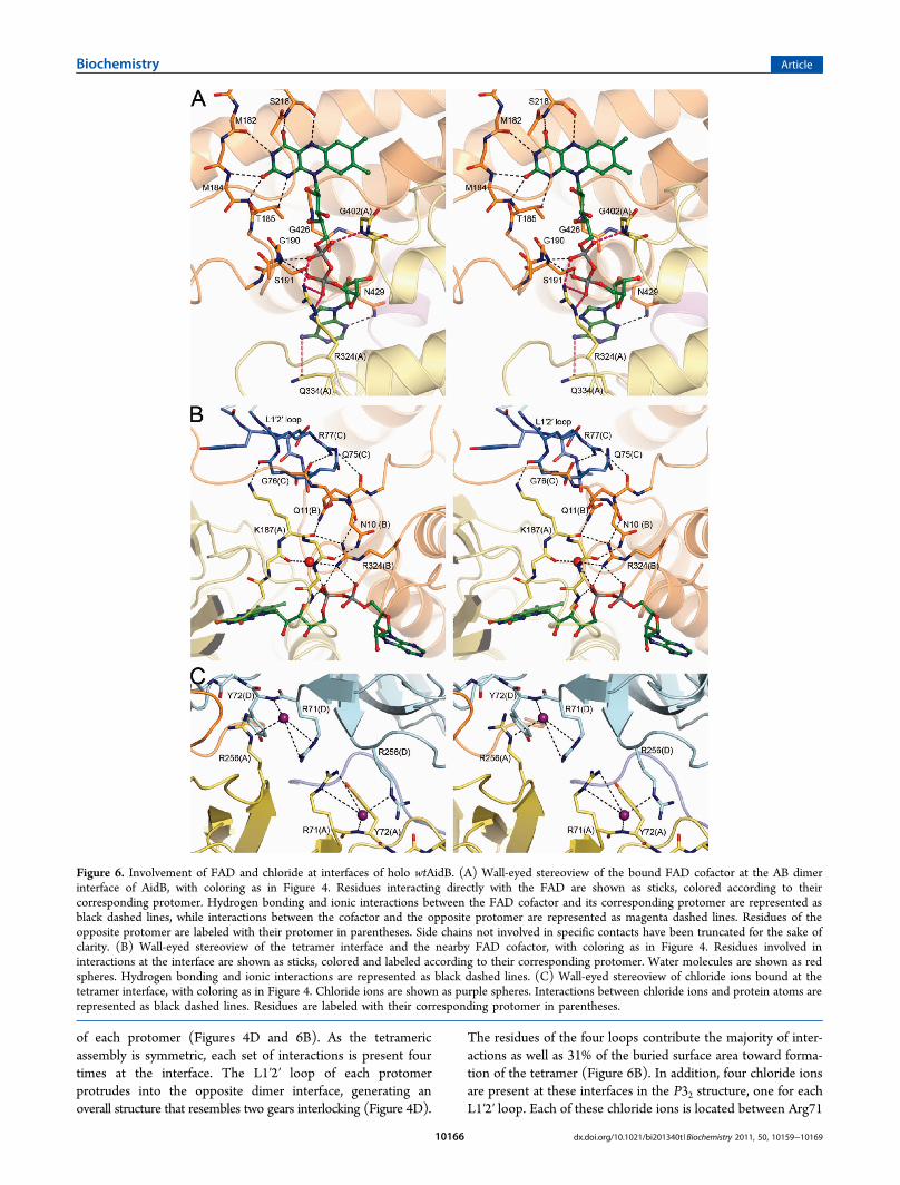
of each protomer (Figures 4D and 6B). As the tetramericassembly is symmetric, each set of interactions is present fourtimes at the interface. The L1′2′ loop of each protomerprotrudes into the opposite dimer interface, generating anoverall structure that resembles two gears interlocking (Figure 4D).
The residues of the four loops contribute the majority of inter-actions as well as 31% of the buried surface area toward forma-tion of the tetramer (Figure 6B). In addition, four chloride ionsare present at these interfaces in the P32 structure, one for eachL1′2′ loop. Each of these chloride ions is located between Arg71
Figure 6. Involvement of FAD and chloride at interfaces of holo wtAidB. (A) Wall-eyed stereoview of the bound FAD cofactor at the AB dimerinterface of AidB, with coloring as in Figure 4. Residues interacting directly with the FAD are shown as sticks, colored according to theircorresponding protomer. Hydrogen bonding and ionic interactions between the FAD cofactor and its corresponding protomer are represented asblack dashed lines, while interactions between the cofactor and the opposite protomer are represented as magenta dashed lines. Residues of theopposite protomer are labeled with their protomer in parentheses. Side chains not involved in specific contacts have been truncated for the sake ofclarity. (B) Wall-eyed stereoview of the tetramer interface and the nearby FAD cofactor, with coloring as in Figure 4. Residues involved ininteractions at the interface are shown as sticks, colored and labeled according to their corresponding protomer. Water molecules are shown as redspheres. Hydrogen bonding and ionic interactions are represented as black dashed lines. (C) Wall-eyed stereoview of chloride ions bound at thetetramer interface, with coloring as in Figure 4. Chloride ions are shown as purple spheres. Interactions between chloride ions and protein atoms arerepresented as black dashed lines. Residues are labeled with their corresponding protomer in parentheses.
Biochemistry Article
dx.doi.org/10.1021/bi201340t |Biochemistry 2011, 50, 10159−1016910166
of one chain and Arg256 of the other, thereby stabilizing thesetwo adjacent positive charges (Figure 6C). The L1′2′ loops haveno direct interactions with the FAD cofactors. However, Ala74,Gln75, Gly76, and Arg77 are positioned within 13 Å of theFAD cofactor, and a network of hydrogen bonds connects theseresidues to the pyrophosphate moiety and the isoalloxazine ringsystem of the cofactor (Figure 6B).
■ DISCUSSIONThe function of AidB in the adaptive response has been asubject of debate.16,17,19−21 With no catalytic activity reportedoutside of the residual IVD activity, it remains unclear if therole of AidB in the adaptive response involves the use of thebound flavin in catalysis. The flavoprotein literature reportsseveral cases in which flavins play structural roles and are notdirectly involved in catalysis.40 Glyoxylate carboligases, forexample, do not appear to use their flavins to catalyze redoxchemistry but instead display flavin-dependent transitions inthe oligomerization state.41,42 For both Pseudomonas oxalaticusand E. coli glyoxylate carboligases, deflavination leads tooligomer disassembly and subsequent inactivation.41,42 Withthis precedent in the literature, here we investigate thepossibility that FAD serves a structural role in AidB.With the first crystal structure of AidB showing FAD bound
at the AB and the CD dimer interfaces,20 it was tempting topropose that FAD might play a structural role in thedimerization. To test this idea, deflavinated AidB (apo AidB)was generated to measure the FAD dissocation constant and toinvestigate whether AidB is monomeric or even unfolded withoutflavin. Using CD spectroscopy, we found that removal of flavindoes not unfold the protein (Figure S2B of the SupportingInformation), and by fluorescence quenching, we measured anapparent Kd for FAD of 402.1 ± 35.1 nM and a Hill coefficient(h) of 1.73 ± 0.21, consistent with each FAD making extensiveinteractions with two AidB protomers. With the knowledge thatFAD binds tightly and cooperatively to AidB, along with crystalstructures showing FAD engaged in numerous interactions at theAB dimer interface, AUC experiments were conducted todetermine if AidB is monomeric in the absence of flavin.Surprisingly, we found that both apo wtAidB and apo mtAidBexhibit a dimeric state instead of the expected monomeric state,while holo wtAidB is a tetramer.Because the AB interface is most directly affected by FAD
binding (Figures 5 and 6A), we considered whether the dimericstructure of apo AidB could be represented by AC or ADdimers, such that FAD binding would yield tetramers bybringing the A protomer of AC together with the B protomer ofBD to yield ABCD tetramers. To determine which protomerscreate the apo AidB dimers, we calculated s20,w values from thecrystal structure and compared them to the experimental s20,wvalues for apo wtAidB and apo mtAidB. The excellent agreementbetween the calculated value for the AB dimer (s20,w of 7.0 S) withexperiment (s20,w of 7.0 and 7.1 S) provides strong evidence thatdeflavinated AidB has the form of an AB dimer. Thus, even thoughthe flavin cofactor appears to contribute stabilizing interactions tothe AB dimer, our data indicate that the AB dimer interface ispresent even in the absence of bound flavin cofactor. In contrast,the AC and AD interfaces are disintegrated upon deflavinationdespite not being directly involved in flavin binding. Although anunexpected result if one considers only direct FAD−proteininteractions, this result makes sense from the perspective of totalburied surface area. When a contact area is small, as is the case forthe AC and AD interfaces (1782 and 1319 Å2, respectively), even a
few changes in hydrogen bonds or packing interactions can make adramatic difference in interface stability, whereas when the interfaceis extensive, such as the case with the AB dimer (6500 Å2),hydrogen bonds and packing interactions can be lost withouthaving an impact on stability.With a rationale at hand for why AidB is an AB dimer in the
absence of FAD, we then considered how FAD bindinggenerates AidB tetramers when the cofactor does not appear tobe directly involved in creating the tetramer interface. To morecarefully evaluate the tetramer interface, it was important toobtain a crystal structure in which more than one protomer waspresent in the asymmetric unit. In the P32 structure presentedhere, the asymmetric unit contains three copies of the tetramerinterface, none of which are restrained by crystallographicsymmetry. With independent tetramers to analyze, we find thatbinding of flavin to dimers of AidB could order residues thatcreate an intricate network of hydrogen bonds, thereby exertinga long-range stabilization on the tetramer interface (Figure 6B).Importantly, the L1′2′ loops (residues 69−80) that protrudeacross the tetramer interface (Figure 4D) and make themajority of contacts at the interface are involved in thishydrogen bonding network. With so little buried surface area atthe tetramer interface, the loss or disruption of theseinteractions due to the absence of FAD must be enough toshift the balance from tetramer to dimer, while the AB interfaceis strong enough to exist without FAD. Formation of thetetramer interface could also be dependent on ionic strength, asfour chloride molecules are present at the interface, againcontacting the L1′2′ loop (Figure 6C). AidB remains a tetramerat NaCl concentrations as low as 100 mM (data not shown),but the instability of the protein precluded experiments at lowersalt concentrations.Notably, while AidB shares sequence homology and
structural similarity with members of the ACAD family suchas IVD and medium chain acyl-CoA dehydrogenase(MCAD),16,43,44 the AidB L1′2′ loop and the DNA-bindingregion are not conserved in any other members of the ACADfamily. Presumably, as a result of these differences, thestructures of IVD and MCAD reveal a completely differenttetrameric arrangement compared to that observed for AidB(Figure S4 of the Supporting Information). The DNA-bindingdomain prevents AidB from forming the IVD-type tetramer,while the lack of the L1′2′ loop in IVD would restrict theformation of an AidB-like tetramer in that protein. Thus, AidBcould have diverged from the ACAD family by the addition ofthis DNA-binding domain, allowing it to perform its function inthe adaptive response, while the addition of the L1′2′ β-hairpinloop would still allow for tetramer formation. With so fewalterations in the secondary structure between the ACADfamily and AidB, the adaptation of the L1′2′ loop suggests thattetramerization is important to the function of AidB.We also found that the transition from a tetrameric state to a
dimeric state is fully reversible and does not depend on theflavin oxidation state, as reconstitution of apo wtAidB byoxidized FAD restores the tetrameric state and reduced holowtAidB is a tetramer. Notably, incorporation of free FAD intoapo wtAidB is a highly cooperative process as indicated by bothAUC and fluorescence quenching studies. During stepwisereconstitution experiments, regardless of the amount of FADused, all FAD-containing AidB molecules are tetramers.Although we cannot predict the exact mechanism oftetramerization, all of these results support a model in whichbinding of substoichiometric amounts of FAD promotes
Biochemistry Article
dx.doi.org/10.1021/bi201340t |Biochemistry 2011, 50, 10159−1016910167

tetramer formation for wtAidB. Thus, while the individualstructures of AidB protomers are likely to be retained duringdeflavination, we observe a reversible and flavin-dependentchange in the oligomeric state, indicating that flavin, regardlessof oxidation state, does play a structural role in the creation ofthe AidB tetramer.Despite the switch in oligomeric state upon deflavination, the
in vitro DNA binding capability of apo AidB was notsignificantly affected, with a <2-fold effect compared to thatof holo AidB. These results also suggest that tetramerization isnot essential for the interaction of AidB with double-strandedDNA. In agreement with these findings, it was recentlyreported that a truncated version of AidB that contains only theDNA-binding domain, and thus is unlikely to be tetrameric,retains the capability of binding DNA in vitro.21 In vivo,however, tetramerization could allow AidB to bind DNA atmultiple locations simultaneously.Interestingly, the higher-order AidB oligomer observed in the
I222 crystal structure and proposed to sequester and therebyprotect DNA20 is not present in the P32 crystal form, nor do weobserve any higher-order AidB oligomers in our solutionstudies. The distinct architecture of the crystal lattice could bedue to the absence of DNA under our crystallizationconditions; however, no electron density for DNA wasobserved in the previously reported structure.20 More likely,AidB can crystallize in multiple lattice systems, as is frequentlyobserved for other proteins (for examples, see refs 45 or 46 and47), with these two AidB crystal structures representing two ofthe possible lattice systems. While an AidB “mega complex”exists at least in the I222 crystal lattice, a role for this complexin sequestering DNA is not supported by our studies. Withrespect to the idea of protection by DNA sequestration ingeneral, recent data from the Volkert laboratory oppose thismodel.17 Their data show that deletion of AidB’s DNA-bindingdomain does not result in the loss of AidB’s protective function;the protective ability is independent of DNA binding.17
Protection in the absence of DNA binding is also inconsistentwith AidB serving as a DNA repair protein. While our studiesclearly show FAD-dependent oligomerization of AidB, they donot address whether FAD also has a catalytic function.However, the picture of AidB that is emerging invokes a rolefor the DNA-binding domain in localization of AidB to specificgenes, while the protective function appears to reside elsewhereon the protein.17,21 Whether this protective function resideswith FAD or whether FAD was retained in the evolutionaryprocess solely for its ability to stabilize the AidB tetramerremains to be determined.
■ ASSOCIATED CONTENT
*S Supporting InformationPrimers for site-directed mutagenesis, crystallographic datacollection and refinement statistics, UV−vis and CD spectra ofdifferent AidB forms, and additional figures of the crystalstructure. This material is available free of charge via theInternet at http://pubs.acs.org.
Accession CodesThe atomic coordinates have been deposited with the ProteinData Bank as entry 3U33.
■ AUTHOR INFORMATION
Corresponding Author*S.J.E.: telephone, (617) 358-2816; fax, (617) 353-6466;e-mail, [email protected]. C.L.D.: telephone, (617) 253-5622;fax, (617) 258-7847; e-mail, [email protected].
Present Address@Johnson & Johnson Pharmaceutical Research & Develop-ment, 930 Route 202 South, Raritan, NJ 08869.
Author ContributionsM.J.H. and M.J. contributed equally to this work.
FundingThis work was supported by National Institutes of HealthGrants R01-GM072663 (to S.J.E.) and P30-ES002109 (toC.L.D.) and National Science Foundation Grant MCB-0543833 (to C.L.D.). C.L.D. is a Howard Hughes MedicalInstitute Investigator.
■ ACKNOWLEDGMENTSWe thank Dr. Nozomi Ando for assistance with AUCexperiments and data analysis. We thank Dr. Michael Volkertfor providing information about the ability of AidB to bind toUP elements prior to publication. Use of the MassachusettsInstitute of Technology Biophysical Instrumentation Facilityfor the Study of Complex Macromolecular Systems (NationalScience Foundation Grant 0070319 and National Institutes ofHealth Grant GM68762) is gratefully acknowledged. Use of theAdvanced Photon Source, an Office of Science User Facilityoperated for the U.S. Department of Energy (DOE) Office ofScience by Argonne National Laboratory, was supported by theU.S. DOE under Contract No. DE-AC02-06CH11357.
■ ABBREVIATIONSMNNG, N-methyl-N′-nitro-N-nitrosoguanidine; FAD, flavinadenine dinucleotide; ACAD, acyl-coenzyme A dehydrogenase;IVD, isovaleryl-coenzyme A dehydrogenase; Kd, dissociationconstant; β-ME, β-mercaptoethanol; EDTA, ethylenediamine-tetraacetic acid; h, Hill coefficient; NCS, noncrystallographicsymmetry; CD, circular dichroism; AUC, analytical ultra-centrifugation; s20,w, sedimentation coefficient normalized to20 °C and water; rmsd, root-mean-square deviation; MCAD,medium chain acyl-coenzyme A dehydrogenase.
■ REFERENCES(1) Sedgwick, B. (2004) Repairing DNA-methylation damage. Nat.
Rev. Mol. Cell Biol. 5, 148−157.(2) Volkert, M. R., and Nguyen, D. C. (1984) Induction of specific
Escherichia coli genes by sublethal treatments with alkylating agents.Proc. Natl. Acad. Sci. U.S.A. 81, 4110−4114.(3) Volkert, M. R. (1988) Adaptive response of Escherichia coli to
alkylation damage. Environ. Mol. Mutagen. 11, 241−255.(4) Samson, L., and Cairns, J. (1977) A new pathway for DNA repair
in Escherichia coli. Nature 267, 281−283.(5) Sedgwick, B., Robins, P., Totty, N., and Lindahl, T. (1988)
Functional domains and methyl acceptor sites of the Escherichia coliAda protein. J. Biol. Chem. 263, 4430−4433.(6) Demple, B., Sedgwick, B., Robins, P., Totty, N., Waterfield, M.
D., and Lindahl, T. (1985) Active site and complete sequence of thesuicidal methyltransferase that counters alkylation mutagenesis. Proc.Natl. Acad. Sci. U.S.A. 82, 2688−2692.(7) Landini, P., and Volkert, M. R. (2000) Regulatory responses of
the adaptive response to alkylation damage: A simple regulon withcomplex regulatory features. J. Bacteriol. 182, 6543−6549.
Biochemistry Article
dx.doi.org/10.1021/bi201340t |Biochemistry 2011, 50, 10159−1016910168
(8) Sedgwick, B., and Lindahl, T. (2002) Recent progress on the Adaresponse for inducible repair of DNA alkylation damage. Oncogene 21,8886−8894.(9) Evensen, G., and Seeberg, E. (1982) Adaptation to alkylation
resistance involves the induction of a DNA glycosylase. Nature 296,773−775.(10) Karran, P., Hjelmgren, T., and Lindahl, T. (1982) Induction of a
DNA glycosylase for N-methylated purines is part of the adaptiveresponse to alkylating agents. Nature 296, 770−773.(11) Trewick, S. C., Henshaw, T. F., Hausinger, R. P., Lindahl, T.,
and Sedgwick, B. (2002) Oxidative demethylation by Escherichia coliAlkB directly reverts DNA base damage. Nature 419, 174−178.(12) Falnes, P. O., Johansen, R. F., and Seeberg, E. (2002) AlkB-
mediated oxidative demethylation reverses DNA damage in Escherichiacoli. Nature 419, 178−182.(13) Falnes, P. O. (2004) Repair of 3-methylthymine and 1-
methylguanine lesions by bacterial and human AlkB proteins. NucleicAcids Res. 32, 6260.(14) Delaney, J. C., Smeester, L., Wong, C., Frick, L. E., Taghizadeh,
K., Wishnok, J. S., Drennan, C. L., Samson, L. D., and Essigmann, J. M.(2005) AlkB reverses etheno DNA lesions caused by lipid oxidation invitro and in vivo. Nat. Struct. Mol. Biol. 12, 855−860.(15) Frick, L. E., Delaney, J. C., Wong, C., Drennan, C. L., and
Essigmann, J. M. (2007) Alleviation of 1,N 6-ethanoadenine genotox-icity by the Escherichia coli adaptive response protein AlkB. Proc. Natl.Acad. Sci. U.S.A. 104, 755−760.(16) Landini, P., Hajec, L. I., and Volkert, M. R. (1994) Structure
and transcriptional regulation of the Escherichia coli adaptive responsegene aidB. J. Bacteriol. 176, 6583−6589.(17) Rippa, V., Duilio, A., di Pasquale, P., Amoresano, A., Landini, P.,
and Volkert, M. R. (2011) Preferential DNA damage prevention bythe E. coli AidB gene: A new mechanism for the protection of specificgenes. DNA Repair 10, 934−941.(18) Volkert, M. R., Nguyen, D. C., and Beard, K. C. (1986)
Escherichia coli gene induction by alkylation treatment. Genetics 112,11−26.(19) Rohankhedkar, M. S., Mulrooney, S. B., Wedemeyer, W. J., and
Hausinger, R. P. (2006) The AidB component of the Escherichia coliadaptive response to alkylating agents is a flavin-containing, DNA-binding protein. J. Bacteriol. 188, 223−230.(20) Bowles, T., Metz, A. H., O’Quin, J., Wawrzak, Z., and Eichman,
B. F. (2008) Structure and DNA binding of alkylation responseprotein AidB. Proc. Natl. Acad. Sci. U.S.A. 105, 15299−15304.(21) Rippa, V., Amoresano, A., Esposito, C., Landini, P., Volkert, M.,
and Duilio, A. (2010) Specific DNA binding and regulation of its ownexpression by the AidB protein in Escherichia coli. J. Bacteriol. 192,6136−6142.(22) Mulrooney, S. B., Howard, M. J., and Hausinger, R. P. (2011)
The Escherichia coli alkylation response protein AidB is a redox partnerof flavodoxin and binds RNA and acyl carrier protein. Arch. Biochem.Biophys. 513, 81−86.(23) Kelly, S. M., Jess, T. J., and Price, N. C. (2005) How to study
proteins by circular dichroism. Biochim. Biophys. Acta 1751, 119−139.(24) Curley, G. P., Carr, M. C., Mayhew, S. G., and Voordouw, G.
(1991) Redox and flavin-binding properties of recombinant flavodoxinfrom Desulfovibrio vulgaris (Hildenborough). Eur. J. Biochem. 202,1091−1100.(25) Laue, T. M., Shah, B. D., Ridgeway, T. M., and Pelletier, S. L.
(1992) Computer-aided interpretation of analytical sedimentation datafor proteins. In Analytical ultracentrifugation in biochemistry and proteinscience (Harding, S. E., Rowe, A. J., and Horton, J. C., Eds.) pp 90−125, Royal Society of Chemistry, Cambridge, U.K.(26) Garcia De La Torre, J., Huertas, M. L., and Carrasco, B. (2000)
Calculation of hydrodynamic properties of globular proteins from theiratomic-level structure. Biophys. J. 78, 719−730.(27) Lebowitz, J., Lewis, M. S., and Schuck, P. (2002) Modern
analytical ultracentrifugation in protein science: A tutorial review.Protein Sci. 11, 2067−2079.
(28) Philo, J. S. (2000) A method for directly fitting the timederivative of sedimentation velocity data and an alternative algorithmfor calculating sedimentation coefficient distribution functions. Anal.Biochem. 279, 151−163.(29) Otwinowski, Z., and Minor, W. (1997) Processing of X-ray
diffraction data collected in oscillation mode. In Methods inEnzymology (Carter, C. W., Jr., and Sweet, R. M., Eds.) pp 307−326, Academic Press, New York.(30) McCoy, A. J., Grosse-Kunstleve, R. W., Adams, P. D., Winn, M.
D., Storoni, L. C., and Read, R. J. (2007) Phaser crystallographicsoftware. J. Appl. Crystallogr. 40, 658−674.(31) Brunger, A. T., Adams, P. D., Clore, G. M., DeLano, W. L.,
Gros, P., Grosse-Kunstleve, R. W., Jiang, J. S., Kuszewski, J., Nilges, M.,Pannu, N. S., Read, R. J., Rice, L. M., Simonson, T., and Warren, G. L.(1998) Crystallography & NMR system: A new software suite formacromolecular structure determination. Acta Crystallogr. D54, 905−921.(32) Brunger, A. T. (2007) Version 1.2 of the Crystallography and
NMR system. Nat. Protoc. 2, 2728−2733.(33) Emsley, P., Lohkamp, B., Scott, W. G., and Cowtan, K. (2010)
Features and development of Coot. Acta Crystallogr. D66, 486−501.(34) Adams, P. D., Afonine, P. V., Bunkoczi, G., Chen, V. B., Davis, I.
W., Echols, N., Headd, J. J., Hung, L. W., Kapral, G. J., Grosse-Kunstleve, R. W., McCoy, A. J., Moriarty, N. W., Oeffner, R., Read, R.J., Richardson, D. C., Richardson, J. S., Terwilliger, T. C., and Zwart, P.H. (2010) PHENIX: A comprehensive Python-based system formacromolecular structure solution. Acta Crystallogr. D66, 213−221.(35) Painter, J., and Merritt, E. A. (2006) Optimal description of a
protein structure in terms of multiple groups undergoing TLS motion.Acta Crystallogr. D62, 439−450.(36) Chen, V. B., Arendall, W. B. III, Headd, J. J., Keedy, D. A.,
Immormino, R. M., Kapral, G. J., Murray, L. W., Richardson, J. S., andRichardson, D. C. (2010) MolProbity: All-atom structure validationfor macromolecular crystallography. Acta Crystallogr. D66, 12−21.(37) The PyMOL Molecular Graphics System, version 1.3r1 (2010)
Schrodinger, LLC, New York.(38) Hefti, M. H., Vervoort, J., and van Berkel, W. J. (2003)
Deflavination and reconstitution of flavoproteins. Eur. J. Biochem. 270,4227−4242.(39) Krissinel, E., and Henrick, K. (2007) Inference of macro-
molecular assemblies from crystalline state. J. Mol. Biol. 372, 774−797.(40) Bornemann, S. (2002) Flavoenzymes that catalyse reactions
with no net redox change. Nat. Prod. Rep. 19, 761−772.(41) Chung, S. T., Tan, R. T., and Suzuki, I. (1971) Glyoxylate
carboligase of Pseudomonas oxalaticus. A possible structural role forflavine-adenine dinucleotide. Biochemistry 10, 1205−1209.(42) Cromartie, T. H., and Walsh, C. T. (1976) Escherichia coli
glyoxalate carboligase. Properties and reconstitution with 5-deazaFADand 1,5-dihydrodeazaFADH2. J. Biol. Chem. 251, 329−333.(43) Tiffany, K. A., Roberts, D. L., Wang, M., Paschke, R., Mohsen,
A. W., Vockley, J., and Kim, J. J. P. (1997) Structure of humanisovaleryl-CoA dehydrogenase at 2.6 Å resolution: Basis for substratespecificity. Biochemistry 36, 8455−8464.(44) Kim, J. J., Wang, M., and Paschke, R. (1993) Crystal structures
of medium-chain acyl-CoA dehydrogenase from pig liver mitochondriawith and without substrate. Proc. Natl. Acad. Sci. U.S.A. 90, 7523−7527.(45) Higgins, L. J., Yan, F., Liu, P. H., Liu, H. W., and Drennan, C. L.
(2005) Structural insight into antibiotic fosfomycin biosynthesis by amononuclear iron enzyme. Nature 437, 838−844.(46) Nordlund, P., Sjoberg, B. M., and Eklund, H. (1990) 3-
Dimensional Structure of the Free-Radical Protein of RibonucleotideReductase. Nature 345, 593−598.(47) Sommerhalter, M., Saleh, L., Bollinger, J. M., and Rosenzweig,
A. C. (2005) Structure of Escherichia coli ribonucleotide reductase R2in space group P6122. Acta Crystallogr. D61, 1649−1654.
Biochemistry Article
dx.doi.org/10.1021/bi201340t |Biochemistry 2011, 50, 10159−1016910169


![by Joseph Bruchac illustrated by Teresa Flavin · illustrated by Teresa Flavin by Joseph Bruchac illustrated by Teresa Flavin Pushing Up)&(the Sky FjZhi^dc d[ i]Z LZZ` =dl Yd eZdeaZ](https://img.dokumen.tips/doc/110x75/5f8e7f4dad596368cb63a9b9/by-joseph-bruchac-illustrated-by-teresa-flavin-illustrated-by-teresa-flavin-by-joseph.jpg)