Embed Size (px)
Citation preview
Otorhinolaryngology Clinics: An International Journal, May-August 2013;5(2):63-76 63
Flap Selection in Head and Neck Cancer Reconstruction
AIJOC
REVIEW ARTICLE
Flap Selection in Head and Neck Cancer ReconstructionAmresh S Baliarsing, Tushar S Thorat, Arunesh Gupta, Uday Bhat, Sanchit Garg, Diptarka Bhattacharyya
10.5005/jp-journals-10003-1112
ABSTRACT
Advances in head and neck reconstruction techniques haveimproved the results in function and the aesthetic outcome.Several flaps with different composition are available for specificreconstruction to achieve optimum result. Sensate free tissuetransfer, dental rehabilitation and epiphyseal transfer for pediatricmandible are also now possible to achieve better function. Thespecific choice of the flap according to the region of defect andimportant keypoints in harvesting and reconstruction strategyfor head and neck cancer are based on our experience in thelast two decades.
Keywords: Local flaps, Pedicled flaps, Free flaps, Maxilla andmandible reconstruction, Pharynx reconstruction.
How to cite this article: Baliarsing AS, Thorat TS, Gupta A,Bhat U, Garg S, Bhattacharyya D. Flap Selection in Head andNeck Cancer Reconstruction. Int J Otorhinolaryngol Clin2013;5(2):63-76.
Source of support: Nil
Conflict of interest: None declared
INTRODUCTION
In India, oral malignancies account for 35% of totalmalignancies.1 Surgical excision of tumor and neckdissection forms the mainstay of treatment in addition toadjuvant chemotherapy and radiotherapy. The resultinganatomical defect, functional loss, cosmetic disfigurementand the accompaning psychosocial effects can bedevastating to the patient. Reconstructive surgery plays acrucial role in improving the quality of life by restoringanatomical defect, achieving functional rehabilitation andaesthetic outcome. In 1963, McGregor introducedtemporalis muscle flap for midface and lower face defectcoverage.2 In 1965, Bakamjian discribed the deltopectoralflap for coverage of the lower third of the face as well as ofthe oral and esophageal defects.3 In 1979, Ariyan describedpectoralis major myocutaneous flap for lower third of face.4
These regional restrictions were abolished by free radialforearm flap which was discribed by Yang in 1981.5
Thereafter, the use of microvascular tissue transfer hasrevolutionized the field of head and neck cancerreconstruction. Recent techniques like sensate free tissuetransfer, dental rehabilitation and epiphyseal transfer forpediatric mandible can help us to achieve the ultimate goalof ‘replacing like with like’.
MATERIALS, METHODS AND OBSERVATIONS
We retrospectively reviewed patients undergoing head andneck reconstruction for cancer and grouped them according
to the regions (1) scalp, (2) upper face, (3) midface, (4) lip,(5) oral cavity, (6) mandible, and (7) neck and pharynx andchoice of reconstruction (1) local flap, (2) pedicled flap,and (3) free flap.
Scalp
Vasculature of scalp ascends from periphery toward center(below upward) and it is essential to base the flapperipherally. Skin is raised in the loose areolar tissue planeabove the galea aponeurotica layer. Recommended choiceof reconstruction for scalp defects are as follows (Table 1).
Rotation Flap for Scalp Defect6
It is the most common flap done for small defects of thescalp. An isosceles triangle ABC is created. Pivot point Dis located on the projection of line AC. Line CD must be50% larger than AC. Midpoint of AD designed whichbecomes center of arc drawn from B to D (Fig. 1).
Upper Face-Eyelid
Eyelid reconstruction requires, both skin cover andconjunctival lining. Recommended choice of reconstructionfor eyelid defects are as follows (Table 2).
Forehead Flap for Total Eyelid Defect13
Supratrochlear artery based median forehead flap is raisedfor eyelid reconstruction. Distal one-third of flap is raisedin the subcutaneous plane. Frontalis muscle is included inthe middle one-third of the flap. Subperiosteal plane isachieved in proximal one-third of flap to include thesupratrochlear artery into the flap. Depilation is performedif flap is planned beyond the frontal hair line. Buccal mucosais used for prelamination of forehead flap to replaceconjunctiva. Flap inset is done over defect and division isdone after 3 weeks (Fig. 2).
Midface
Nose
Nasal reconstruction involves restoration of the tip, dorsum,columella, and paired alae, sidewalls, and soft trianglesubunits. Recommended choice of reconstruction for nosedefects are as follows (Table 3).
Forehead Flap for Large Defect of Nose18
Exact pattern of the defect is marked over median foreheadregion. For reconstruction for total loss of nose,

64
Amresh S Baliarsing et al
consideration is given for projection of the nose and flapdesign is made little larger. Short flap will result inforeshortened nose. Due consideration is given to providenasal mucosal lining. Flap based on supratrochlear artery israised and sutured over the defect. After 3 to 4 weeks, pedicleof the forehead flap is divided and contoured at the superior
Fig. 1: Rotation flap for scalp defect
Fig. 2: Forehead flap for lower eyelid reconstruction after excision of basal cell carcinoma
Fig. 3: Forehead flap for basal cell carcinoma over left nasal ala
aspect of the defect. The proximal pedicle is untubed andrepositioned back to the medial brow and sutured as inverted‘V’ (Fig. 3).
Cheek
There is excess laxity of skin over cheek, thus localadvancement flaps are preffered unless there is insufficientsurrounding skin. Recommended choice of reconstructionfor cheek defects are as follows (Table 4):
Rhomboid Flap for Superficial Cheek Defect21
Small superficial defects of cheek are managed withrhomboid flap. The defect is converted to rhomboid shapewith 60° and 120° angles. The flap is planned in an areawith loose skin to allow direct closure of the wound. Theshort diagonal BD is of the same length as each side and isextended by same length to point E. The line EF is drawnparallel to AD’ and is of the same length. After the flapmargins have been incised, the flap is transposed into therhomboid defect. Primary closure of the donor site is donealong the resting skin tension lines (Fig. 4).

Otorhinolaryngology Clinics: An International Journal, May-August 2013;5(2):63-76 65
Flap Selection in Head and Neck Cancer Reconstruction
AIJOC
Fig. 4: Rhomboid flap for basal cell carcinoma of cheek
Table 2: Choice of reconstruction for eyelid defects
Site Size of defect10 Choice of reconstruction10
Upper eyelid Partial thickness, <50% Primary closure, V-Y advancementPartial thickness, >50% Full thickness skin graftFull thickness, <25% Primary closure with canthotomy and
advancementFull thickness, <75% Hughes sliding tarsoconjunctival flap,11
cutler-beard advancement flap,12
Lower lid switch flap10
Full thickness, >75% Lower lid switch flap, forehead flapLower eyelid Partial thickness, <50% Primary closure, V-Y advancement
Partial thickness, >50% Full thickness skin graft, myocutaneous unipedicledFricke flap and the bipedicled Tripier flap13
Full thickness, <50% Primary closure with canthotomy andadvancement,Tessier’s flap14
Full thickness, >50% Hughes sliding tarsoconjunctival flap, Mustardecheek advancement flap,13 nasolabial flap, forehead flap13
Table 1: Choice of reconstruction for scalp defects
Site Size of defect Choice of reconstruction
Scalp <3 cm Primary closureUp to 50 cm² Rotation flap,6 transposition flap,
pinwheel flap, bipedicleadvancement flaps, doubleopposing rotation flaps,7Orticochea four flap8
>50 cm² Free anterolateral thigh flap orLatissimus dorsi free flap9
Submental Flap for Intraoral and Cheek Defect24
Submental artery, direct branch of the facial artery suppliesthe submental flap and is located 5 to 6.5 cm distal to theorigin of the facial artery. Inferior mandible border forms
the upper flap margin. Size of the flap varying from 4 × 5 to15 × 7 cm can be raised. Average flap extends from theipsilateral to contralateral mandible angle. However, largeflap than this dimension is also possible. The incision ismade in the inferior margin of the flap directly through theplatysma muscle. Then, the dissection is carried out withdivision of the anterior belly of the digastric muscle, whichis included in the flap to ensure inclusion of the submentalperforator (Fig. 5).
Deltopectoral Pedicled Flap for Cheek Defect25
Vascular supply of the flap is derived from the second tofourth musculocutaneous perforators of the internal thoracic

66
Amresh S Baliarsing et al
Table 4: Choice of reconstruction for cheek defects
Site Size of defect Choice of reconstruction
Cheek Superficial Full thickness skin graft, rhomboid flap,21
bilobed flap,20 cervicofacial advancement flap22
Soft tissue defects Temporoparietal fascia flap,23 temporalismuscle flap23
Small full thickness defects Submental flap,24 deltopectoral flap,25
forehead flap,26 free radial forearm flap27
Large full thickness defects Free radial forearm flap,27 free anterolateralthigh flap28
Table 3: Choice of reconstruction for nose defects
Site Size of defect15 Choice of reconstruction
Nose Small (<1.5 cm) Full thickness skin graftSuperficial defect (skin and Bilobed flap,16 V-Y advancement flapsubcutaneous tissue)Adversely located defects Nasolabial flap,17 forehead flap18
(near nostril opening)Large (>1.5 cm) Nasolabial flap, forehead flapComposite defect including Free radial artery forearm flap19
adjacent structures
Fig. 5: Submental flap for intraoral and cheek defect
Fig. 6: Deltopectoral pedicled flap for cheek defect with strategic delay after excision of squamous cell carcinoma of oral cavity
artery. Flap extends horizontally from 2 cm lateral to theparasternal border to the anterior aspect of shoulder. Theupper border follows the infraclavicular line beyond thedeltopectoral groove onto the anterior shoulder. The inferiorborder is parallel to the superior border and may lie on the4th costochondral junction. Flap is raised in the subfascial
plane from distal to proximal direction. Delay of the flap isessential if it is required to extend beyond the anterior deltoidborder. That portion of the flap extending beyond thedeltopectoral groove is then elevated and resutured into thedonor site before 10 days of definitive surgery (StandardDelay), or the outline of flap is incised and undermining

Otorhinolaryngology Clinics: An International Journal, May-August 2013;5(2):63-76 67
Flap Selection in Head and Neck Cancer Reconstruction
AIJOC
done in the triangle of infraclavicular fossa to divide thecutaneous branch of thoracoacromial artery (StrategicDelay). Extensive dissection toward feeding vessels of flapnear the parasternal area is avoided. After 3 weeks of insetflap is divided (Fig. 6).
Superficial Temporal Artery Forehead Flap forCheek and Intraoral Defect26
The forehead flap is based on frontal branch of superficialtemporal artery. Entire forehead skin can be raised. Flapelevation begins at the distal flap margin located at outercanthus of the contralateral eye. Flap is raised in the looseareolar tissue plane above the galea aponeurotica layer. Flapincludes skin from the anterior hairline to superior edge ofthe eyebrow. At the base of the flap near the outer canthusof the ipsilateral eye, the dissection proceeds from thesuperior aspect of the flap edge at the hairline toward thezygomatic arch. The parietal branch of the superficialtemporal artery and associated vein are divided anddissection proceeds deep to the temporoparietal fasciainferiorly. It is also possible to elevate the flap superficialto the frontalis and corrugator muscles to preserve facialexpression, however dissection is difficult and requirespreservation of the frontal branch of the facial nerve. Flapdivision is done after 3 weeks of inset (Fig. 7).
Maxilla
Maxilla forms part of the midfacial elements and isconnected to orbit, zygoma and nasal unit. It supports theoverlying structures and contribute to the facial appearanceand assists in functions such as mastication, deglutition,speech and orbital integrity. Reconstruction requiresrestoration of shape of the face, support to eyeball, barrierbetween the nasal sinuses and the anterior cranial fossa,palate and alveolus. Recommended choice of reconstructionfor maxilla defects are as follows (Table 5).
Deep Circumflex Iliac Artery Flap for Maxilla Defect29
A line is drawn from the femoral artery at the midinguinalpoint to the inferior angle of the scapula. The cutaneouspaddle (average size 8 × 18 cm) can be raised with the flap.Skin paddle is placed one-third caudal and two-thirdcephalad along the above line begining at the anterior-superior iliac spine extending posteriorly. Skin incision isfirst made in the upper margin of the flap, exposing theexternal oblique muscle fibers. Care is taken not to injurethe musculocutaneous perforators. External oblique muscleis then incised 2 to 3 cm parallel to the iliac crest.
The incision is then continued medially and inferior tothe anterior-superior iliac spine over the inguinal ligamentto the inguinal canal. Medial and upward retraction of the
Fig. 7: Pedicled forehead flap for full thickness defect of cheek and buccal mucosa after excision of squamouscell carcinoma of buccal mucosa
Table 5: Choice of reconstruction for maxilla defects
Site Site of defect Choice of reconstruction
Maxilla Alveolectomy Obturator, iliac bone graft29
Palatal defect Free radial forearm flap, free anterolateralthigh flap,30
Maxillectomy with intact orbital floor Free fibula flap, free deep circumflex iliacartery flap,29 free radial forearmosteocutaneous flap31
Total maxillectomy with intact Osteocutaneous free fibula flap30
orbital contentMaxillectomy with orbital exenteration Free anterolateral thigh flap30

68
Amresh S Baliarsing et al
spermatic cord or round ligament of the uterus exposes theexternal iliac artery and vein. The deep circumflex iliacartery is identified as it takes origin from the external iliacartery. The fibers of the internal oblique and transversusmuscle are divided and the vessel traced laterally to thelevel of the anterior superior iliac spine where the ascendingbranch is easily identified and divided. The artery isdissected as it courses laterally along the curvature of theiliac crest. Just below the vessel the iliac fascia and muscleare divided, thus exposing the inner surface of the ilium.The dissection continues until the desired length of iliaccrest is reached.
The distal end of the deep circumflex iliac artery isdivided and the borders of the iliac crest to be harvested areidentified. Care is taken to avoid injury to the lateralcutaneous nerve of the thigh. Osteotomy sites are marked.Lower border skin incision is then taken and deepenedthrough deep fascia of the thigh, gluteus maximus andmedius and tensor fascia lata muscles 1 to 2 cm from theirorigin at iliac crest. Osteotomies are done to completeelevation of the bone. Deep circumflex iliac artery basedcomposite flap is with skin, abdominal wall muscles andiliac crest bone. It can be used for reconstruction of themaxilla and mandible defect. Anatomical variation of
vascular pedicle is common. Precaution to be taken duringdissection of the flap. Flap vessels are anastomosed to facialvessels (Fig. 8).
Lip
Complex anatomic structure of the lips consists of skin,mucosa, minor salivary glands, muscles and neurovascularstructures. The lips are an important esthetic feature of thelower face and are essential for maintenance of oralcompetence, speech articulation, facial expression and oralphase of deglutition. Aim of reconstruction of lip is to restoreskin,muscle and mucosa, preserve competence andalignment of vermillion. Recommended choice forreconstruction of lip defects are as follows (Table 6).
Abbe Flap for Two-Third of Upper Lip Defect32
This inferior Labial artery based pedicled flap contains skin,orbicularis oris muscle and mucosa of lower lip. Width ofthe flap is half the width of the upper lip defect. Height ofthe flap should be the same as height of the defect. Pedicleof Abbé flap is placed at the midpoint of the defect. Donordefect is closed primarily. Pedicle division is done at 14 to21 days.
Fig. 8: Deep circumflex iliac artery composite osteomyocutaneous flap for maxilla reconstruction in operated case of squamous cellcarcinoma maxilla. Patient had dental rehabilitation with osteointegrated implants
Fig. 9: Estlander flap for lower lip and commissural defect after excision of squamous cell carcinoma of lip

Otorhinolaryngology Clinics: An International Journal, May-August 2013;5(2):63-76 69
Flap Selection in Head and Neck Cancer Reconstruction
AIJOC
Estlander Flap for Lower Lip andCommissure Defect33
This superior labial artery based flap is raised from lateralaspect of upper lip. Flap pedicle lies medially. Flap is rotateddownward into lateral defect of the lower lip resulting in arounded commissure (Fig. 9).
Karapandzic Flap for Lower Lip Defect33
Bilateral facial artery based flap is raised. Defect measuring3/4th of the length of lower lip can be closed with this flap.The neurovascular bundle is preserved laterally on both sides(Fig. 10).
Free Radial Forearm Flap for Extensive Lip Defect27
Radial artery forearm flap is harvested from the flexor aspectof the forarm. Skin paddle is supplied by multiple perforatorsfrom the radial artery. Venae commitantes with the radialartery are sufficient for veinous drainage. However,superficial veins also can be reliably used for venousdrainage. Forearm flap can be harvested with palmarislongus tendon which can be used for achieving oralcompetence.37 Part of the radius bone also can be harvestedwith the flap for maxilla reconstruction.31 Adequacy ofvascular supply of the hand by ulnar artery should beconfirmed by Allen’s test prior to harvest of the flap.5 Flapdissection is initiated in the subfascial plane from the ulnarside up to the septum near flexor carpi radialis tendon.
Subsequent dissection is carried from the radial side in thesubfascial plane up to the border of brachioradialis tendon.Care is taken to preserve the superficial veins with the flapand avoid damage to the superficial radial nerve passingfrom the undersurface of the brachioradialis tendon. Septumis then separated from the radius bone with sharp dissection.Radial artery is then divided at the distal edge of the flap.After dividing the distal pedicle at the wrist, the dissectionis then continued from distal to proximal direction. Vascularpedicle can be dissected proximally up to the desired legth.A suprafascial dissection will avoid exposure of the tendonand damage to superficial radial nerve and reduce the donorsite morbidity38 (Fig. 11).
Lateral Arm Flap for Total Lower Lip Defect39
Multiple skin perforators from the posterior radial collateralartery supplies skin over the lateral aspect of arm. Linedrawn from the deltoid insertion to lateral epicondyle ofhumerus forms the axis of the flap. An elliptical skin flapof average 12 × 6 cm can be raised along central axis offlap. Flap is elevated from the posterior margin in subfacialplane till lateral intermuscular septum. The triceps musle isseparated from septum to expose posterior radial collateralartery. The posterior cutaneous nerve of the forearm iselevated with the pedicle to make it a sensate flap. Pedicleis dissected proximally till origin from the radial collateralartery. Anterior margin of flap is raised in subfacial planeabove the brachialis muscle. The origin of brachioradialis
Table 6: Choice for reconstruction of lip defects
Site Site of defect Choice of reconstruction
Upper lip <25% defect Direct closureIntermediate (up to two-third) Abbe flap,32 Abbe-Estlander flap, Gillis
flap,33 Karapandzic flap, Webstercrescentic advancement flap34
Total Free radial forearm flap
Lower lip <30% Primary closureIntermediate Estlander flap, Karapandzic flap, Webster-
Bernard flap, step flap,35 Schuchardt flap,Unilateral nasolabial Fuzimori gate flap36
Total Free radial forearm flap,33 lateral armflap, bilateral nasolabial Fuzimori gate flap
Fig. 10: Karapandzic flap for squamous cell carcinoma of lower lip excision
70
Amresh S Baliarsing et al
is dissected from lateral septum. Distal end of the posteriorradial collateral vessel is divided at the distal end of theflap. Unassumingly flap is bulky and will requiredefattening. This flap can also be used for reconstructionof small defect involving oral mucosa.
Oral Cavity
Recommended choice of reconstruction for oral cavitydefects are as follows (Table 7):
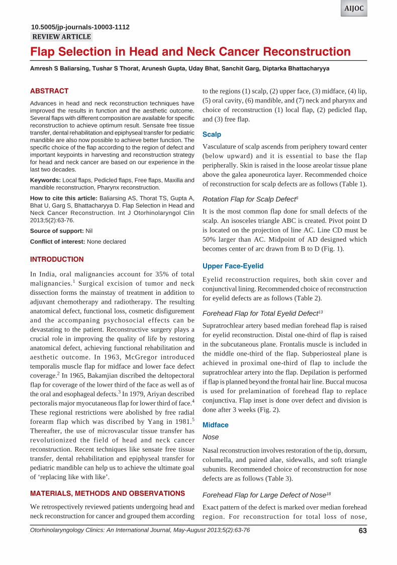
Nasolabial Flap for Intraoral Mucosa Defect40
Inferiorly based nasolabial flap is raised from nasolabialfold in subcutaneous plane. The flap is usually based onperpendicular branches of the angular artery that coursethrough the muscle and into the overlying skin. Facial arteryis preserved during dissection and adequate tunnel is madein buccal mucosa to deliver the flap inside oral cavity. Partof the flap is deepithelialized at the base of the flap where itenters oral cavity to avoid a second operation for closure ofthe orocutaneous fistula (Fig. 12).
Pectoralis Major Myocutaneous Flap forIntraoral Mucosa Defect41
Thoracoacromial vessels supplying the flap traverses alongthe line perpendicular from midpoint of clavicle to the linejoining the acromion process to xiphisternum and then
courses along this line toward xiphisternum. Entire skin overthe pectoralis major muscle can be raised. However, smallto moderate size flap is raised depending on the requirementfor reconstruction of the oral mucosa defect alone or formucosa and cheek skin defect where flap is folded to coverboth the defect.
Commonly 6 × 7 cm skin paddle is raised with the flap.Caution is taken to avoid hair bearing skin in males andbreast tissue in females. In female the flap can be located atthe inframammary fold or on the medial side of the breastand in male it is located at the lower part of the parasternallocation. Flap elevation is started from a lateral incision toexpose the lateral border of the pectoralis major muscle.Dissection is then carried out under the pectoralis majormuscle to include the thoracoacromial vessel. After the
Table 7: Choice of reconstruction for oral cavity defects
Site Site of defect Choice of reconstruction
Buccal mucosa <2 cm Primary closure>2 cm Nasolabial flap,40 submental flap, forehead flap,Large Pectoralis major myocutaneous flap,41 free radial
forearm flap, free anterolateral thigh flap
Fig. 11: Free radial forearm flap for extensive upper lip defect and nasal ala
Fig. 12: Nasolabial flap for buccal mucosa defect after excisionof squamous cell carcinoma

Otorhinolaryngology Clinics: An International Journal, May-August 2013;5(2):63-76 71
Flap Selection in Head and Neck Cancer Reconstruction
AIJOC
identification of the muscle and location of the feedingvessel is confirmed, the planned skin paddle is incised. Themuscle is then detached medially and laterally preservingthe vascular pedicle. In case deltopectoral flap is harvestedat the same time with this flap, internal mammaryperforators are preserved by leaving sufficient amount ofpectoralis muscle near the parasternal border (Fig. 13).
Tongue
It is necessary to achieve three-dimensional reconstructionof tongue for function of deglutition and speech. Functionof the tongue after reconstruction depends mainly on theremaining residual tongue tissue. Recommended choice ofreconstruction for tongue defects are as follows (Table 8).
Free Radial Forearm Flap for Hemiglossectomy42
Radial artery forearm flap can be used for hemiglossectomyto subtotal glossectomy defect. Flap designed in omegashape () with wider base for the base of the tongue andfloor of the mouth. Total glossectomy requiresreconstruction of the floor of the mouth with a pentagonalshaped flap.43 Anterolateral thigh flap can also be used forfloor of the mouth reconstruction in total glossectomy(Fig. 14).
Mandible
Mandible reconstruction is essential restoration facialcontour, adequate oral continence and deglutition and dental
rehabilitation. Fibula flap is workhorse for mandiblereconstruction. Fibula flap can be raised along with skin(osteocutaneous) or with skin and muscle (osteomusculo-cutaneous) depending on the requirement. Deep circumflexiliac artery composite flap can be used for lateral orhemimandibulectomy defect. However, recommendedchoice of reconstruction for mandible defects are as follows(Table 9).
Free Fibula Osteocutaneous Flap forMandible Reconstruction44,45
Flap is harvested under tourniquet with patient in supineposition and knee flexed at 135° and hip at 60° and leginternally rotated. Skin perforators are marked in the middlethird of the leg along the axis about 3 cm parallel and behindthe line drawn from fibula head to lateral malleolus. Skinpaddle is marked around the perforator. Incision is taken
Fig. 13: Bipaddle pectoralis major myocutaneous flap for skin and buccal mucosal defect
Table 8: Choice of reconstruction for tongue defects
Site Size of defect Choice of reconstruction
Tongue Hemiglossectomy Free radial forearm flap,40
Subtotal glossectomy Free anterolateral thigh flapTotal glossectomy with floor of mouth Pentagonal free anterolateral thigh flap41
Fig. 14: Free radial forearm flap for hemiglossectomy and marginalmandibulectomy in case of squamous cell carcinoma of tongue

72
Amresh S Baliarsing et al
along the posterior border of the flap margin and extendeddownward direction in curvilinear manner for betterexposure. Soleus muscle is separated from fibula andseptocutaneous perforators preserved while cutting posteriorintermuscular septum . Flexor hallucis muscle is dissectedfrom the fibula leaving a cuff of muscle over it. Perforatorssupplying the skin paddle are traced to their origin at theperoneal vessels. Anterior skin incision along the flapmargin is then taken and flap is raised in the subfascial planeup to the septum. Peroneal muscles dissected off from thefibula in the similar manner and peroneal nerve is protectedat the upper end.
Anterior intermuscular septum is then incised andextensor digitorum longus and extensor hallucis longusmuscle is separated from the fibula. Interosseous membraneis then incised at the distal osteotomy site and peronealvessels are exposed and divided. Osteotomy of the fibula isperformed at the distal and proximal site and interosseousmembrane is incised along the divided fibula segment.Tibialis posterior muscle is then divided in layers exposingthe peroneal vessels. Peroneal vessels dissected proximallyup to the desired length. Osteotomies are planned accordingto mandible defect. Contoured plate is fixed to fibula andpedicle is divided. Peroneal vessels are anastomosed to facialvessels according to side of defect (Fig. 15).
Neck
Recommended choice of reconstruction for neck defectsare as follows (Table 10).
Anterolateral Thigh Flap for Large Defect ofNeck and Intraoral Mucosa28
Flap is based on septocutaneous (or musculocutaneous)perforators of the descending branch of lateral circumflexfemoral artery. Average flap size of 10 × 8 cm can be raisedand the defect can be closed primarily. Large flap withdimension of 30 × 12 cm can also be raised. Straight line
from the anterior superior iliac spine to the superolateralborder of the patella is marked. A circle with radius of 3 cmdrawn at the mid-point of this line. The perforators supplyingthe skin are in the lower outer quadrant of the circle. Anteriorincision is taken first and the perforators are identified andtraced to the origin from the descending branch of lateralcircumflex femoral artery in the intermuscular septumbetween the vastus lateralis and rectus femoris muscle. Thedescending branch of lateral circumflex femoral vessels aredissected to the origin for gaining the desired vascularpedicle length. Posterior incision is then taken and the flapis raised in the suprafascial plane up to the entry of theperforforator. Cuff of fascia is included with the flap aroundthe perforator.
This flap can be used for small to large oral mucosaldefect or for both mucosa and skin defect. Large extensivedefect can easily be reconstructed with this flap.
Table 9: Choice of reconstruction for mandible defects
Site Size of defect Choice of flap
Mandible Central 1/3rd Free fibula flap44,45
Lateral defect Free fibula flap, deep circumflex iliac artery flapHemimandibulectomy Free fibula flap, pectoralis major myocutaneous flapTotal mandibulectomy Free fibula flap
Table 10: Choice of reconstruction for neck defects
Site Size of defect Flap of choice
Neck Small Platysma flap, deltopectoral flapLarge Free anterolateral thigh flap,28 pedicled latissimus
dorsi myocutaneous flap, pectoralis majormyocutaneous flap
Fig. 15: Mandible reconstruction for right hemimandibulectomyin squamous cell carcinoma of lower gingivobuccal sulcus
Otorhinolaryngology Clinics: An International Journal, May-August 2013;5(2):63-76 73
Flap Selection in Head and Neck Cancer Reconstruction
AIJOC
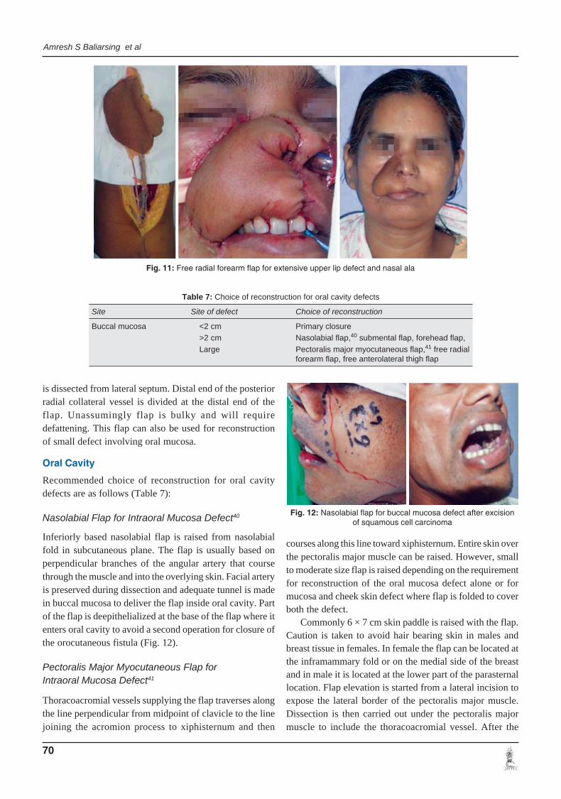
Anterolateral thigh flap in most patients particularly infemales is thick. It can be thinned by excision of excesssubcutaneous fat at the periphery without jeopardizing theperfusion for better in setting of the flap. Flap is bulky andrequires thinning at the time of primary operation or later.The vastus lateralis muscle is nourished by the same pedicleand can also be harvested with the flap needed in variousreconstructions (Fig. 16).
Pharynx
Recommended choice reconstruction for pharynx defectsare as follows (Table 11).
Table 11: Choice reconstruction for pharynx defects
Site Size of defect Choice of reconstruction
Pharynx Anterior wall Free anterolateral thigh flap,46 pectoralismajor myocutaneous flap, free radialforearm flap
Circumferential Free anterolateral thigh flap, free radialforearm flap, free jejunal flap
Fig. 16: Free anterolateral thigh flap for neck and intraoral mucosa defect after failed pectoralis major myocutaneous flap
Anterolateral Thigh Flap for Anterior WallDefects of Pharynx46
Anterolateral thigh flap will require thinning at the time ofprimary operation to allow water tight closure. Proximallyflap is anastomosed with the base of tongue and posteriorpharyngeal mucosa. The anterior wall of the cervicalesophagus is split longitudinally for approximately 1.5 cmto spatulate the distal anastomosis to enlarge the distalanastomosis and minimize the risk of ring stricture. Atriangular lip is created at the distal margin of the flap andinserted into the longitudinal split of the esophagus tocomplete the spatulation.
74
Amresh S Baliarsing et al
Anterolateral thigh flap for pharynx reconstruction ispreferably raised with two perforators with two separateskin paddles. One skin paddle is used for pharynxreconstruction and the second skin paddle is turned outwardto resurface the neck for flap monitoring. If two skin paddlesare not available, then monitoring of the flap can be donewith a implantable Doppler or with a hand held Doppler.
DISCUSSION
Choice of reconstruction is based on defect size, requirementfor type of tissue, function and appearance, associatedphysical condition of the patient and availability ofresources. Primary reconstruction has several benefits. Softand pliable tissue allows reconstruction with better functionand appearance. Secondary reconstruction at times inspecific condition may have limited advantage. However,primary reconstruction is preferred for reasons mentionedabove and to reduce morbidity.
The smaller tissue defects may enable primary closureor can be satisfactorily reconstructed with local randompattern flaps or axial flaps. Limited reach and downwardpull of the pedicled flap may lead to distal flap necrosis andwound gape. It is also difficult to achieve three-dimensionalreconstruction or cover of extensive tissue defects withpedicle flap. It also at times demands for a multistageprocedure and may result in delay in implementing theadjuvant therapy like radiotherapy in the postoperativeperiod.
Microsurgical free tissue transfer overcomes all thesedrawbacks. However, it requires surgical expertize,prolonged operating time and vigorous monitoring. But withavailability of adequate microsurgical training, safeanesthesia techniques, two team approach can overcomethese disadvantages in tertiary care center. Reliable singlestage with two team approach in head and neck recons-truction result in better predictable outcome. However,co-morbid factors in some patients may restrict its use andcompel to use alternative methods of reconstruction.
Several flaps with various composition are available forreconstruction. Reconstruction can be planned with aparticular flap depending on the reconstruction need for boneand soft tissue. Osseous flaps will require osteotomy atappropriate site for contouring to fit in to the defect (asosteotomy of fibula bone in to different segments to achieveproper contour in mandible reconstruction) and skin flapwill need thinning and molding to fit in to three-dimensionalcontour of the defect.
Advances have occured in head and neck reconstructiondue to better understanding anatomical composition ofvarious flaps. Sensory recovery at the reconstructed site is
essential for better function. Oral mucosa sensation helpsin improving patient’s quality of life by giving sensaryfeedback during mastication, swallowing and phonation.Radial forearm free flap can be made sensate by coaptingthe lateral or medial antebrachial cutaneous nerve with theflap to the donor sensory nerve in the neck.47 Similarly,anterolateral thigh flap can be made sensate by incorporatingthe lateral cutaneous nerve of the thigh with the flap.
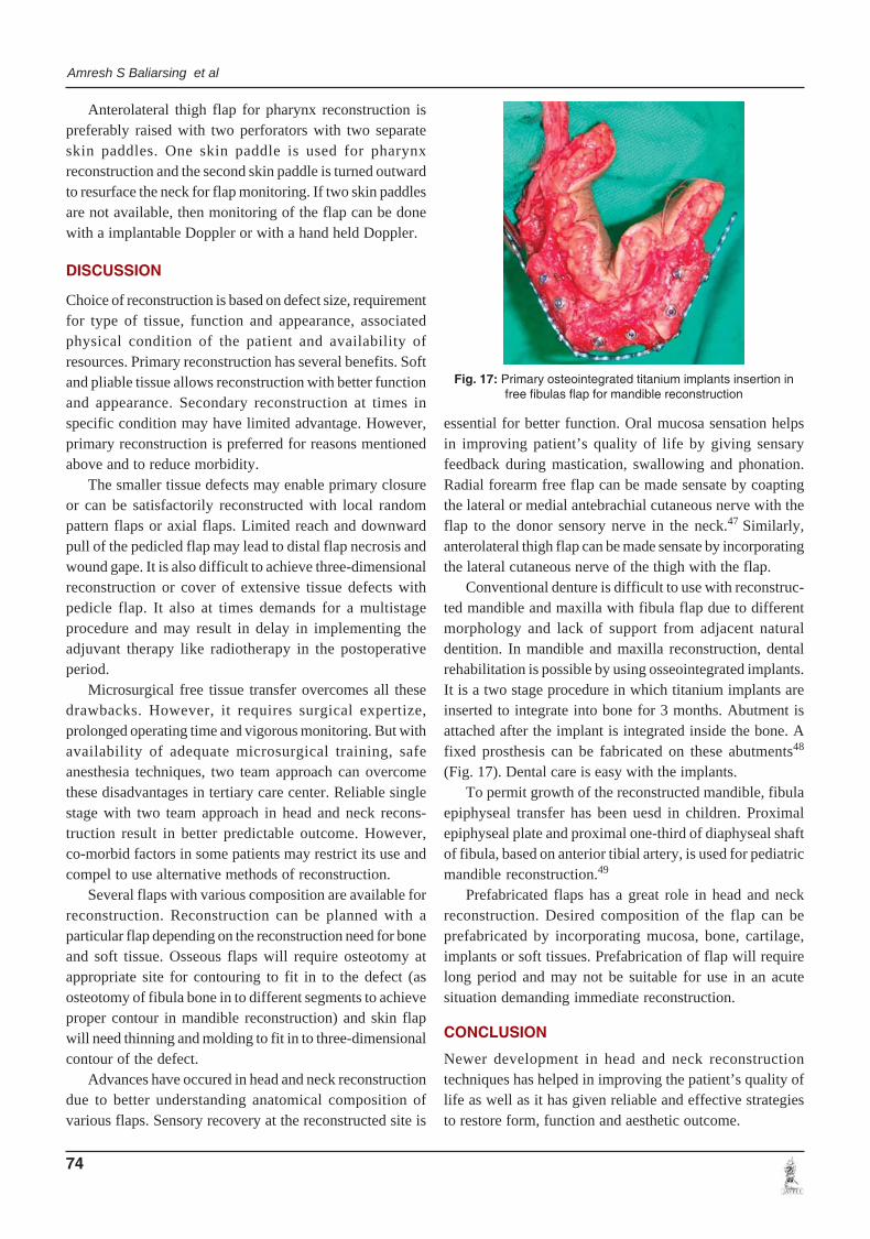
Conventional denture is difficult to use with reconstruc-ted mandible and maxilla with fibula flap due to differentmorphology and lack of support from adjacent naturaldentition. In mandible and maxilla reconstruction, dentalrehabilitation is possible by using osseointegrated implants.It is a two stage procedure in which titanium implants areinserted to integrate into bone for 3 months. Abutment isattached after the implant is integrated inside the bone. Afixed prosthesis can be fabricated on these abutments48
(Fig. 17). Dental care is easy with the implants.To permit growth of the reconstructed mandible, fibula
epiphyseal transfer has been uesd in children. Proximalepiphyseal plate and proximal one-third of diaphyseal shaftof fibula, based on anterior tibial artery, is used for pediatricmandible reconstruction.49
Prefabricated flaps has a great role in head and neckreconstruction. Desired composition of the flap can beprefabricated by incorporating mucosa, bone, cartilage,implants or soft tissues. Prefabrication of flap will requirelong period and may not be suitable for use in an acutesituation demanding immediate reconstruction.
CONCLUSION
Newer development in head and neck reconstructiontechniques has helped in improving the patient’s quality oflife as well as it has given reliable and effective strategiesto restore form, function and aesthetic outcome.
Fig. 17: Primary osteointegrated titanium implants insertion infree fibulas flap for mandible reconstruction

Otorhinolaryngology Clinics: An International Journal, May-August 2013;5(2):63-76 75
Flap Selection in Head and Neck Cancer Reconstruction
AIJOC
REFERENCES
1. Larson DL. Tumours of the lips, oral cavity and oropharynx. InMathes SJ editor. Plastic surgery. 2nd ed. volume 1. Philadelphia:Saunders Elsevier 2006:159-187.
2. McGregor IA. The temporal flap in intra-oral cancer: its use inrepairing the post-excisional defect. Br J Plast Surg 1963;16:318.
3. Bakamjian VY. A technique for primary reconstruction of thepalate after radical maxillectomy for cancer. Plast Reconstr Surg1963;31:103.
4. Ariyan S. The pectoralis major myocutaneous flap. A versatileflap for reconstruction in the head and neck. Plast Reconstr Surg1979;63:73-81.
5. Yang GF, Chen BJ, Gao YZ. The free forearm flap. Chin MedJ 1981;61:4.
6. Worthen EF. Repair of forehead defects by rotation flaps. PlastReconstr Surg 1976;57;204.
7. Millard R. Principlization of plastic surgery. Boston: Little,Brown; 1986:250-350.
8. Orticochea M. Four flap scalp reconstruction technique. Br JPlast Surg 1967;20:159-171.
9. Beasley NJ, Gilbert RW, Gullane PJ, et al. Scalp and foreheadreconstruction using free revascularized tissue transfer. ArchFacial Plast Surg 2004;6:16-20.
10. Spinelli HM, Jelks GW. Periocular reconstruction: a systematicapproach. Plast Reconstr Surg 1993;91(6):1017-1024.
11. Hughes WL. Reconstructive surgery of eyelids. St Louis: Mosby,1943.
12. Cutler NL, Beard CA. Method for partial and total upper lidreconstruction. Am J Ophthalmol 1955;39:1.
13. Mustarde JC. Repair and reconstruction of orbital region. 2nded. Edinburg: Churchill Livingstone, 1980:150-160.
14. Tessier P. Eyelid reconstruction or blepharopoesis in plasticsurgery of the orbit and eyelids. New York: Mason; USA Inc,1981.
15. Burget G, Menick F. Aesthetic reconstruction of the nose.St Louis, MO: Mosby; 1993:30-57.
16. Zitelli JA. The bilobed flap for nasal reconstruction. ArchDermatol 1989;125:957.
17. Hagerty RF, Smith W. The nasolabial cheek flap. Am Surg1958;24;506.
18. Millard DR Jr. Reconstructive rhinoplasty for the lower half ofnose. Plast Reconstr Surg 1974;53:133.
19. Pribaz J, Weiss D, Mulliken J, et al. Prelaminated free flapreconstruction of complex central facial defects. Plast ReconstrSurg 1999;104:357-365.
20. Jackson IT. Local flaps in head and neck reconstruction, 2nded. St Louis: Quality Medical; 2007:134-140.
21. Limberg AA. Design of local flaps. In Gibson T, editor. Moderntrends of plastic surgery (Vol. 2). London: Butterworth; 1966;38-61.
22. Kroll S, Reece G, Robb G, et al. Deep-plane cervicofacialrotation advancement flap for reconstruction of large cheekdefects. Plast Reconstr Surg 1994;94:88.
23. Bakamjian VY, Souther SG. Use of temporal muscle flap forreconstruction after orbitomaxillary resections for cancer. PlastReconstr Surg 1975;56:171.
24. Chow TL, Chan TT, Chow TK, et al. Reconstruction withsubmental flap for aggressive orofacial cancer. Plast ReconstrSurg 2007;120:431-436.
25. Bakamjian VY, Poole M. Maxillofacial and palatal recons-truction with deltopectoral flap. Br J Plast Surg 1977;30:17.
26. Wilson JSP, Breach NM. Forehead skin flaps, Ch105. In Grabb’sEncyclopedia of flaps, 3rd ed volume 1, Philadelphia, LippincottWilliams & Wilkins. 2009:294-305.
27. Soutar DS, McGregor IA. The radial forearm flap in intraoralreconstruction: the experience of 60 consecutive cases. PlastReconstr Surg 1986;78:1-8.
28. Song YG, Chen GZ, Song YL. The free thigh flap: a new freeflap concept based on the septocutaneous artery. Br J Plast Surg1984;37:149-1598.
29. Bitter K. Bone transfer from iliac creast to maxillofacial defectby microsurgical technique. J Maxillofac Surg 1980;8:210.
30. Wells MD, Luce EA. Reconstruction of midfacial defects aftersurgical resection of malignancies. Clin Plast Surg1995;22(1):79-89.
31. Cordeiro PG, Bacilious N, Schantz S, et al. The radial forearmosteocutaneous “sandwich” free flap for reconstruction of thebilateral subtotal maxillectomy defect. Ann Plast Surg1998;40(4):397-402.
32. Abbe R. A new plastic operation for the relief of deformity dueto double harelip. Plast Reconstr Surg 1968;42(5):481-483.
33. Neligan PC. Strategies in lip reconstruction. Clin Plast Surg2009;36(3):477-485.
34. Webster J. Crescentic peri-alar cheek excision for upper lip flapadvancement with a short history of upper lip repair. PlastReconstr Surg 1955;16:434-464.
35. Sullivan D. ‘Staircase’ closure of lower lip defects. Ann PlastSurg 1978;1:392-397.
36. Fujimori R. Gate flap for the total reconstruction of the lowerlip. Br J Plast Surg 1980;33(3):340-345.
37. Carroll CM, Pathak I, Irish J, et al. Reconstruction of total lowerlip and chin defects using the composite radial forearm—palmaris longus tendon free flap. Arch Facial Plast Surg2000;2(1):53-56.
38. Chang SC, Miller G, Halbert CF, et al. Limiting donor sitemorbidity by suprafascial dissection of the radial forearm flap.Microsurgery 1996;17:136-140.
39. Katsaros J, Schusterman M, Beppu M, et al. The lateral upperarm flap: Anatomy and clinical applications. Ann Plast Surg1984;12:489.
40. Herbert DC, Harrison RG. Nasolabial subcutaneous pedicledflap. Rr J Plast Surg 1975;28;85.
41. Koh KS, Eom JS, Kirk I, et al. Pectoralis major musculo-cutaneous flap in oropharyngeal reconstruction: revisited. PlastReconstr Surg 2006;118:1145-1149.
42. Hsiao HT, Leu YS, Lin CC. Tongue reconstruction with freeradial forearm flap after hemiglossectomy: a functionalassessment. J Reconstr Microsurg 2003;19:137-142.
43. Engel H, Huang JJ, Lin CY, et al. A strategic approach for tonguereconstruction to achieve predictable and improved functionaland aesthetic outcomes. Plast Reconstr Surg 2010;126;1967-1977.
44. Hidalgo DA. Fibula free flap: a new method of mandiblereconstruction. Plast Reconstr Surg 1989;84:71-79.
45. Wei FC, Seah CS, Tsai YC, et al. Fibula osteoseptocutaneousflap for reconstruction of composite mandibular defects. PlastReconstr Surg 1994;93:294-304; discussion 305-306.
46. Yu P, Robb GL. Pharyngoesophageal reconstruction with theanterolateral thigh flap: a clinical and functional outcomes study.Plast Reconstr Surg 2005;116:1845-1855.

76
Amresh S Baliarsing et al
47. Kurlakose MA, Loree TR. Sensate free radial forearm flap fortongue reconstruction; Arch Otolaryngol Head and Neck Surg2001;127;1463-1466.
48. Guriek A, Miller MJ, Jacob RF, Functional result ofosteointegrated dental implants into free fibula for mandiblereconstruction. Plast Reconstr Surg 1998;102;680-688.
49. Phillips JH, Rechner B, Tompson BD. Mandibular growthfollowing reconstruction using free fibula graft in paediatricfacial skeleton. Plast Reconstr Surg 2005;116;419-426.
ABOUT THE AUTHORS
Amresh S Baliarsing (Corresponding Author)
Professor and Head, Department of Plastic Surgery, Topiwala NationalMedical College and BYL Nair Charitable Hospital, MumbaiMaharashtra, India, e-mail: [email protected]
Tushar S Thorat
Resident, Department of Plastic Surgery, Topiwala National MedicalCollege and BYL Nair Charitable Hospital, Mumbai, Maharashtra, India
Arunesh Gupta
Assistant Professor, Department of Plastic Surgery, Topiwala NationalMedical College and BYL Nair Charitable Hospital, MumbaiMaharashtra, India
Uday Bhat
Associate Professor, Department of Plastic Surgery, Topiwala NationalMedical College and BYL Nair Charitable Hospital, MumbaiMaharashtra, India
Sanchit Garg
Resident, Department of Plastic Surgery, Topiwala National MedicalCollege and BYL Nair Charitable Hospital, Mumbai, MaharashtraIndia
Diptarka Bhattacharyya
Resident, Department of ENT and Head and Neck Surgery, TopiwalaNational Medical College and BYL Nair Charitable Hospital, MumbaiMaharashtra, India


![Complex reconstructions in head and neck cancer surgery ...branch and became a workhorse for head and neck reconstruction. Aryian was first to describe this flap in 1979 [3]. The limitation](https://img.dokumen.tips/doc/110x75/600204a8b262377b6076f298/complex-reconstructions-in-head-and-neck-cancer-surgery-branch-and-became-a.jpg)