Embed Size (px)
Citation preview
Flabelliforma montana (Phylum Microsporidia) fromPhlebotomus ariasi (Diptera, Psychodidae): ultrastructuralobservations and phylogenetic relationships
Elizabeth U. Canning1,*, A. Curry2, Sarah A. Cheney1, Nathalie J. Lafranchi-Tristem1,D. Ebert3, D. Rifardt3, M. Killick-Kendrick1 and R. Killick-Kendrick1
1Department of Biology, Imperial College of Science, Technology and Medicine, London SW7 2AZ, U.K.2Public Health Laboratory, Withington Hospital, Manchester M20 2LR, U.K.3Zoologisches Institut, Universität Basel, Rheinsprung 9, CH -4 051 Basel, Switzerland
Received: 29 January 2001; 30 April 2001. Accepted: 4 May 2001
Ultrastructural observations are presented on Flabelliforma montana (type species) (Phylum Mi-crosporidia) from sandflies Phlebotomus ariasi (Diptera, Psychodidae). All stages have isolated nuclei.The sporophorous vesicle (SV) arises by separation of a 5 nm layer, via small blisters, from the 15 nm sur-face membrane of sporonts. Sporoblasts form by deep invagination of the sporont surface, the SV at firstfolding inwards around the invaginations but later unfolding to form a rounded vesicle enclosing spores.The vesicle contains tubules with bulbous terminal expansions. Spores have a deeply domed anchoringdisc within a polar sac which covers the anterior polaroplast, consisting of membranes acutely angled tothe longitudinal axis around the straight section of the polar tube. Posterior polaroplast membranes, inthe region where the polar tube changes course towards the periphery, appear almost transverse insagittal section. The 130 nm thick endospore is overlain by several membrane-like layers, which togetherwith amorphous material constitute the exospore.There are 3.5–4.0 coils of the polar tube. Flabelliformaostracodae, Flabelliforma diaptomi and Flabelliforma magnivora conform well in morphology with thetype species. However, when F. magnivora was included in parsimony and maximum likelihood analyses,using 16S rDNA sequences of 20 microsporidia, it emerged as unrelated to F. montana. Disparities werefound between morphological characters and molecular groupings of several of the microsporidia inves-tigated. Within clades, nuclei could be isolated or diplokaryotic, sporophorous vesicles present or absentand life cycles simple or complex.
Key words: Flabelliforma spp.; Sandflies; Microcrustaceans; Ultrastructure; Phylogeny; 16S rDNA.
those hatching from eggs laid by an uninfected fe-male (Canning et al. 1991). A dichotomous keywas presented to distinguish Flabelliforma fromtwelve other polysporous microsporidia whichhad been described previously. F. montana wasshown to have isolated nuclei throughout its de-velopment and to undergo sporogony by divisionof a sporogonial plasmodium via deep lobes withina sporophorous vesicle (SV), which at first fol-lowed the contours of the lobes but finally becamerounded at the time of sporoblast separation. The
0932-4739/01/37/02-207 $ 15.00/0
Introduction
The polysporous microsporidium Flabelliformamontana was described from midgut epithelialcells of sandflies, Phlebotomus ariasi, and wastransmitted orally to larvae of P. ariasi, including
*Address for correspondence: Prof. E. U. Canning, ImperialCollege at Silwood Park, Ascot, Berks SL5 7PY, U.K.; Fax:020 7594 22339, E.mail: [email protected]
Europ. J. Protistol. 37, 207–221 (2001)© Urban & Fischer Verlaghttp://www.urbanfischer.de/journals/ejp

SV was a fragile structure, containing about 30sporoblasts. Spore preservation was poor but amembranous polaroplast and 3.5 coils of the polartube (= polar filament) were observed.
Three further species have since been ascribedto the genus: Flabelliforma ostracodae from Can-dona sp. (Crustacea, Ostracoda) (Bronnvall andLarsson 1994), Flabelliforma diaptomi, a newcombination proposed by Voronin (1996) forStempellia diaptomi Voronin, 1977 from Diapto-mus gracilis (Crustacea, Copepoda) (Voronin1977) and Flabelliforma magnivora from Daphniamagna (Crustacea, Cladocera) (Larsson et al.1998). F. magnivora had previously been referredto as Tuzetia sp. (Mangin et al. 1995). The 3 specieswere distinguished from the type species by fea-tures such as number of sporoblasts produced by asporont, spore size and number of coils of thepolar tube. Larsson et al. (1998) placed F. mag-nivora in the family Duboscqiidae adopting thesystematic position used by Bronnvall and Lars-son (1994) for F. ostracodae. The pathogenicityand epidemiology of infections of F. magnivora inD. magna have been described by Mangin et al.(1995) and Ebert et al. (2000).
In the summer of 1998 we collected adult P. ari-asi at a site in Laumède in the Cevennes mountains,France and obtained several new infections ofF. montana. We have used this material and sam-ples of D. magna infected with F. magnivora to ob-tain 16S rDNA sequences. We present new ultra-structural data on F. montana to add to the descrip-tion of the type species and assess the systematicpositions of the other species currently attributedto Flabelliforma, using the ultrastructural data anda molecular phylogeny based on 22 sequences ofthe 16S rDNA from 20 species of microsporidia,derived mainly from invertebrate hosts. We alsoobtained sequences of Cystosporogenes operoph-terae and Orthosomella operophterae from wintermoth Operophtera brumata and included these se-quences in the phylogenetic analysis.
Materials and methods
Sources of Flabelliforma spp., O. operophteraeand C. operophterae
Adult male and female P. ariasi were caught in lighttraps set in the cellar, stable and gardens of a house inLaumède, Cevennes mountains, France between
16/08/98 and 20/08/98. The gut of each fly was dissect-ed into phosphate buffered saline (PBS) within 24 h oftrapping and examined by microscopy. Guts showingsigns of infection with F. montana were either fixed in96% ethanol for DNA extraction or were stored in PBSbefore being fed to larvae of P. ariasi. Spores from oneheavily infected fly were placed in a pot with 126 P. ari-asi eggs laid by two sandflies and spores from one heav-ily infected and one lightly infected sandfly were placedin a pot with 258 eggs laid by five sandflies. Some emer-gent larvae were examined at the fourth instar. The re-mainder entered diapause and were examined as adultseight months after exposure to spores. Infected gutsfrom the experimental infections were preserved in 96%ethanol for DNA extraction. A few remaining sporeswere added to a new batch of P. ariasi larvae and, fromthese, two heavily infected guts were fixed inKarnovsky’s fixative for electron microscopy.
Daphnia magna infected with F. magnivora were col-lected from ponds, one in Oxfordshire, UK and theother near Moscow, Russia in 1992 (Larsson et al. 1998;Ebert et al. 2000). Females were kept singly in filteredpond water and their parthenogenetic offspring weretransferred to fresh pond water within 10 hours ofbirth. Clones were raised from these offspring, all ofwhich were infected and infected hosts have been main-tained in the laboratory by transovarial transmissionsince 1993. After maintenance for 3 years infectedD. magna were preserved in 96% ethanol for extractionof DNA.
Winter moth larvae were collected from WythamWood, Oxford, UK and Wistman’s Wood, Dartmoor,UK in 1997. Salivary glands were dissected and thoseinfected with O. operophterae or C. operophterae werefixed whole in 96% ethanol or suspended in PBS forDNA extraction.
Electron microscopy of F. montanaThe previously fixed guts were transferred to 0.1 M
cacodylate buffer pH 7.2, post fixed in 1% Os O4 in ca-codylate buffer and dehydrated in an ethanol series.After transfer to propylene oxide, then mixtures of70:30 and 30:70 propylene oxide : Agar 100 resin (AgarScientific, Stansted, UK), the material was embedded inAgar 100 resin. Sections were stained with uranyl ac-etate and lead citrate and examined under an AE1EM801 electron microscope.
Nucleic acid preparationDNA extraction was carried out on guts of infected
P. ariasi, whole infected D. magna and salivary glands ofO. brumata. Samples were suspended in 300 µl of TEbuffer, pH 7.2, and shaken with 300 mg of 0.5 mm Zir-conium beads in a Mini-Bead beater (Biospec ProductsLtd.) at low speed for 30 s. A 15 µl aliquot of a 10
208 E. U. Canning et al.

mg/ml–1 Proteinase K stock solution in H2O was addedand the samples were incubated at 55 °C for2 h. After further bead-beating for 30 s, the suspensionwas extracted twice with phenol :chloroform (1:1) andthe DNA was precipitated with sodium acetate and icecold ethanol (Sambrook et al. 1989). The DNA was sus-pended in TE buffer, pH 8.0 and stored at –20 °C.
PCR amplification and sequencingThe 16S rDNA was amplified using the 18f (5′-
CACCAGGTTGATTCTGCC-3′) and 1537r (5′-TTATGATCCTGCTAATGGTTC-3′) primers de-signed by Baker et al. (1995). PCR amplification wascarried out in 20 µl volumes, using about 10 ng DNA, 5pmol of each primer, 0.2 mM of each dNTP, 2 mMMgCl2 and 1 unit of Taq polymerase (Life Technologies
Ltd., Inchinnan, Paisley). The reactions were comprisedof 92 °C denaturation for 2 min, followed by 30–40 cy-cles of 92 °C denaturation for 30 s, 48–58 °C annealingfor 30 s and 72 °C extension for 45 s, with a final exten-sion of 5 min. PCR products were cleaned using theWizard PCR preps (Promega, UK) and cloned into thepGem-T Easy Vector system (Promega, UK). One tothree clones were selected, amplified with SP6 and T7vector system primers, and cleaned, as before, for se-quencing with the Thermo Sequenase dye terminatorcycle sequencing pre-mix kit (Amersham, UK). Sixprimers were used to obtain the whole sequence in bothdirections: SP6, T7, 3F (= 5′-GTCCAAGGA/TC/GGCAGCAGGC-3′), 4F (= 5′-CACCACCAGGAGTGGAGTGTG-3′), 5R (= 5′-CACA/AC/TCCACTCCTTGTG G-3′) and 6R (= 5′-GCCTGCTGCTGTCCTTG-GAC-3′).
Ultrastructure and phylogeny of Flabelliforma 209
Table 1. Species and isolates of microsporidia used in the phylogenetic analyses, their hosts, the accession numbersand sequence lengths of the 16S rDNA.
Microsporidia Hosts Accession Number Sequence (Systematic Group1) Length (bp)
Amblyospora stimuli Aedes stimulans (DC) AF027685 1361Ameson sp.2 Litopenaeus setiferus (CD) AJ252959 1305Bacillidium sp. Lumbriculus sp. (O) AF104087 1386Caudospora palustris Cnephia ornithophilia (DS) AF132544 1374Culicosporella lunata Culex pilosus (DC) AF027683 1343Cystosporogenes operophterae Operophtera brumata (LG) AJ302320 1258Edhazardia aedis Aedes aegypti (DC) AF027684 1448Endoreticulatus schubergi Lymantria dispar (LL) L39109 1252Flabelliforma magnivora Daphnia magna (CC) AJ302318 1197(C1, Russia)Flabelliforma magnivora Daphnia magna (CC) AJ302319 1304(E-11-18, Oxford)Flabelliforma montana Phlebotomus ariasi (DP) AJ2052962 1115(p)3
Janacekia debaisieuxi Simulium sp. (S. ornatum or AJ252950 1417S. equinum) (DS)
Nosema algerae Anopheles stephensi (DC) AF069063 1365Nosema tyriae Tyria jacobaeae (LA) AJ012606 1233(p)Orthosomella operophterae Operophtera brumata (LG) AJ302316 1197(p)(Dartmoor)Orthosomella operophterae Operophtera brumata (LG) AJ302317 1280(Oxford)Polydispyrenia simulii Simulium sp. (S. ornatum or AJ252960 1291(p)
S. equinum) (DS)Thelohania solenopsis Solenopsis invicta (H) AF031538 1382Trachipleistophora hominis Homo sapiens (PH) AJ002605 1364Vavraia culicis Aedes albopictus (DC) AJ252961 1364Vairimorpha imperfecta Plutella xylostella (LY) AJ131645 1231Vittaforma corneae Homo sapiens (PH) U11046 1237Giardia lamblia (outgroup) (Portland 1 strain) M54878 1453
1CC = Crustacea, Cladocera; CD = Crustacea, Decapoda; DC = Diptera, Culicidae; DP = Diptera, Psychodi-dae; DS = Diptera, Simuliidae; H = Hymenoptera; LA = Lepidoptera, Arctiidae; LG = Lepidoptera, Ge-ometridae; LL = Lepidoptera, Lymantriidae; LY = Lepidoptera, Yponomeutidae; O = Oligochaeta, Lumbricul-idae; PH = Primates, Hominidae. 2Referred to as Pleistophora sp. (LS) in Cheney et al. (2000). 3p = partial

Phylogenetic analysis
For phylogenetic analysis, the 16S rDNA sequencesof F. montana and F. magnivora were aligned with thoseof 18 other species of microsporidia (two sequences ofO. operophterae), which are listed with their hosts inTable 1. The corresponding sequence of Giardia lam-blia (Portland 1 strain) was used as outgroup.
Sequences were aligned using the CLUSTAL W pro-gramme (Thompson et al. 1994), which gave an align-ment of 1559 characters. Only those sites which couldbe unambiguously aligned among all the microsporidiaand the outgroup were used to construct phylogenetictrees (1168 bp). The aligned sequences were analyzed byboth parsimony and maximum likelihood methodsusing PAUP version 4.0b2a (Swofford 1998). For theparsimony tree, an initial heuristic search was carriedout on the 23 CLUSTAL aligned sequences, using thetree bisection and reconnection swapping algorithmand 1000 bootstrap resamplings with 10 subreplicatesand random addition of the sequences. The resultingbootstrap tree was used to construct a 90% majorityrule (majrule) consensus tree. The nexus file from thealigned sequences was then imported into MacCladeversion 3.07 (Maddison and Maddison 1997), togetherwith the 90% majrule tree, and the rescaled consistency(RC) index used to weight the data, treating the poly-tomies as soft. The weighted data set was then importedback into PAUP and the branch and bound option ofPAUP was used to generate the most parsimonious tree,using the 90% majrule consensus tree as a constraintand the MacClade calculated RC character weights.Bootstrap analysis (100 replicates) was performed togive a measure of the confidence that can be placed inthe resulting gene tree. Maximum Likelihood analysiswas carried out on a subset of 10 microsporidian speciesand G. lamblia, using either a heuristic search or abranch and bound search, both with 100 bootstrap re-sampling, but using a 90% majrule consensus tree gen-erated from an initial parsimony analysis as a constraintfor the latter. A topological constraint tree, which con-strained the Flabelliforma spp. together, was construct-
ed using the same search strategy as that used to find theminimum (unconstrained) trees (a heuristic search with100 bootstrap resamplings and 10 subreplicates on un-weighted data) and the winning-sites of Kishino-Hasegawa test options in PAUP.
Results
Prevalence of F. montana
F. montana was found in 6 wild caught sandflies,3 males and 3 females out of 331 dissected. On ascale of 1+ to 5+ (low to high intensities of infec-tion): 2 were 2+, 2 were 4+ and 2 were 5+.
Ultrastructure of F. montana
Undivided meronts (Figs. 1, 2) and meronts di-viding by plasmotomy were observed with up tofour nuclei in one plane. Structures, appearing asrows of 45 nm diameter beads lying in host cell cy-toplasm close to meront surfaces (Figs. 1, 2), weresometimes seen emanating from the meront sur-face (Fig. 2). Microtubules, 25 nm diameter, werecommon in the host cell cytoplasm surrounding allstages of the parasite (Fig. 3).
Sporogony was initiated by deposition of a thicksurface coat on the sporont (Fig. 4) at the time ofseparation of a fine membrane from the plasmamembrane to form a sporophorous vesicle (SV)(Fig. 4), within which spores were eventually held.The mode of formation of the vesicle envelope wasnot determined with certainty but there was strongevidence that it was parasite derived as follows: thesurface structure of the meront measured 15 nmand was thick enough to have incorporated twotightly apposed membranes. These appeared toseparate (Figs. 5–7) in preparation for deposition
210 E. U. Canning et al.
Figs. 1–9. Flabelliforma montana: merogony and early sporogony. p = parasite, n = nuclei, m = host cell mitochon-dria. Bar on Fig. 1 applies to all figures. 1, 2. Meronts with one and three nuclei in the plane of section, are in directcontact with host cell cytoplasm. Beaded structures indicated by arrowheads appear to emanate from the parasite’ssurface in Fig. 2. The boxed area in Fig. 1 is enlarged in Fig. 6. Bar = 400 nm (Fig. 1), 625 nm (Fig. 2). 3. Microtubulesin host cell cytoplasm near surface (arrowhead) of parasite. Bar = 200 nm. 4. Binucleate sporont with electron dense,pitted surface coat (arrow) surrounded by a very fine vesicle envelope (arrowheads). Enlargements of (a) and (b) areshown in Figs. 8 and 9. Bar = 625 nm. 5–7. Formation of sporophorous vesicle envelope. The electron dense surfacelayer of the parasite appears to split into two leaflets (large arrowheads). Junctions of three host cells (small arrow-heads) are included in Fig. 7 to show the appearance of unit membrane for comparison with the parasite’s surface.Fig. 6 is an enlargement of the boxed area in Fig. 1 and shows the beaded structures (arrow) close to the parasite’ssurface. Bar = 275 nm (Fig. 5) and 220 (Figs. 6, 7). 8. Enlargement of (a) from Fig. 4 showing pitted surface coat ofsporont. Bar = 333 nm. 9. Enlargement of (b) from Fig. 4, showing tubular structures emanating from surface ofparasite and lying within the fine sporophorous vesicle envelope arrowheads. Bar = 333 nm.

Ultrastructure and phylogeny of Flabelliforma 211

of the sporont surface coat which was secreted, atfirst in patches (Fig. 12), then as a continuous coat(Fig. 4). In patches lacking the surface coat(Figs. 10, 12) the SV envelope remained attached tothe plasma membrane, indicating that it is the se-cretion of the surface coat that finally forces theseparation of the SV envelope from the parasite’ssurface. Tangential sections revealed the surfacecoat to be a pitted layer (Figs. 4, 8, 10) even up tothe stage when sporoblasts were being separated(Fig. 16). Tubules, measuring about 40 nm in diam-eter running between the SV envelope and thesporont surface coat appeared to connect with thesporont plasma membrane possibly through thepits in the surface coat (Fig. 9). In its final form onthe sporont, the surface coat was a layered struc-ture (Fig. 11) consisting of a 10 nm amorphouscentre separating less dense 5 nm outer and innerlayers, giving a total thickness of 20 nm. The coatwas separated from the 7.5 nm plasma membraneby a 5–10 nm gap. The SV envelope became sepa-rated in places as two leaflets (Figs. 10, 12).
Nuclear division continued during sporogony.A centriolar plaque was surmounted by two ormore double-membrane vesicles (Figs. 12, 13) andwas the point from which spindle microtubules ra-diated into the nucleoplasm and terminated inchromosomes (Fig. 10). Division of the sporontoccurred by constriction of the cytoplasm arounduninucleate segments. In one sporont prominentparamural bodies were present at the bases of theinvaginations, at points where the surface coat was
absent (Figs. 14, 15). In the early stages of sporob-last separation some host cell cytoplasm, in contactwith the SV envelope, was carried in with the con-stricting sporont surface (Figs. 16–18) but finallythe contours of the vesicle smoothed out (Figs. 19,21) so that the sporoblasts and spores lay in arounded vesicle. The vesicle envelope remained indirect contact with host cell cytoplasm at the endof sporogony. Some fragments of the sporont con-tained two nuclei (Fig. 16) before final divisioninto uninucleate sporoblasts. A complex of 20 nmdiameter tubules was seen in tangential sectionwithin or beneath the surface coat of one sporob-last (Fig. 21).
Vesicles containing spores almost invariablyalso contained tubules consisting of an outer60–100 nm cylinder and inner 30–50 nm tube(Fig. 23). The tubules terminated in bulbous ex-pansions, involving an increase in diameter of theinner tube to 70 nm and outer tube up to 140 nm.The maximum length of a tubule observed in sec-tion was 1.2 µm (Fig. 22). Spores were only seenfresh in gut squashes and were not measured.They were flattened on one side, convex on theother with both ends tapering. The exospore de-rived from the sporont surface coat was a layeredstructure (Fig. 25), consisting of an outer 8 nmamorphous coat, three or four parallel mem-branes together measuring about 16 nm and aninner 11 nm amorphous layer, measuring about35 nm in total. The endospore measured about130 nm, except over the anchoring disc where it
212 E. U. Canning et al.
Figs. 10–18. Flabelliforma montana : stages of sporogony. n = nuclei. Bar on Fig. 10 applies to all figures. 10. Earlysporont, with pitted surface coat, enclosed by a fine sporophorous vesicle envelope (arrowheads) which has separat-ed in places into 2 leaflets (star). In a patch lacking the surface coat, the sporophorous vesicle envelope is still close-ly apposed to the plasma membrane (arrow). Nuclei show chromosomes and spindle of mitosis. Bar = 680 nm. 11.Surface of a mature sporont showing plasma membrane (pm) surface coat of three layers (a, b, c) and sporophorousvesicle envelope (sv). Bar = 110 nm. 12. Formation of a cytoplasmic bud in a sporont with incomplete surface coat.Chromosomes (arrowheads) close to a spindle pole indicate end of karyokinesis. Note that the sporophorous vesi-cle (large arrows) follows the contours of the bud and is attached to the plasma membrane in patches where the sur-face coat is lacking (small arrows). In other places (star) it has separated into 2 leaflets. Bar = 375 nm. 13. Enlarge-ment of spindle plaque from Fig. 12 showing electron dense plaque in nuclear envelope, surmounted by double-walled vesicles. Bar = 200 nm. 14, 15. Complex tubular paramural bodies (arrowheads) at points on the surface ofsporonts where buds emerge to form sporoblasts. 16. Almost complete division of sporont into sporoblasts (thebuds are connected in another plane of section). At this stage the sporophorous vesicle, accompanied by host cellcytoplasm, follows the contours of the sporoblast buds (arrows). Note that one branch still has two nuclei, imply-ing further division will take place. The surface coat is now almost devoid of pits but some pits remain (arrowhead).Areas a and b are enlarged in Figs. 17, 18. Bar = 1.1 µm. 17. Enlargement of (a) from Fig. 16. Sporophorous vesicleenvelope (arrowheads) and host cell cytoplasm occupying the space between sporoblast buds (s). Bar = 680 nm. 18.Enlargement of (b) from Fig. 16 showing host cell cytoplasm and sporophorous vesicle envelope (arrowheads) be-tween sporoblast buds (s). Bar = 280 nm.

Ultrastructure and phylogeny of Flabelliforma 213

214 E. U. Canning et al.
Ultrastructure and phylogeny of Flabelliforma 215
Figs. 19–26. Flabelliforma montana : sporogony and spores. n = nuclei. Bar on Fig. 19 applies to all figures. 19. Latesporogonic division. The sporophorous vesicle envelope (arrowheads) and host cell cytoplasm (hc) has now retract-ed, leaving the dividing sporont in a rounded cavity. The surface coat is almost complete but the layered structureover the plasma membrane is not yet clear. Bar = 880 nm. 20. Sporoblast showing tubular structures (arrow) withinor beneath the surface coat. Arrowhead indicates sporophorous vesicle envelope. Bar = 670 nm. 21. Sporophorousvesicle containing mature spores and tubules. Bar = 1.7 µm. 22, 23. Detail of tubules in the sporophorous vesiclecavity showing inner tubule (arrowheads) and termination in a bulb (arrow). Bar = 400 nm (Fig. 22) and 240 nm(Fig. 23). 24. Mature spore showing deeply domed anchoring disc (ad), polar sac (ps) polaroplast of membranes an-gled (a) and appearing transverse (t) to the long axis, nucleus and 3.5 coils of the polar tube. Bar = 333 nm. 25. Ante-rior end of mature spore showing: exospore (arrow) composed of three or four membranous layers bounded exter-nally and internally by amorphous coats; lucent endospore (en), plasma membrane (1), cytoplasmic layer (2), mem-brane of polar sac/anchoring disc complex (3), polar tube (pt) with denser core terminating in the anchoring disc(ad). a = acutely angled polaroplast membranes. Bar = 110 nm. 26. Transverse sections of polar tube. The middle sec-tion shows the full range of layers consisting of a central core (1), lucent ring (2) containing longitudinal fibrils andouter two membranes (3, 4). Bar = 125 nm.
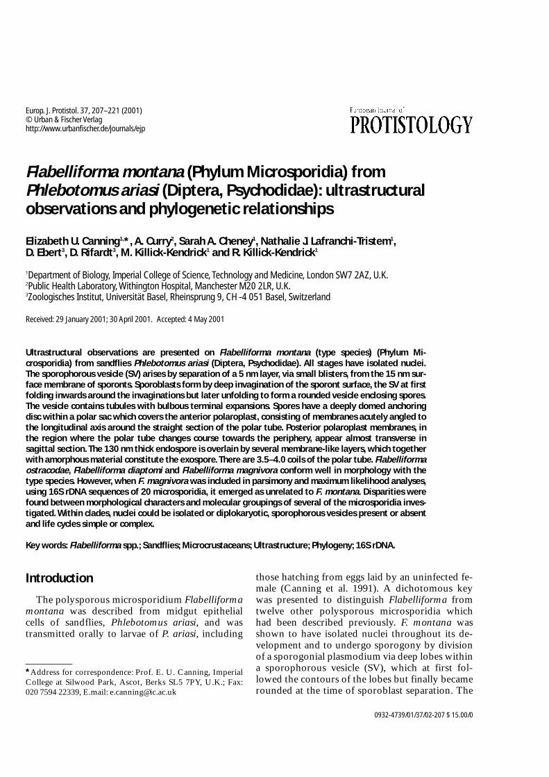
Fig. 27. The most parsimonious tree (tree length = 1192.72), found using the branch and bound option of PAUP4.0b2a. Bootstrap analysis (100 replicates) was performed to give a measure of the confidence that can be placed inthe resulting tree, with bootstrap values being located on the tree branches.

was thinned to 30 nm or less (Figs. 24, 25). Theanterior end of the polar tube terminated in a 200nm diameter deeply domed central region (an-choring disc) of the polar sac, which was uni-formly dense except for a slightly denser 11 nmlayer immediately beneath its anterior border(Fig. 25). The sac itself was separated from theendospore by a narrow band of cytoplasm 11 nmthick and the plasma membrane. Laterally thepolar sac extended back over the polaroplast,which was entirely of lamellar construction with
membranes 16–20 nm apart separated by amor-phous material. The anterior region was arrangedas a system of folds at an acute angle to thestraight part of the polar tube (Fig. 25). The po-laroplast membranes probably maintained thisangled relationship with the polar tube when thetube changed direction towards the periphery, sothat the membranes appeared transverse to thelongitudinal axis in some planes of section (Fig.24). The single nucleus lay immediately posteriorto the polaroplast, surrounded by cytoplasm
216 E. U. Canning et al.
Fig. 28. A maximum likelihood tree (-ln Likelihood 7467.27) of a subset of 11 species. The same tree topology wasgenerated from both heuristic and branch and bound searches, both carried out with 100 bootstrap resamplings ofthe data. Bootstrap values indicated on the tree branches are taken from the heuristic search, with those in parenthe-ses representing the values obtained from the branch and bound search where these differed from heuristic analysis.

packed with ribosomes and the 3.5–4.0 coils ofthe polar tube (Figs. 24). A membrane-boundposterior vacuole with amorphous contents wasobserved only rarely. Cross sections of the polartube revealed several concentric layers, includinga wide electron lucent layer in which a circlet offibres ran longitudinally (Fig. 26).
Phylogenetic Relationships
The accession numbers of the 22 16S rDNA se-quences of the microsporidia used in the phyloge-netic analyses and their sequence lengths are givenin Table 1. The parsimony tree (Fig. 27) gave goodresolution of the terminal branches but poor reso-lution of the clades with respect to one another, re-sulting in a polytomy. The 2 sequences of F. mag-nivora were not close to the clade containingF. montana, Polydispyrenia simulii and Caudospo-ra palustris. Whereas there was about 0.5% se-quence difference between F. montana andP. simulii and 0.7% between F. montana andC. palustris, the distance between F. montana andF. magnivora was 20%. The sequence differencebetween the two isolates of F. magnivora was 1.8%and between the two isolates of Orthosomellaoperophterae was 0.9%. When F. magnivora wasconstrained to cluster with F. montana in the parsi-mony tree, 19 more steps were required than in theoriginal tree. This was significant (p = <0.039) butnot highly significant. The ML tree (Fig. 28) con-structed on a subset of the data, placed all but oneof the species examined in one clade, albeit withlow bootstrap support. The exception was F. mag-nivora.
As found previously (Canning et al. 1999),Nosema tyriae paired with Vairimorpha imperfecta
with strong bootstrap support but these specieswere distant from Nosema algerae which pairedwith Thelohania solenopsis with equally strongsupport. N. algerae has been transferred to thegenus Brachiola (Lowman et al. 2000). Of the newsequences obtained in this study, that of C. oper-ophterae was close to those of Vittaforma corneaeand Endoreticulatus schubergi and a sister to thisclade was O. operophterae. A surprisingly close re-lationship, with 100% bootstrap support, wasfound between Janacekia debaisreuxi and Bacillid-ium sp. Finally the clades containing Amblyosporastimuli, Edhazardia aedis and Culicosporella luna-ta on the one hand and Trachipleistophora hominisand Vavraia culicis on the other were as found re-spectively by Alder et al. (2000) and Cheney et al.(2000). The species previously designated asPleistophora sp. LS and found to be close to Ame-son michaelis (see Cheney et al. 2000) has sincebeen identified on ultrastructural data as a speciesof Ameson (unpublished data).
Discussion
The new material of F. montana has permitted usto elucidate some of the features which were in-completely described in the original descriptionand to compare it, as far as data permits, with themore recently described species, F. ostracodaeBronnvall and Larsson, 1994 from Candona sp.(Crustacea, Ostracoda), F. diaptomi (Voronin1977) from Diaptomus gracilis (Crustacea,Calanoidea) and F. magnivora Larsson, Ebert,Mangin and Vávra, 1998 from Daphnia magna(Crustacea, Cladocera).
Important generic characters of Flabelliformawere confirmed, especially the unpaired nucleithroughout development, merogony in direct con-tact with host cell cytoplasm and multisporoussporogony in a sporophorous vesicle (SV) by for-mation and final separation of deep lobes in thesporogonial plasmodium. It was also confirmedthat the SV envelope separates from the sporogo-nial plasmodium at the time of deposition of thesurface coat, at first following the contours of thelobes and later rounding off to enclose the groupsof sporoblasts and spores. New data ofgeneric/specific importance were provided onmode of SV envelope formation by separationfrom the meront plasma membrane and the occur-rence of tubules with a terminal bulbous expansion
Ultrastructure and phylogeny of Flabelliforma 217
Table 2. Summary of topological constraint analyses.
Topological Number p value 2Constraint of steps
required 1 Kishino- Winning-Hasegawa sites
No constraint 3088 – –(best tree)Monophyly of all 3107 <0.0393 <0.0509Flabelliforma spp.
1 Tree length from a heuristic search with 100 bootstrap re-samplings and 10 subreplicates on unweighted data2 p<0.001 highly significant; p>0.1 not significant

in the SV cavity, similar to the type 1 tubules de-scribed by Takvorian and Cali (1983). Also onspore structure with details of the anchoring disc,the polaroplast composed entirely of lamellae andthe exospore made up of 3 or 4 membrane-like lay-ers bounded on either side by amorphous material.The spindle plaques and paramural bodies are ofmore widespread microsporidian occurrence,while the significance of the beaded structures em-anating from the meront surface is not clear.
F. ostracodae conforms closely with the genericcharacters, notably in the unpaired nuclei, the sep-aration of the very fine (8 nm) SV envelope as tinyblisters from the surface of the sporogonial plas-modium and sporoblast formation from a distinct-ly lobed sporogonial plasmodium. Its polaroplastis also of lamellar type only but in the posterior re-gion the membranes are distinctly more widelyseparated than in F. montana. Other featureswhich differ are the exospore structure of twomembrane-like layers surmounted by an amor-phous layer, the 13–16 coils of the polar tube andthe large amount of “debris” in the SV.
Stempellia diaptomi Voronin, 1977 was trans-ferred to the genus Flabelliforma by Voronin(1996) on the basis of unpaired nuclei, meronts indirect contact with host cell cytoplasm and divi-sion of the sporogonial plasmodium into sporob-lasts via deep lobes. It is also likely that the SV en-velope is formed as in F. montana but no evidenceis available that it accompanies the invaginations ofthe plasmodium during early sporogony. At anearly stage the SV cavity contains prominent, in-clusions which appear as double-walled tubules asin F. montana. Although spore preservation wasnot good there is some evidence that the polaro-plast is lamellar only but, in contrast to F. montana,has anterior and posterior close-packed lamellaeand a middle region of widely separated lamellae.The exospore differs from that of F. montana, as itis composed of two prominent dense layers sepa-rated by a more lucent layer of the same thickness,with amorphous material disposed externally andinternally.
F. magnivora conforms with the genus Flabelli-forma in having isolated nuclei and in the generalmodes of division of meronts and sporogonial plas-modia and probably in the mode of formation ofthe SV envelope. However, the surface coat (futureexospore) of the sporogonial plasmodium is con-structed of a continuous inner layer and a compact-ly-folded outer layer, like a frill. Excess of this ma-
terial, extruded into the SV cavity, is organised asthick frilled tubules or balls. As well as these, thecavity becomes packed with thin-walled tubules.The exospore eventually resolves as 5 layers at thesurface of spores in an arrangement quite unlikethat on spores of F. montana. The polaroplast issimilar in being entirely lamellar but in the anteriorregion the membranes are tightly-packed, givingway to more widely spaced membranes posteriorly.
As outlined above the assignments of the threenew species to the genus Flabelliforma were basedon reasonable morphological grounds, especiallyas there were deficiencies in the description of thetype species, notably about its spore structure(Canning et al. 1991). However the present phylo-genetic study, based on 16S rDNA sequences of 20microsporidia, of which 18 including F. montanaand F. magnivora were from invertebrate hosts,has indicated that these two species are not closelyrelated. F. montana was found to be most closelyrelated to P. simulii and C. palustris with 100%bootstrap support. These are both parasites ofblackflies. Cheney et al. (2000) also found thatF. montana paired with P. simulii with 100% boot-strap support, in a parsimony tree constructedwith a different set of microsporidia of which13/21 were from vertebrate hosts.
The parsimony tree (Fig. 27) failed to resolve therelationships of the various clades to one another.Resolution was marginally improved in the MLtree (Fig. 28) based on a subset of the species and itwas again clear that F. magnivora was not close toF. montana. This was supported by the 20% differ-ence in their sequences and the topological con-straints analysis which required 19 more steps thanthe original tree when F. montana and F. magnivo-ra were constrained to be monophyletic. On thebasis of the present molecular data, as opposed tomorphological data, F. magnivora should not beplaced in the genus Flabelliforma. We were notable to include F. diaptomi or F. ostracodae in ourmolecular study. These, and F. magnivora are para-sites of freshwater microcrustaceans and it wouldbe interesting, if material becomes available for 16SrDNA sequencing of F. diaptomi and F. ostracodae,to determine whether these form a clade whichevolved in the crustacean groups.
Groups I–III in the ML tree (Fig. 28), reflectingthe same groups in the parsimony tree, containgenera of markedly different morphology. Ingroup I P. simulii is diplokaryotic in merogony butundergoes meiosis in sporogony to give rise to nu-
218 E. U. Canning et al.
merous haploid spores in SVs (Canning and Haz-ard 1982). C. palustris is diplokaryotic in mero-gony and remains diplokaryotic in sporogony (ap-parently without meiosis), dividing into 4–8diplokaryotic sporoblasts, not enclosed in SVs(Alder et al. 2000). F. montana has unpaired nucleithroughout development, producing about 30 un-inucleate spores in SVs. As parasites ofhaematophagous Diptera producing masses ofspores in the aquatic larval stage, it is likely thatP. simulii, and possibly also C. palustris, have com-plex life cycles involving alternation of hosts, as isknown for several species of Amblyospora infect-ing mosquitoes and copepods (Andreadis 1985;Sweeney et al. 1985, 1988; Becnel 1992). F. mon-tana can be transmitted directly by feeding sporesto larvae (Canning et al. 1991) and, as its host,P. ariasi, does not have an aquatic stage, similarcomplexities in the life cycle are unlikely. In theparsimony tree generated by Alder et al. (2000),C. palustris branched independently and was notincluded in the clade containing microsporidianparasites of Culicidae (Diptera). In our analyses,C. palustris was again distinct from the group para-sitizing Culicidae but, by including P. simulii, C.palustris was found to cluster with it, indicatingthat these two taxa from Simuliidae are related.
In view of the morphological characters sharedby F. montana and F. magnivora and the differ-ences between F. montana and P. simulii, it is re-markable that 1.8% nucleotide difference wasfound in the 16S rDNA sequences obtained forisolates of F. magnivora collected in Oxford, UKand Russia (confirmed by independent sequencingof F. magnivora, D. Rifardt personal communica-tion), while only 0.5% nucleotide difference wasfound between species of different genera, i.e.F. montana and P. simulii. No ultrastructural dif-ferences to suggest even different species werefound between the Oxford and Russian isolates ofF. magnivora (Larsson et al. 1998) but geographicalisolation could have been responsible for diver-gence in the 16S rDNA. It has not been possible toobtain new material of F. montana but its sequencewas obtained four months after that of P. simulii,rendering it unlikely that there was cross contami-nation. As morphological differences at least asgreat as those exhibited by the Group III organ-isms (Fig. 28) are found in microsporidia inGroups I and II (discussed below), it is not entirelysurprising that Group III organisms are related.
In Group II the pairing of Janacekia and Bacil-
lidium cannot be reconciled on morphologicalgrounds. The polysporous genus Janacekia ex-hibits diplokarya which undergo meiosis at theonset of sporogony so that each sporoblast andspore has one haploid nucleus (Larsson 1983). TheSV enclosing the sporogonial plasmodium divideswith the plasmodium so that each spore is enclosedin its own SV. In contrast, the disporous genusBacillidium is diplokaryotic throughout the lifecycle and produces Bacillus-like spores fromwhich a double membrane-like exospore layermay separate as a sac around individual spores(Larsson 1994). The sacs resemble SVs but have adifferent origin. The morphological differencescoupled with host differences – blackflies forJanacekia, oligochaetes for Bacillidium suggest anuneasy partnership.
In Group III Cystosporogenes and Endoreticula-tus are morphologically similar. Both have un-paired nuclei in all stages and develop in para-sitophorous vacuoles in host cell cytoplasm. Thetwo membranes forming the boundary of the vac-uole enclosing Endoreticulatus were unequivocallyidentified as host endoplasmic reticulum (ER), asthe outer membrane was ribosome studded(Brooks et al. 1988). The origin of the single mem-brane enclosing stages of Cystosporogenes was notdetermined when the genus was established (Can-ning et al. 1985) but recent ultrastructural observa-tions (unpublished) show that the vacuoles canharbour merogonic and sporogonic stages concur-rently (as in Endoreticulatus) and cannot thereforebe SVs. The presence of one vacuolar membrane inCystosporogenes and two in Endoreticulatus de-fines the difference between the genera. In contrastto these two genera, Vittaforma is diplokaryoticthroughout development (Shadduck et al. 1990).Each stage is encased in host ER but merogonicand sporogonic stages do not coexist in the samevacuole. The feature common to this clade is,therefore, the envelopment of parasites by hostER. Orthosomella, the other genus in this group,which emerged as a sister taxon, has unpaired nu-clei and develops directly in host cell cytoplasmwith no intervening SV or parasitophorous vac-uole (Canning et al. 1985). Sequence difference of0.9% between the isolates of O. operophterae fromtwo different woodlands in England was alsogreater than the distance between F. montana andP. simulii. Ultrastructural data are only availablefor the Dartmoor isolate (Canning et al. 1985) butlight microscopy has revealed no differences (un-
Ultrastructure and phylogeny of Flabelliforma 219

published observations) which might suggestgeneric or specific differences.
The disparity between morphological andmolecular data for the organisms in Groups I-IIIimplies either that SVs, diplokarya and complexlife cycles have evolved and been lost several timesin microsporidian evolution or that 16S rDNA, asa highly conserved gene, does not contain suffi-cient information to discriminate between generictaxa. However, in two species, F. magnivora andO. operophterae, the gene varied considerably be-tween isolates. To date, 16S rDNA sequences havebeen obtained for only about 30 of the 140 or soknown genera of microsporidia and data for moreof these taxa may resolve the apparent anomalies.At present, there is no clear guidance as to themorphological characters which are useful in sys-tematics.
Acknowledgements: The authors are grateful to Dr.V.N. Voronin (St. Petersburg) for providing originalphotomicrographs of F. diaptomi for comparison withF. montana, Dr. Lionel Cole (Oxford) for collection ofO. brumata from Wytham Wood, Ms. Trish Rowlandfor assistance with electron microscopy and the MedicalResearch Council for financial support (Grant No.G9603050).
References
Alder P.H., Becnel J.J. and Moser B. (2000): Molecular charac-terization and taxonomy of a new species of Caudospori-dae (Microsporidia) from black flies (Diptera: Simuliidae),with host-derived relationships of the North AmericanCaudosporids. J. Invertebr. Pathol. 75, 133–143.
Andreadis T.G. (1985): Experimental transmission of a mi-crosporidian pathogen from mosquitoes to an alternatecopepod host. Proc. Natl. Acad. Sci. USA, 82, 5574–5577.
Baker M.D., Vossbrinck C.R., Didier E.S., Maddox J.V. andShadduck J.A. (1995): Small subunit ribosomal DNA phy-logeny of various microsporidia with emphasis on AIDSrelated forms. J. Euk. Microbiol. 47, 564–570.
Becnel J.J. (1992): Horizontal transmission and subsequentdevelopment of Amblyospora californica (Microsporida:Amblyosporidae) in the intermediate and definitive hosts.Dis. Aquat. Org. 13, 17–28.
Bronnvall A.M. and Larsson J.I.R. (1994): Flabelliforma ostra-codae n. sp. (Microspora, Duboscqidae), a new mi-crosporidian parasite of Candona sp. (Crustacea, Ostraco-da). Europ. J. Protistol. 30, 280–287.
Brooks W.M., Becnel J.J. and Kennedy G.G. (1988): Estab-lishment of Endoreticulatus N.G. for Pleistophora fidelis(Hostounsky & Weiser, 1975) (Microsporidia:Pleistophoridae) based on the ultrastructure of a mi-
crosporidium in the Colorado potato beetle, Leptinotarsadecemlineata (Say) (Coleoptera: Chrysomelidae). J. Pro-tozool. 35, 481–488.
Canning E.U. and Hazard E.I. (1982): Genus PleistophoraGurley, 1893. An assemblage of at least three genera. J.Protozool. 29, 39–49.
Canning E.U., Barker R.J., Nicholas J.P. and Page A.M.(1985): The ultrastructure of three microsporidia fromwinter moth, Operophtera brumata (L.) and the estab-lishment of a new genus Cystosporogenes n.g. forPleistophora operophterae (Canning, 1960). Syst. Para-sitol. 7, 213–225.
Canning E.U., Curry A., Cheney S.A., Lafranchi-TristemN.J., Kawakami Y., Hatakeyama Y., Iwano H. and Ishi-hara R. (1999): Nosema tyriae n.sp. and Nosema sp. mi-crosporidian parasites of cinnabar moth Tyria jacobaeae. J.Invert. Pathol. 74, 29–38.
Canning E.U., Killick-Kendrick R. and Killick-Kendrick M.(1991): A new microsporidian parasite Flabelliformamontana n.g., n. sp. infecting Phlebotomus ariasi(Diptera, Psychodidae) in France. J. Invert. Pathol. 57,71–81.
Cheney S.A., Lafranchi-Tristem N.J. and Canning E.U.(2000): Phylogenetic relationships of Pleistophora-like mi-crosporidia based on small subunit ribosomal DNA se-quences and implications for the source of Tra-chipleistophora hominis infections. J. Euk. Microbiol. 47,280–287.
Ebert D., Lipsitch M. and Mangin K. (2000): The effect of par-asites on host population density and extinction: experi-mental epidemiology with Daphnia and six micropara-sites. Am. Naturalist 156, 459–477.
Larsson R. (1983): A revisionary study of the taxon TuzetiaMaurand, Fize, Fenwick and Michel, 1971, and relatedforms (Microspora, Tuzetiidae) Protistologica 19,323–355.
Larsson J.I.R. (1994): Characteristics of the genus BacillidiumJanda, 1928 (Microspora, Mrazekiidae) – reinvestigationof the type species B. criodrili and improved diagnosis ofthe genus. Europ. J. Protistol. 30, 85–96.
Larsson J.I.R., Ebert D., Mangin K.L. and Vávra J. (1998): Ul-trastructural study and description of Flabelliforma mag-nivora sp. n. (Microspora: Duboscqiidae) a microsporidi-an parasite of Daphnia magna (Crustacea: Cladocera:Daphniidae). Acta Protozool. 37, 41–52.
Lowman P.M., Takvorian P.M. and Cali A. (2000): The effectsof elevated temperatures and time-temperature combina-tions on the development of Brachiola (Nosema) algeraeN. Comb. in mammalian tissue culture. J. Euk. Microbiol.47, 221–234.
Maddison W.P. and Maddison D.R. (1997): MacClade: analy-sis of phylogeny and character evolution, version 3.07,Sunderland, MA.
Mangin K.L., Lipsitch M. and Ebert D. (1995): Virulence andtransmission modes of two microsporidia in Daphniamagna. Parasitology 111, 133–142.
Sambrook J., Fritsch E.F. and Maniatis T. (1989): MolecularCloning. A laboratory manual. 2nd ed. Cold Spring Har-bor Laboratory Press, N.Y.
Shadduck J.A., Meccoli R.A., Davis R. and Font R.L. (1990):Isolation of a microsporidian from a human patient. J. In-fect. Dis. 162, 773–776.
220 E. U. Canning et al.

Sweeney A.W., Hazard E.I. and Graham M.F. (1985): Inter-mediate host for an Amblyospora sp. (Microspora) infect-ing the mosquito Culex annulirostris. J. Invertebr. Pathol.46, 98–102.
Sweeney A.W., Hazard E.I. and Graham M.F. (1988): Lifecycle of Amblyospora dyxenoides sp. nov. in the mosquitoCulex annulirostris and the copepod Mesocyclops albicans.J. Invertebr. Pathol. 51, 46–57.
Swofford D.L. (1998): PAUP*, phylogenetic analysis usingparsimony (*and other methods), version 4.0b2a. SinaurAssociates, Sunderland, MA.
Takvorian P.M. and Cali A. (1983): Appendages associatedwith Glugea stephani, a microsporidian found in flounder.J. Protozool. 30, 251–256.
Thompson J.D., Higgins D.G. and Gibson T.J. (1994):CLUSTAL W: improving the sensitivity of progressivemultiple alignment through sequence weighting, position-specific gap penalties and weight matrix choice. Nucl.Acids Res. 22, 4673–4680.
Voronin V.N. (1977): Microsporidians (Protozoa, Mi-crosporidia) of lower crustaceans (Entomostraca) in thewaters of the Leningrad region. Parazitologiya 11,505–512. In Russian with English summary.
Voronin V.N. (1996): Ultrastructure of Stempellia diaptomi(Protozoa, Microsporidia) with the revision of generic po-sition of the species. Parazitologiya 30, 59–63. In Russianwith English summary.
Ultrastructure and phylogeny of Flabelliforma 221