Embed Size (px)
Citation preview
Loh, R. and Landos M. (2011) Fish Vetting Essentials. Richmond Loh Publishing, Perth.
0 - 0 -
Loh, R. and Landos M. (2011) Fish Vetting Essentials. Richmond Loh Publishing, Perth.
1 - 1 -
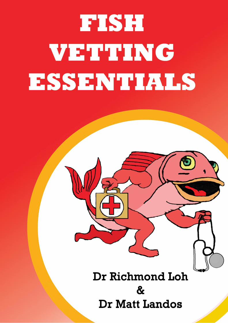
© 2011 Richmond Loh Publishing ISBN 978-0-9871571-0-2 This work is copyright. Apart from any use permitted under the Copyright Act 1968, no part may be reproduced by any process, nor may any other exclusive right be exercised, without the permission of the author. Requests and enquiries concerning reproduction and rights should be addressed to Dr Richmond Loh. Published by: Richmond Loh Publishing Perth, Western Australia, Australia. For orders Phone: +61 (0)421 822 383 Email: [email protected] Web: http://www.thefishvet.com.au Preferred way to cite this publication: Loh, R. and Landos M. (2011) Fish Vetting Essentials. Richmond Loh Publishing, Perth. Publication designed and typeset by Richmond Loh. Front cover: The Fish Vet’s logo. Part fish, part vet.
Loh, R. and Landos M. (2011) Fish Vetting Essentials. Richmond Loh Publishing, Perth.
3 - 3 -
FOREWORD This is a revised version of the self-published “Australian Fish Vetting Essentials” (2007) by Drs Richmond Loh & Matt Landos. The purpose of this manual is to collate the knowledge that aquarists, aquaculturalists, public aquaria, local fish shops and veterinarians already have, and to filter out misinformation and then provide this information in a readily digestible form. The information contained in this publication has been in the process of compilation since 2001. This manual is not prescriptive, but rather, it is a collection from our combined knowledge to promote to the industry that veterinarians are best equipped to deal with aquatic animal health. Worthy of note is that many diseases found in aquatics can be classified as emerging diseases since an “emerging disease” is one that has appeared in a population for the first time, or that may have existed previously but is rapidly increasing in incidence of geographic range. The Authors The Fish Vet Dr Richmond Loh BSc, BVMS, MPhil, MANZCVSc (Aquatics & Pathobiology), DipPM, CMAVA +61 (0)421 822 383 [email protected] http://www.thefishvet.com.au Future Fisheries Veterinary Service Dr Matt Landos BVSc HonsI, MANZCVSc (Aquatics) +61 (0)437 492 863 [email protected]
Contributors Dr Michael Chia
Dr Stephen Pyecroft
Dr Judith Handlinger
Dr Shane Raidal
Dr Fran Stephens
Loh, R. and Landos M. (2011) Fish Vetting Essentials. Richmond Loh Publishing, Perth.
5 - 5 -
ABOUT THE AUTHORS Dr Richmond Loh Dr Loh has always been interested in animals, nature and medicine, so naturally he studied to become a veterinarian at Murdoch University. However, his passion for all things fish was strong and so his first job was as a veterinary fish pathologist for the Tasmanian state laboratory, providing diagnostic services for the large aquaculture farms including species such as salmon, trout, ornamental fishes, abalone and oysters. At the same time, he was offering veterinary services to owners of ornamental fishes. In 2006, he passed the examinations for Aquatic Animal Health for the Australian & New Zealand College of Veterinary Scientists (ANZCVS). In the same year, he was awarded a Master of Philosophy degree for cancer research in Tasmanian devils, publishing the seminal papers on Devil Facial Tumour Disease in Veterinary Pathology. To increase his depth of knowledge in the area of diseases, he studied for and passed the examinations for Pathobiology for the ANZCVS in 2009. So far, he has given numerous talks at seven National Veterinary Conferences and also to the Pet Industry Australia Association delegates and at the New Zealand Companion Animal Conference. He regularly writes for aquarium and pet publications. These are an initiative to generate interest within the respective professions and industry to apply scientific reasoning for the better health and management of fishes. Through his veterinary career, he has appeared on TV (Creature Features, Stateline, Catalyst, ABC news), been interviewed on radio (Curtin FM), appeared in newspapers (The Sunday Times UK, Herald Sun, The Examiner, Sunday Tasmanian, The Cairns Post, Canning Times), magazines (Australian Aquarium Magazine, Aquarium Keeper Australia, TIME Australia Magazine, Your Pet Magazine, Woman’s Day, Pets – Taking Care of Your Family’s Best Friend, Animals’ Voice) and appears on several local and international websites (ABC Online). He is the consultant veterinarian to AQWA (the Aquarium of WA), is an adjunct lecturer at Murdoch University, is a founding member of the World Aquatic Veterinary Medical Association (WAVMA), is the secretary for the Aquatic Animal Health Chapter of the ANZCVSc and provides advice on fish health and welfare to several universities and the RSPCA. His clients are diverse and range from individual pet fish owners, to retailers, farmers (ornamental and food cultured fishes) and exporters.

Loh, R. and Landos M. (2011) Fish Vetting Essentials. Richmond Loh Publishing, Perth.
6 - 6 -
Dr Matt Landos Dr Landos is the Founding Director of Future Fisheries Veterinary Service, is an honorary lecturer in aquatic animal health and associate researcher at the University of Sydney, Faculty of Veterinary Science and in 2011 he was the president of the Aquatic Animal Health Chapter of the Australian & New Zealand College of Veterinary Scientists. Dr Landos commenced his consultancy practice in aquatic animals in 2005 after a 5 year stint with the NSW DPI as the Veterinary Officer in Aquatic Animal Health. The client base is located throughout Australia, and it ranges from small native fish hatcheries to 3,000 tonne sea cage operations. He works with all aquatic species including molluscs, crustacea and finfish. He reviews emergency disease preparedness plans and develops health management plans for aquaculture industries. He has had a prominent media profile in recent years associated with investigation of the impacts of environmental pollutants on fisheries in relation to the notorious two-headed Australian bass larvae case from the Noosa River.
Loh, R. and Landos M. (2011) Fish Vetting Essentials. Richmond Loh Publishing, Perth.
19 - 19 -
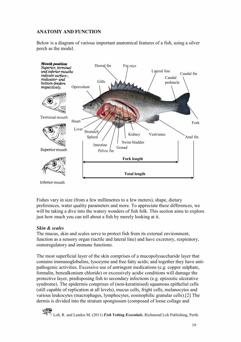
ANATOMY AND FUNCTION Below is a diagram of various important anatomical features of a fish, using a silver perch as the model. Fishes vary in size (from a few millimetres to a few meters), shape, dietary preferences, water quality parameters and more. To appreciate these differences, we will be taking a dive into the watery wonders of fish folk. This section aims to explore just how much you can tell about a fish by merely looking at it. Skin & scales The mucus, skin and scales serve to protect fish from its external environment, function as a sensory organ (tactile and lateral line) and have excretory, respiratory, osmoregulatory and immune functions. The most superficial layer of the skin comprises of a mucopolysaccharide layer that contains immunoglobulins, lysozyme and free fatty acids; and together they have anti-pathogenic activities. Excessive use of astringent medications (e.g. copper sulphate, formalin, benzalkonium chloride) or excessively acidic conditions will damage the protective layer, predisposing fish to secondary infections (e.g. epizootic ulcerative syndrome). The epidermis comprises of (non-keratinised) squamous epithelial cells (still capable of replication at all levels), mucus cells, fright cells, melanocytes and various leukocytes (macrophages, lymphocytes, eosinophilic granular cells).[2] The dermis is divided into the stratum spongiosum (composed of loose collage and
Pelvic fin
Total length
Fork length
Intestine Swim bladder Gonad
Caudal fin
Dorsal fin Fin rays
Anal fin
Caudal peduncle Gills
Operculum
Heart
Stomach
Vent/anus Kidney
Liver
Lateral line
Spleen
Fork

Loh, R. and Landos M. (2011) Fish Vetting Essentials. Richmond Loh Publishing, Perth.
29 - 29 -
Catfish Origin: various Aquarium system: tropical freshwater. Characteristics: have down-turned mouths and “whiskers” (barbels);; have scutes or are scaleless. General types: Corydoras (Bronze cats or Corys); Suckermouths (plecostomus – e.g. bristlenose), Shark catfish. *If fish is stuck/tangled in the net, leave net in container of water and fish will free itself. Corydoras
Have large scutes (heavily armoured); The leading ray of the dorsal and pectoral fins are robust and act as defensive spines.
Loricariids Come from fast-moving waters (hence their sucker mouth); Have rough skin; Vegetarian. Do well with some drift wood (ballasts are good for their digestive system). Common disease issues: Bloat (bacterial septicaemia) and fungal infection. Shark catfish
Live in brackish water (only the very young do well in pure freshwater as most are thought to be on their migratory path); Grow to a very large size;
Have large mouths (beware of other fish being eaten!). Common disease issues: Over eating, intestinal foreign body. Loaches Characteristics: have forked tail and no scales. General types: Botia (Clown loach), Loach (Kuhli loach). Botia Special requirements: caves/hiding places. Loach Burrows into the gravel (they can disappear for days!).
Loh, R. and Landos M. (2011) Fish Vetting Essentials. Richmond Loh Publishing, Perth.
98 - 98 -
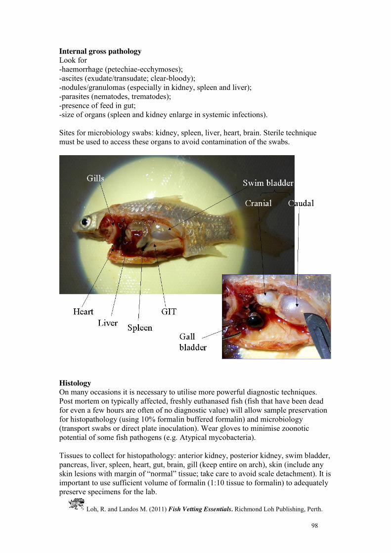
Internal gross pathology Look for -haemorrhage (petechiae-ecchymoses); -ascites (exudate/transudate; clear-bloody); -nodules/granulomas (especially in kidney, spleen and liver); -parasites (nematodes, trematodes); -presence of feed in gut; -size of organs (spleen and kidney enlarge in systemic infections). Sites for microbiology swabs: kidney, spleen, liver, heart, brain. Sterile technique must be used to access these organs to avoid contamination of the swabs.
Histology On many occasions it is necessary to utilise more powerful diagnostic techniques. Post mortem on typically affected, freshly euthanased fish (fish that have been dead for even a few hours are often of no diagnostic value) will allow sample preservation for histopathology (using 10% formalin buffered formalin) and microbiology (transport swabs or direct plate inoculation). Wear gloves to minimise zoonotic potential of some fish pathogens (e.g. Atypical mycobacteria). Tissues to collect for histopathology: anterior kidney, posterior kidney, swim bladder, pancreas, liver, spleen, heart, gut, brain, gill (keep entire on arch), skin (include any skin lesions with margin of “normal” tissue;; take care to avoid scale detachment). It is important to use sufficient volume of formalin (1:10 tissue to formalin) to adequately preserve specimens for the lab.
Loh, R. and Landos M. (2011) Fish Vetting Essentials. Richmond Loh Publishing, Perth.
99 - 99 -
Fixatives 10% Neutral Buffered Formalin (fish, freshwater invertebrates) Formalin (40% w/v formaldehyde) 100mL Sodium phosphate, monobasic monohydrate 4g Sodium phosphate, dibasic, anhydrous 6.5g Distilled water to 1L This solution is stable for many months at room temperature. Fix small blocks of tissue (10x10x3mm) for up to 24 hours. Seawater Formalin (marine shellfish and crustaceans) As for 10% NBF, but fill with seawater to 1L. Davidson’s Fixative (marine shellfish and crustaceans) 600ml Seawater 600ml 95% ethanol 400ml 37% formaldehyde Add 200ml glacial acetic acid prior to use. Virology Preservation of tissues for viral isolation: best to send tissues fresh on ice on express courier to laboratory. Your local laboratory may be able to help with making transport media (containing antibiotics and anti-fungals). Matching samples of formalin, glutaraldehyde and 70% ethanol fixed tissues should be sent separately, to allow processing for histology, electron microscopy and molecular assays. Molecular Biology Material for PCR testing needs to be preserved in 95% ethanol. There is also a product called “RNA Later” that can preserve genetic material, including RNA material, for long periods. It is important not to cross-contaminate samples. Remember that PCR tests only provides you with evidence for the presence or absence of that organism’s DNA. A positive result does not necessarily mean there is disease per se. And a negative result could mean that you may not have selected your primers properly. Results should be interpreted in light of clinical signs and histopathology findings.

Loh, R. and Landos M. (2011) Fish Vetting Essentials. Richmond Loh Publishing, Perth.
112 - 112 -
Wasting Disease & Fish Tuberculosis (Mycobacteriosis) Clinical Signs Mycobacteria and Nocardia tend to cause systemic infections that may manifest themselves as skin lesions and weight loss despite being well-fed (gaunt appearance to the goldfish pictured below).
The most common bacterial species include: Mycobacterium marinum, Mycobacterium fortuitum & Mycobacterium platypoecilus. Fish suffering from these chronic infections tend to develop granulomatous nodules throughout their internal organs and occasionally on the skin. This leads to a variety of organ-related signs such as: inappetence, wasting, ascites, loss of colour, exophthalmia, loss of scales, spinal cord deformity, loss of balance, listless behaviour, finrot and external ulceration of the body. Skin abscesses that appear to erupt/originate from beneath can occur. Diagnosis A tentative diagnosis is made based on clinical signs. Unfortunately, a definitive diagnosis can only be made via necropsy. On necropsy, there may be widespread granulomatous, grey-yellow nodules in most organs (especially liver, kidney and spleen). Acid-fast bacilli can be visualised using ZN stains on heat-dried smears or on histology.

Loh, R. and Landos M. (2011) Fish Vetting Essentials. Richmond Loh Publishing, Perth.
113 - 113 -
Risk Factors
Carrier fish Poor tank hygiene Overcrowding Cannibalism of infected fish
Treatment Mycobacteria tends to pass from fish to fish by way of cannibalism of infected dead fish. Thus it is a very important aspect of mycobacterial control - that dead fish are promptly removed from tanks so that cannibalism is not allowed to occur. It is also possible that vertical transmission from parent to offspring occurs in live-bearing species of fish. Like many other diseases of fish, it is likely that a carrier state exists and that apparently healthy fish may harbour the disease. Outbreaks of mycobacteriosis may then occur if these carriers are subjected to poor environmental conditions and excessive stress. If the condition is diagnosed early enough, drugs such as doxycycline and sulphafurazole can be administered systemically (IM) but with limited success. Affected fish should be isolated from other fish and if their condition deteriorates during treatment, they should be euthanased. As there may be a zoonotic risk associated with these pathogens (below, see granulomatous nodule on the arm of this patient), it is essential to avoid contact with uncovered skin when handling affected stock or contaminated equipment and water.
Some believe that following an outbreak, disinfect all equipment, tanks and other facilities and dispose of all affected fish properly. Some disinfectants recommended include oxidizing agents (chlorine, Virkon), iodophores, alcohol and phenols. However, some consider Mycobacteria to be ubiquitous. Therefore, improving aquarium conditions, decreasing stressors and improving nutrition are the measures to be taken. Prevention Avoid overcrowding Do not allow organic matter to build up Maintain parasite-free fish Avoid unnecessary handling Quarantine new fish for >2-4 weeks.
Loh, R. and Landos M. (2011) Fish Vetting Essentials. Richmond Loh Publishing, Perth.
117 - 117 -
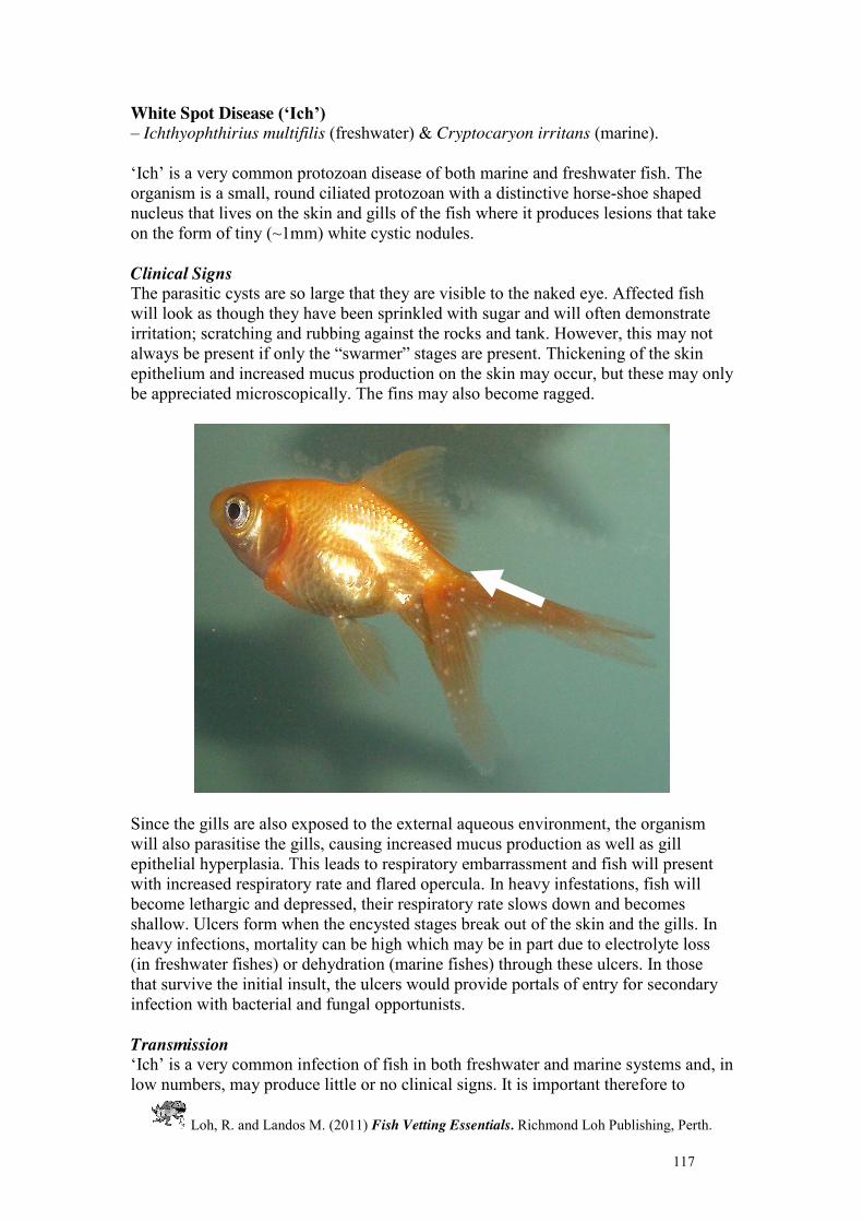
White Spot Disease (‘Ich’) – Ichthyophthirius multifilis (freshwater) & Cryptocaryon irritans (marine). ‘Ich’ is a very common protozoan disease of both marine and freshwater fish. The organism is a small, round ciliated protozoan with a distinctive horse-shoe shaped nucleus that lives on the skin and gills of the fish where it produces lesions that take on the form of tiny (~1mm) white cystic nodules. Clinical Signs The parasitic cysts are so large that they are visible to the naked eye. Affected fish will look as though they have been sprinkled with sugar and will often demonstrate irritation; scratching and rubbing against the rocks and tank. However, this may not always be present if only the “swarmer” stages are present. Thickening of the skin epithelium and increased mucus production on the skin may occur, but these may only be appreciated microscopically. The fins may also become ragged.
Since the gills are also exposed to the external aqueous environment, the organism will also parasitise the gills, causing increased mucus production as well as gill epithelial hyperplasia. This leads to respiratory embarrassment and fish will present with increased respiratory rate and flared opercula. In heavy infestations, fish will become lethargic and depressed, their respiratory rate slows down and becomes shallow. Ulcers form when the encysted stages break out of the skin and the gills. In heavy infections, mortality can be high which may be in part due to electrolyte loss (in freshwater fishes) or dehydration (marine fishes) through these ulcers. In those that survive the initial insult, the ulcers would provide portals of entry for secondary infection with bacterial and fungal opportunists. Transmission ‘Ich’ is a very common infection of fish in both freshwater and marine systems and, in low numbers, may produce little or no clinical signs. It is important therefore to

Loh, R. and Landos M. (2011) Fish Vetting Essentials. Richmond Loh Publishing, Perth.
118 - 118 -
always look for other pathogens (bacteria, fungi, other protozoa etc.) when presented with a sick fish with only a small burden of Ich, as this may be an incidental problem. ‘Ich’ replicates quickly and so a minor burden in one fish tank can rapidly become a serious burden to a tank full of fishes if left untreated. Mature Ich parasites that have been feeding on the host eventually fall off the host to the bottom of the tank. There they secrete protective cysts around themselves and begin to divide, producing many hundreds of swarmers (infective stages) which swim off in search of a host. The transmission of ‘ich’ is facilitated by high stocking densities of fish. Individual encysted stages on fish may go unnoticed by fish-owners and yet be a major source of infection to other fish. Temperature also plays a big role in disease transmission with the lifecycle being completed more rapidly at higher temperatures (3-4 days at 21oC vs 5 weeks at 10oC). This factor is particularly important when dealing with Cryptocaryon outbreaks as it is more temperature-governed than Ichthyophthirius. Diagnosis Diagnosis is made by performing a skin or gill scraping in the region of one of the white lesions and by identification of the spherical ciliate organisms, with their characteristic slow spinning motion, in a fresh wet preparation. Notice also that the parasites can be of various sizes, which is pathognomonic for “Ich” (other ciliated parasites are of uniform size and shape). Notice the horse-shoe shaped nucleus (inset).
Treatment Formalin may be used, however, because it displaces dissolved oxygen, it is not recommended if fish exhibit severe respiratory embarrassment. Malachite green + formalin combination is the most effective treatment since the mixture has a “synergistic effect” and a smaller concentration of each ingredient is used. Dip treatments and osmotic challenges will only be effective against the non-encysted
Loh, R. and Landos M. (2011) Fish Vetting Essentials. Richmond Loh Publishing, Perth.
119 - 119 -
stages of the parasites. Thus, this will need to be repeated every 2-3 days for 10 days. Thermal challenge by raising the water temperature to at least 32oC for a few hours every 3 to 5 days (provided the water is well-aerated and that the fishes will tolerate it) is another method used. The high temperature interferes with the reproduction of the parasites. Since the organisms are obligate parasites, allowing the aquarium or pond to be left fish-free for at least 7 days at >20oC usually eliminates the white spot parasites. It has been reported that some fishes that recover from the infection will develop immunity to the disease. See the medication section under “Protozoa – General”. <><

Loh, R. and Landos M. (2011) Fish Vetting Essentials. Richmond Loh Publishing, Perth.
201 - 201 -
AQUARIUM PET ADVICE FORM SUBMITTER DETAILS
FISH DETAILS WATER QUALITY PARAMETERS
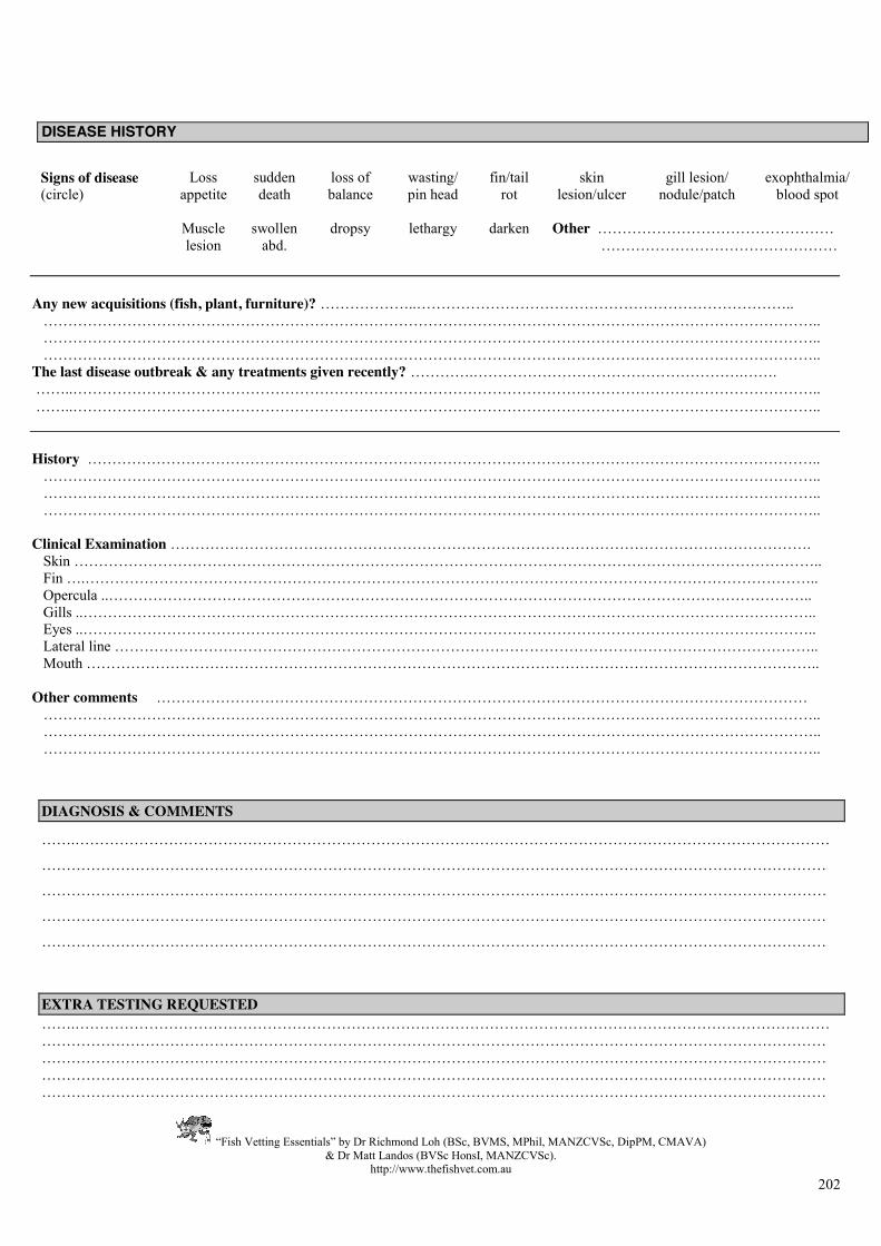
DISEASE HISTORY
TheFishVet Website: www.thefishvet.com.au E-mail: [email protected] Mob: 0421 822 383
Name …………………………………………………………………. Address ……………………………………………………………….. ……………………………………………………...………………..… E-mail …………………………………………………………………
Name …………………………………………………….. Species……………………………………………………………….……………..…………………………………………………….….…………………………………………. Size: ….……mm Weight : …….…… g Age: ……….. Tank mates & population size ……………………………………………………………….……………..……………………………………………….…….….……………………………………………………. Duration of problem…………..days/weeks
Tank dimensions ………………………..…………….
Sex (circle) Male Female Unknown
Stress factors (circle) new fish maturation overcrowding temperature anoxia algae aggression
Water data (specify) freshwater seawater brackish SG ……… or salinity ….……% Filter type under-gravel canister trickle other …………………….….. Aeration air pump power head other ……….………….…….. Substrate gravel (rough / fine) shell grit pebbles other ………………………… Furnishings plant cover …….% bog wood caves other …………………….…… Lighting incandescent fluorescent metal Halide other ………………..……….. Miscellaneous heater protein skimmer UV/ozone other …………………………. Diet ………………………………………………………………………………………………………………….
PRELIMINARY TESTS NH4
+ ……………mg/L NO3
- …………...mg/L NO2 ……………mg/L pH ………………… Temp ……………..oC Frequency of water changes ………………… % water change ………..
Date ………………………………………… Phone (H) ……………………..………….. (W) ……………………………..…. Mob …..……………………………....……
ADDITIONAL TESTS PO4
-3 ..………..……mg/L Ca+2 ………..……..mg/L Fe (NC) ….…...……mg/L (C) ……..……...mg/L Hardness (G) ….……….…mg/L (CO3
-) ………….mg/L Cl2 ………….………. Cu+2 ………………… Other ………………..
“Fish Vetting Essentials” by Dr Richmond Loh (BSc, BVMS, MPhil, MANZCVSc, DipPM, CMAVA) & Dr Matt Landos (BVSc HonsI, MANZCVSc).
http://www.thefishvet.com.au 202
DISEASE HISTORY
DIAGNOSIS & COMMENTS
…….………………………………………………………………………………………………………………………………………
……………………………………………………………………………………………………………………………………………
……………………………………………………………………………………………………………………………………………
……………………………………………………………………………………………………………………………………………
……………………………………………………………………………………………………………………………………………
EXTRA TESTING REQUESTED …….……………………………………………………………………………………………………………………………………………………………………………………………………………………………………………………………………………………………………………………………………………………………………………………………………………………………………………………………………………………………………………………………………………………………………………………………………………………………………………………………………………………………………………………………
Signs of disease (circle)
Loss appetite
sudden death
loss of balance
wasting/ pin head
fin/tail rot
skin lesion/ulcer
gill lesion/ nodule/patch
exophthalmia/ blood spot
Muscle
lesion swollen
abd. dropsy lethargy darken Other …………………………………………
…………………………………………
Any new acquisitions (fish, plant, furniture)? ………………..………………………………………………………………….. ………………………………………………………………………………………………………………………………………….. ………………………………………………………………………………………………………………………………………….. ………………………………………………………………………………………………………………………………………….. The last disease outbreak & any treatments given recently? ………….……………………………………………….……. ……..…………………………………………………………………………………………………………………………………….. ……..……………………………………………………………………………………………………………………………………..
History ………………………………………………………………………………………………………………………………….. ………………………………………………………………………………………………………………………………………….. ………………………………………………………………………………………………………………………………………….. ………………………………………………………………………………………………………………………………………….. Clinical Examination …………………………………………………………………………………………………………………. Skin …………………………………………………………………………………………………………………………………….. Fin ….………………………………………………………………………………………………………………………………….. Opercula ..…………………………………………………………………………………………………………………………….. Gills ..………………………………………………………………………………………………………………………………….. Eyes ..………………………………………………………………………………………………………………………………….. Lateral line …………………………………………………………………………………………………………………………….. Mouth ………………………………………………………………………………………………………………………………….. Other comments …………………………………………………………………………………………………………………… ………………………………………………………………………………………………………………………………………….. ………………………………………………………………………………………………………………………………………….. …………………………………………………………………………………………………………………………………………..

216