Embed Size (px)
Citation preview

PRENATAL DIAGNOSISPrenat Diagn 2008; 28: 319–322.Published online 3 March 2008 in Wiley InterScience(www.interscience.wiley.com) DOI: 10.1002/pd.1843
First-trimester maternal serum progesterone in aneuploidpregnancies
Michael Christiansen*, Tina Lindvig Sørensen, Severin Olesen Larsen and Bent Nørgaard-PedersenDepartment of Clinical Biochemistry, Statens Serum Institut, Copenhagen, Denmark
Background First-trimester maternal serum screening for Down syndrome (DS) can be improved by theuse of additional serum markers. We examined whether progesterone (P), synthesized by placenta, might be afirst-trimester maternal serum marker for fetal DS.
Materials and Methods P was quantified in first-trimester maternal serum from 42 DS, six trisomy 18 andtwo trisomy 13 pregnancies and 115 controls. Log-regression of P versus gestational age in days was used toconvert P concentrations into multiples of the median (MoM).
Results The P concentrations in controls increased with gestational age (p = 9.5 × 10⁻⁷). The log10MoM Pdistribution in DS pregnancies was not significantly different from that in controls. However, from day 58–67,the log10MoM P was elevated in DS pregnancies (n = 10) with a mean (SD) of 0.1040 (0.0956), comparedto a mean (SD) of −0.0109 (0.1661) in controls (n = 24) (p = 0.05). Five out of six trisomy 18 and bothtrisomy 13 pregnancies had a P MoM <1.
Conclusion P is not a useful marker for DS in first trimester, except perhaps in a narrow gestational agewindow from day 58 to 67. P is a trisomy 18/13 marker. Copyright 2008 John Wiley & Sons, Ltd.
KEY WORDS: progesterone; prenatal screening; first trimester; aneuploidy; placenta
INTRODUCTION
At present, the gold standard of first-trimester screen-ing for chromosomal abnormalities is the combined useof serum screening by PAPP-A and hCGβ and ultra-sound screening using nuchal translucency as a marker(Avgidou et al., 2005; Spencer, 2007; Wojdemann et al.,2005). However, as the detection rate (DR) for Downsyndrome (DS) is not higher than ca 90% for a falsepositive rate (FPR) of ca 5% ((Nicolaides et al., 2005;Spencer, 2007) there is still room for improvement ofperformance. The FPR may be reduced to 2–3% for aDR of 90% by adding a number of first-trimester ultra-sound markers, e.g. absent nasal bone or tricuspid regur-gitation (Nicolaides et al., 2005). Another approach isthe inclusion of additional serum markers. Several can-didates have been identified, e.g. ADAM 12 (Laigaardet al., 2003; 2006), hPL (Christiansen et al., 2007b), SP1(Qin et al., 1997a), ProMBP (Chen et al., 2002; Chris-tiansen et al., 1999; 2000) and inhibin A (Christiansenand Norgaard-Pedersen, 2005).
Progesterone (P) is a steroid hormone initially syn-thesized by the corpus luteum and later—from week7 to 9 (the luteal-placental shift) (Csapo and Pulkki-nen, 1978) - by the placenta. P is necessary to establishand maintain pregnancy (Rebar and Cedars, 1992) andthe synthesis is stimulated by hCG (Kratzer and Taylor,1990). Thus, P seems to be a candidate maker for Downsyndrome in first trimester. Previous studies of P in first
*Correspondence to: Michael Christiansen, Department of Clin-ical Biochemistry, Statens Serum Institut, Artillerivej 5 DK2300S, Denmark. E-mail: [email protected]
trimester have not been made with present day state-of-the-art statistical methodology (Larsen et al., 1998),but a study in gestational week 9–12 found P to beslightly, albeit significantly, reduced in DS pregnanciesand clearly reduced in trisomy 18 and 13 pregnancies(Kratzer et al., 1991).
In the present study we examine the potential ofP as a first-trimester maternal serum marker of DSin first trimester. Furthermore, we examined whetherprevious findings of low P values in trisomy 18 and13 pregnancies could be confirmed.
MATERIALS AND METHODS
Serum samples
Serum samples from 42 pregnant women (median age:38 years, range: 25–43 years, sampled at median gesta-tional age 74 days, range: 56–95 days) with a DS fetus,six pregnant women, sampled at median gestational age66 days, range 62–90 days, with a trisomy 18 fetus,and two pregnant women with a trisomy 13 fetus, sam-pled at 72 and 84 days, respectively, were retrieved fromsamples received as part of routine first-trimester serumscreening (PAPP-A and hCGβ) program performed atStatens Serum Institute (SSI), Copenhagen, and storedin a biobank as part of the quality control programat the Pregnancy Screening Registry at the SSI. Sam-ples from pregnancies with a chromosomally abnormalfetus and controls were matched for maternal age (+/−5 years) and sampled at the same time period and storedat −20 ◦C prior to analysis. All DS cases were identi-fied by cross-referencing with the Danish Cytogenetic
Copyright 2008 John Wiley & Sons, Ltd. Received: 20 May 2007Revised: 28 July 2007
Accepted: 3 August 2007Published online: 3 March 2008

320 M. CHRISTIANSEN ET AL.
Figure 1—Progesterone concentration values in maternal sera fromDS pregnancies (black circle) and control pregnancies (open circles)
Central Registry, and none of the controls were regis-tered with chromosomal disease. All cases were single-ton pregnancies. None of the pregnant women examinedhere received progesterone supplementation. Informa-tion about the gestational age, based on the crown rumplength (CRL) and maternal age, as well as drug treat-ment, was obtained from referral sheets.
Quantification of progesterone
The maternal serum concentration of progesterone wasdetermined using a commercially available TRACEassay Progesterone KRYPTOR (cat.no. 815.050,BRAHMS, Henningsdorf, Germany), used on the Kryp-tor platform (BRAHMS). The analytical range was0.2–100 nanomoles/L. The intra-assay CV was <7%and the inter-assay CV was <10%.
Data analysis
A normal median of P was established from P con-centrations measured in control pregnancies using log-regression of P on gestational age in days. The normalmedians were used to transform P concentrations in bothcontrols and affected pregnancies into multiples of themedians (MoMs) and the distribution of log10MoM Pwas established in all groups. Compatibility with thenormal distribution was assessed by the Shapiro–Wilk’stest. Means were compared using the Mann–WhitneyU-test.
RESULTS
The distribution of individual P concentrations in bothcontrols and DS pregnancies are given in Figure 1 as afunction of gestational age. A log-linear regression of Pon gestational age in days showed a highly significantincrease of log10 P with gestational age, r = 0.4384,p = 9.5∗10−7, n = 115. The regression line was: log10P = 0.0078∗days + 1.7371. The residuals were normallydistributed, W = 0.9718, p = 0.1478.
The log-regression line given above was used toconvert the P concentration values into MoM. Thedistribution of log10MoM P values as a function ofgestational age is shown in Figure 2 for both DS andcontrol pregnancies. In neither controls (r = 0.0040,p = 0.9655) nor DS pregnancies (r = −0.1073, p =0.4987) was there any significant change in log10MoMP with gestational age. The mean log10MoM P was0.0049 with a SD of 0.1506 in controls and 0.0181 witha SD of 0.1256 in DS pregnancies. The difference wasnot significant, p = 0.7361.
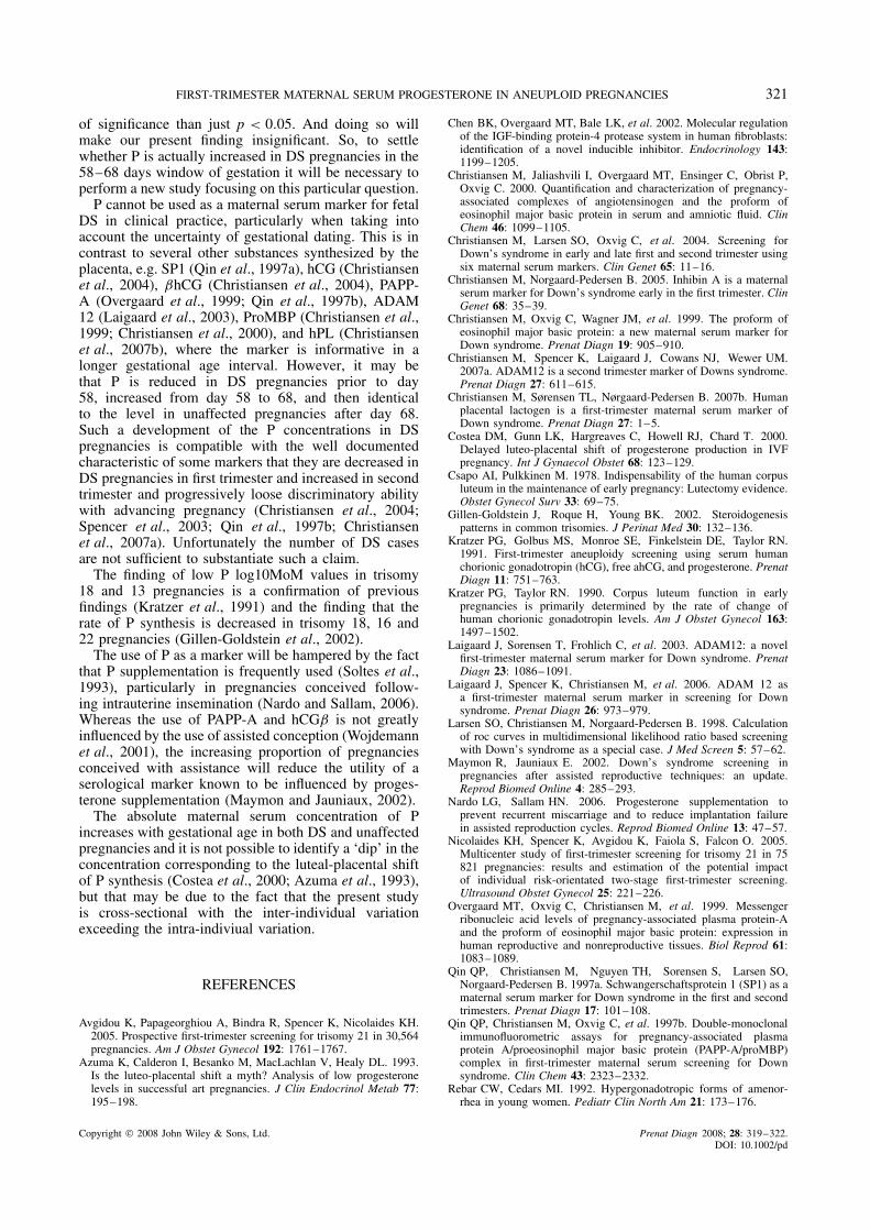
Despite the fact that the difference between thelog10MoM P values in DS pregnancies and controlswas not significant when all samples were included,a visual inspection of Figure 2 revealed that in asmall gestational age window from day 58 to 68, thelog10MoM P values were higher in DS pregnanciesthan in controls. In between the 58 and 68 day windowthere were 10 DS pregnancies with a mean log10MoMP of 0.1040 and a SD of 0.0956, marginally significantlyhigher than the mean log10MoM P of −0.0109 and SDof 0.1661 seen in 24 controls (p = 0.05).
The P log10MoM values in trisomy 18 and 13 casesare shown in Figure 2. In five out of six trisomy 18 casesand both of the trisomy 13 cases the P MoM was lessthan 1. The low number of cases preclude a statisticalanalysis.
DISCUSSION
The main finding in this paper is that the maternalserum concentration of P does not differ between DSpregnancies and unaffected pregnancies in first trimester,except perhaps for a short interval between 58 and68 days. The latter finding is, however, the result ofsplitting up the data in several subgroups and doingso makes it reasonable to use a more stringent level
Figure 2—Log10MoM values of progesterone in DS pregnancies(black circles) and control pregnancies (open circles). The brokenline marks the 1MoM level. The ellipse marks the cases where theprogesterone log10MoM values are higher than in control pregnancies.The six trisomy 18 cases are marked with open triangles and the twotrisomy 13 cases are marked by dark triangles
Copyright 2008 John Wiley & Sons, Ltd. Prenat Diagn 2008; 28: 319–322.DOI: 10.1002/pd
FIRST-TRIMESTER MATERNAL SERUM PROGESTERONE IN ANEUPLOID PREGNANCIES 321
of significance than just p < 0.05. And doing so willmake our present finding insignificant. So, to settlewhether P is actually increased in DS pregnancies in the58–68 days window of gestation it will be necessary toperform a new study focusing on this particular question.
P cannot be used as a maternal serum marker for fetalDS in clinical practice, particularly when taking intoaccount the uncertainty of gestational dating. This is incontrast to several other substances synthesized by theplacenta, e.g. SP1 (Qin et al., 1997a), hCG (Christiansenet al., 2004), βhCG (Christiansen et al., 2004), PAPP-A (Overgaard et al., 1999; Qin et al., 1997b), ADAM12 (Laigaard et al., 2003), ProMBP (Christiansen et al.,1999; Christiansen et al., 2000), and hPL (Christiansenet al., 2007b), where the marker is informative in alonger gestational age interval. However, it may bethat P is reduced in DS pregnancies prior to day58, increased from day 58 to 68, and then identicalto the level in unaffected pregnancies after day 68.Such a development of the P concentrations in DSpregnancies is compatible with the well documentedcharacteristic of some markers that they are decreased inDS pregnancies in first trimester and increased in secondtrimester and progressively loose discriminatory abilitywith advancing pregnancy (Christiansen et al., 2004;Spencer et al., 2003; Qin et al., 1997b; Christiansenet al., 2007a). Unfortunately the number of DS casesare not sufficient to substantiate such a claim.
The finding of low P log10MoM values in trisomy18 and 13 pregnancies is a confirmation of previousfindings (Kratzer et al., 1991) and the finding that therate of P synthesis is decreased in trisomy 18, 16 and22 pregnancies (Gillen-Goldstein et al., 2002).
The use of P as a marker will be hampered by the factthat P supplementation is frequently used (Soltes et al.,1993), particularly in pregnancies conceived follow-ing intrauterine insemination (Nardo and Sallam, 2006).Whereas the use of PAPP-A and hCGβ is not greatlyinfluenced by the use of assisted conception (Wojdemannet al., 2001), the increasing proportion of pregnanciesconceived with assistance will reduce the utility of aserological marker known to be influenced by proges-terone supplementation (Maymon and Jauniaux, 2002).
The absolute maternal serum concentration of Pincreases with gestational age in both DS and unaffectedpregnancies and it is not possible to identify a ‘dip’ in theconcentration corresponding to the luteal-placental shiftof P synthesis (Costea et al., 2000; Azuma et al., 1993),but that may be due to the fact that the present studyis cross-sectional with the inter-individual variationexceeding the intra-indiviual variation.
REFERENCES
Avgidou K, Papageorghiou A, Bindra R, Spencer K, Nicolaides KH.2005. Prospective first-trimester screening for trisomy 21 in 30,564pregnancies. Am J Obstet Gynecol 192: 1761–1767.
Azuma K, Calderon I, Besanko M, MacLachlan V, Healy DL. 1993.Is the luteo-placental shift a myth? Analysis of low progesteronelevels in successful art pregnancies. J Clin Endocrinol Metab 77:195–198.
Chen BK, Overgaard MT, Bale LK, et al. 2002. Molecular regulationof the IGF-binding protein-4 protease system in human fibroblasts:identification of a novel inducible inhibitor. Endocrinology 143:1199–1205.
Christiansen M, Jaliashvili I, Overgaard MT, Ensinger C, Obrist P,Oxvig C. 2000. Quantification and characterization of pregnancy-associated complexes of angiotensinogen and the proform ofeosinophil major basic protein in serum and amniotic fluid. ClinChem 46: 1099–1105.
Christiansen M, Larsen SO, Oxvig C, et al. 2004. Screening forDown’s syndrome in early and late first and second trimester usingsix maternal serum markers. Clin Genet 65: 11–16.
Christiansen M, Norgaard-Pedersen B. 2005. Inhibin A is a maternalserum marker for Down’s syndrome early in the first trimester. ClinGenet 68: 35–39.
Christiansen M, Oxvig C, Wagner JM, et al. 1999. The proform ofeosinophil major basic protein: a new maternal serum marker forDown syndrome. Prenat Diagn 19: 905–910.
Christiansen M, Spencer K, Laigaard J, Cowans NJ, Wewer UM.2007a. ADAM12 is a second trimester marker of Downs syndrome.Prenat Diagn 27: 611–615.
Christiansen M, Sørensen TL, Nørgaard-Pedersen B. 2007b. Humanplacental lactogen is a first-trimester maternal serum marker ofDown syndrome. Prenat Diagn 27: 1–5.
Costea DM, Gunn LK, Hargreaves C, Howell RJ, Chard T. 2000.Delayed luteo-placental shift of progesterone production in IVFpregnancy. Int J Gynaecol Obstet 68: 123–129.
Csapo AI, Pulkkinen M. 1978. Indispensability of the human corpusluteum in the maintenance of early pregnancy: Lutectomy evidence.Obstet Gynecol Surv 33: 69–75.
Gillen-Goldstein J, Roque H, Young BK. 2002. Steroidogenesispatterns in common trisomies. J Perinat Med 30: 132–136.
Kratzer PG, Golbus MS, Monroe SE, Finkelstein DE, Taylor RN.1991. First-trimester aneuploidy screening using serum humanchorionic gonadotropin (hCG), free ahCG, and progesterone. PrenatDiagn 11: 751–763.
Kratzer PG, Taylor RN. 1990. Corpus luteum function in earlypregnancies is primarily determined by the rate of change ofhuman chorionic gonadotropin levels. Am J Obstet Gynecol 163:1497–1502.
Laigaard J, Sorensen T, Frohlich C, et al. 2003. ADAM12: a novelfirst-trimester maternal serum marker for Down syndrome. PrenatDiagn 23: 1086–1091.
Laigaard J, Spencer K, Christiansen M, et al. 2006. ADAM 12 asa first-trimester maternal serum marker in screening for Downsyndrome. Prenat Diagn 26: 973–979.
Larsen SO, Christiansen M, Norgaard-Pedersen B. 1998. Calculationof roc curves in multidimensional likelihood ratio based screeningwith Down’s syndrome as a special case. J Med Screen 5: 57–62.
Maymon R, Jauniaux E. 2002. Down’s syndrome screening inpregnancies after assisted reproductive techniques: an update.Reprod Biomed Online 4: 285–293.
Nardo LG, Sallam HN. 2006. Progesterone supplementation toprevent recurrent miscarriage and to reduce implantation failurein assisted reproduction cycles. Reprod Biomed Online 13: 47–57.
Nicolaides KH, Spencer K, Avgidou K, Faiola S, Falcon O. 2005.Multicenter study of first-trimester screening for trisomy 21 in 75821 pregnancies: results and estimation of the potential impactof individual risk-orientated two-stage first-trimester screening.Ultrasound Obstet Gynecol 25: 221–226.
Overgaard MT, Oxvig C, Christiansen M, et al. 1999. Messengerribonucleic acid levels of pregnancy-associated plasma protein-Aand the proform of eosinophil major basic protein: expression inhuman reproductive and nonreproductive tissues. Biol Reprod 61:1083–1089.
Qin QP, Christiansen M, Nguyen TH, Sorensen S, Larsen SO,Norgaard-Pedersen B. 1997a. Schwangerschaftsprotein 1 (SP1) as amaternal serum marker for Down syndrome in the first and secondtrimesters. Prenat Diagn 17: 101–108.
Qin QP, Christiansen M, Oxvig C, et al. 1997b. Double-monoclonalimmunofluorometric assays for pregnancy-associated plasmaprotein A/proeosinophil major basic protein (PAPP-A/proMBP)complex in first-trimester maternal serum screening for Downsyndrome. Clin Chem 43: 2323–2332.
Rebar CW, Cedars MI. 1992. Hypergonadotropic forms of amenor-rhea in young women. Pediatr Clin North Am 21: 173–176.
Copyright 2008 John Wiley & Sons, Ltd. Prenat Diagn 2008; 28: 319–322.DOI: 10.1002/pd

322 M. CHRISTIANSEN ET AL.
Soltes B, Molo MW, Binor Z, Rawlins RG, Radwanska E. 1993.Hormonal profiles of early gestations with abnormal karyotype.Fertil Steril 59: 810–814.
Spencer K. 2007. Aneuploidy screening in the first trimester. Am JMed Genet C Semin Med Genet 145: 18–32.
Spencer K, Crossley JA, Aitken DA, Nix AB, Dunstan FD, WilliamsK. 2003. The effect of temporal variation in biochemical markersof trisomy 21 across the first and second trimesters of pregnancyon the estimates of individual patient-specific risks and detectionrates for Down’s syndrome. Ann Clin Biochem 40: 219–231.
Wojdemann KR, Larsen SO, Shalmi A, Sundberg K, Christiansen M,Tabor A. 2001. First trimester screening for Down syndrome andassisted reproduction: no basis for concern. Prenat Diagn 21:563–565.
Wojdemann KR, Shalmi AC, Christiansen M, et al. 2005. Improvedfirst-trimester Down syndrome screening performance by loweringthe false-positive rate: a prospective study of 9941 low-risk women.Ultrasound Obstet Gynecol 25: 227–233.
Copyright 2008 John Wiley & Sons, Ltd. Prenat Diagn 2008; 28: 319–322.DOI: 10.1002/pd