Embed Size (px)
Citation preview
Finish Cancer Lecture
Tissues and Cell-Cell Interactions
Cancer Cells Are Created when Certain Genes are Mutated
Mutations can be Inherited, Introduced by Viruses, or Result of DNA Damage
1. Oncogenes - Gene whose presence can trigger inappropriate cell proliferation.Example: ras, bcl-2(Normal version of gene: Proto-oncogene)
2. Tumor Suppressors- Gene whose absence or inactivation can lead to cancerUsually Function to Block Cell Cycle ProgressionExample: p53, Rb
DNA Repair Genes- Increase Rate of Mutation, provide opportunity for mutation in growth controlling genes, increase rate of tumor progression
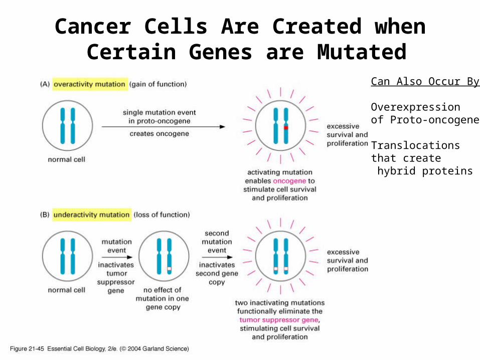
Can Also Occur By:
Overexpression of Proto-oncogene
Translocations that create hybrid proteins
Cancer Cells Are Created when Certain Genes are Mutated

Oncogenes are Found in Mitogen andGrowth Factor Signal Transduction Pathways
Mutation of Proto-oncogene- Constitutively Active DownstreamSignal Transduction Pathway

Inactivation of Tumor Suppressor Rb
Common Target for Viruses that Cause Tumors (along with p53)

Cancer Cells Exhibit Unlimited Proliferative Ability
Cancer cells avoid Senescence by inactivating tumor suppressor genes, p53 and Rb.
Cancer Cells will continue to divide for a period of time
Crisis Point – Large number of Cancer Cells Die- Result of catastrophic rearrangements- due to lack of telomerase
Rare Occasion A Cell Survives- It is Immortalized.At some point- de-repressed telomerase expression
~ 90% of cancer cells express significant levels of telomerase

Structural and Functional Framework of Animal Tissues: Formation and Maintenance
4 Main Types of Tissue- Nervous, Muscle, Epithelial and Connective
Tissues Are Composed of: 1) Organized Groups of Cells with Similar Function 2) Extracellular Matrix (ECM)- Network of Proteins and Sugars that
Lies in the Intercellular Spaces
To Create and Maintain Tissues Cells Need to Adhere to One Another or to the ECM1) Structure of the ECM
2) Cell-Cell and Cell-ECM JunctionsSpecialized Protein Complexes that Provide Specific Means of Joining Cells
in Long Term Association
2) Cell-Cell and Cell-ECM Adhesion MoleculesTransient Interactions involving Transmembrane Proteins Do Not form Stable Cell Junctions
Proteins of ECM and Cell Junctions Control 3-D Organization of Cells in Tissues and Growth, Movement, Shape and Differentiation

ECM of Loose Connective Tissue
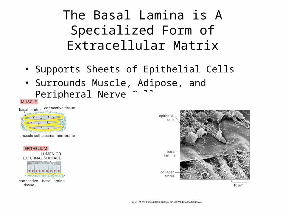
• Supports Sheets of Epithelial Cells
• Surrounds Muscle, Adipose, and Peripheral Nerve Cells
The Basal Lamina is A Specialized Form of Extracellular Matrix

The ECM of Animal Cells
Composed of :1) Structural Proteins-
Give Strength and FlexibilityExamples: Collagen and Elastin
2) Proteoglycans- Protein Polysaccharide ComplexesProvide the Gel-Like Matrix in which Structural Proteins are Embedded
3) Adhesive Glycoproteins- Attach Cells to the Matrix and Matrix Proteins to Each OtherExample: Fibronectins and Laminins
Differences in Types of ECM Reflect Differences in :
1) The Types of Structural Proteins and Kinds of Proteoglycans Present
2) The Ratio of Structural Protein to Proteoglycans Present

Structural Proteins of the ECM
1)Collagens- Major Proteins of the ECM
– Long, Stiff, Triple Stranded Collagen Super Helix– High Tensile Strength, Provide Mechanical Strength– Nearly all Animal Cells Synthesize and Secrete at least
One Form of Collagen (27 Families of Collagen Exist)
2) Elastic Fibers – Impart Elasticity and Flexibility– Fibers Formed from Elastin and a Glycoprotein that
forms a Sheath around Elastin

Assembly and Structure of Collagen
Forms: Fibrillar, Fibril Associated, or Network Forming
Once Assembled- OftenLarger than Cells that Secreted Them!
Three StrandsSelf AssembleIn ER
Fibrils and FibersOnly Assemble After Secretion

Elastic Fibers Impart Elasticity and Flexibility to the ECM
-Strength Arises from Covalent Crosslinks-Abundant in:Blood vessels, skin, lungs-Only Assemble into FibersAfter Secretion
Blood Vessel Elastic Fibers

Collagen and Elastic Fibers Are Embedded in a Matrix of Proteoglycans
ProteoglycansProtein Core with Glycosaminoglycans (GAGs)-
Unbranched polysaccharide chains- Covalently Attached
Function:• Make Up the Hydrated Gel Like Network• Resist Compressive Forces• GAG Bristles Act as Filters- Limit Diffusion • Can Bind Growth Factors, Structural Proteins, and Cell
Surface Receptors• Some are Transmembrane Proteins

Glycosaminoglycans (GAGs) Are Long Unbranched Polysaccharides of Repeating Disaccharides
Example: Hyaluronan

Proteoglycans and GAGs Can Form Large Aggregates
Aggregates from Cartilage

Third Component of ECM:Adhesive Glycoproteins
Attach Cells to ECM and ECM components to One Another-Bind Collagen and Proteoglycans- Organize Matrix-Binding Site for Cell Surface Receptors called Integrins
Fibronectins- Prototype for Adhesive Glycoproteins– Most Common Adhesive Glycoprotein– Approximately 50% Carbohydrate– Bind Cells to the Matrix and Guides Cellular Movement
Laminins-– Distinct Adhesive Component of Specialized Basal Lamina– Function as a Structural Support and as a Permeability Barrier

Fibronectin binds Cell Surface Receptors Called Integrins Linking Cell Surface to ECM
Fibronectin Focal Adhesions Involved in
Cell Migration
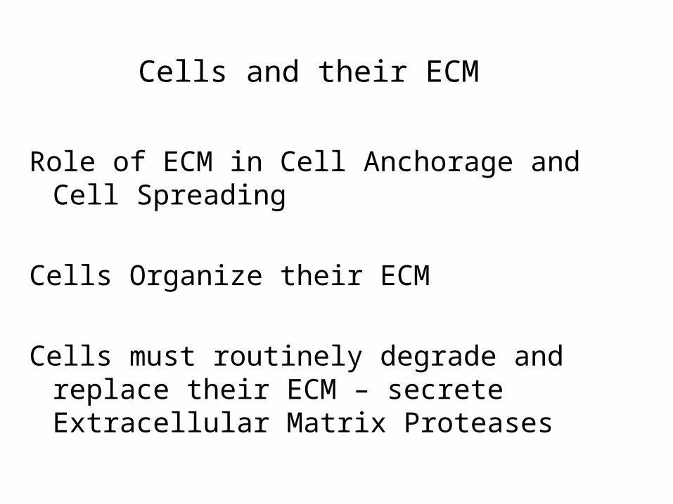
Cells and their ECM
Role of ECM in Cell Anchorage and Cell Spreading
Cells Organize their ECM
Cells must routinely degrade and replace their ECM – secrete Extracellular Matrix Proteases

Cell-Cell Adhesion and Cell-Matrix Adhesion

The Cadherin Superfamily
• Responsible for Ca2+ Dependent Cell-Cell Adhesion in Vertebrates
• Main Adhesion Molecule Holding Cells Together in Early Embryonic Development
• Primarily Responsible for Stable Junctions involving Cytoskeleton
• Typically A Single Pass Transmembrane Glycoproteins
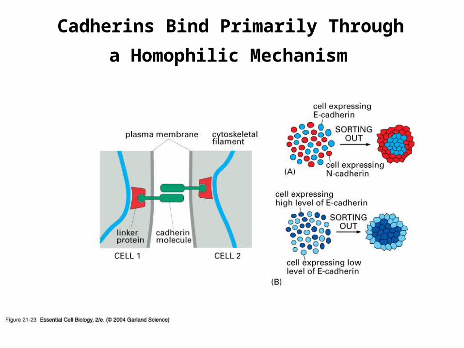
Cadherins Bind Primarily Through
a Homophilic Mechanism
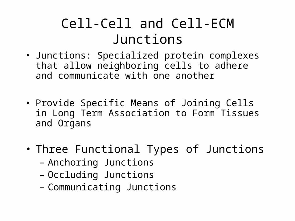
Cell-Cell and Cell-ECM Junctions
• Junctions: Specialized protein complexes that allow neighboring cells to adhere and communicate with one another
• Provide Specific Means of Joining Cells in Long Term Association to Form Tissues and Organs
• Three Functional Types of Junctions – Anchoring Junctions – Occluding Junctions– Communicating Junctions
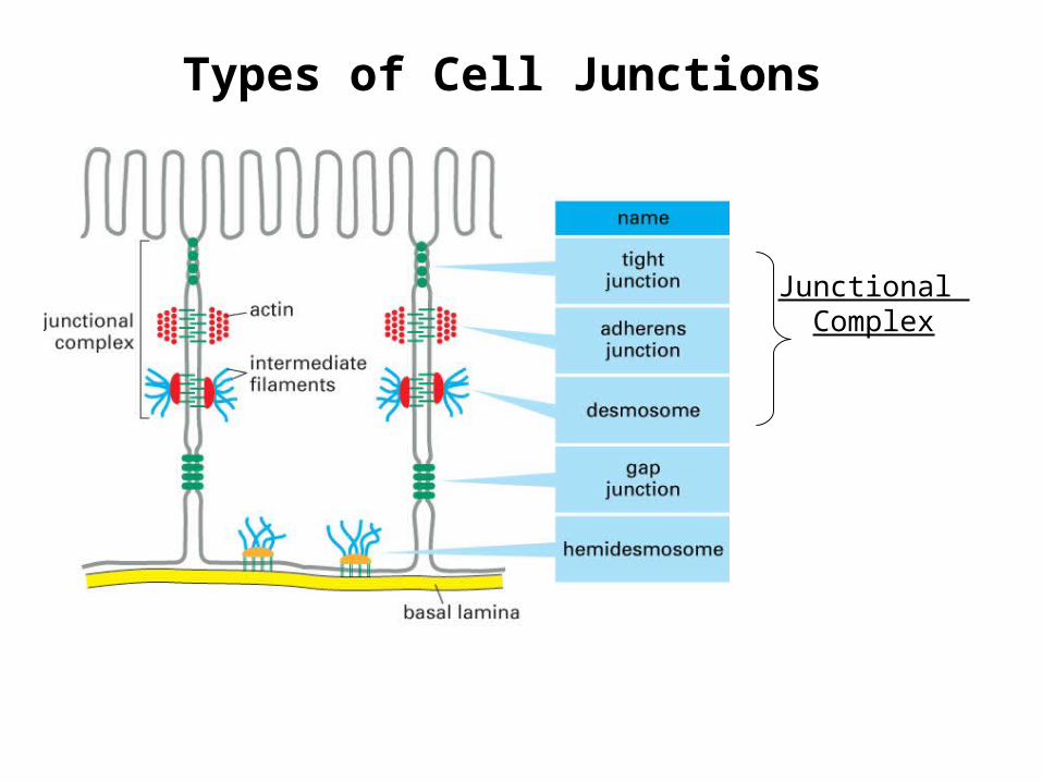
Types of Cell Junctions
Junctional Complex

Anchoring Junctions
Link Cells Together into Tissues
Enable Cells to Function as a Unit
- Involve Anchoring Cytoskeleton of One Cell to the Cytoskeleton of Neighboring Cell or to ECM
- Important for Tissues Subjected to Mechanical Stress
- Three Types of Anchoring Junctions:- Adherens Junctions - Desmosomes- Hemidesmosomes

Anchoring Junctions:Adherens Junctions Link Actin Cytoskeleton of Adjacent Cells
Function: To Hold Neighboring Cells Together
Epithelial and Non-Epithelial Tissues
1) Involve Transmembrane Proteins called Cadherins
2) Provide Enough Strength for Tissue to Resist Stress/Change Shape

Anchoring Junctions:Desmosomes Indirectly Connect Intermediate
Filaments of Adjacent Cells
Key Function:Resisting Physical Stresses(Particularly to Epithelial Sheets)
Epithelial and Nonepithelial Tissue
Involve Non-Traditional Cadherin ProteinsAnchor Proteins and Intermediate Filaments
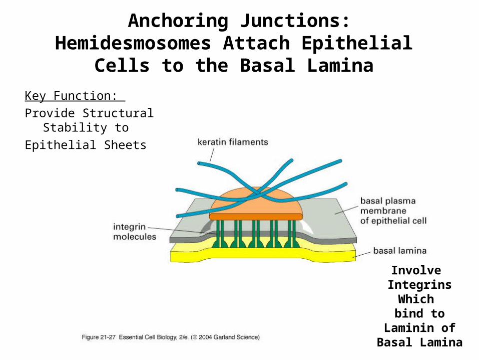
Involve IntegrinsWhich bind to
Laminin ofBasal Lamina
Key Function:
Provide Structural Stability to
Epithelial Sheets
Anchoring Junctions:Hemidesmosomes Attach Epithelial
Cells to the Basal Lamina

Occluding Junctions: Tight Junctions
Form Selectively Permeable
Barriers between Epithelial and Endothelial Cells
Functions:
1) Regulate Paracellular Transport
(Transport in space between cells)
2) Prevent Diffusion of Membrane
Proteins- Preserve Cell Polarity
Involves Transmembrane
Proteins from Adjacent Cells
-Claudins , Occludins, and Junctional Adhesion Molecules (JAM)
-Structural proteins

Experimental Evidence Demonstrating Tight Junctions Create a Selectively Permeable Barrier
-The Seal is Not Absolute!-Variations in Selectivity of Barrier Exist

Communication Junctions:Gap Junctions
Found in Most Animal Cells
Function:Allow Electrical and Chemical Communication Between Cells
Open Channel- Provides Point of Cytoplasmic Contact Between Two Adjacent Cells
Formed by Connexons
Like Ion Channels- Regulated Opening/Closing
No Macromolecules - Just Inorganic Ions, Small Molecules

Communication Junctions:Gap Junctions
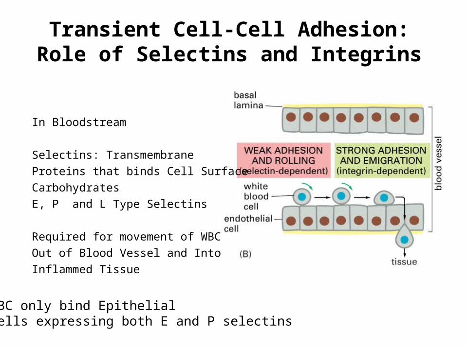
Transient Cell-Cell Adhesion:Role of Selectins and Integrins
In Bloodstream
Selectins: Transmembrane
Proteins that binds Cell Surface
Carbohydrates
E, P and L Type Selectins
Required for movement of WBC
Out of Blood Vessel and Into
Inflammed Tissue
WBC only bind Epithelial Cells expressing both E and P selectins

Office Hours This Week
Tuesday
B430 Nelson
5:30- 7:30 pm


![Chapter 4 cell & tissues (1) [compatibility mode]](https://img.dokumen.tips/doc/110x75/55a439a01a28ab3f5c8b468b/chapter-4-cell-tissues-1-compatibility-mode.jpg)