Embed Size (px)
Citation preview

Fine taxonomic sampling of nervous systemswithin Naididae (Annelida: Clitellata) revealsevolutionary lability and revised homologies ofannelid neural componentsZattara and Bely
Zattara and Bely Frontiers in Zoology (2015) 12:8 DOI 10.1186/s12983-015-0100-6

Zattara and Bely Frontiers in Zoology (2015) 12:8 DOI 10.1186/s12983-015-0100-6
RESEARCH Open Access
Fine taxonomic sampling of nervous systemswithin Naididae (Annelida: Clitellata) revealsevolutionary lability and revised homologies ofannelid neural componentsEduardo E Zattara1,2* and Alexandra E Bely1
Abstract
Introduction: An important goal for understanding how animals have evolved is to reconstruct the ancestralfeatures and evolution of the nervous system. Many inferences about nervous system evolution are weak becauseof sparse taxonomic sampling and deep phylogenetic distances among species compared. Increasing samplingwithin clades can strengthen inferences by revealing which features are conserved and which are variable withinthem. Among the Annelida, the segmented worms, the Clitellata are typically considered as having a largelyconserved neural architecture, though this view is based on limited sampling.
Results: To gain better understanding of nervous system evolution within Clitellata, we used immunohistochemistryand confocal laser scanning microscopy to describe the nervous system architecture of 12 species of the basallybranching family Naididae. Although we found considerable similarity in the nervous system architecture of naididsand that of other clitellate groups, our study identified a number of features that are variable within this family,including some that are variable even among relatively closely related species. Variable features include theposition of the brain, the number of ciliary sense organs, the presence of septate ventral nerve cord ganglia,the distribution of serotonergic cells in the brain and ventral ganglia, and the number of peripheralsegmental nerves.
Conclusions: Our analysis of patterns of serotonin immunoreactive perikarya in the central nervous systemindicates that segmental units are not structurally homogeneous, and preliminary homology assessmentssuggest that whole sets of serotonin immunoreactive cells have been gained and lost across the Clitellata.We also found that the relative position of neuroectodermal and mesodermal segmental components issurprisingly evolutionarily labile; in turn, this revealed that scoring segmental nerves by their position relativeto segmental ganglia rather than to segmental septa clarifies their homologies across Annelida. We concludethat fine taxonomic sampling in comparative studies aimed at elucidating the evolution of morphologicaldiversity is fundamental for proper assessment of trait variability.
Keywords: Ancestral character estimation, Annelida, Clitellata, Comparative morphology, Evolution, Homology,Naididae, Nervous systems, Neurophylogeny
* Correspondence: [email protected] of Biology, University of Maryland, College Park, MD 20740, USA2Current address: Department of Biology, Indiana University, 915 E. ThirdStreet, Myers Hall 150, Bloomington, IN 47405-7107, USA
© 2015 Zattara and Bely; licensee BioMed Central. This is an Open Access article distributed under the terms of the CreativeCommons Attribution License (http://creativecommons.org/licenses/by/4.0), which permits unrestricted use, distribution, andreproduction in any medium, provided the original work is properly credited. The Creative Commons Public DomainDedication waiver (http://creativecommons.org/publicdomain/zero/1.0/) applies to the data made available in this article,unless otherwise stated.

Zattara and Bely Frontiers in Zoology (2015) 12:8 Page 2 of 20
IntroductionComplex nervous systems are characteristic of eume-tazoan taxa and, because their study can help to under-stand organismal function and evolution, they have beenof particular interest to zoologists for several centuries[1-4]. Nervous systems play crucial roles integrating in-ternal and external information into physiological andbehavioral responses [2]. While incredibly diverse acrossmajor animal groups, nervous system architectures tendto be, by comparison, relatively well conserved withinphyla [1,2]. As a result, many studies aimed at under-standing the evolution of animal nervous systems havedrawn conclusions from comparisons of only a few rep-resentatives from widely distant groups (e.g., flies andmice) [3-6]. Inferences from such studies are typicallybased on the similarities identified across these distantlyrelated species, but these inferences hinge on the as-sumption that the traits in question are invariable atlower taxonomic levels. In order to make strong infer-ences about the evolution of animal nervous systems,their structure needs to be investigated in a broad arrayof taxa and with fine taxonomic sampling.The nervous system of the phylum Annelida (seg-
mented worms) comprises a central nervous system(CNS), composed of an anterior dorsal brain linked viacircumesophageal connectives to a ventral nerve cordthat is segmentally ganglionated, and a peripheral ner-vous system (PNS) composed of nerves branching off ofthe CNS components (Figure 1). Based on descriptionsfrom a limited number of primarily polychaete species(summarized by Bullock and Horridge [2]), the annelid
Figure 1 Overview of the naidid ground plan. A) Basic annelid body plan.composed of the prostomium (pr) and peristomium (pe), followed by a varegion, the pygidium (py). In front of the pygidium is the posterior growthof the nervous system in naidids. This schematic shows the anterior centraperipheral nervous system (green). Anterior is to the left in this and all figuconnective; con: interganglion connective; dch: dorsal chaetae; gut: ciliatedpharynx; pnI-IV: peripheral segmental nerve I-IV; pr: prostomium; prn: prostomganglion; sep: intersegmental septum; vch: ventral chaetae.
nervous system was originally inferred to have a highlyconserved ground plan. However, more recent studieson a broader range of annelids have revealed enormousvariation of the annelid nervous system, especially re-garding the morphology of the ventral nerve cord andthe number and pattern of peripheral nerves, raisingnew questions about the ancestral architecture and evo-lution of the annelid nervous system [7].The Clitellata are a large annelid subclade to which
most freshwater and terrestrial annelids belong. The ner-vous system of clitellates has often been considered tobe a simpler and less variable version of the nervous sys-tem typical of the primarily marine polychaetes; how-ever, this inference is based on studies of a few clitellatespecies, mostly earthworms and leeches, with rather spe-cialized morphology [8-10] and which may not closelyreflect the ancestral clitellate condition. Clitellates com-prise Naididae (water nymph worms), Crassiclitellata(earthworms), Enchytraeidae (pot worms), Lumbriculi-dae (blackworms) and Hirudina (leeches). The Naididae(sensu Erséus et al. [11]) are the sister clade to mostother clitellates [9,11,12] and knowledge of naidid ner-vous system architecture is thus of particular importancefor inferring how the nervous system has evolved withinthe clitellates, what the ancestral clitellate nervous sys-tem was like, and how it relates to the nervous system ofclosely related polychaetes.Available studies of nervous system structure in naidids
are few and are difficult to analyze comparatively. Olderdescriptions based on direct observation, light microscopy,and histological sectioning [13-15] provide different kinds
The annelid body consists of an anterior non-segmental regionriable number of segments (grey bars), and a posterior non-segmentalzone (pgz), where new segments are made. B) Generalized structurel nervous system (blue), ventral nerve cord neuropil (yellow) andres unless otherwise indicated. Labels: br: brain; cec: circumesophagealgut; mo: mouth; pe: peristomium; pgz: posterior growth zone; phx:ial nerves; py: pygidium; sXg: segment x ganglion; seg: subesophageal

Zattara and Bely Frontiers in Zoology (2015) 12:8 Page 3 of 20
of information than newer studies using immunohisto-chemistry and whole-mount confocal laser scanning mi-croscopy [16-19]. Studies using consistent techniques,sampling at a fine taxonomic scale, and analyzing data in aphylogenetic framework are needed in order to reconstructthe ancestral naidid nervous system architecture and how ithas evolved. Such studies can identify conservative andvariable elements of the nervous system and should be par-ticularly useful in identifying possible homologies betweenneural elements (e.g., nerves, cell types) across species, atask usually made challenging by the high degree of serialduplication characteristic of nervous system evolution.In this paper, we describe and compare the nervous
system architecture of 12 species of Naididae Ehrenberg,1828 (sensu Erseus et al. [11]), representing four out ofseven naidid subfamilies: Tubificinae - Tubifex tubifex;Pristininae - Pristina leidyi and Pristina æquiseta; Rhya-codrilinae - Monopylephorus rubroniveus; and Naidinae -Dero digitata, Dero furcata, Allonais paraguayensis,Paranais litoralis, Amphichaeta sp., Chaetogaster dia-phanus, Nais stolci and Stylaria lacustris. We base ourdescriptions on adult individuals immunostained foracetylated-alpha-tubulin and serotonin, known to label asignificant fraction of the neurites and some perikarya[16-21], along with labeled phalloidin to visualize mus-cular F-actin and DAPI as a nuclear counterstain, andimaged using confocal laser scanning microscopy. Wefocus in particular on the location and organization ofimmunoreactive elements of the brain and ventral nervecord, the topological relationship between the ventralganglia and the mesodermal septa, and the number andbranching architecture of peripheral nerves. Based onour new descriptions and available published data, weidentify conserved and variable elements of the naididnervous system and propose possible homologies forsome of these elements. We discuss our findings in thecontext of current knowledge about the phylogenetic re-lationships within this family, as well as relationshipswithin the Clitellata and Annelida more broadly, provid-ing insight into the evolution of the nervous systemwithin these groups.
Results and discussionAn important goal for understanding how animals haveevolved is to reconstruct the ancestral features and evo-lution of the nervous system. Many inferences aboutnervous system evolution are weak, though, becausetaxonomic sampling is sparse and phylogenetic distancesbetween species compared are deep. Increasing samplingwithin specific clades can strengthen such inferences byrevealing which features are conserved and which arevariable within these groups. In the Annelida, the seg-mented worms, considerable variation in nervous systemarchitecture has been reported for marine polychaete
families [7] but the terrestrial and freshwater Clitellataare typically viewed as having a simple and conservednervous system [7,15]. However, this view is based on in-formation from a limited number of species spanningthis clade and, importantly, no detailed comparativestudies within subgroups, such as within families, areavailable to provide insight into variability and conserva-tion of neural architecture.To address this gap, we characterized the morphology
of the nervous system in 12 species of naidids using im-munohistochemistry and confocal laser scanning mi-croscopy. In the interest of brevity, we provide detaileddescriptions and diagrams as Supplementary Informa-tion, including diagrams of the nervous system of 10species (Additional file 1: Figure S1, Additional file 2:Figure S2, Additional file 3: Figure S3, Additional file 4:Figure S4, Additional file 5: Figure S5, Additional file 6:Figure S6, Additional file 7: Figure S7, Additional file 8:Figure S8, Additional file 9: Figure S9 and Additionalfile 10: Figure S10), an overview of a generalized naididbody segment (Additional file 11: Figure S11), imagepanels showing data for all species (Additional file 12:Figure S12, Additional file 13: Figure S13, Additionalfile 14: Figure S14, Additional file 15: Figure S15, Additionalfile 16: Figure S16, Additional file 17: Figure S17 andAdditional file 18: Figure S18), and morphological de-scriptions for each species (Additional file 19). In ourdescriptions, we use whenever possible the terminologydefined by Richter et al. [22]. A summary of the char-acter states for all neural traits we found to be variableis provided in Table 1.Below, we first synthesize the results of our compara-
tive analysis of the nervous system morphology of the 12naidid species we studied, giving an overview of the com-mon patterns of nervous system components, followed byremarks on their variability. We then discuss the implica-tions of our findings for understanding the stability or la-bility of neural traits, and the consequences of finding theappropriate homology criteria for inferring the naidid,clitellate and annelid ancestor.
Overview of naidid nervous system componentsThe general body and nervous system morphology of allnaidid species examined follows the basic clitellate plan(Figure 1A, B and Additional file 11: Figure S11). Thenervous system of naidids has three main components:the anterior brain and associated peripheral nervous sys-tem, the ganglionated ventral nerve cord, and the seg-mental peripheral nerves (Figure 1B). The brain, locateddorsal to the mouth, is a paired bilobed structure com-posed of an outer cell cortex (comprising neuron cellbodies and supporting cells) surrounding an innerneuropil (formed by cell free neurites, or neuronal pro-cesses), and is linked to the ventral nerve cord by paired

Table 1 Character state of variable traits in the naidid nervous sytem
Species Tubifextubifex
Pristinaaequiseta
Pristinaleidyi
Monopyleporusrubroniveus
Derodigitata
Derofurcata
Allonaisparaguayensis
Paranaislitoralis
Amphichaetasp.
Chaetogasterdiaphanus
Naisstolci
Stylarialacustris
Subfamily T P P R N N N N N N N N
Traits
Anteriornervous system
brain, anterior edge pr pr pr s1 pe pr/pe pe s1 pr s1 pr/pe pr/pe
brain, posterioredge
pe/s1 pe/s1 pe/s1 s2 s1 s1 s1 s2 pe s1 s1 pe/s1
#brain SIR cells 2/6 2 2 2 4 4/6 2 10 2 0 8 4
#ciliary senseorgans
1 2 2 0* 2 2 2 2 2 4/6 2 2
position ciliarysense organs
br br br NA* br br br pr pr br/pr br br
prostomium shape cone prob. prob. blunt cone cone cone blunt cone lips cone prob.
eyes no no no no no no no no no no yes yes
Ventral nervecord ganglia
#parachaetal 0-3 0-2 1-2 1-4 1-4 3-4 2-6 1 2 1-2 1-2 1-2
#axillar 0-2 1 1 1(2) 1 1 1-2 1-2 1 1 1 1
#central 0-1 0-2 1-3 1-2 1-3 2 1-2 0-2 0 1-2 1-2 1-2
#rear 0-many 0-1 0-1 1-2 1 1 1 1 1 1 1 1
#segments withant. SIR pattern
3 4 4 4 4 4 4 4 4 3 4 4
#medullary ant.segments
4 4 4 ? 5 4 5 3 2 2 3 + 2 4
first septum 2/3 2/3 2/3 3/4 3/4 3/4 3/4 3/4 2/3 3, 4/5 3/4 3/4
ganglion type non-sept sept sept non-sept* non-sept
sept non-sept sept non-sept non-sept sept non-sept
PNS #seg. nerves 4 4 4 4* 4 4 4 4 4 5 4 4
#segments withant. PNS
2 4 4 0 4 4 4 1 2 4 4 2
Summary of main nervous system traits found to be variable across the twelve species of Naididae presented in this study. Character states with an asterisk (*) are based on observations of poor quality images and should notbe considered as confirmed. See Main Text and Additional file 19 for explanation of traits. Abbreviations by row: Subfamily - T: Tubificinae; P: Pristininae; R: Rhyacodrilinae; N: Naidinae; brain - pr: prostomium, pe: peristomium,s1: chaetigerous segment 1; s2: chaetigerous segment 2; x/y: boundary between x and y; prostomium shape - prob: proboscis; ganglion type – non-sep: non-septate, sep: septate.
Zattaraand
BelyFrontiers
inZoology
(2015) 12:8 Page
4of
20
Zattara and Bely Frontiers in Zoology (2015) 12:8 Page 5 of 20
circumesophageal connectives, which also connect topaired sets of prostomial peripheral nerves. The prosto-mium is usually cone-shaped, but may be blunt (Monopyle-phorus, Paranais), elongated into a proboscis (Pristina,Stylaria), or very reduced (Chaetogaster). Stylaria and Naishave a pair of lateral pigment-cup eyes located near theposterior edge of the prostomium; other species we studiedare eyeless. The ventral nerve cord runs longitudinallydown the length of the animal, between the ventral bloodvessel and the ventral body wall (Additional file 11:Figure S11). It is composed of clusters of cell bodies(ganglia) linked by short connectives. There is oneganglion per segment, plus a subesophageal ganglion at theanterior end of the cord (Figure 1B); the cell cortex istrough-shaped and a neuropil runs through the trough(Additional file 11: Figure S11). In each segment, a numberof peripheral segmental nerves (variously referred to in theliterature as ring, circular, peripheral or segmental nerves)branch off perpendicular to the nerve cord (Figure 1B,Additional file 11: Figure S11A). These nerves, designatednerves I to IV based on the antero-posterior order of theirroots along the ganglion, pass through the body wall’smuscle layers and run between the muscle and outer epi-dermis to the dorsal side of the animal.The following sections describe and compare the anter-
ior nervous system, ventral nerve cord and peripheral ner-vous system across our study species; within each section,common patterns and conserved elements are describedfirst, followed by a description of the variable elements.
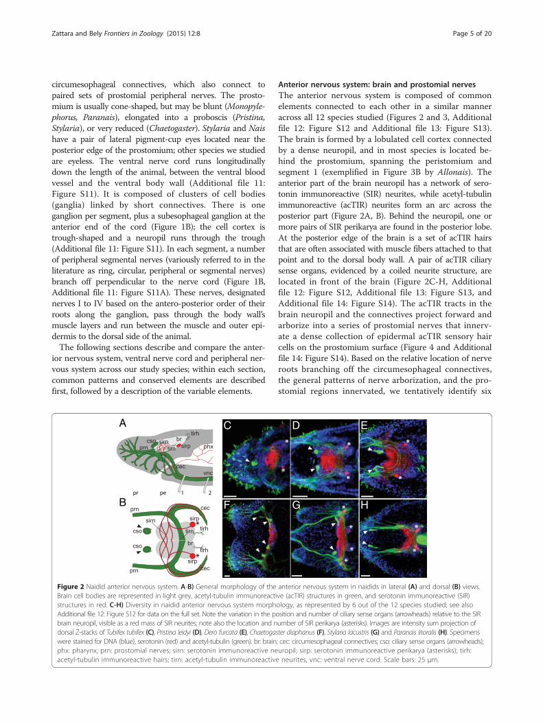
Figure 2 Naidid anterior nervous system. A-B) General morphology of theBrain cell bodies are represented in light grey, acetyl-tubulin immunoreactistructures in red. C-H) Diversity in naidid anterior nervous system morphAdditional file 12: Figure S12 for data on the full set. Note the variation in the pobrain neuropil, visible as a red mass of SIR neurites; note also the location and ndorsal Z-stacks of Tubifex tubifex (C), Pristina leidyi (D), Dero furcata (E), Chaetogaswere stained for DNA (blue), serotonin (red) and acetyl-tubulin (green). br: brainphx: pharynx; prn: prostomial nerves; sirn: serotonin immunoreactive neacetyl-tubulin immunoreactive hairs; tirn: acetyl-tubulin immunoreactive
Anterior nervous system: brain and prostomial nervesThe anterior nervous system is composed of commonelements connected to each other in a similar manneracross all 12 species studied (Figures 2 and 3, Additionalfile 12: Figure S12 and Additional file 13: Figure S13).The brain is formed by a lobulated cell cortex connectedby a dense neuropil, and in most species is located be-hind the prostomium, spanning the peristomium andsegment 1 (exemplified in Figure 3B by Allonais). Theanterior part of the brain neuropil has a network of sero-tonin immunoreactive (SIR) neurites, while acetyl-tubulinimmunoreactive (acTIR) neurites form an arc across theposterior part (Figure 2A, B). Behind the neuropil, one ormore pairs of SIR perikarya are found in the posterior lobe.At the posterior edge of the brain is a set of acTIR hairsthat are often associated with muscle fibers attached to thatpoint and to the dorsal body wall. A pair of acTIR ciliarysense organs, evidenced by a coiled neurite structure, arelocated in front of the brain (Figure 2C-H, Additionalfile 12: Figure S12, Additional file 13: Figure S13, andAdditional file 14: Figure S14). The acTIR tracts in thebrain neuropil and the connectives project forward andarborize into a series of prostomial nerves that innerv-ate a dense collection of epidermal acTIR sensory haircells on the prostomium surface (Figure 4 and Additionalfile 14: Figure S14). Based on the relative location of nerveroots branching off the circumesophageal connectives,the general patterns of nerve arborization, and the pro-stomial regions innervated, we tentatively identify six
anterior nervous system in naidids in lateral (A) and dorsal (B) views.ve (acTIR) structures in green, and serotonin immunoreactive (SIR)ology, as represented by 6 out of the 12 species studied; see alsosition and number of ciliary sense organs (arrowheads) relative to the SIRumber of SIR perikarya (asterisks). Images are intensity sum projection ofter diaphanus (F), Stylaria lacustris (G) and Paranais litoralis (H). Specimens; cec: circumesophageal connectives; cso: ciliary sense organs (arrowheads);uropil; sirp: serotonin immunoreactive perikarya (asterisks); tirh:neurites, vnc: ventral nerve cord. Scale bars: 25 μm.

Figure 3 Variation in position of the brain and ciliary sense organs,as represented by 4 naidid species. Images are intensity sumprojection of sagittal Z-stacks. Brain boundaries are shown by pairedbrackets; approximate prostomium/peristomium and peristomium/segment 1 boundaries are marked by dashed lines; ciliary senseorgans are indicated by arrowheads. The brain is located almostcompletely within the prostomium and peristomium in Tubifextubifex (A), peristomium and segment 1 in Allonais paraguayensis (B),segment 1 in Chaetogaster diaphanus (C) and back in segments 1and 2 in Paranais litoralis (D). Specimens were stained for DNA(blue), serotonin (red), acetyl-tubulin (green), and F-actin (white). Thedense acetyl-tubulin staining near the center of the animal in A, B,and D corresponds to the heavily ciliated pharynx. Scale bars: 25 μm.
Zattara and Bely Frontiers in Zoology (2015) 12:8 Page 6 of 20
major prostomial nerve branches in the naidids(branches A to F, color coded in Figure 4B). Given thelimited number of specimens and range of ages examinedhere, these homology assignments are necessarily prelim-inary, but should prove useful as a guide for future studies.While this general pattern of anterior nervous system
elements is shared by most species, we found consider-able variation in the location of the brain, number ofSIR perikarya, number and location of acTIR ciliarysense organs, and structure and origin of acTIR prosto-mial nerves across the species examined in this study.
With respect to the position of the brain (Figure 3 andAdditional file 13: Figure S13), we found that Tubifexhas a brain that is displaced anteriorly, straddling theprostomium/peristomium (Figure 3A and Additional file13: Figure S13A); Chaetogaster, which has a reducedprostomium, has a relatively small brain located levelwith the chaetae of segment 1 (Figure 3C and Additionalfile 13: Figure S13J); and Monopylephorus and Paranaishave a brain that is displaced posteriorly into segments 1and 2 (Figure 3D, Additional file 13: Figure S13D andS13H). The number and location of SIR perikarya in thebrain also varies among species, even between close rela-tives (Figure 4A; see also Table 1). We detected no SIRperikarya in Chaetogaster, one pair in both Pristina spe-cies, Monopylephorus, Amphichaeta and Allonais, twopairs in Stylaria and Dero digitata, three pairs in Tubifexand Dero furcata, four pairs in Nais and five pairs inParanais. Interestingly, we only detected a single pair ofSIR perikarya in the brain of a recently hatched Tubifex(data not shown), instead of the three pairs scored inolder worms, suggesting that the number of SIR peri-karya may also vary with developmental stage. All spe-cies examined have a pair of acTIR ciliary sense organslocated at the anterior edge of the brain with the excep-tion of Tubifex, which has a single, medial organ whichmay represent fusion of the original pair, and Chaetoga-ster, which has two pairs of ciliary sense organs (Figures 2,3, 4A, Additional file 12: Figure S12, Additional file 13:Figure S13, and Additional file 14: Figure S14). Paranais,Amphichaeta and Chaetogaster also differ from the rest ofthe species in that the acTIR ciliary sense organs are lo-cated at a short distance from the brain, rather thanagainst the anterior edge, and are connected to the mainneuropil by acTIR neurite bundles. Although the num-ber of major prostomial nerve branches is largelysimilar across species, the location of roots of theprostomial acTIR nerves differs according to differ-ences in the shape of the prostomium, and somenerves are absent (Figure 4 and Additional file 14:Figure S14): for example, the dorsal projecting nerveF is not found in Nais and Stylaria. In species wherethe prostomium elongates into a proboscis, namelyPristina and Stylaria (which likely evolved prosto-mium elongation independently), nerve D projectsforward to innervate this structure (Figure 4B). Whilenerve A is closest to the eye in Nais and Stylaria, wecould not verify whether these were actually con-nected due to signal masking by the eye’s pigment.
Ventral nerve cordThe ventral nerve cord has a similar architecture in allstudied species (Figures 5, 6, 7, Additional file 15: FigureS15, Additional file 16: Figure S16 and Additional file 17:Figure S17). It is formed by a continuous neuropil running

Figure 4 Variation in the architecture of the anterior nervous system across 10 naidid species. A) Schematic drawings of the anterior nervoussystem in dorsal view. Brain lobes are shown in grey, serotonin immunoreactive perikarya and neurites in red, ciliary sense organs in light green,and other acetyl-tubulin immunoreactive nerves in dark green. B) Schematic drawings of the prostomial/peristomial nerves in lateral view,color-coded to highlight putative homology assignments. The phylogenetic relationships among the species are shown to the right and arebased on recent molecular analyses, as described in the Methods section. Brain is shown in grey; black patches in Nais stolci and Stylarialacustris are lateral pigmented eyespots.
Zattara and Bely Frontiers in Zoology (2015) 12:8 Page 7 of 20
through segmentally iterated ganglia, with the neuropilcontaining acetyl-tubulin immunoreactive (acTIR) andserotonin immunoreactive (SIR) neurites that form longi-tudinal nerve tracts. The acTIR neurites are found latero-ventrally while SIR neurites tend to be medial and dorsal(Figure 5A, A1 and Additional file 15: Figure S15). Thesenerve tracts are linked by segmentally iterated transverse
commissures of variable acTIR neurite density. Phalloidinstaining indicates that the ventral cord is sheathed by athin muscular tunic (Figure 5A1). In all ventral nerve cordganglia a number of SIR perikarya are found connected tothe longitudinal SIR nerve tracts. Based on their locationin the ganglion relative to the peripheral nerve roots,we recognize four positional types of SIR perikarya

Figure 5 (See legend on next page.)
Zattara and Bely Frontiers in Zoology (2015) 12:8 Page 8 of 20

(See figure on previous page.)Figure 5 Conservation and variation of the segmental nervous system in naidids. A) Structure of a ventral nerve cord ganglion. Image A is anintensity sum projection of a ventral view of a trunk segment from Allonais paraguayensis, with transverse reconstructions to show the structureof the connective (A1) and ventral ganglia at two levels (A2, A3). Specimen was stained for DNA (blue), serotonin (red), acetyl-tubulin (green)),and F-actin (white). Segmental nerves are labeled I-IV; serotonin immunoreactive perikarya are within the parachaetal (p), central (c), axillar (a) orrear (r) group; ventral chaetae (vch) are visible due to birefringence. The paired arrowheads mark the position of the mesodermal septum. Thelooping, acetyl-tubulin positive structures in the lower and right part of the image correspond to a ciliated nephridium (nf: nephridial funnel; nt:nephrotubule). Scale bar: 25 μm. B) Diagram of non-septate and septate ganglia. Dashed vertical lines represent the mesodermal septa, andhorizontal bars indicate the span of a “neural segment” (defined as an entire ganglion and the interganglionic space anterior to it) and an“interseptal segment” (defined as the region between two consecutive septa). C) Generalized pattern of serotonin immunoreactive perikaryain a generic naidid trunk segment. Full circles represent cells that are always or almost always present, while half-circles represent cells whosepresence varies among species, individuals and/or segments. D) Nervous system structure of a typical trunk segment for each of the 12 species studied.Diagrams show typical pattern of serotonin immunoreactive perikarya of the ganglion (colored according to putative homology group assignmentsshown in C), location of peripheral nerve roots and location of septa. The phylogenetic relationships among the species are shown to the right and arebased on recent molecular analyses, as described in the Methods section.
Zattara and Bely Frontiers in Zoology (2015) 12:8 Page 9 of 20
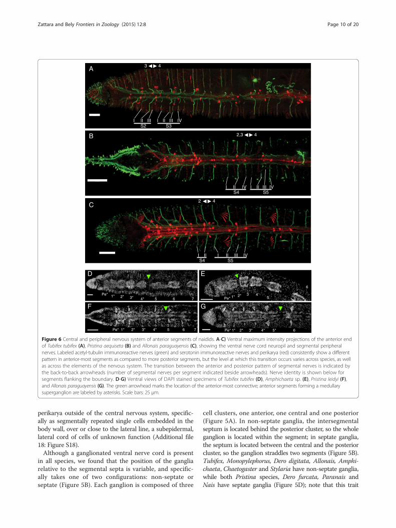
(Figure 5C): 1) parachaetal cells, located within the an-terior third of the ganglion, approximately medial tothe neuropil and level with the ventral chaetae, betweennerve roots I and II; 2) axillar cells, located within themiddle third of the ganglion, lateroventrally outside ofthe neuropil, either to the left or right, and always be-hind nerve root II; 3) central cells, located more medi-ally within the middle third of the ganglion and closerto the neuropil but level with the axillar cells; and 4)rear cells, located within the posterior third of the ganglionand medial to the neuropil, between nerve roots III and IV.Notably, in all species examined, the anterior-most gangliahave a pattern of SIR perikarya that is clearly different frommore posterior segments (Figure 6A-C and Additional file16: Figure S16). Within a single trunk ganglion, SIR cellsare asymmetrically distributed, mostly evidenced by thepresence of axillar cells at only one side; however, consecu-tive ganglia show alternating mirror symmetry with respectto the mid-sagittal plane. In contrast, anterior ganglia havea larger number of SIR perikarya and these are arrangedsymmetrically with respect to the mid-sagittal plane. Basedon counts from 1–4 individuals per species (~30 specimensacross all species), the following general patterns emerge: a)on average, there are approximately 80% more SIR peri-karya in anterior ganglia than in more posterior ganglia; b)the subesophageal ganglion has fewer SIR perikarya thanthe ganglia of the anterior segments, but more than moreposterior ganglia; c) ganglia of segments 1 and 2 have aboutthe same number of SIR perikarya per segment, and thisnumber is the highest in the body; d) ganglia of segments 3and 4 have fewer perikarya than segments 1 and 2, butmore than more posterior ganglia. In all cases, SIR peri-karya of the anterior ganglia are arranged in a bilaterallysymmetric pattern resembling what would be obtained bythe superposition of perikarya from two consecutive moreposterior ganglia. In all species, the ganglia of the peristo-mium and anterior-most segments adjoin one another,forming a superganglion that adopts a medullary configur-ation (Figure 6D-G and Additional file 17: Figure S17).
These ganglia, especially the first three, are displaced pos-teriorly relative to other segmental organs, as compared totheir position in more posterior segments.Despite the common architecture of the ventral nerve
cord, we found considerable variation among species inthe specific location of SIR perikarya in the ganglia, thelocation along the body where perikarya switch from theabove described anterior pattern to the general distribu-tion pattern found in the rest of the segments, and thenumber of anterior segments forming a medullary super-ganglion. We also noticed unexpected inter-species vari-ability in the position of the mesodermal segmentalsepta relative to the ventral nerve cord ganglia. A gen-eral pattern of the ganglion SIR cells for each species isshown in Figure 5D; however, these patterns should beconsidered as approximate, since there is considerablevariation from segment to segment even within the sameworm. Interpretation is further complicated by the factthat some perikarya show weaker serotonin signal insome segments, and their detection may depend both ontheir developmental state and the quality of immuno-staining. Despite these caveats, we found that the asym-metric trunk pattern of SIR perikarya begins at segment5 in all species studied (Figure 6B-C and Additional file16: Figure S16), with the exception of Tubifex and Amphi-chaeta, where it starts at segment 4 (Figure 6A, Additionalfile 16: Figure S16F). The extent of the anterior medullarysuperganglion is more variable: it includes two ganglia inChaetogaster (Additional file 17: Figure S17E) and Amphi-chaeta (Figure 6E), three ganglia in Paranais (Additionalfile 17: Figure S17D), four ganglia in Tubifex (Figure 6D),Pristina (Figure 6F and Additional file 17: Figure S17A),Dero furcata (Additional file 17: Figure S17C), and Sty-laria (Additional file 17: Figure S17G), and five ganglia inDero digitata (Additional file 17: Figure S17B) and Allo-nais (Figure 6G). In Nais, we observed a gap between gan-glion 3 and 4, and another between 5 and 6, but notbetween 4 and 5 (Additional file 17: Figure S17F). Naisalso was the only species in which we detected SIR
IIS2
IIII II IIIS3
IVI
S4I IV IIII IIII
S5IV
S5I IIIII IVII
S4I
A
B
C2 4
2,3 4
3 4
Pe* 1* 2* 3* 4* 5 6 7
Pe* 1* 2* 3* 4* 5 6 7
Pe* 1*2* 3 4
5
6
Pe* 1* 2* 3* 4* 5* 6
D
F
E
G
Figure 6 Central and peripheral nervous system of anterior segments of naidids. A-C) Ventral maximum intensity projections of the anterior endof Tubifex tubifex (A), Pristina aequiseta (B) and Allonais paraguayensis (C), showing the ventral nerve cord neuropil and segmental peripheralnerves. Labeled acetyl-tubulin immunoreactive nerves (green) and serotonin immunoreactive nerves and perikarya (red) consistently show a differentpattern in anterior-most segments as compared to more posterior segments, but the level at which this transition occurs varies across species, as wellas across the elements of the nervous system. The transition between the anterior and posterior pattern of segmental nerves is indicated bythe back-to-back arrowheads (number of segmental nerves per segment indicated beside arrowheads). Nerve identity is shown below forsegments flanking the boundary. D-G) Ventral views of DAPI stained specimens of Tubifex tubifex (D), Amphichaeta sp. (E), Pristina leidyi (F),and Allonais paraguayensis (G). The green arrowhead marks the location of the anterior-most connective; anterior segments forming a medullarysuperganglion are labeled by asterisks. Scale bars: 25 μm.
Zattara and Bely Frontiers in Zoology (2015) 12:8 Page 10 of 20
perikarya outside of the central nervous system, specific-ally as segmentally repeated single cells embedded in thebody wall, over or close to the lateral line, a subepidermal,lateral cord of cells of unknown function (Additional file18: Figure S18).Although a ganglionated ventral nerve cord is present
in all species, we found that the position of the gangliarelative to the segmental septa is variable, and specific-ally takes one of two configurations: non-septate orseptate (Figure 5B). Each ganglion is composed of three
cell clusters, one anterior, one central and one posterior(Figure 5A). In non-septate ganglia, the intersegmentalseptum is located behind the posterior cluster, so the wholeganglion is located within the segment; in septate ganglia,the septum is located between the central and the posteriorcluster, so the ganglion straddles two segments (Figure 5B).Tubifex, Monopylephorus, Dero digitata, Allonais, Amphi-chaeta, Chaetogaster and Stylaria have non-septate ganglia,while both Pristina species, Dero furcata, Paranais andNais have septate ganglia (Figure 5D); note that this trait

Figure 7 Phylogenetic distribution of serotonin immunoreactive perikarya and peripheral nerve patterns. A) Reconstruction of the basalarchitecture of a trunk ventral ganglion across clitellate groups, based on this study and previous reports [20,27,29,32,35,36]. Colored circlesrepresent serotonin immunoreactive perikarya, with color representing putative homologous groups; A to G cells in Crassiclitellata refer to cellgroups defined for Lumbricus terrestris [35]; anteromedial (am), dorsolateral (dl), ventrolateral (vl), posteromedial (pm) and Retzius cells in Hirudinarefer to cell groups defined for leech species [27,29,32]. Segmental peripheral nerves are shown aligned according to their position along theganglion. B) Phylogenetic mapping of the number of segmental peripheral nerves across Annelida, and maximum likelihood estimations of theancestral state at each node. Pie charts illustrate the relative likelihood of a given node having had each character state, and were calculated in R[63] using the ace function from the ape package [64]. Annelid relationships are based on recent phylogenetic studies [50,52]. Labeled ancestralnodes are for Errantia (E) polychaetes, Sedentaria (S) polychaetes and Clitellata (C); the dashed box highlights data from this paper. Nerve number foreach group is based on information from one or a few species, except for Naididae, where ancestral condition was estimated from our 12 studyspecies. References are indicated on the right, in brackets for published works; *: data from this study; **R. Hessling, personal communications in [7];***E.E. Zattara, unpublished observations.
Zattara and Bely Frontiers in Zoology (2015) 12:8 Page 11 of 20
varies even between phylogenetically close species pairs(e.g. Stylaria and Nais; Dero furcata and Dero digitata).
Peripheral nervous systemThe main component of the naidid peripheral nervoussystem is a system of segmentally iterated acTIR nervesthat originate from the neuropil of the ventral nervecord, exit through the ganglia, pass ventrally through thebody wall muscle layer and run subepidermally towardsthe segment’s dorsum (Figures 1B, 5A and Additionalfile 11: Figure S11A). Each nerve innervates a number ofepidermal sensory hairs. We found that certain nervescan be found in all species examined, while others ap-pear to have been recently gained or lost; we also foundthat the number of segmental nerves is reduced in theanterior-most segments of most species.All examined species have four segmental nerves in
each trunk segment, except for Chaetogaster diaphanuswhich has five (Figure 5D, Additional file 15: FigureS15). We designated the segmental nerves as nerves I to
IV, according to the antero-posterior order in which theybranch from the ganglion. The largest nerves are nerveI, located in the anterior half of the segment, and nerveII, which is always located just posterior to the chaetalplane (defined by the ventral and dorsal chaetal bundles)and which sends branches anteriorly to a set of epidermalsensory structures closely associated with the chaetae.Nerves III and IV are usually smaller, and one of the twois generally associated with the intersegmental septum;which nerve this is depends on whether the ganglion isseptate or not (Figure 5B). Some species diverge from thisgeneral pattern, however (Figure 5D): in Pristina leidyi(Additional file 15: Figure S15C), Pristina aequiseta(Additional file 15: Figure S15B), and Allonais para-guayensis (Figure 5A) nerve III is very small or absent;in Amphichaeta sp. (Additional file 15: Figure S15G)and Chaetogaster diaphanus (Additional file 15: FigureS15H) there is a small fifth nerve located betweennerves I and II (labeled as “2” in arabic numerals inAdditional file 15: Figure S15G, H); in Amphichaeta sp.

Zattara and Bely Frontiers in Zoology (2015) 12:8 Page 12 of 20
we could not detect nerve IV and nerve III is displacedbackwards.We performed an ancestral character estimation ana-
lysis to reconstruct the evolution of trunk segment nervenumber across annelids, using our data and publishedinformation from across the phylum. This analysis indi-cates that four segmental nerves is the most likely statefor the last common ancestor of the naidids (Figure 7B).Our analysis also suggests that the number of segmentalnerves has been evolutionarily labile, increasing and de-creasing at several points during the evolution of anne-lids, and that basal stem annelids most likely had threesegmental nerves.The segmental nerve patterns described above are
consistent across all segments within a species with theexception of the anterior-most segments, which have fewernerves the closer they are to the anterior end (Figure 6A-C,Additional file 16: Figure S16). Species vary considerably inthe degree of nerve number reduction and in the numberof anterior segments that show reduced nerve number. Inmost species, the full complement of nerves begins in seg-ment 5. However, it begins in segment 3 in Tubifex, Amphi-chaeta and Stylaria, segment 2 in Paranais and in segment1 (no reduction) inMonopylephorus.
Fine taxonomic sample of Naididae reveals conservationand lability of neural traitsTo gain better understanding of nervous system evolutionwithin Clitellata, we described and made a comparativeanalysis of the nervous system of 12 species of Naididae, abasally branching clitellate family [9,23-25]. Althoughwe found many similarities between nervous systemarchitecture in naidids and that of other clitellate groups[15,20,23], our study identified many features that are vari-able within this family of clitellates, including some thatare variable even among relatively closely related species(Table 1). Variable features of the nervous system includethe location of the brain, the number of ciliary sense or-gans, the presence of non-septate or septate ganglia alongthe ventral nerve cord, the distribution of serotonergiccells in the brain and ventral ganglia, and the number ofperipheral nerves. Below, we discuss interspecific andintra-individual variation in the distribution of serotoninimmunoreactive perikarya in the central nervous systemand potential homologies between Naididae and other cli-tellates; we address the unexpected interspecific variabilityin the position of segmental septa relative to ventral gan-glia; and we show how using ganglia rather than body seg-ments to identify peripheral nerves can help reveal nervehomologies across Annelida. We end by highlighting theimportance of fine taxonomic sampling in comparativestudies aimed at elucidating the evolution of morpho-logical diversity.
Patterns of serotonin immunoreactive perikarya in theannelid central nervous system indicate that segmentalunits are not structurally homogeneousThe central nervous system of annelids typically includesserotonin immunoreactive (SIR) cells, which are putativelyassociated with motor neurons [17,26-31]. The distribu-tion patterns of these cells vary among species and bodyregions, and have been suggested to be potentially usefultaxonomic traits [17]. Our data on 12 species of naidid an-nelids show that SIR perikarya distribution patterns in thebrain and ventral nerve cord can vary considerably acrossspecies and even within individuals, both along theantero-posterior body axis and potentially between devel-opmental stages. Despite this variability, the positions ofserotonin-positive perikarya in the ventral nerve cord gan-glia show consistent enough patterns to suggest putativehomologies both within naidids and between naidids andother clitellate groups.We found that the number of paired serotonin immu-
noreactive (SIR) perikarya in the brain varies across nai-dids. While a single pair of SIR perikarya is the mostcommon arrangement for the group, the range is quitebroad, from no SIR perikarya in Chaetogaster to 5 pairsin Paranais. The number of perikarya does not appearto be related in any obvious manner to species attributessuch as size, habitat or preferred movement type. Whilein most species the number of serotonin-positive peri-karya is lower in the brain than in the body ganglia, wefound that the reverse is true in both Nais and Paranais.Thus, the number of perikarya in the brain is not alwaysless than that in the ventral ganglia, as had been previouslysuggested [17]. Our observation that Tubifex juveniles hadonly two serotonin-positive cells in the brain while adultworms have six cells suggests that this number can changepost-embryonically; caution is recommended when usingthe number of serotonin-positive cells in the brain as adiagnostic feature for species identification.In all but the anterior-most segments, serotonin im-
munoreactive (SIR) perikarya in ventral ganglia have anasymmetrical distribution, with consecutive segmentshaving patterns that are mirror-images of one another.This alternating pattern has been previously described inthree naidid species, all three being naidines [17]; ourstudy extends the distribution of this pattern in naididsand close relatives to seven other naidines, a rhyacodriline,two pristinines, and a tubificine. Such an alternating pat-tern is also present in leeches [27,29,32], a more distantlyrelated group of clitellates, and even the polychaetous Aeo-losoma sp. has a single yet alternating serotonergic cell ineach ventral ganglion [33]. Thus, the alternating pattern isnot an autapomorphy of Naididae, as suggested elsewhere[17], but a feature common to many clitellates and relatedannelids. The developmental processes that generate thispattern have not been studied in naidids, but during

Zattara and Bely Frontiers in Zoology (2015) 12:8 Page 13 of 20
development of the leech species Helobdella triserialis,Theromizon rude and Hirudo medicinalis paired seroto-nergic precursor cells in consecutive segments make con-tact with each other and one member of each pairundergoes cell death in a pattern that alternates acrosssegments, setting up a similar alternating pattern of un-paired cells as seen in the naidids [27,29,32]. Whether asimilar mechanism is responsible for the SIR perikarya dis-tribution in naidids remains to be determined.In contrast to trunk ganglia, serotonin immunoreactive
(SIR) perikarya in the anterior-most ganglia of naidids aremore numerous and show a symmetrical pattern thatroughly matches the overlay of two consecutive trunk gan-glia. In naidines, anterior segments often develop post-embryonically by paratomic fission [34], and it has beensuggested that the symmetrical pattern of SIR perikarya inthose segments results from such segments being at anearlier developmental stage in which differential cell deathin consecutive segments has not yet taken place [17].However, our study indicates that anterior segmentshave a symmetrical pattern not only in fissioning spe-cies but in two non-fissioning species as well, Tubifexand Monopylephorus. Furthermore, in the pristinines,six anterior segments form during fission [19] yet onlythe four anterior-most segments show symmetricallyarranged SIR perikarya, indicating that an asymmetricalpattern can be established during fission as well.Our finding that naidids have a different distribution
of serotonin immunoreactive (SIR) perikarya in anteriorsegments as compared to more posterior segments isconsistent with data from several other annelid groups.In the crassiclitellate earthworm Lumbricus terrestris, asubesophageal medullary superganglion formed by twofused anterior ganglia has over three times as many SIRperikarya as trunk ganglia, while segments 4–10 have twiceas many [28]. In the leech Hirudo medicinalis, eight SIRperikarya per ganglion are found in the six anterior ganglia,with four of these being fused into a subesophageal medul-lary ganglion and two remaining as free ganglia; in contrast,only seven SIR perikarya per ganglion are found in moreposterior cells [26]. In the freshwater polychaete Aeolosomasp., the anterior-most two ganglia have four SIR perikarya,while remaining trunk ganglia have just a single SIR cell[33]. Thus, the presence of two clearly different distributionpatterns of SIR perikarya along the body, with a clear breakat a specific point along the antero-posterior axis, is wide-spread among clitellates and is even found outside of thisclade. The location of this transition between the two pat-terns is different for different groups, however. It would beinteresting to investigate whether other nervous systemcomponents and other organ systems also show a similarantero-posterior break, and whether the presence of suchboundaries indicates some degree of “cephalization” of theanterior-most segments.
Preliminary homology assessments suggest gains andlosses of serotonin immunoreactive cell sets across theClitellataBased on our studies of naidids, serotonin immunoreac-tive (SIR) perikarya within the ventral nerve cord gangliaappear to show a common pattern across species. Thesecells form four spatially segregated sets of cells, whichwe termed parachaetal, central, axillar and rear cells(Figure 5C). When the naidid pattern is compared with SIRcell patterns reported for representative crassiclitellates(Lumbricus terrestris and Eisenia foetida) [35,36], enchy-traeids (Enchytraeus crypticus) [20], hirudines (Helobdellatriserialis, Theromizon rude and Hirudo medicinalis)[27,29,32] and lumbriculids (Lumbriculus variegatus,E.E.Z. unpublished observations), a very preliminaryhomology assignment across the clitellates is possible, usingas criteria the axial position of the cells along the length ofthe ganglion and the topological relationship of the cells tothe peripheral nerve roots (Figure 7A). By these criteria, thenaidid parachaetal cells might be homologous to Lumbri-cus’ B cells, the leech’s anteromedial or Retzius cells, andunnamed anterior cells found in Enchytraeus and Lumbri-culus; the central cells might be homologous to E cells inLumbricus and to unnamed midline cells in Enchytraeusand Lumbriculus, while being absent in hirudines; the axil-lar cells might be homologous to D cells in Lumbricus, ven-tro and dorsolateral cells in hirudines, and unnamed lateralcells in Enchytraeus and Lumbriculus; and the rear cellsmight be homologous to G cells in Lumbricus, posterome-dial cells in hirudines, and unnamed midline posterior cellsin Enchytraeus and Lumbriculus. Interestingly, Lumbricusterrestris seems to have a number of SIR cell sets clearly ab-sent in more distantly related groups (namely, A, C and Fcells). The total number of cells per trunk ganglion variessignificantly among groups: naidids reported here have 4–11 cells, leeches [26,27,29], enchytraeids [20] and lumbricu-lids (E.E.Z. unpublished observations) have 7–9 cells, whileearthworms have 30–80 cells [35,36]. The homology as-signments we propose here are necessarily tentative, andfuture studies including developmental and neuronal con-nectivity studies are needed to confirm them; nonetheless,even this preliminary comparison across clitellates high-lights that whole sets of serotonin immunoreactive cellshave been gained or lost throughout the evolution of thisgroup of annelids.
The relative position of neuroectodermal and mesodermalsegmental components is evolutionarily labileAn unexpected finding from our study is that the positionof the segmental ganglia relative to the septa (which areused to define morphological segment boundaries) variesacross species, and even among close relatives. We foundthat some naidids have non-septate ganglia (in which theentire ganglion falls between consecutive septa) while

Zattara and Bely Frontiers in Zoology (2015) 12:8 Page 14 of 20
others have septate ganglia (in which the ganglion strad-dles two consecutive segments, with a septum fallingacross the ganglion). Among our study species, we foundexamples of ganglion type being variable among closely re-lated genera (Nais and Stylaria), and even within a singlegenus (Dero digitata and Dero furcata). Among other cli-tellates, ganglion type is also variable. Currently availabledescriptions indicate that Lumbricus and Eisenia (both inCrassiclitellata) as well as Lumbriculus (Lumbriculidae)have non-septate ganglia [15,23], while Enchytraeus(Enchytraeidae) has septate ganglia [20]; leeches cannot becategorized since they have no septa at all [23]. Amongpolychaetes, septate ganglia have been described in thenereids Neanthes, Platynereis and Hediste [37] and thisganglion type is not uncommon in other polychaete groupsas well [7]. Although variability in ganglion type can be in-ferred from the literature, sampling density remains sparseoutside of the naidids and none of these previously pub-lished observations have been made within a comparativeframework; to our knowledge, this is the first report show-ing that ganglion type can vary even between closely relatedspecies. Given our observations, we conclude that the rela-tive position of neuroectodermal elements (i.e., neural gan-glia) and mesodermal elements (i.e., septa) of each segmentcan experience frequent shifts over evolutionary time, andthese elements should not be assumed to be in the sameregister within a group.We hypothesize that the evolutionary lability of the
relative position of neural ganglia and mesodermal septain clitellates likely reflects the considerable developmentalindependence of ectodermal and mesodermal teloblasticbandlets during embryogenesis [38,39]. As indicated bywork done in Tubifex hattai [38-40] and several leech spe-cies [41-44], in clitellates much of a segment’s ectodermaland mesodermal tissues arises through teloblastic growth,in which a small set of large stem cells (four ectodermaland one mesodermal teloblast pairs) divide asymmetricallyleaving behind bandlets of founder cells that will eachform components of one or two consecutive segments.Ablation experiments in Tubifex suggest that ectodermalsegmentation comprises an initial autonomous morpho-genetic stage, followed by mesoderm-dependent alignmentof segmental elements [39]. Mesodermal segmentation, onthe other hand, does not require segmented ectoderm[38]. Similar results have been reported for leeches[45-47]. Given this degree of independence in the develop-ment of these two tissue layers, evolutionary changes inectoderm/mesoderm alignment may arise relatively easilywithin clitellates.Interestingly, even though the relative position of septa
and ganglia varies across species, we found that all septateganglia are septate in a similar manner, with the septumconsistently located at two-thirds of the ganglion length.This uniformity in the configuration of septate ganglia
suggests that there may be developmental constraintsrestricting the possible locations of the septa relative tothe ganglia. Furthermore, the septate/non-septate condi-tion appears to be fixed within a species, suggesting astrong genetic control. The developmental mechanismsthat keep ectoderm and mesoderm in the same registerintraspecifically while allowing for interspecific shifts inNaididae are unknown and warrant further investigation.
The homologies of segmental nerves are clarified byscoring their position relative to segmental ganglia ratherthan to segmental septaAmong our study species, the most common configurationof the peripheral nervous system was four segmental nervesper trunk ganglion. We did find variation in this pattern,however, with Chaetogaster having a fifth small nerve lo-cated between nerves I and II, Pristina spp. and Allonaishaving nerve III very reduced, and Amphichaeta having nodetectable nerve IV but a posteriorly displaced nerve III.Given the variation in ganglion septation we found in
our study, we determined that the common approach ofnaming nerves based on their position within the seg-ment [19,20,37], that is, relative to segmental septa,might not reflect underlying nerve homologies. Instead,we based our naming scheme for segmental nerves onthe order of nerve roots along the segmental ganglion(rather than position within the segment), such thatnerve I is the anterior-most nerve emanating from theganglion, nerve II is the next most anterior nerve, etc. Inaddition to the order of nerve roots along the ganglion,we also considered in our naming scheme the relative sizesand innervation patterns of these nerves, to account for thepossibility that certain nerves may be gained or lost overevolutionary time. Using this approach, the arrangement,relative size and innervated structures of nerve roots alongthe ganglion are almost identical in Dero furcata and Derodigitata (which, respectively, have septate and non-septateganglia), whereas a septum-based naming scheme wouldentail nerves with quite different sizes and innervation pat-terns being assigned the same nerve number, and nervesassociated with the same structures being assigned differentnerve numbers (e.g., the nerve innervating the chaetaewould be the third nerve in D. furcata and the secondnerve in D. digitata). The homologies of segmental nervesacross species that are implied by our naming scheme fol-low from our expectation that relative slippage of the meso-dermal/ectodermal boundaries (see above) is more likelythan concerted change in size and innervation patterns ofall segmental nerves; thus, naming nerves based on theirposition along the ganglion rather than relative to mesoder-mal septa is more likely to reflect nerve homologies. Wewould discourage others from using septum boundaries toidentify segmental nerves in annelids, and encourage theuse of ganglion boundaries instead; such an approach

Zattara and Bely Frontiers in Zoology (2015) 12:8 Page 15 of 20
should facilitate efforts to trace how segmental nervesevolved in clitellates and other annelids.Based on our data and ancestral character estimation
(ace) analysis, four peripheral nerves per trunk ganglionis the most likely ancestral state for Naididae. Outside ofthe naidids but still among the clitellates, the number ofnerves described is four for Lumbriculus [48], five forEnchytraeus [7,18,20], three for Lumbricus [23], and twofor adult leeches [23,26,27] (although four nerves are de-scribed in embryos of Erpobdella octoculata; R. Hessling,unpublished observations in [7]). Under the assumptionthat these species are representative of their respectivegroups, our ancestral character estimation analysis sup-ports four peripheral nerves as the ancestral state forClitellata as a whole (Figure 7B). Out of the four nerves,nerve II is larger and associates with the chaetae inmembers of Naididae, in Lumbriculus variegatus [48]and in Lumbricus terrestris [15]. Nerve II is also associatedwith chaetae in Enchytraeus crypticus [20], but it is smallerthan the following nerve (Figure 7A). Since five rather thanfour nerves are present in this species, we speculate thatenchytraeids must have either evolved a novel nerve (II’ inFigure 7A), intercalated between nerves I and II, or dupli-cated nerve II (II’ and II in Figure 7A).How does this basal clitellate state for segmental nerve
number relate to the rest of Annelida? Within polychaet-ous annelids, the number of nerves reported ranges fromnone (Trilobodrilus hermaphroditus) to eight or more(Protodrilus sp.) [7,49]. Putative nerve homologies amongtaxa have been difficult to establish, in part due to a poorunderstanding of deep phylogenetic relationships amongannelid taxa. However, recent progress in understandingannelid phylogeny [50-52] now provides a stronger frame-work for mapping of variation in segmental nerves acrossthe phylum, including for ancestral character estimation toreconstruct the most likely number of nerves at the mainnodes of the annelid tree, as we have done here (Figure 7B)[7,18,20,23,37,48,49,53-57]. Our analysis supports a previ-ous claim that three segmental nerves represents theancestral condition for the phylum [2,7]. Interestingly,the analysis suggests that a fourth nerve evolved eitherat the base of the Sedentaria, the clade of mostly lessmotile worms within which the clitellates are nested, orshortly thereafter, after the cirratulids + orbinids lineagebranched off. According to this reconstruction, the two-nerve state of capitellids and echiurids and the five-nervestate of sabellids and spionids would both be derived froma four-nerve condition. Even if our specific results regardingancestral character estimation for nerve number at eachnode are later revised, it is clear that segmental nerves un-questionably have been gained and lost several times duringannelid evolution. Finer taxonomic sampling would allow amore precise mapping of novel origins and losses of seg-mental nerves, which in turn could reveal useful groups in
which to study the evolution of neuronal elements withinannelids.
ConclusionsOur comparative description of the nervous system ofseveral species of Naididae and the resulting identificationof common patterns and differences in nervous systemarchitecture in this group highlight the potential insightsthat can be gleaned from comparative morphological stud-ies made at a fine taxonomic scale. Such studies have thepower to confirm the deep conservation of certain wide-spread traits, whose evolution may be strongly constrainedby functional or developmental constraints, but also toreveal highly labile traits that can change readily duringevolution. Furthermore, such fine-scale comparative studiescan contradict prior inferences about which characters areunlikely to vary, as we have shown here, for example,for the septate/non-septate ganglion condition. Obtain-ing strong data for the conservation or lability of char-acters is particularly important since traits thought tobe well conserved tend to be used as landmarks for hom-ology assignments; thus we recommend ensuring thatsuch conservation has been evaluated at a relatively finetaxonomic scale before using such traits more broadly as abasis for determining homologies.
MethodsAnimal samplesSpecimens of Tubifex tubifex, Pristina leidyi, Pristinaaequiseta, Dero digitata, Dero furcata, Allonais para-guayensis, Paranais litoralis, Chaetogaster diaphanus,Nais stolci and Stylaria lacustris were obtained fromestablished laboratory cultures [58,59] and specimens ofMonopylephorus rubroniveus and Amphichaeta sp. werefield collected. Table 2 provides worm sources, cultureconditions, and NCBI accession numbers for availableCOI and 16S sequence barcodes for the strains used.New COI or 16S sequences were obtained for Paranaislitoralis, Dero furcata, Pristina leidyi and Pristina aequiseta;primers, PCR parameters, and sequencing methods are asdescribed elsewhere [58].
ImmunocytochemistrySamples were relaxed 10 min in cold (4°C) relaxant solu-tion (10 mM MgCl2/5 mM NaCl/1 mM KCl/8% etha-nol), fixed 30 min in 4% formaldehyde in 0.75x PBS, andrinsed in 1x PBS. Then they were permeabilized with0.1% Triton-X in PBS (PBTx), blocked 1 h in 10% nor-mal goat serum (NGS) in PBTx, and incubated 15–20 hat 4°C with mouse anti-acetylated-α-tubulin monoclonalantibody (T6793, Sigma, St. Louis, MO, USA) and rabbitanti-serotonin polyclonal antibodies (S5545, Sigma),both diluted 1:100 in blocking solution. Specimens werethen washed in PBTx and incubated 15–20 h at 4°C in
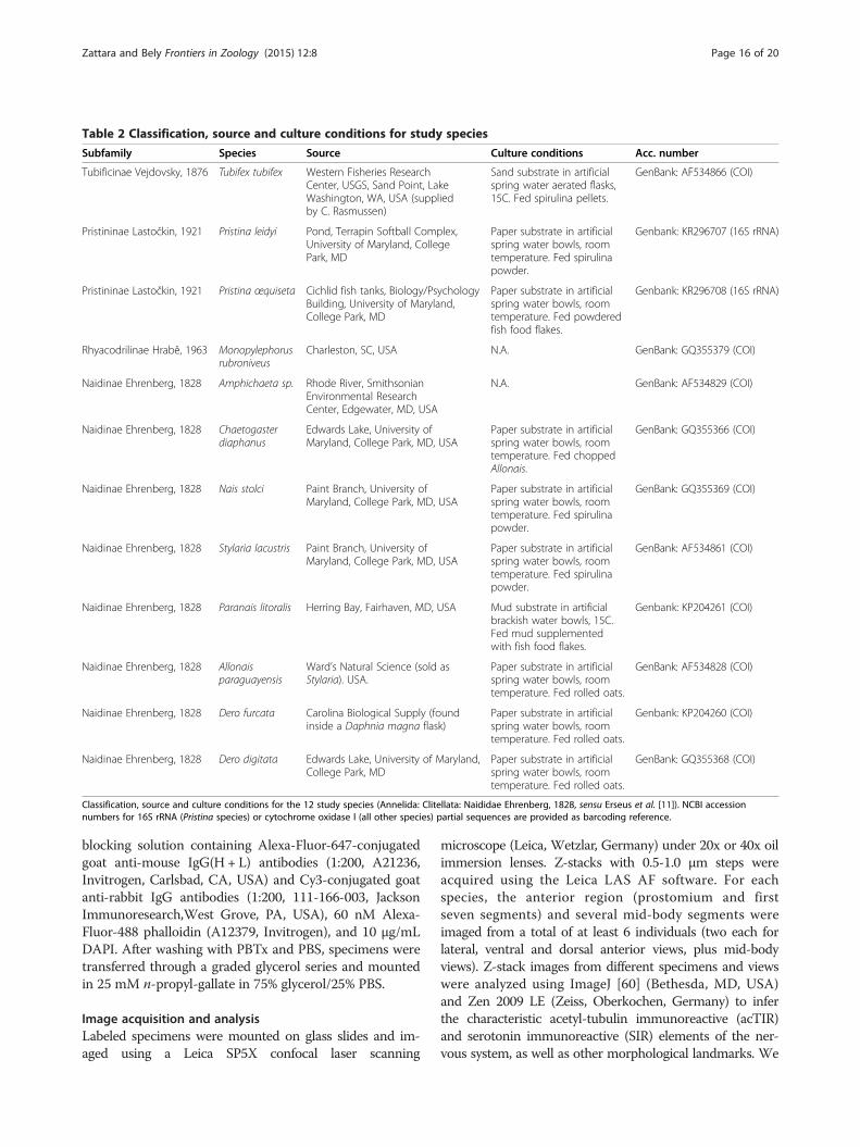
Table 2 Classification, source and culture conditions for study species
Subfamily Species Source Culture conditions Acc. number
Tubificinae Vejdovsky, 1876 Tubifex tubifex Western Fisheries ResearchCenter, USGS, Sand Point, LakeWashington, WA, USA (suppliedby C. Rasmussen)
Sand substrate in artificialspring water aerated flasks,15C. Fed spirulina pellets.
GenBank: AF534866 (COI)
Pristininae Lastočkin, 1921 Pristina leidyi Pond, Terrapin Softball Complex,University of Maryland, CollegePark, MD
Paper substrate in artificialspring water bowls, roomtemperature. Fed spirulinapowder.
Genbank: KR296707 (16S rRNA)
Pristininae Lastočkin, 1921 Pristina œquiseta Cichlid fish tanks, Biology/PsychologyBuilding, University of Maryland,College Park, MD
Paper substrate in artificialspring water bowls, roomtemperature. Fed powderedfish food flakes.
Genbank: KR296708 (16S rRNA)
Rhyacodrilinae Hrabě, 1963 Monopylephorusrubroniveus
Charleston, SC, USA N.A. GenBank: GQ355379 (COI)
Naidinae Ehrenberg, 1828 Amphichaeta sp. Rhode River, SmithsonianEnvironmental ResearchCenter, Edgewater, MD, USA
N.A. GenBank: AF534829 (COI)
Naidinae Ehrenberg, 1828 Chaetogasterdiaphanus
Edwards Lake, University ofMaryland, College Park, MD, USA
Paper substrate in artificialspring water bowls, roomtemperature. Fed choppedAllonais.
GenBank: GQ355366 (COI)
Naidinae Ehrenberg, 1828 Nais stolci Paint Branch, University ofMaryland, College Park, MD, USA
Paper substrate in artificialspring water bowls, roomtemperature. Fed spirulinapowder.
GenBank: GQ355369 (COI)
Naidinae Ehrenberg, 1828 Stylaria lacustris Paint Branch, University ofMaryland, College Park, MD, USA
Paper substrate in artificialspring water bowls, roomtemperature. Fed spirulinapowder.
GenBank: AF534861 (COI)
Naidinae Ehrenberg, 1828 Paranais litoralis Herring Bay, Fairhaven, MD, USA Mud substrate in artificialbrackish water bowls, 15C.Fed mud supplementedwith fish food flakes.
Genbank: KP204261 (COI)
Naidinae Ehrenberg, 1828 Allonaisparaguayensis
Ward’s Natural Science (sold asStylaria). USA.
Paper substrate in artificialspring water bowls, roomtemperature. Fed rolled oats.
GenBank: AF534828 (COI)
Naidinae Ehrenberg, 1828 Dero furcata Carolina Biological Supply (foundinside a Daphnia magna flask)
Paper substrate in artificialspring water bowls, roomtemperature. Fed rolled oats.
Genbank: KP204260 (COI)
Naidinae Ehrenberg, 1828 Dero digitata Edwards Lake, University of Maryland,College Park, MD
Paper substrate in artificialspring water bowls, roomtemperature. Fed rolled oats.
GenBank: GQ355368 (COI)
Classification, source and culture conditions for the 12 study species (Annelida: Clitellata: Naididae Ehrenberg, 1828, sensu Erseus et al. [11]). NCBI accessionnumbers for 16S rRNA (Pristina species) or cytochrome oxidase I (all other species) partial sequences are provided as barcoding reference.
Zattara and Bely Frontiers in Zoology (2015) 12:8 Page 16 of 20
blocking solution containing Alexa-Fluor-647-conjugatedgoat anti-mouse IgG(H + L) antibodies (1:200, A21236,Invitrogen, Carlsbad, CA, USA) and Cy3-conjugated goatanti-rabbit IgG antibodies (1:200, 111-166-003, JacksonImmunoresearch,West Grove, PA, USA), 60 nM Alexa-Fluor-488 phalloidin (A12379, Invitrogen), and 10 μg/mLDAPI. After washing with PBTx and PBS, specimens weretransferred through a graded glycerol series and mountedin 25 mM n-propyl-gallate in 75% glycerol/25% PBS.
Image acquisition and analysisLabeled specimens were mounted on glass slides and im-aged using a Leica SP5X confocal laser scanning
microscope (Leica, Wetzlar, Germany) under 20x or 40x oilimmersion lenses. Z-stacks with 0.5-1.0 μm steps wereacquired using the Leica LAS AF software. For eachspecies, the anterior region (prostomium and firstseven segments) and several mid-body segments wereimaged from a total of at least 6 individuals (two each forlateral, ventral and dorsal anterior views, plus mid-bodyviews). Z-stack images from different specimens and viewswere analyzed using ImageJ [60] (Bethesda, MD, USA)and Zen 2009 LE (Zeiss, Oberkochen, Germany) to inferthe characteristic acetyl-tubulin immunoreactive (acTIR)and serotonin immunoreactive (SIR) elements of the ner-vous system, as well as other morphological landmarks. We

Zattara and Bely Frontiers in Zoology (2015) 12:8 Page 17 of 20
used a combination of maximum intensity projections,depth-color-coded projections and 3D volume reconstruc-tions to guide our interpretation. Based on these imagedata, we generated hand-drawn representative diagrams(lateral and ventro-dorsal views) of the morphology ofanterior and mid-body regions of each species. Represen-tative drawings of the nervous system and associatedstructures for each species were traced and colored usingAdobe Illustrator CS3. We used these summary drawingsalong with the actual Z-stacks to compare the morphologyof all twelve species.
Phylogenetic analyses and ancestral character estimationPhylogenetic relationships among the 12 study specieswere established primarily based on previously publishedstudies [10,12,58,61,62]; where prior studies conflict,phylogenetic positions were resolved according to theresults of a recent analysis using the largest dataset ofnaidid sequences yet analyzed (C. Erséus, personal com-munications). Relationships among clitellate and poly-chaete groups were also established based on previousstudies [50-52]. Ancestral character estimation of per-ipheral nerve numbers was made using R [63] with theace function from the ape package [64]; the trait value foreach group was based on results from either this study orfrom existing reports for representatives of the group[2,7,15,16,20,37,49,55-57,65-67]. We used maximumlikelihood estimation with a custom symmetrical transi-tion rate matrix allowing single-step changes in characterstate.
Availability of supporting dataThe data supporting the results of this article are includedwithin the article and its additional files.
Additional files
Additional file 1: Figure S1. Nervous system of Tubifex tubifex (Clitellata:Naididae: Tubificinae). Drawings based on observations of individuals stainedusing anti-acetylated-alpha-tubulin and anti-serotonin antibodies, fluorescentlylabeled phalloidin and DAPI, imaged as Z-stacks under a confocal laserscanning microscope. Acetylated-tubulin immunoreactive (acTIR)neuropil shown in green, serotonin immunoreactive (SIR) neurites andperikarya shown in red, brain shown in dark gray in A and B, ventralnerve cord ganglia shown in dark grey in C and D, and intersegmentalsepta shown as dashed dark red lines. A) Lateral view of the anteriorend showing ventral nerve cord, segmental peripheral nerves, prostomialnerves, circumesophageal connective and brain. SIR structures shown onlyfor brain. B) Dorsal view of the anterior end, showing same structures as A,plus SIR elements in ventral nerve cord and pharyngeal plexus. C) Lateralview of a typical trunk body segment showing localization of peripheralnerve roots relative to ganglia, septa and chaetae; SIR elements not shown.D) Schematic of the structure of the ventral nerve cord showing localizationof peripheral nerve roots relative to ganglia, septa and chaetae. Abbreviations:br: brain; cec: circumesophageal connectives; cso: ciliary sense organ; dch:dorsal chaetae (notochaetae); eye: pigment cup eye (not present in all species);gut: digestive tract; mo: mouth; php: pharyngeal plexus; phx: pharynx; prb:
proboscis (not present in all species); prn: prostomial nerve; psn: peripheralsegmental nerve; sep: mesodermal septum; sirl: serotonin immunoreactivelateral neuron (not present in all species); sirn: serotonin immunoreactiveneuropil; sirp: serotonin immunoreactive perikaryon; tirn: acetyl-tubulinimmunoreactive neuropil; vch: ventral chaetae (neurochaetae); vg: ventralganglion; vnc: ventral nerve cord.
Additional file 2: Figure S2. Nervous system of Pristina aequiseta(Clitellata: Naididae: Pristininae). Drawings based on specimens preparedand labeled as in Additional file 1: Figure S1. A) Lateral view of theanterior end showing ventral nerve cord, segmental peripheral nerves,prostomial nerves, circumesophageal connective and brain. Serotoninimmunoreactive (SIR) structures shown only for brain and pharyngealplexus. B) Dorsal view of the anterior end, showing same structures as A,plus SIR elements in ventral nerve cord and pharyngeal plexus. C) Lateralview of a typical trunk body segment showing localization of peripheralnerve roots relative to ganglia, septa and chaetae; SIR elements notshown. D) Schematic of the structure of the ventral nerve cord showinglocalization of peripheral nerve roots relative to ganglia, septa andchaetae. Abbreviations as in Additional file 1: Figure S1.
Additional file 3: Figure S3. Nervous system of Pristina leidyi (Clitellata:Naididae: Pristininae). Drawings based on specimens prepared andlabeled as in Additional file 1: Figure S1. A) Lateral view of the anteriorend showing ventral nerve cord, segmental peripheral nerves, prostomialnerves, circumesophageal connective and brain. Serotonin immunoreactive(SIR) structures shown only for brain and pharyngeal plexus. B) Dorsal view ofthe anterior end, showing same structures as A, plus SIR elements in ventralnerve cord and pharyngeal plexus. C) Lateral view of a typical trunk bodysegment showing localization of peripheral nerve roots relative to ganglia,septa and chaetae; SIR elements not shown. D) Schematic of the structure ofthe ventral nerve cord showing localization of peripheral nerve roots relativeto ganglia, septa and chaetae. Abbreviations as in Additional file 1: Figure S1.
Additional file 4: Figure S4. Nervous system of Dero digitata (Clitellata:Naididae: Naidinae). Drawings based on specimens prepared and labeledas in Additional file 1: Figure S1. A) Lateral view of the anterior endshowing ventral nerve cord, segmental peripheral nerves, prostomialnerves, circumesophageal connective and brain. Serotonin immunoreactive(SIR) structures shown only for brain and pharyngeal plexus. B) Dorsal view ofthe anterior end, showing same structures as A, plus SIR elements in ventralnerve cord and pharyngeal plexus. C) Lateral view of a typical trunk bodysegment showing localization of peripheral nerve roots relative to ganglia,septa and chaetae; SIR elements not shown except for lateral subepidermalperikarya. D) Schematic of the structure of the ventral nerve cord showinglocalization of peripheral nerve roots relative to ganglia, septa and chaetae.Abbreviations as in Additional file 1: Figure S1.
Additional file 5: Figure S5. Nervous system of Dero furcata (Clitellata:Naididae: Naidinae). Drawings based on specimens prepared and labeledas in Additional file 1: Figure S1. A) Lateral view of the anterior endshowing ventral nerve cord, segmental peripheral nerves, prostomialnerves, circumesophageal connective and brain. Serotonin immunoreactive(SIR) structures shown only for brain and pharyngeal plexus. B) Dorsal view ofthe anterior end, showing same structures as A, plus SIR elements in ventralnerve cord and pharyngeal plexus. C) Lateral view of a typical trunk bodysegment showing localization of peripheral nerve roots relative to ganglia,septa and chaetae; SIR elements not shown except for lateral subepidermalperikarya. D) Schematic of the structure of the ventral nerve cord showinglocalization of peripheral nerve roots relative to ganglia, septa and chaetae.Abbreviations as in Additional file 1: Figure S1.
Additional file 6: Figure S6. Nervous system of Allonais paraguayensis(Clitellata: Naididae: Naidinae). Drawings based on specimens prepared andlabeled as in Additional file 1: Figure S1. A) Lateral view of the anterior endshowing ventral nerve cord, segmental peripheral nerves, prostomial nerves,circumesophageal connective and brain. Serotonin immunoreactive (SIR)structures shown only for brain and pharyngeal plexus. B) Dorsal view of theanterior end, showing same structures as A, plus SIR elements in ventralnerve cord and pharyngeal plexus. C) Lateral view of a typical trunk bodysegment showing localization of peripheral nerve roots relative to ganglia,septa and chaetae. D) Schematic of the structure of the ventral nerve cordshowing localization of peripheral nerve roots relative to ganglia, septa andchaetae. Abbreviations as in Additional file 1: Figure S1.

Zattara and Bely Frontiers in Zoology (2015) 12:8 Page 18 of 20
Additional file 7: Figure S7. Nervous system of Paranais litoralis (Clitellata:Naididae: Naidinae). Drawings based on specimens prepared and labeled as inAdditional file 1: Figure S1. A) Lateral view of the anterior end showing ventralnerve cord, segmental peripheral nerves, prostomial nerves, circumesophagealconnective and brain. Serotonin immunoreactive (SIR) structures shown onlyfor brain and pharyngeal plexus. B) Dorsal view of the anterior end, showingsame structures as A, plus SIR elements in ventral nerve cord and pharyngealplexus. C) Lateral view of a typical trunk body segment showing localization ofperipheral nerve roots relative to ganglia, septa and chaetae. D) Schematic ofthe structure of the ventral nerve cord showing localization of peripheralnerve roots relative to ganglia, septa and chaetae. Abbreviations as inAdditional file 1: Figure S1.
Additional file 8: Figure S8. Nervous system of Chaetogaster diaphanus(Clitellata: Naididae: Naidinae). Drawings based on specimens preparedand labeled as in Additional file 1: Figure S1. A) Lateral view of theanterior end showing ventral nerve cord, segmental peripheral nerves,prostomial nerves, circumesophageal connective and brain. Serotoninimmunoreactive (SIR) structures shown only for brain. B) Dorsal view ofthe anterior end, showing same structures as A, plus SIR elements inventral nerve cord and pharyngeal plexus. C) Lateral view of a typicaltrunk body segment showing localization of peripheral nerve rootsrelative to ganglia, septa and chaetae; SIR elements not shown. D)Schematic of the structure of the ventral nerve cord showing localizationof peripheral nerve roots relative to ganglia, septa and chaetae.Abbreviations as in Additional file 1: Figure S1.
Additional file 9: Figure S9. Nervous system of Nais stolci (Clitellata:Naididae: Naidinae). Drawings based on specimens prepared and labeledas in Additional file 1: Figure S1. A) Lateral view of the anterior endshowing ventral nerve cord, segmental peripheral nerves, prostomialnerves, circumesophageal connective and brain. Serotonin immunoreactive(SIR) structures shown only for brain and pharyngeal plexus. B) Dorsal view ofthe anterior end, showing same structures as A, plus SIR elements in ventralnerve cord and pharyngeal plexus. C) Lateral view of a typical trunk bodysegment showing localization of peripheral nerve roots relative to ganglia,septa and chaetae; SIR elements not shown except for lateral subepidermalperikarya. D) Schematic of the structure of the ventral nerve cord showinglocalization of peripheral nerve roots relative to ganglia, septa and chaetae.Abbreviations as in Additional file 1: Figure S1.
Additional file 10: Figure S10. Nervous system of Stylaria lacustris(Clitellata: Naididae: Naidinae). Drawings based on specimens prepared andlabeled as in Additional file 1: Figure S1. A) Lateral view of the anterior endshowing ventral nerve cord, segmental peripheral nerves, prostomial nerves,circumesophageal connective and brain. Serotonin immunoreactive (SIR)structures shown only for brain and pharyngeal plexus. B) Dorsal view of theanterior end, showing same structures as A, plus SIR elements in ventral nervecord and pharyngeal plexus. C) Lateral view of a typical trunk body segmentshowing localization of peripheral nerve roots relative to ganglia, septa andchaetae. D) Schematic of the structure of the ventral nerve cord showinglocalization of peripheral nerve roots relative to ganglia, septa and chaetae.Abbreviations as in Additional file 1: Figure S1.
Additional file 11: Figure S11. Naidid segment ground plan. A)Stereogram of a generic naidid segmental unit. B) Internal structure of a bodysegment. Parasagittal maximum intensity projection of a lateral Z-stack ofStylaria lacustris stained for DNA (blue - nuclei), F-actin (red - muscle), acetyl-tubulin (green - primarily peripheral nervous system and ciliated structures(e.g., nephridia and gut)) and serotonin (yellow - primarily ventral nerve cord).Labels: bw: body wall; chl: chloragogen cells; cm: circular muscle; dbv: dorsalblood vessel; dbw: dorsal body wall; dch: dorsal chaetae; ep: epidermis; gut:ciliated gut; ll: lateral line; lm: longitudinal muscle; nf: nephridial funnel; nph:nephridium; nt: nephrotubule; pnI-IV: peripheral segmental nerve I-IV; sep:intersegmental septum; vbv: ventral blood vessel; vbw: ventral body wall;vch: ventral chaetae; vg: ventral ganglion; vnc: ventral nerve cord.
Additional file 12: Figure S12. Diversity in anterior nervous systemmorphology of 12 species of Naididae. Note the variation in the position andnumber of ciliary sense organs (arrowheads) relative to the SIR brain neuropil,visible as a red mass of SIR neurites; note also the location and number of SIRperikarya (asterisks). Images are intensity sum projections of dorsal Z-stacks ofTubifex tubifex (A), Pristina aequiseta (B), Pristina leidyi (C), Monopylephorusrubroniveus (D), Dero digitata (E), Dero furcata (F), Allonais paraguayensis (G),
Paranais litoralis (H), Amphichaeta sp. (I), Chaetogaster diaphanus (J), Naisstolci (K) and Stylaria lacustris (L). Specimens were stained for DNA (blue),serotonin (red) and acetyl-tubulin (green), except for Monopylephorus (D),where DNA was degraded and failed to stain. Scale bars: 25 μm.
Additional file 13: Figure S13. Variation in position of the brain andciliary sense organs of 12 species of Naididae. Images are intensity sumprojection of sagittal Z-stacks. Brain boundaries are shown by pairedbrackets; approximate prostomium/peristomium and peristomium/segment1 boundaries are marked by dashed lines; ciliary sense organs are indicatedby arrowheads. Tubifex tubifex (A), Pristina aequiseta (B), Pristina leidyi (C),Monopylephorus rubroniveus (D), Dero digitata (E), Dero furcata (F), Allonaisparaguayensis (G), Paranais litoralis (H), Amphichaeta sp. (I), Chaetogasterdiaphanus (J), Nais stolci (K) and Stylaria lacustris (L). Specimens werestained for DNA (blue), serotonin (red) and acetyl-tubulin (green), exceptfor Monopylephorus (D), where DNA was degraded and failed to stain.Scale bars: 25 μm.
Additional file 14: Figure S14. Diversity in prostomial innervation in 12species of Naididae. Images are maximum intensity projections of sagittalZ-stacks, color coded by depth along the stack (relative color reference scaleat bottom right). All panels show staining for acetylated-tubulin. Ciliary senseorgans are indicated by arrowheads. Images are of Tubifex tubifex (A), Pristinaaequiseta (B), Pristina leidyi (C), Monopylephorus rubroniveus (D), Derodigitata (E), Dero furcata (F), Allonais paraguayensis (G), Paranais litoralis(H), Amphichaeta sp. (I), Chaetogaster diaphanus (J), Nais stolci (K) andStylaria lacustris (L). Scale bars: 25 μm.
Additional file 15: Figure S15. Diversity of ventral nerve cord ganglionarchitecture in 10 species of Naididae. Images are intensity sum projections ofventral Z-stacks. Specimens were stained for DNA (blue), serotonin (red) andacetyl-tubulin (green). Segmental nerves are labeled I-IV; alternativearabic numerals are shown in G and H. Ventral chaetae (ch) are visibledue to birefringence. The paired arrowheads mark the position of themesodermal septum. The looping, acetyl-tubulin positive structures inthe image corresponds to ciliated nephridia (nf: nephridial funnel; nt:nephrotubule). Tubifex tubifex (A), Pristina aequiseta (B), Pristina leidyi (C),Dero digitata (D), Dero furcata (E), Paranais litoralis (F), Amphichaeta sp.(G), Chaetogaster diaphanus (H), Nais stolci (I) and Stylaria lacustris (J).Monopylephorus is not included here due to failure of DAPI stainingand low quality of acITR detection. Scale bars: 25 μm.
Additional file 16: Figure S16. Central and peripheral nervous systemof anterior segments of 9 species of Naididae. Images are intensity sumprojections of ventral Z-stacks showing the ventral nerve cord neuropiland segmental peripheral nerves. Labeled acetyl-tubulin immunoreactivenerves (green), and serotonin immunoreactive nerves and perikarya (red)consistently show a different pattern in anterior-most segments relative tomore posterior segments, but the level at which this transition occurs variesacross species, as well as across the elements of the nervous system.The transition between the anterior and posterior pattern of segmentalnerves is indicated by occurs the opposed arrowheads (number ofsegmental nerves per segment indicated beside arrowheads). Nerveidentity is shown below for segments flanking the boundary. Images arefrom Pristina leidyi (A), Monopylephorus rubroniveus (B), Dero digitata (C),Dero furcata (D), Paranais litoralis (E), Amphichaeta sp. (F), Chaetogasterdiaphanus (G), Nais stolci (H) and Stylaria lacustris (I). Scale bars: 25 μm.
Additional file 17: Figure S17. Diversity in architecture of anteriorventral nerve cord ganglia. Images are intensity sum projections of ventralZ-stacks showing cell nuclei. The green arrowhead marks the location of theanterior-most connective. Images are from Pristina aequiseta (A), Dero digitata(B), Dero furcata (C), Paranais litoralis (D), Chaetogaster diaphanus (E), Nais stolci(F) and Stylaria lacustris (G). Scale bars: 25 μm.
Additional file 18: Figure S18. Single serotonin-immunoreactive cellsin the lateral body wall of Nais stolci. A,B) Representative images oftwo different segments from different individuals; maximum intensityprojections of sagittal Z-stacks of representing thick optical sectionslevel to the lateral body wall of mid-trunk segments. Specimens werestained for DNA (blue), serotonin (red) and acetyl-tubulin (green). ll: lateralline; psn: peripheral segmental nerve; sirl: serotonin-immunoreactive cell;vch: ventral chaetae. Scale bars: 25 μm.
Additional file 19: Supplementary results.

Zattara and Bely Frontiers in Zoology (2015) 12:8 Page 19 of 20
AbbreviationsacTIR: acetyl-tubulin immunoreactive; CNS: Central nervous system;PNS: Peripheral nervous system; SIR: Serotonin immunoreactive.
Competing interestsThe authors declare that they have no competing interests.
Authors’ contributionsEEZ conceived the study, conducted the fluorescent stainings, the CLSMimaging and 3D reconstruction, made interpretative hand and digitaldrawings, and wrote the first draft of the manuscript and assembled thefigures. AEB contributed substantially to the writing of the manuscript. Bothauthors read and approved the final manuscript.
AcknowledgmentsWe are thankful to Amy E. Beaven for her continual support during confocalimaging at the UMCP-CMNS-CBMG Imaging Core Facility. E.E.Z. would like towarmly thank Kyomu Magoya for diligently taking care of so many tasks noone else was willing to do. The authors also thank two anonymous reviewerswhose comments and suggestions greatly enhanced our original manuscript.This research was supported in part by National Science Foundation grantIOS-0920502 to A.E.B. and by scholarships from the University of MarylandGraduate School and the Behavior, Ecology, Evolution, and SystematicsGraduate Program to E.E.Z.
Received: 21 November 2014 Accepted: 27 March 2015
References1. Brusca RC, Brusca GJ. Invertebrates. Sinauer: Sunderland, MA; 1990.2. Bullock TH, Horridge GA. Structure and Function in the Nervous System of
Invertebrates. London: W.H. Freeman and Company; 1965.3. Arendt D, Denes AS, Jékely G, Tessmar-Raible K. The evolution of nervous
system centralization. Philos Trans R Soc B Biol Sci. 2008;363:1523–8.4. Holland LZ, Carvalho JE, Escriva H, Laudet V, Schubert M, Shimeld SM, et al.
Evolution of bilaterian central nervous systems: a single origin? EvoDevo.2013;4:27.
5. Engelbrecht van Z D. The annelid ancestry of the chordates and the originof the chordate central nervous system and the notochord. J Zool Syst EvolRes. 1969;7:18–30.
6. Northcutt RG. Evolution of centralized nervous systems: Two schools ofevolutionary thought. Proc Natl Acad Sci. 2012;109:10626–33.
7. Orrhage L, Müller MCM. Morphology of the nervous system of Polychaeta(Annelida). Hydrobiologia. 2005;535/536:79–111.
8. McHugh D. Molecular phylogeny of the Annelida. Can J Zool. 2000;78:1873–84.9. Erséus C. Phylogeny of oligochaetous Clitellata. Hydrobiologia.
2005;535–536:357–72.10. Erséus C, Envall I, Marchese M, Gustavsson L. The systematic position of
Opistocystidae (Annelida, Clitellata) revealed by DNA data. Mol PhylogenetEvol. 2010;54:309–13.
11. Erséus C, Wetzel MJ, Gustavsson LN. IZCD rules - a farewell to Tubificidae(Annelida, Clitellata). Zootaxa. 2008;1744:66–8.
12. Envall I, Källersjö M, Erséus C. Molecular evidence for the non-monophyleticstatus of Naidinae (Annelida, Clitellata, Tubificidae). Mol Phylogenet Evol.2006;40:570–84.
13. Dehorne L. Les naidimorphes et leur reproduction asexuée. Arch Zool ExpGénérale. 1916;56:25–157.
14. Brode HS. A contribution to the morphology of Dero vaga. J Morphol.1898;14:141–80.
15. Stephenson J. The Oligochaeta. Oxford: Oxford University Press; 1930.16. Muller MCM. Polychaete nervous systems: Ground pattern and variations–cLS
microscopy and the importance of novel characteristics in phylogenetic analysis.Integr Comp Biol. 2006;46:125–33.
17. Hessling R, Müller MC, Westheide W. CLSM analysis of serotonin-immunoreactiveneurons in the central nervous system of Nais variabilis, Slavina appendiculataand Stylaria lacustris (Oligochaeta: Naididae). Hydrobiologia. 1999;406:223–33.
18. Müller M. Nerve development, growth and differentiation duringregeneration in Enchytraeus fragmentosus and Stylaria lacustris (Oligochaeta).Dev Growth Differ. 2004;46:471–8.
19. Zattara EE, Bely AE. Evolution of a novel developmental trajectory: fission isdistinct from regeneration in the annelid Pristina leidyi. Evol Dev. 2011;13:80–95.
20. Hessling R, Westheide W. CLSM analysis of development and structure ofthe central nervous system of Enchytraeus crypticus (“Oligochaeta”,Enchytraeidae). Zoomorphology. 1999;119:37–47.
21. Müller M, Berenzen A, Westheide W. Experiments on anterior regenerationin Eurythoe complanata (“Polychaeta”, Amphinomidae): reconfiguration ofthe nervous system and its function for regeneration. Zoomorphology.2003;122:95–103.
22. Richter S, Loesel R, Purschke G, Schmidt-Rhaesa A, Scholtz G, Stach T, et al.Invertebrate neurophylogeny: suggested terms and definitions for aneuroanatomical glossary. Front Zool. 2010;7:29.
23. Bullock TH. Annelida. In: Bullock TH, Horridge GH, editors. Structure andFunction in the Nervous System of Invertebrates. Volume Vol. 1. San Francisco:Freeman; 1965. p. 661–789.
24. Erséus C, Kallersjo M. 18S rDNA phylogeny of Clitellata (Annelida). Zool Scr.2004;33:187–96.
25. Marotta R, Ferraguti M, Erseus C, Gustavsson LM. Combined-data phylogeneticsand character evolution of Clitellata (Annelida) using 18S rDNA and morphology.Zool J Linn Soc. 2008;154:1–26.
26. Marsden CA, Kerkut GA. Fluorescent microscopy of the 5HT- andcatecholamine-containing cells in the central nervous system of the leechHirudo medicinalis. Comp Biochem Physiol. 1969;31:851–62.
27. Stuart DK, Blair SS, Weisblat DA. Cell lineage, cell death, and thedevelopmental origin of identified serotonin- and dopamine-containingneurons in the leech. J Neurosci. 1987;7:1107–22.
28. Spörhase-Eichmann U, Gras H, Schürmann F-W. Patterns of serotonin-immunoreactive neurons in the central nervous system of the earthwormLumbricus terrestris L. II. Rostral and caudal ganglia. Cell Tissue Res. 1987;249:625–32.
29. Macagno ER, Stewart RR. Cell death during gangliogenesis in the leech:competition leading to the death of PMS neurons has both random andnonrandom components. J Neurosci. 1987;7:1911–8.
30. Anctil M, Waele J-PD, Miron M-J, Pani AK. Monoamines in the nervous system ofthe tube-worm Chaetopterus variopedatus (Polychaeta): Biochemical detectionand serotonin immunoreactivity. Cell Tissue Res. 1990;259:81–92.
31. Reglödi D, Lubics A, Szelier M, Lengvári I. Serotonin immunoreactivity inthe peripheral nervous system of oligochaeta. Acta Biol Hung.1997;48:439–51.
32. Stewart RR, Gao WQ, Peinado A, Zipser B, Macagno ER. Cell death duringgangliogenesis in the leech: bipolar cells appear and then degenerate in allganglia. J Neurosci. 1987;7:1919–27.
33. Hessling R, Purschke G. Immunohistochemical (cLSM) and ultrastructuralanalysis of the central nervous system and sense organs in Aeolosomahemprichi (Annelida, Aeolosomatidae). Zoomorphology. 2000;120:65–78.
34. Zattara EE, Bely AE. Investment choices in post-embryonic development:Quantifying interactions among growth, regeneration, and asexual reproductionin the annelid Pristina leidyi. J Exp Zoolog B Mol Dev Evol. 2013;320:471–88.
35. Spörhase-Eichmann U, Gras H, Schürmann F-W. Patterns of serotonin-immunoreactive neurons in the central nervous system of the earthwormLumbricus terrestris L. Cell Tissue Res. 1987;249:601–14.
36. Koza A, Wilhelm M, Hiripi L, Elekes K, Csoknya M. Embryogenesis of theserotonergic system in the earthworm Eisenia fetida (Annelida, Oligochaeta):Immunohistochemical and biochemical studies. J Comp Neurol. 2006;497:451–67.
37. Smith JE. The Nervous Anatomy of the Body Segments of Nereid Polychaetes.Philos Trans R Soc Lond B Biol Sci. 1957;240:135–96.
38. Goto A, Kitamura K, Shimizu T. Cell lineage analysis of pattern formation in theTubifex embryo. I. Segmentation in the mesoderm. Int J Dev Biol. 1999;43:317–27.
39. Nakamoto A, Arai A, Shimizu T. Cell lineage analysis of pattern formation in theTubifex embryo. II. Segmentation in the ectoderm. Int J Dev Biol. 2000;44:797–805.
40. Goto A, Kitamura K, Arai A, Shimizu T. Cell fate analysis of teloblasts in the Tubifexembryo by intracellular injection of HRP. Dev Growth Differ. 1999;41:703–13.
41. Fernández J, Stent GS. Embryonic development of the hirudinid leechHirudo medicinalis: structure, development and segmentation of thegerminal plate. J Embryol Exp Morphol. 1982;72:71–96.
42. Irvine SM, Martindale MQ. Cellular and molecular mechanisms ofsegmentation in annelids. Semin Cell Dev Biol. 1996;7:593–604.
43. Seaver EC, Shankland M. Leech Segmental Repeats Develop Normally in theAbsence of Signals from either Anterior or Posterior Segments. Dev Biol.2000;224:339–53.
44. Gline SE, Kuo D-H, Stolfi A, Weisblat DA. High resolution cell lineage tracingreveals developmental variability in leech. Dev Dyn. 2009;238:3139–51.
45. Blair SS. Interactions between mesoderm and ectoderm in segmentformation in the embryo of a glossiphoniid leech. Dev Biol. 1982;89:389–96.

Zattara and Bely Frontiers in Zoology (2015) 12:8 Page 20 of 20
46. Gleizer L, Stent GS. Developmental origin of segmental identity in the leechmesoderm. Development. 1993;117:177–89.
47. Shain DH, Stuart DK, Huang FZ, Weisblat DA. Segmentation of the centralnervous system in leech. Development. 2000;127:735–44.
48. Isossimow VV. Zur Anatomie des Nervensystems der Lumbriculiden. Zool JbAnat. 1926;48:365–404.
49. Müller, M. C. Das Nervensystem der Polychaeten: ImmunhistochemischeUntersuchungen an ausgewählten Taxa. Dissertation. Univ. Osnabrück; 1999.
50. Struck TH, Paul C, Hill N, Hartmann S, Hosel C, Kube M, et al. Phylogenomicanalyses unravel annelid evolution. Nature. 2011;471:95–8.
51. Zrzavy J, Riha P, Pialek L, Janouskovec J. Phylogeny of Annelida(Lophotrochozoa): total-evidence analysis of morphology and six genes.BMC Evol Biol. 2009;9:189.
52. Weigert A, Helm C, Meyer M, Nickel B, Arendt D, Hausdorf B, et al.Illuminating the base of the annelid tree using transcriptomics. Mol BiolEvol. 2014;31(6):1391–401.
53. Hessling R. Novel aspects of the nervous system of Bonellia viridis (Echiura)revealed by the combination of immunohistochemistry, confocal laser-scanning microscopy and three-dimensional reconstruction. Hydrobiologia.2003;496:225–39.
54. Müller MCM, Henning L. Ground plan of the polychaete brain—I. Patternsof nerve development during regeneration in Dorvillea bermudensis(Dorvilleidae). J Comp Neurol. 2004;471:49–58.
55. Müller MCM, Westheide W. Comparative analysis of the nervous systems inpresumptive progenetic dinophilid and dorvilleid polychaetes (Annelida) byimmunohistochemistry and cLSM. Acta Zool. 2002;83:33–48.
56. Müller MC, Westheide W. Structure of the nervous system of Myzostomacirriferum (Annelida) as revealed by immunohistochemistry and cLSManalyses. J Morphol. 2000;245:87–98.
57. Martin N, Anctil M. The nervous system of the tube-worm Chaetopterusvariopedatus (Polychaeta). J Morphol. 1984;181:161–73.
58. Bely AE, Sikes JM. Latent regeneration abilities persist following recentevolutionary loss in asexual annelids. Proc Natl Acad Sci. 2010;107:1464–9.
59. Zattara EE. Regeneration, fission and the evolution of developmental novelty innaid annelids. In: Ph.D. Thesis. College Park: University of Maryland; 2012.
60. Abramoff MD, Magalhaes PJ, Ram SJ. Image processing with ImageJ.Biophotonics Int. 2004;11:36–42.
61. Erséus C, Prestegaard T, Kallersjo M. Phylogenetic analysis of Tubificidae(Annelida, Clitellata) based on 18S rDNA sequences. Mol Phylogenet Evol.2000;15:381–9.
62. Bely AE, Wray GA. Molecular phylogeny of naidid worms (Annelida: Clitellata)based on cytochrome oxidase I. Mol Phylogenet Evol. 2004;30:50–63.
63. R Development Core Team. R: A Language and Environment for StatisticalComputing. Vienna, Austria: R Foundation for Statistical Computing; 2011.
64. Paradis E, Claude J, Strimmer K. APE: Analyses of Phylogenetics andEvolution in R language. Bioinformatics. 2004;20:289–90.
65. Nicol J a C. The Giant Nerve-Fibres in the Central Nervous System of Myxicola(Polychaeta, Sabellidae). Q J Microsc Sci. 1948;89(3):1–45.
66. Filippova AV, Tzetlin AB, Purschke G. Morphology and ultrastructure of theanterior end of Diplocirrus longisetosus Marenzeller, 1890 (Flabelligeridae,Polychaeta, Annelida). Hydrobiologia. 2003;496:215–23.
67. Winchell CJ, Valencia JE, Jacobs DK. Confocal analysis of nervous systemarchitecture in direct-developing juveniles of Neanthes arenaceodentata(Annelida, Nereididae). Front Zool. 2010;7:17.
Submit your next manuscript to BioMed Centraland take full advantage of:
• Convenient online submission
• Thorough peer review
• No space constraints or color figure charges
• Immediate publication on acceptance
• Inclusion in PubMed, CAS, Scopus and Google Scholar
• Research which is freely available for redistribution
Submit your manuscript at www.biomedcentral.com/submit


![Biologische Untersuchungen in Seitengewässern der Aare ... · Oligochaeta NAIDIDAE Naididae [Fam] Gen. sp. Wenigborster 8 1 Arachnida HYDRACARINA Hydracarina Wassermilbe 1 1 Amphipoda](https://img.dokumen.tips/doc/110x75/606172dc3535325b9c5a1a5b/biologische-untersuchungen-in-seitengewssern-der-aare-oligochaeta-naididae.jpg)


