Embed Size (px)
Citation preview

Finding the causes of Autism Spectrum Disorders: the
Trigeminal Factor Dwight Jennings, DDS, MICCMO
Alameda, California
February 2010
Emerging clinical evidence indicates that jaw malignment is a major factor in the
development of autism. In addition, review of the medical literature strongly supports
this conclusion.
Current working model for the development of autism:
Preliminary clinical evidence supports the following findings:
1. the mother has a cranio-mandibular disorder that evidence shows is likely due
to multigenerational dietary insufficiencies.
2. cranio-mandibular disorder causes a shift in neuropeptides with subsequent
compromised neurodevelopment and epigenetic shifts.
3. mother gestates infant in an altered neurochemical environment.
4. baby is born with elevated neuropeptide levelsl neuroplasticity shifts, and
altered epigenetic functions.
5. baby inherits poor cranio-mandibular relationship, perhaps further degraded
than mothers by modern diet, which contributes further toward developmental
abnormalities.
6. when teeth erupt, traumatic cranio-mandibular dysfunction causes shifts in
tonicity of reticular formation, altered neuropeptide levels, shifts in immune
function, and alterations in endocrine function, contributing to overt onset of
autistic symptoms.
Note: cranio-mandibular dysfunction manifests as a multitude of disorders, affecting
everyone differently. The breadth of pathology created by cranio-mandibular
dysfunction is best viewed from the perspective of “chaos theory”(see Dental Physician,
Fonder, 1976).
The evidence in medical literature that cranio-mandibular dysfunction contributes
significantly to the development of autism is indirect but extensive. The following list
includes many, but not all associations between cranio-mandibular dysfunction and
autism.
1. Somatic sensory abnormalities are common to both temporomandibular disorders (TMJ) and autism
spectrum disorders (ASD)i. Bite abnormalities found in TMJ cause the trigeminal nerve to become
hypertonic leading to a number of conditions that promote hyper sensitization. Some of those
mechanisms include:
a. The trigeminal nerve is anatomically and functionally a spinal nerve, hence sitting atop the
spine it has the ability to modulate the ascending spinal signal (that is why biting the bullet
works). Judith Bluestone in her work with autism believed that the primary defect in autism
was a hypersensitization of the trigeminal nerve.

b. Bite dysfunction impacts autonomic tone leading to acidosis, which increases production of
pain neuropeptides production.
c. Elevated trigeminal tonicity leads to increase production of substance P. The trigeminal
nerve has an enormous density of C fibers which produce substance P; substance P is known
to systemically sensitize all sensory neurons.
2. Hyperacusis (noise hypersensitivity) is common in both ASD and cranio-mandibular disorders.ii
The1 purpose of the tensor tympani muscle in the ear is to dampen sounds. It is innervated by the
trigeminal nerve. Hence, trigeminal nerve hypertonicity from jaw malalignment can cause the
muscle to malfunction, thus causing hyperacusis.
3. Hearing loss is common to both ASD and cranio-mandibular disorders.iii
The relationship between
temporomandibular joint dysfunction and hearing disorders has long been recognized by some
healthcare providers. Fonder reported that "chronic low-grade otitis media is a constant finding in
patients who have a disturbance of the stomatognathical structures due to malocclusion"
4. Increased otitis media is common to both ASD and cranio-mandibular disorders and the severity of
the otitis media matches age onset of ASD.iv
Multiple studies have found that bite correction
through posterior build up of primary teeth is approximately 95% effective at eliminating otitis
media.
5. Both ASD and cranio-mandibular disorders are associated with sleep disturbance.v The trigeminal
nerve is a major input into the brain stem (reticular activating system) which controls activity level
of the brain. Hence, over stimulation of the trigeminal nerve can cause sleep disturbance.
6. Both ASD and cranio-mandibular disorders are associated with oculomotor dysfunction.vi
The
primary afferent cell somata subserving extraocular muscle proprioception are located within the
medial portion of the ipsilateral trigeminal ganglion. Hence, eye muscle coordination is easily
influenced by trigeminal disturbance as found in cranio-mandibular dysfunction.
7. Both ASD and cranio-mandibular disorders are associated with pigment disorders.vii
My clinical
observations indicate that facial pigmentation disorders are very common with cranio-mandibular
dysfunction, though I have found no published accounts of my observations. This I suspect is due to
the common embryological origin of trigeminal proprioception cells and melanocytes from neural
crest cells.
8. Both ASD and cranio-mandibular disorders have altered plasma levels of amino acids.viii
Research
has shown that the trigeminal nerve has the ability to modulate nutrient levels in the blood. This
manifests clinically with increased stability in blood sugar, calcium, etc. levels with bite therapy.
9. Both ASD and cranio-mandibular disorders are associated with seizures.ix
The connection with jaw
alignment and seizures is one that I happened on twenty years ago. I published an article on it which
is included. The mechanism of action I suspect is likely through the impact of bite on neuropeptides
levels. To date I have over 60 cases of seizures that have resolved with bite therapy.
1

10. Both ASD and cranio-mandibular disorders are associated with elevated
neuropeptides.x In particular, substance P levels are elevated with cranio-mandibular
dysfunction. This is manifested by the large number of neurogenic inflammatory
disorders that occur with cranio-mandibular disorders, and that respond to bite therapy.
With an elevation in substance P, many other neuropeptides levels will be altered as
well as levels of neurotrophins and neurotransmitters.
11. Both ASD and cranio-mandibular disorders are associated with elevated familial history of
autoimmune disorders.xi
The trigeminal nerve is known to modulate sensory input into the limbic
brain, hence has the ability to modulate the neuroimmune complex. Levels of a neuropeptide co-
secreted with substance P (“calcitonin gene related peptide”) is known to correlate with autoimmune
disorders.
12. Cranio-mandibular disorders are known to have a high incidence of thyroid dysfunction; ASD is
hypothesized to have been associated with thyroid dysfunction.xii
Alread Fonder in his book The
Dental Physician, found thyroid dysfunction to be a constant finding with cranio-mandibular
dysfunction.
13. Dopamine disregulation is found in both ASD and cranio-mandibular disorders.xiii
In persons with
craniomandibular disorders there will be an elevated substance P, whose primary effect on the brain
is the stimulation of dopamine levels.
14. Both ASD and cranio-mandibular disorders are associated with abnormalities in the inflammatory
response system.xiv
Substance P, which becomes elevated with cranio-mandibular dysfunction, has
two primary effects on the body: increased inflammatory response and hypersensitization of all
sensory neurons.
15. xvIn autism it is hypothesized that over activity of the amygdale could account for much of the
behavioral changes. The trigeminal nerve is known to direct neural effects on the amygdale, as well
as possible modulatory effects through neuropeptides.
16. Rimland in 1964 hypothesized that there was a relationship between the cognitive dysfunction in
autism and the reticular formation of the brain stem. Griffin (1964) demonstrated that the trigeminal
proprioceptors were a major influence on tonicity of the reticular formation. Recent advances in
effective treatment of Parkinsons symptoms has demonstrated that jaw orthopedic therapy is
effective at impacting the reticular formation (Jennings 2008, 2010).
17. It has been thought that the observed excess affect on twins implied a genetic link to autism, but I
believe this may have been misinterpreted. I believe it in fact supports the idea that the mother has
elevated substance P, which is known to regulate cell division (i.e. the mothers elevated substance P
is the cause of twins).xvi
Treatment:
Assuming the foregoing to be somewhat relevant, the treatment for the autistic condition would
benefit from optimal biomechanical repositioning of the lower jaw. This treatment is generally

understood by dentists that do TMJ or functional jaw orthopedic therapy, though my 30 years
experience in pain management indicates that the current dental protocol for this type of treatment
fails to understand a few significant nuances.
Case Histories:
Generally what is found is that the vertical dimension is the one off the most (excess freeway space),
though the other dimensions are off significantly. Treatment has been through orthopedic
repositioning of the mandible with twin block appliances on light wire crozat appliances and
craniosacral support as needed. The mandible has been repositioned to criteria set foth in
Biomechanical Principles of Occlusion (Jennings 2007).
Of the limited number of autism cases that I have treated, I have seen a very favorable response.
Generally, they very rapidly improve in executive function (i.e. ability to hear and respond). I have
seen many forms of movement disorders correct and multiple forms of hypersensitivity resolve (food
texture, noise hypersensitivity, etc.). Their ability to socially interact typically significantly
improves.
Case History (s.d.): 5 year old Downs Syndrome with major autistic symptoms: impaired gait and
balance, no speech, no eye focus, limited response to commands, nonresponsive to surrounding
noises, constant rocking, microcephalic, retruded occlusion. Due to presence of sleep apnea and
microcephalia, he was fitted with a removable Herbst appliance which placed his mandible in a class
III relationship. Within 3 months he reacted to the noise of an airplane flying over for the first time
in his life, and rocking had stopped. At 6 months his balance and strength had significantly
improved so as he could open heavy doors. At 12 months he had developed fantastic eye contact,
able to make multiple sounds on command, give high five immediately on request, and was being
commended daily at school for his attentiveness. At 13 months he was able to say bye-bye.
Dwight Jennings, DDS, MICCMO
2187 Harbor Bay Parkway
Alameda, Ca 94502
510.522.6828
i J Autism Dev Disord. 2007 May;37(5):894-910.
Describing the sensory abnormalities of children and adults with
autism.
Leekam SR, Nieto C, Libby SJ, Wing L, Gould J.
Department of Psychology, Science Laboratories, University of Durham,
South Road,
Durham, UK. [email protected]
Patterns of sensory abnormalities in children and adults with autism
were
examined using the Diagnostic Interview for Social and Communication
Disorders
(DISCO). This interview elicits detailed information about
responsiveness to a
wide range of sensory stimuli. Study 1 showed that over 90% of children
with
autism had sensory abnormalities and had sensory symptoms in multiple
sensory
domains. Group differences between children with autism and clinical
comparison
children were found in the total number of symptoms and in specific
domains of
smell/taste and vision. Study 2 confirmed that sensory abnormalities
are
pervasive and multimodal and persistent across age and ability in
children and
adults with autism. Age and IQ level affects some sensory symptoms
however.
Clinical and research implications are discussed.
PMID: 17016677
Rogers SJ, Ozonoff S.
M.I.N.D. Institute & Department of Psychiatry and Behavioral Sciences,
University of California-Davis, CA 95817, USA. [email protected]
BACKGROUND: Unusual responses to sensory stimuli are seen in many
children with autism. Their presence was highlighted both in early
accounts of autism and in more recent first-person descriptions. There
is a widespread belief that sensory symptoms characterize autism and
differentiate it from other disorders. This paper examines the
empirical evidence for this assumption. METHOD: All controlled
experimental laboratory investigations published since 1960 were
identified through systematic searches using Medline/PubMed and
PsycInfo search engines. A total of 48 empirical papers and 27
theoretical or conceptual papers were reviewed. RESULTS: Sensory
symptoms are more frequent and prominent in children with autism than
in typically developing children, but there is not good evidence that
these symptoms differentiate autism from other developmental disorders.
Certain groups, including children with fragile X syndrome and those
who are deaf-blind, appear to demonstrate higher rates of sensory
symptoms than children with autism. In reviewing the evidence relevant
to two theories of sensory dysfunction in autism, over- and under-
arousal theory, we find that there is very little support for hyper-
arousal and failure of habituation in autism. There is more evidence
that children with autism, as a group, are hypo-responsive to sensory
stimuli, but there are also multiple failures to replicate findings and
studies that demonstrate lack of group differences. CONCLUSIONS: The
use of different methods, the study of different sensory modalities,
and the changing scientific standards across decades complicate
interpretation of this body of work. We close with suggestions for
future research in this area.
PMID: 16313426
ii J Autism Dev Disord. 1999 Oct;29(5):349-57.

Autism and hearing loss.
Rosenhall U, Nordin V, Sandström M, Ahlsén G, Gillberg C.
Department of Audiology, Karolinska Hospital, Karolinska Institute,
Stockholm,
Sweden. [email protected]
A group of 199 children and adolescents (153 boys, 46 girls) with
autistic
disorder was audiologically evaluated. Mild to moderate hearing loss
was
diagnosed in 7.9% and unilateral hearing loss in 1.6% of those who
could be
tested appropriately. Pronounced to profound bilateral hearing loss or
deafness
was diagnosed in 3.5% of all cases, representing a prevalence
considerably above
that in the general population and comparable to the prevalence found
in
populations with mental retardation. Hearing deficits in autism
occurred at
similar rates at all levels of intellectual functioning, so it does not
appear
that the covariation with intellectual impairment per se can account
for all of
the variance of hearing deficit in autism. Hyperacusis was common,
affecting
18.0% of the autism group and 0% in an age-matched nonautism comparison
group. In
addition, the rate of serous otitis media (23.5%) and related
conductive hearing
loss (18.3%) appeared to be increased in autistic disorder. The study
emphasizes
the need for auditory evaluation of individuals with autism in order to
refer
those with pronounced to profound hearing loss for aural habilitation
and to
follow those with mild to moderate hearing loss because of the risk of
deterioration.
Publication Types:
Research Support, Non-U.S. Gov't
PMID: 10587881
Otolaryngol Head Neck Surg. 1980 Jul-Aug;88(4):361-7.
Myofascial pain-dysfunction syndrome: the role of nonmasticatory
muscles in 91 patients.
Curtis AW.
Ninety-one new patients with myofascial pain-dysfunction (MPD) syndrome
were studied prospectively. The patients experienced aural fullness,
tinnitus, vertigo, odynophagia, and headache in addition to the

cardinal symptoms of otalgia, muscle tenderness, temporomandibular
joint (TMJ) click, and trismus. Some nonmasticatory muscles were found
to be tender as frequently as the masticatory muscles. It is proposed
that MPD syndrome as seen clinically involves more than just the
masticatory musculature and is a composite of several head and neck
myofascial pain syndromes including tensor tympani syndrome, muscle
tension headache, cervical syndrome, and hyoid syndrome.
Med Oral Patol Oral Cir Bucal. 2007 Mar 1;12(2):E96-100.
Tensor tympani muscle: strange chewing muscle.
Ramírez LM, Ballesteros LE, Sandoval GP.
Universidad Javeriana, Santa fe de Bogota, Colombia. [email protected]
This work seeks to alert medical and odontological staff to
understanding and using interdisciplinary handling for detecting
different pathologies common otic symptoms. It offers better tools for
this shared symptomatology during therapy s conservative phase. Tensor
tympani muscle physiology and function in the middle ear have been
veiled, even when their dysfunction and anatomical relationships may
explain a group of confused otic symptoms during conventional clinical
evaluation. Middle ear muscles share a common embryological and
functional origin with chewing and facial muscles. This article
emphasizes that these muscles share a functional neurological and
anatomical dimension with the stomatognathic system; these muscles
increased tonicity ceases to be a phenomenon having no logical
connections. It offers functionality and importance in understanding
referred otic symptoms in common with other extra-otical symptom
pathologies. Tinnitus, vertigo, otic fullness sensation, hyperacusia,
hypoacusia and otalgia are not only primary hearing organ symptoms.
They should be redefined and related to the neighboring pathologies
which can produce them. There is a need to understand temporomandibular
disorders and craniofacial referred symptomatology from
neurophysiologic and muscle-skeletal angles contained in the
stomatognathic system. Common symptomatology is frequently observed in
otic symptoms and temporomandibular disorders during daily practice;
this should be understood by each discipline from a broad, anatomical
and clinical perspective.
Tensor tympani
From Wikipedia, the free encyclopedia
(Redirected from Tensor tympani muscle)
• Ten things you didn't know about images on Wikipedia •
Jump to: navigation, search
Tensor tympani

The right membrana tympani with the hammer
and the chorda tympani, viewed from
within, from behind, and from above.
The medial wall and part of the posterior
and anterior walls of the right tympanic
cavity, lateral view. (Label for "Tensor
tympani muscle" is at right, second from
bottom.)

Malleus
Tensor Tympani
Incus
Stapedius
Labyrinth
Stapes
Auditory Canal
Tympanic Membrane
(Ear Drum)
Eustachian Tube
Tympanic cavity
Bones and muscles in the tympanic cavity
in the middle ear
Latin musculus
tensor
tympani
Gray's subject #231
1046
Origin: auditory tube
Insertion: handle of the
malleus
Artery: superior
tympanic
artery
Nerve: medial
pterygoid
nerve from
the
mandibular
nerve (V)
Action:
Dorlands/Elsevier m_22/12551096
The tensor tympani, the larger of the two muscles of the tympanic
cavity, is contained in the bony canal above the osseous portion of the
auditory tube, from which it is separated by the septum canalis
musculotubarii.

Contents
[hide]
1 Origin and insertion
2 Function
3 Innervation
4 Additional images
5 External links
Origin and insertion
It arises from the cartilaginous portion of the auditory tube and the
adjoining part of the great wing of the sphenoid, as well as from the
osseous canal in which it is contained.
Passing backward through the canal, it ends in a slender tendon which
enters the tympanic cavity, makes a sharp bend around the extremity of
the septum, and is inserted into the manubrium of the malleus, near its
root.
Function
When tensed, the action of the muscle is to pull the malleus medially,
tensing the tympanic membrane, damping vibration in the ear ossicles
and thereby reducing the amplitude of sounds. This muscle is contracted
primarily to dampen the noise produced by chewing. (Compare to the more
general dampening function of the stapedius muscle.)
[edit] Innervation
Innervation of the muscle is from branches of the mandibular division
of the trigeminal nerve (V), by way of the Otic ganglion.
iii J Autism Dev Disord. 1999 Oct;29(5):349-57.
Autism and hearing loss.
Rosenhall U, Nordin V, Sandström M, Ahlsén G, Gillberg C.
Department of Audiology, Karolinska Hospital, Karolinska Institute,
Stockholm,
Sweden. [email protected]
A group of 199 children and adolescents (153 boys, 46 girls) with
autistic
disorder was audiologically evaluated. Mild to moderate hearing loss
was
diagnosed in 7.9% and unilateral hearing loss in 1.6% of those who
could be
tested appropriately. Pronounced to profound bilateral hearing loss or
deafness
was diagnosed in 3.5% of all cases, representing a prevalence
considerably above
that in the general population and comparable to the prevalence found
in
populations with mental retardation. Hearing deficits in autism
occurred at
similar rates at all levels of intellectual functioning, so it does not
appear
that the covariation with intellectual impairment per se can account
for all of
the variance of hearing deficit in autism. Hyperacusis was common,
affecting
18.0% of the autism group and 0% in an age-matched nonautism comparison
group. In
addition, the rate of serous otitis media (23.5%) and related
conductive hearing
loss (18.3%) appeared to be increased in autistic disorder. The study
emphasizes
the need for auditory evaluation of individuals with autism in order to
refer
those with pronounced to profound hearing loss for aural habilitation
and to
follow those with mild to moderate hearing loss because of the risk of
deterioration.
Publication Types:
Research Support, Non-U.S. Gov't
PMID: 10587881
Am J Orthod Dentofacial Orthop. 2003 Jun;123(6):620-3.
Prevalence of otologic complaints in patients with
temporomandibular disorder.
Tuz HH, Onder EM, Kisnisci RS.
Department of Oral and Maxillofacial Surgery, Ankara University,
Faculty of Dentistry, Turkey.
The prevalence and rank of order of 4 otologic complaints in 200
temporomandibular disorder (TMD) patients, as well as the
relationship between the complaints and TMD subgroups, were
investigated and compared with an asymptomatic control group. No
subjective otologic complaints were reported by 45 (22.5%) TMD
patients; the remaining 155 (77.5%) patients had at least 1
otologic complaint. Otalgia, tinnitus, vertigo, and hearing loss
were reported by 63.6%, 59.1%, 50%, and 36.4%, respectively, of
the subjects with myofascial pain and dysfunction; by 46.1%,
44.2%, 32.5%, and 22% of the patients with internal derangement;
and by 62.5%, 45.8%, 41.6%, and 20.8% of the patients with both
myofascial pain and dysfunction and internal derangement.
However, the incidence of otalgia (8%), tinnitus (26%), vertigo
(14%), and hearing loss (14%) was found to be lower for the
control group. Statistically, the control group had fewer
otologic complaints. Patients in the TMD groups had high
incidences of otologic complaints compared with the control
subjects without TMD signs or symptoms. Aural symptoms in
patients with internal derangement or myofascial pain and
dysfunction, or their combination, were nonspecific.

Funct Orthod. 1995 Jan-Feb;12(1):26-9.
Documented instance of restored conductive hearing loss.
Bubon MS.
The relationship between temporomandibular joint dysfunction and
hearing disorders has long been recognized by some healthcare
providers (1,2). Fonder reports that "chronic low-grade otitis
media is a constant finding in patients who have a disturbance of
the stomatognathical structures due to malocclusion" (3).
Fingeroth stated that "a constricted maxillary dental arch
frequently results in a decrease in nasal permeability...and
within this environment a conductive hearing loss may be present"
(4). Histological studies confirm the intimate relationship
between the TMJ, the tympanic cavity and the eustachian tube
(5,6). Nevertheless, craniomandibular origins are frequently
overlooked in the medical profession as possible causes for
hearing problems. The following case illustrates this point.
iv J Dev Behav Pediatr. 2006 Apr;27(2 Suppl):S120-7.
Early medical history of children with autism spectrum disorders.
Niehus R, Lord C.
University of Michigan Autism and Communication Disorders Center
University of
Michigan, Ann Arbor, Michigan 48109, USA.
Previous studies have suggested that children with autism spectrum
disorders
(ASD) may have different medical histories than nonspectrum children in
several
areas: their reactions to vaccinations, number of ear infections,
chronic
gastrointestinal problems, and use of antibiotics. Furthermore, some
studies have
found associations between regressive autism and gastrointestinal (GI)
symptoms.
The present study analyzes the medical records from birth to the age of
2 years
of 99 children (24 typically developing; 75 with ASD, of whom 29 had
parent-reported regression). Data were coded in the following areas:
frequency
and purpose of pediatrician visits, frequency and type of illnesses and
medications, type and chronicity of GI complaints, date of
vaccinations, growth
data, and whether the pediatrician noted behaviors indicative of an ASD
before
the age of 2 years. Children with ASD were found to have significantly
more ear
infections than the typically developing children as well as to use
significantly
more antibiotics. Typically developing children had significantly more
illness-related fevers. There was a nonsignificant trend toward the ASD
group
having more chronic gastrointestinal problems. There were no
significant
differences between the groups for the age of vaccination or for number
of
pediatrician visits. Finally, pediatricians noted symptoms of onset of
possible
autism, including language delay, for 44 of the 75 children with ASD
and 2 of the
24 typical children. Results are discussed in terms of needs for future
research.
Publication Types:
Multicenter Study
Research Support, N.I.H., Extramural
PMID: 16685178
J Autism Dev Disord. 1987 Dec;17(4):585-94.
Ear infections in autistic and normal children.
Konstantareas MM, Homatidis S.
Clarke Institute of Psychiatry, Toronto, Ontario, Canada.
The frequency of ear infections, ear tube drainage, and deafness was
examined
through parental reports in autistic and yoke-matched, normal children.
For the
autistic group these difficulties were additionally examined as a
function of the
children's cognitive and communication abilities, verbal versus
nonverbal status,
sex, and degree of autistic symptomatology. Autistic children had a
greater
incidence of ear infections than matched normal peers. Lower-
functioning children
had an earlier onset of ear infections than their higher-functioning
autistic
peers. Ear infections coexisted with low-set ears, and with a higher
autistic
symptomatology score. The findings are discussed in terms of greater
CNS
vulnerability in the autistic children, which is likely present since
embryogenesis. The possible adverse consequences of intermittent
hearing loss on
language, cognitive, and socioaffective development are considered.
Publication Types:
Research Support, Non-U.S. Gov't
PMID: 3680158
Cranio. 1991 Apr;9(2):169-73.
The relationship between craniomandibular disorders and otitis
media in children.
Youniss S.
Most of the literature written about temporomandibular joint
(TMJ) or craniomandibular dysfunction has looked at the problem
in adults, probably because most of the patients we see with
problems are adults. This article first establishes the fact that
young children also exhibit signs and symptoms of
craniomandibular dysfunction, almost at the same percentage as
seen in adults. A review of otitis media with effusion (OME) in
children establishes that malfunction of the eustachian tube is
the underlying cause of this disease process. Because of the
close anatomical and embryological relationship between the TMJ
and the middle ear, there exists the possibility that a
dysfunctioning TMJ may initiate the bout of OME, primarily by its
relationship to the tensor veli palatini muscle. This muscle
controls the function of the eustachian tube. This author feels
that we might be able to decrease the incidence of OME by
improving the function of the eustachian tube. This could be done
by altering the relationship between the TMJ and the muscles of
mastication, similar to the way we treat craniomandibular (TMJ)
dysfunction in adults.
PMID: 1802427
v Child Psychiatry Hum Dev. 2006 Winter;37(2):179-91.
Sleep disturbances and correlates of children with autism
spectrum disorders.
Liu X, Hubbard JA, Fabes RA, Adam JB.
Department of Psychiatry, University of Pittsburgh School of
Medicine, 134 Webster Hall, Pittsburgh, PA 15213, USA.
This study examined sleep patterns, sleep problems, and their
correlates in children with autism spectrum disorders (ASD).
Subjects consisted of 167 ASD children, including 108 with
autistic disorder, 27 with Asperger's syndrome, and 32 with other
diagnoses of ASD. Mean age was 8.8 years (SD = 4.2), 86% were
boys. Parents completed a self-administered child sleep
questionnaire. Results showed that average night sleep duration
was 8.9 h (SD = 1.8), 16% of children shared a bed with parent.
About 86% of children had at least one sleep problem almost every
day, including 54% with bedtime resistance, 56% with insomnia,
53% with parasomnias, 25% with sleep disordered breathing, 45%
with morning rise problems, and 31% with daytime sleepiness.
Multivariate logistic regression analyses indicated that younger
age, hypersensitivity, co-sleeping, epilepsy, attention-
deficit/hyperactivity disorder (ADHD), asthma, bedtime ritual,
medication use, and family history of sleep problems were related
to sleep problems. Comorbid epilepsy, insomnia, and parasomnias
were associated with increased risk for daytime sleepiness.
Results suggest that both dyssomnias and parasomnias are very
prevalent in children with ASD. Although multiple child and
family factors are associated with sleep problems, other comorbid
disorders of autism may play a major role.
PMID: 17001527
BMC Psychiatry. 2006 Apr 28;6:18.
Insomnia in school-age children with Asperger syndrome or high-
functioning autism.
Allik H, Larsson JO, Smedje H.
Karolinska Institutet, Dept. of Woman and Child Health, Child and
Adolescent Psychiatric Unit, Astrid Lindgren Children's Hospital,
SE-171 76 Stockholm, Sweden. [email protected]
BACKGROUND: Asperger syndrome (AS) and high-functioning autism
(HFA) are pervasive developmental disorders (PDD) in individuals
of normal intelligence. Childhood AS/HFA is considered to be
often associated with disturbed sleep, in particular with
difficulties initiating and/or maintaining sleep (insomnia).
However, studies about the topic are still scarce. The present
study investigated childhood AS/HFA regarding a wide range of
parent reported sleep-wake behaviour, with a particular focus on
insomnia. METHODS: Thirty-two 8-12 yr old children with AS/HFA
were compared with 32 age and gender matched typically developing
children regarding sleep and associated behavioural
characteristics. Several aspects of sleep-wake behaviour
including insomnia were surveyed using a structured paediatric
sleep questionnaire in which parents reported their children's
sleep patterns for the previous six months. Recent sleep patterns
were monitored by use of a one-week sleep diary and actigraphy.
Behavioural characteristics were surveyed by use of information
gleaned from parent and teacher-ratings in the High-Functioning
Autism Spectrum Screening Questionnaire, and in the Strengths and
Difficulties Questionnaire. RESULTS: Parent-reported difficulties
initiating sleep and daytime sleepiness were more common in
children with AS/HFA than in controls, and 10/32 children with
AS/HFA (31.2%) but none of the controls fulfilled our definition
of paediatric insomnia. The parent-reported insomnia corresponded
to the findings obtained by actigraphy. Children with insomnia

had also more parent-reported autistic and emotional symptoms,
and more teacher-reported emotional and hyperactivity symptoms
than those children without insomnia. CONCLUSION: Parental
reports indicate that in childhood AS/HFA insomnia is a common
and distressing symptom which is frequently associated with
coexistent behaviour problems. Identification and treatment of
sleep problems need to be a routine part of the treatment plan
for children with AS/HFA.
PMID: 16646974
Dent Clin North Am. 2001 Oct;45(4):701-13.
Disordered sleep in fibromyalgia and related myofascial facial
pain conditions.
Moldofsky HK.
Sleep Disorders Clinic, Centre for Sleep & Chronobiology, Faculty
of Medicine, University of Toronto, Toronto, Ontario, Canada.
Myofascial pain and fibromyalgia have a recognized relationship
to sleep disturbances. Understanding the comorbidity of these
entities helps the practitioner, physician and dentist alike, be
better prepared to manage the causative factors related to these
conditions rather than treating only the symptoms. The increasing
recognition of the coexistence of fibromyalgia, myofascial pain
in the head and neck region, and the presence of
temporomandibular disorders further increases the need for the
dentist to be aware of sleep as a contributory factor from the
diagnostic and the therapeutic aspects. This awareness results in
more comprehensive management and an improved opportunity for
optimal patient management as well as improved sleep and
diminished pain levels.
PMID: 11699237
vi J Laryngol Otol. 1988 May;102(5):435-9.
Oculomotor findings in autistic children.
Rosenhall U, Johansson E, Gillberg C.
Department of Audiology and Otolaryngology, Sahlgren's Hospital,
Göteborg,
Sweden.
Eleven children with infantile autism or autistic-like conditions were
examined

with oculomotor tests and with auditory brainstem response audiometry.
Measurements of voluntary, horizontal non-predictable saccades showed
that the
eye motor function was abnormal in six (55 per cent) of the eleven
patients. The
saccades were hypometric in all six instances and the saccadic velocity
was
reduced in four instances. The abnormalities observed are consistent
with brain
dysfunction, in most cases probably indicating pontocerebellar
involvement. In
five instances ABR was found to be abnormal which indicates brainstem
dysfunction. Oculomotor dysfunction and/or ABR abnormality was observed
in eight
(73 per cent) of the patients studied.
Publication Types:
Research Support, Non-U.S. Gov't
PMID: 3397639
Neuroscience. 1991;43(2-3):473-81.
The anatomical substrate for cat extraocular muscle proprioception. Porter JD, Donaldson IM.
Department of Anatomy, University of Kentucky Medical Center,
Lexington 40536-0084.
The localization of cell bodies and of the central terminal
projections of extraocular muscle afferent neurons was examined
in adult cats using transport of horseradish peroxidase. The
results confirm that primary afferent cell somata subserving
extraocular muscle proprioception are located within the medial
portion of the ipsilateral trigeminal ganglion. Occasional
labeling of cell bodies in the mesencephalic nucleus of the
trigeminal nerve occurred only in association with evidence of
spread of tracer beyond the eye muscles. These results, taken
together with work of others, make it unlikely that the
trigeminal mesencephalic nucleus participates significantly in
eye muscle proprioception. The central projections of extraocular
muscle afferent neurons were found consistently in a restricted
area in the ventral portion of the pars interpolaris of the
spinal trigeminal nucleus. This corresponds exactly with their
site of termination in the monkey [Porter (1986) J. comp. Neurol.
247, 133-143]. Terminal labeling was restricted to this area in
cases in which there was no evidence of spread of the tracer
beyond the extraocular muscles. In contrast to previous findings
in the monkey, the cat did not exhibit a second muscle afferent
representation in the cuneate nucleus. Though it is known that
extraocular muscle afferent signals interact with both retinal
and vestibular signals, and thus probably are involved in both
visual processing and oculomotor control, the details of their
roles in these processes are not yet clear.(ABSTRACT TRUNCATED AT
250 WORDS)

PMID: 1922779
Ophthalmic nerve
From Wikipedia, the free encyclopedia
• Learn more about using Wikipedia for research •
Jump to: navigation, search
Nerve: Ophthalmic nerve
Oblique section through the cavernous
sinus.
Nerves of the orbit, and the ciliary
ganglion. Side view.
Latin n. ophthalmicus
Gray's subject #200 887
From trigeminal nerve
MeSH Ophthalmic+Nerve
The ophthalmic nerve is one of the three branches of the trigeminal
nerve, the fifth cranial nerve. Like the maxillary branch of the
trigeminal nerve, the ophthalmic branch carries sensory fibers only.
The ophthalmic nerve passes through the cavernous sinus and its

nasociliary branch exits the skull through the superior orbital
fissure.
Contents
[hide]
1 Branches
2 Path
3 Additional images
4 External links
[edit] Branches
Nasociliary nerve
o sensory root of ciliary ganglion
o posterior ethmodial nerve
o long ciliary nerve
o infratrochlear nerve
o anterior ethmoidal nerve
lacrimal nerve
frontal nerve
o supratrochlear nerve
o supraorbital nerve
[edit] Path
The ophthalmic nerve supplies branches to the cornea, ciliary body, and
iris; to the lacrimal gland and conjunctiva; to the part of the mucous
membrane of the nasal cavity; and to the skin of the eyelids, eyebrow,
forehead, and nose.
It is the smallest of the three divisions of the trigeminal, and arises
from the upper part of the semilunar ganglion as a short, flattened
band, about 2.5 cm. long, which passes forward along the lateral wall
of the cavernous sinus, below the oculomotor and trochlear nerves; just
before entering the orbit, through the superior orbital fissure, it
divides into three branches, lacrimal, frontal, and nasociliary.
The ophthalmic nerve is joined by filaments from the cavernous plexus
of the sympathetic, and communicates with the oculomotor, trochlear,
and abducent nerves; it gives off a recurrent filament which passes
between the layers of the tentorium.
vii Eur J Neurol. 2006 Aug;13(8):842-51.
Neurocutaneous syndrome with mental delay, autism, blockage in

intracellular
vescicular trafficking and melanosome defects.
Buoni S, Zannolli R, de Santi M, Macucci F, Hayek J, Orsi A, Scarinci
R,
Buscalferri A, Cuccia A, Zappella M, Miracco C.
Section of Pediatric Neurology, Department of Pediatrics, Policlinico
Le Scotte,
University of Siena, Siena, Italy.
We evaluated a 11-year-old male patient with mental delay, autism and
brownish
and whitish skin spots. The former resembled those of
neurofibromatosis, the
latter those of tuberous sclerosis. The patient received a complete
clinical
work-up to exclude neurofibromatosis, tuberous sclerosis, or any other
known
neurocutaneous disease, with biochemistry, chromosome analysis and
analysis of
skin specimens. Being all the other tests not significant, two main
ultrastructural defects were observed. The first was a blockage in
intracellular
vescicular trafficking with sparing of the mitochondria; the second an
aberrant
presence of melanosomes in vacuoles of several cell lines and abnormal
transfer
of these organelles to keratinocytes. This patient presented with a
unique
clinical picture distinct from neurofibromatosis or tuberous sclerosis
or any
other known neurocutaneous disease. The ultrastructural abnormalities
suggested a
defect in cell trafficking involving several cell lines and
compartments.
Publication Types:
Case Reports
Evaluation Studies
Research Support, Non-U.S. Gov't
PMID: 16879294
Dev Med Child Neurol. 1993 Sep;35(9):826-32.
Autism and hypomelanosis of Ito in twins.
Zappella M.
Department of Child Neuropsychiatry, USL 30, Siena, Italy.
A pair of monozygotic and a pair of dizygotic twins with autism and
hypomelanosis
of Ito skin-abnormalities are described. These observations are further

evidence
of the frequent association between these two conditions, already
demonstrated in
the literature, and suggest a possibly higher incidence of single gene
associations among cases of autism with known genetic basis.
Publication Types:
Case Reports
PMID: 8354433
Neural crest
From Wikipedia, the free encyclopedia
(Redirected from Neural crest cells)
• Have questions? Find out how to ask questions and get answers. •
Jump to: navigation, search
Neural crest
Two stages in the development of the
neural crest in the human embryo.
Gray's subject #184 736
Carnegie stage 9
Precursor ectoderm
MeSH Neural+Crest
The neural crest, a transient component of the ectoderm, is found at in
between the neural tube and the epidermis (or the free margins of the
neural folds) of an embryo during neural tube formation. Neural crest
cells quickly leave this during or shortly after neurulation.
It has been referred to as the fourth germ layer, due to its great
importance. The neural crest can give rise to neurons and glia of the
peripheral nervous system (PNS); some skeletal elements, tendons and

smooth muscle; chondrocytes, osteocytes, melanocytes, chromaffin cells,
and supporting cells and hormone producing cells in certain organs.
Contents
1 Clinical significance
2 History and Nomenclature
3 Induction
4 Categories
o 4.1 Cranial neural crest
o 4.2 Vagal and sacral neural crest
o 4.3 Trunk neural crest
o 4.4 Cardiac neural crest
5 Migration
6 Plasticity
7 See also
8 References
9 External links
Clinical significance
Diseases due to defects in the neural crest induction, formation or
migration are referred to as neurocristopathies, and genes that cause
some of these like piebaldism and Hirschprung's disease have been
cloned in mice models.
History and Nomenclature
In 1868 His described Neural Crest as "zwischenstrang"- a strip of
cells lying between the dorsal ectoderm and the neural tube.[1]
From this time till almost 1950s most of the work on this structure was
done on amphibian embryos, eg a 1950 comprehensive review in a
monograph by Hörstadius.[2] Newth (who also studied it in fishes)[3] in
1951 described it as such by "a remarkable embryonic structure" and
till another decade its origin still remained an enigma!
In 1960s with the invent of cell labeling with tritiated thymidine by
Chibon[4] and Weston[5] gave rise to a major breakthrough in this field
through amphibian and avian studies. But this was a transient method of
cell labeling and the field had to wait till the chick-quail transfer
studies were devised for a definitive confirmation of those results.
These extensive works in 1970s was reviewed extensively in "the Neural
Crest" by Nicole Le Douarin first published in 1982 (and second ed in
1999).[6]
The nomenclature of these cells derives from amphibian and avian
studies which demonstrate migration from the neural crest which forms
on the rostral region of the neurulating ectoderm in the trilaminar
disc. In humans, the cells actually migrate from the lateral margins of
the neural tube however the use of 'crest cells' in this regard is
retained.
Induction
Cells fated to become neural crest tissue are induced by BMP, Wnt and
FGF signaling to express the proteins Fox3D, RhoB and Slug, and to lose
expression of E-cadherin.
RhoB is likely to signal cytoskeletal changes required for
migration. [7]
Slug is a repressor[8] that leads to an activation of factors that
dissociate tight junctions.
Categories
There are several main categories of neural crest based upon function:[9]
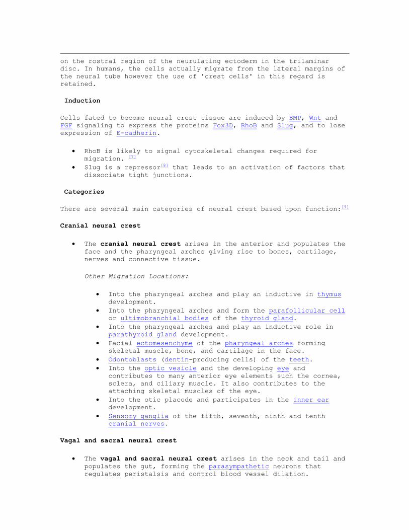
Cranial neural crest
The cranial neural crest arises in the anterior and populates the
face and the pharyngeal arches giving rise to bones, cartilage,
nerves and connective tissue.
Other Migration Locations:
Into the pharyngeal arches and play an inductive in thymus
development.
Into the pharyngeal arches and form the parafollicular cell
or ultimobranchial bodies of the thyroid gland.
Into the pharyngeal arches and play an inductive role in
parathyroid gland development.
Facial ectomesenchyme of the pharyngeal arches forming
skeletal muscle, bone, and cartilage in the face.
Odontoblasts (dentin-producing cells) of the teeth.
Into the optic vesicle and the developing eye and
contributes to many anterior eye elements such the cornea,
sclera, and ciliary muscle. It also contributes to the
attaching skeletal muscles of the eye.
Into the otic placode and participates in the inner ear
development.
Sensory ganglia of the fifth, seventh, ninth and tenth
cranial nerves.
Vagal and sacral neural crest
The vagal and sacral neural crest arises in the neck and tail and
populates the gut, forming the parasympathetic neurons that
regulates peristalsis and control blood vessel dilation.

Other Migration Locations:
Walls of the viscera to become enteric ganglia.
Trunk neural crest
The trunk neural crest lies between the vagal and sacral neural
crest and gives rise to two groups of cells. One group migrates
dorsolateral and populates the skin, forming pigment cells and
the other migrates ventrolateral through the anterior sclerotome
to become the epinephrine-producing cells of the adrenal gland
and the neurons of the sympathetic nervous system. Some cells
remain in the sclerotome to form the dorsal root ganglia
Other Migration Locations:
Proximal to the spinal cord and line up symmetrically to
form the dorsal root ganglia.
Into the skin to form melanocytes and Merkel cells.
Chromaffin cells of the adrenal medulla.
Near the vertebral column and become sympathetic chain
ganglia.
Cardiac neural crest
The cardiac neural crest overlaps the vagal neural crest and
migrates to populate the pharyngeal arches 3, 4 and 6 (producing
structures in the head) and to the heart, forming connective
tissue that separates the great vessels of the heart.
Other Migration Locations:
Into the pharyngeal arches and Truncus arteriosus
(embryology), forming the aorticopulmonary septum and the
smooth muscle of great arteries.
Anterior of the aorta to become the four pre-aortic ganglia
(celiac ganglion, superior mesenteric ganglion, inferior
mesenteric ganglion and aortical renal ganglia)
Migration
Neural crest cells require extracellular matrix to migrate through
interactions between integrins and fibronectin and laminin. Migration
is directed by inhibitory and attractive signals from cells. Ephrin is
an inhibitory ligand in posterior sclerotome that affects ventral
pathway trunk neural crest cells and causes them to migrate through the
anterior sclerotome instead. Thrombospondin promotes migration through
the anterior sclerotome. Another signal, stem cell factor is involved
in specifying the destination of migration. If expressed in the wrong
locations, pigment cells migrate to that site and proliferate there.

Plasticity
Neural crest cells show varying degrees of plasticity. Some trunk
neural crest cells are pluripotent. Cranial neural crest cells can give
rise to trunk neural crest cells if transplanted. However, heart neural
crest cells are committed before migration. Individual neural crest
cells can take on a new fate, however groups of neural crest cells
cannot.
viii Invest Clin. 1996 Jun;37(2):113-28.
Plasma excitatory amino acids in autism.
Moreno-Fuenmayor H, Borjas L, Arrieta A, Valera V, Socorro-Candanoza L.
Servicio de Medicina Genética Perinatal, Hospital Chiquinquirá,
Maracaibo,
Venezuela.
Plasma amino acid levels were measured by high pressure liquid
chromatography
(HPLC) in fourteen autistic children, all below 10 years of age. Mean
glutamic
and aspartic acid valued were elevated (169 +/- 142 uM and 22.1 +/- 13
uM
respectively) together with taurine (90.1 +/- 78.7 uM) (p > 0.1). All
affected
children had low levels of glutamine (241 +/- 166 uM; p < 0.01) and
asparagine
(22.9 +/- 12.9 uM; p < 0.01) as compared to normal values (585 +/- 25
and 59.2
+/- 4.2 uM respectively); eleven children had increased aspartic acid
and eight
children had high levels of glutamate; seven of these children had a
concomitant
increment of taurine. The increment of the three above mentioned
compounds was
observed at the same time only in five children. These findings
demonstrate that
abnormal plasmatic levels of neurotransmitter amino acids may be found
in some
autistic children. Increased glutamatemia may be dietary in origin or
may arise
endogenously for several reasons, among others, metabolic derrangements
in
glutamate metabolism perhaps involving vitamin B6, defects or blockage
of the
glutamate receptor at the neuronal compartment, or alterations in the
function of
the neurotransmitters transporters. Increments of taurine, an
inhibitor, is
likely compensatory and calcium dependent.

Publication Types:
Research Support, Non-U.S. Gov't
PMID: 8718922
ix Brain Dev. 2007 Feb 23; [Epub ahead of print]
Autism and epilepsy: A retrospective follow-up study.
Hara H.
Yokohama Central Area Habilitation Center for Children, Yokohama,
Japan; Kanagawa
Day Treatment & Guidance Center for Children, Japan.
So-called "idiopathic" autism, which exhibited no major complications
before
diagnosis is well-known as one of the risk factors for epilepsy. This
retrospective follow-up study aimed to clarify the characteristics of
epilepsy in
the autism; onset of seizure, seizure types, EEG findings and epilepsy
outcome
and the differences as a group between the autism with epilepsy and
those without
epilepsy. One hundred thirty individuals with autistic disorder or
atypical
autism diagnosed in childhood were followed up over 10years and were
evaluated
almost every year up to 18-35years of age. Their medical records
related to
perinatal conditions, IQ, social maturity scores and several factors of
epilepsy
were reviewed in October 2005. Thirty-three of the follow-up group
(25%)
exhibited epileptic seizures. The onset of epilepsy was distributed
from 8 to
26years of age. Two types of seizure were observed; partial seizure
with
secondarily generalized seizure and generalized seizure. Twenty of the
epileptics
(61%) showed the partial seizure. Although 18% of the non-epileptic
group
exhibited epileptic discharges on EEG, 68% of the epileptic group
revealed
epileptiform EEG findings before the onset of epilepsy. No differences
were
observed concerning the sex ratio, autistic disorder/atypical autism
and past
history of febrile seizures between the epileptic and non-epileptic
groups. Lower
IQ, lower social maturity score and higher frequency of prescribed
psychotropics
were observed in the epileptic group compared to the non-epileptics.

Idiopathic
autism was confirmed as the high risk factor for epilepsy. Epileptiform
EEG
findings predict subsequent onset of epileptic seizures in adolescence.
Epilepsy
is one of negative factors on cognitive, adaptive and
behavioral/emotional
outcomes for individuals with autism.
PMID: 17321709
Epilepsy Behav. 2007 Mar;10(2):344-7. Epub 2007 Feb 14.
Effects of vagus nerve stimulation in a patient with temporal lobe
epilepsy and
Asperger syndrome: case report and review of the literature.
Warwick TC, Griffith J, Reyes B, Legesse B, Evans M.
Department of Internal Medicine, University of California, San
Francisco,University Medical Center, 445 South CedarAvenue, Fresno, CA
93702,
USA. [email protected]
Seizures are a common comorbidity of autism and occur in as many as 30%
of
patients. This case report describes a 23-year-old man diagnosed with
both
Asperger syndrome and bitemporal epilepsy. The patient had behavioral
regression
that correlated with worsening of his intractable seizures. He
subsequently
underwent implantation of a vagus nerve stimulation therapy device for
his
refractory epilepsy. Both his seizures and his behavior were monitored
for 6
months. We describe the efficacy of vagus nerve stimulation therapy in
reducing
seizure severity as well as improving the behavioral components of his
Asperger
syndrome. We also review the current literature regarding epilepsy in
autistic
spectrum disorders.
Publication Types:
Clinical Trial
PMID: 17300990
x Autism. 2002 Sep;6(3):315-28.
Urinary peptides in Rett syndrome.
Solaas KM, Skjeldal O, Gardner ML, Kase FB, Reichelt KL.

Institute of Pediatric Research, The National Hospital, University of
Oslo,
Norway.
Rett syndrome is a neuro-developmental disorder related to autistic
behavior.
Persons with autism have previously been found to have hyperpeptiduria.
We here
report a significantly higher level of peptides in the first fasting
morning
urine from 53 girls with Rett syndrome (both classical and congenital)
compared
with 53 healthy girls. This elevation in urinary peptides was similar
to that in
35 girls with infantile autism. As in persons with autism, the
individual levels
of urinary peptides in the Rett syndrome group varied, and about a
fifth were
within the normal range. Levels of peptides were lower in girls with
classic Rett
syndrome than in girls with congenital Rett syndrome. This may be due
to
different etiological causes or to active and stagnant phases of the
disease.
Urine from girls with Rett syndrome was found to have higher frequency
and higher
levels of some urinary peptides that may cause inhibition of brain
maturation and
epilepsy
Publication Types:
Research Support, Non-U.S. Gov't
PMID: 12212921
Ann Neurol. 2001 May;49(5):597-606.
Neuropeptides and neurotrophins in neonatal blood of children with
autism or
mental retardation.
Nelson KB, Grether JK, Croen LA, Dambrosia JM, Dickens BF, Jelliffe LL,
Hansen
RL, Phillips TM.
National Institute of Neurological Diseases and Stroke, Bethesda, MD
20892-1447,
USA. [email protected]
There has been little exploration of major biologic regulators of
cerebral
development in autism. In archived neonatal blood of children with
autistic
spectrum disorders (n = 69), mental retardation without autism (n =

60), or
cerebral palsy (CP, n = 63) and of control children (n = 54), we used
recycling
immunoaffinity chromatography to measure the neuropeptides substance P
(SP),
vasoactive intestinal peptide (VIP), pituitary adenylate cyclase-
activating
polypeptide (PACAP), calcitonin gene-related peptide (CGRP), and the
neurotrophins nerve growth factor (NGF), brain-derived neurotrophic
factor
(BDNF), neurotrophin 3 (NT3), and neurotrophin 4/5 (NT4/5). Neonatal
concentrations of VIP, CGRP, BDNF, and NT4/5 were higher (ANOVA, all p
values <
0.0001 by Scheffe test for pairwise differences) in children in the
autistic
spectrum and in those with mental retardation without autism than in
control
children. In 99% of children with autism and 97% with mental
retardation, levels
of at least one of these substances exceeded those of all control
children.
Concentrations were similar in subgroups of the autistic spectrum (core
syndrome
with or without mental retardation, other autistic spectrum disorders
with or
without mental retardation) and in the presence or absence of a history
of
regression. Among children with mental retardation, concentrations did
not differ
by severity or known cause (n = 11, including 4 with Down syndrome).
Concentrations of measured substances were similar in children with CP
as
compared with control subjects. SP, PACAP, NGF, and NT3 were not
different by
diagnostic group. No measured analyte distinguished children with
autism from
children with mental retardation alone. In autism and in a
heterogeneous group of
disorders of cognitive function, overexpression of certain
neuropeptides and
neurotrophins was observed in peripheral blood drawn in the first days
of life.
Publication Types:
Research Support, Non-U.S. Gov't
Research Support, U.S. Gov't, P.H.S.
PMID: 11357950
Substance P
From Wikipedia, the free encyclopedia
Jump to: navigation, search
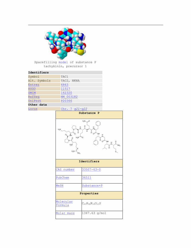
Spacefilling model of substance P
tachykinin, precursor 1
Identifiers
Symbol TAC1
Alt. Symbols TAC2, NKNA
Entrez 6863
HUGO 11517
OMIM 162320
RefSeq NM_003182
UniProt P20366
Other data
Locus Chr. 7 q21-q22
Substance P
Identifiers
CAS number 33507-63-0
PubChem 36511
MeSH Substance+P
Properties
Molecular
formula C63H98N18O13S
Molar mass 1347.63 g/mol

Except where noted otherwise, data
are given for
materials in their standard state
(at 25 °C, 100 kPa)
Infobox disclaimer and references
In neuroscience, Substance P is a neuropeptide: a short-chain
polypeptide that functions as a neurotransmitter and as a
neuromodulator. It belongs to the tachykinin neuropeptide family.
It is an 11-amino acid polypeptide with the sequence: Arg Pro Lys Pro
Gln Gln Phe Phe Gly Leu Met NH2.
Contents
[hide]
1 Receptor
2 Functions
o 2.1 Vomiting
o 2.2 Pain
o 2.3 Stimulating cellular growth
o 2.4 Vasodilation
3 Substance P in gastrointestinal infection
4 Animals without substance P
5 References
Receptor
The endogenous receptor for Substance P is neurokinin 1 receptor (NK1-
receptor, NK1R). It belongs to the tachykinin receptor sub-family of
GPCRs.
Functions
In the central nervous system, substance P has been associated in the
regulation of mood disorders, anxiety, stress, reinforcement,
neurogenesis, respiratory rhythm, neurotoxicity, nausea / emesis and
pain.
Vomiting
The vomiting center in the brainstem contains high concentrations of
substance P and its receptor, in addition to other neurotransmitters
such as choline, histamine, dopamine, serotonin, and opioids. Their
activation stimulates the vomiting reflex. Different emetic pathways
exist, and substance P/NK1R appears to be within the final common
pathway to regulate vomiting. [1]
Substance P antagonist (SPA) aprepitant is available in the market in
the treatment of chemotherapy-induced nausea / emesis.
Pain
Substance P is involved in the transmission of pain impulses from peripheral receptors to
the central nervous system. It has been theorized that it plays a part in
fibromyalgia. Capsaicin has been shown to reduce the levels of Substance P probably
by reducing the number of C-fibre nerves or causing these nerves to be more tolerant.
Stimulating cellular growth
Substance P has been shown to stimulate cellular growth in cell culture [2], and it was shown that Substance P could promote wound healing of
non-healing ulcers in humans. [3] It has also been shown to reverse
diabetes in mice. [4]
Vasodilation
It also has effects as a potent vasodilator. This is caused by the
release of nitric oxide from the endothelium. Its release can cause
hypotension.
Substance P in gastrointestinal infection
Entamoeba histolytica is a single-celled parasitic protozoan that
infects the lower gastrointestinal tract of humans, producing symptoms
of diarrhea, constipation, and abdominal pain.[5][6] This protozoan was
found to secrete serotonin[7], as well as substance P and neurotensin.[8]
xi J Autism Dev Disord. 2006 Apr;36(3):317-24.
Familial autoimmune thyroid disease as a risk factor for regression in
children
with Autism Spectrum Disorder: a CPEA Study.
Molloy CA, Morrow AL, Meinzen-Derr J, Dawson G, Bernier R, Dunn M,
Hyman SL,
McMahon WM, Goudie-Nice J, Hepburn S, Minshew N, Rogers S, Sigman M,
Spence MA,
Tager-Flusberg H, Volkmar FR, Lord C.
Center for Epidemiology and Biostatistics, Cincinnati Children's
Hospital Medical
Center, University of Cincinnati College of Medicine, Ohio 45229-3039,
USA.
A multicenter study of 308 children with Autism Spectrum Disorder (ASD)

was
conducted through the Collaborative Programs of Excellence in Autism
(CPEA),
sponsored by the National Institute of Child Health and Human
Development, to
compare the family history of autoimmune disorders in children with ASD
with and
without a history of regression. A history of regression was determined
from the
results of the Autism Diagnostic Interview-Revised (ADI-R). Family
history of
autoimmune disorders was obtained by telephone interview. Regression
was
significantly associated with a family history of autoimmune disorders
(adjusted
OR=1.89; 95% CI: 1.17, 3.10). The only specific autoimmune disorder
found to be
associated with regression was autoimmune thyroid disease (adjusted
OR=2.09; 95%
CI: 1.28, 3.41).
Publication Types:
Multicenter Study
PMID: 16598435
xii J Autism Dev Disord. 2006 Apr;36(3):317-24.
Familial autoimmune thyroid disease as a risk factor for regression in
children
with Autism Spectrum Disorder: a CPEA Study.
Molloy CA, Morrow AL, Meinzen-Derr J, Dawson G, Bernier R, Dunn M,
Hyman SL,
McMahon WM, Goudie-Nice J, Hepburn S, Minshew N, Rogers S, Sigman M,
Spence MA,
Tager-Flusberg H, Volkmar FR, Lord C.
Center for Epidemiology and Biostatistics, Cincinnati Children's
Hospital Medical
Center, University of Cincinnati College of Medicine, Ohio 45229-3039,
USA.
A multicenter study of 308 children with Autism Spectrum Disorder (ASD)
was
conducted through the Collaborative Programs of Excellence in Autism
(CPEA),
sponsored by the National Institute of Child Health and Human
Development, to
compare the family history of autoimmune disorders in children with ASD
with and
without a history of regression. A history of regression was determined
from the

results of the Autism Diagnostic Interview-Revised (ADI-R). Family
history of
autoimmune disorders was obtained by telephone interview. Regression
was
significantly associated with a family history of autoimmune disorders
(adjusted
OR=1.89; 95% CI: 1.17, 3.10). The only specific autoimmune disorder
found to be
associated with regression was autoimmune thyroid disease (adjusted
OR=2.09; 95%
CI: 1.28, 3.41).
Publication Types:
Multicenter Study
PMID: 16598435
xiii Med. Hypotheses. 2007;68(1):46-60. Epub 2006 Sep 7.
Prenatal influences on brain dopamine and their relevance to the rising
incidence
of autism.
Previc FH.
10906 Whispering Wind, San Antonio, TX 78230, USA.
The incidence of autism has risen 10-fold since the early 1980s, with
most of
this rise not explainable by changing diagnostic criteria. The rise in
autism is
paradoxical in that autism is considered to be one of the most
genetically
determined of the major neurodevelopmental disorders and should
accordingly
either be stable or even declining. Because a variety of epigenetic
influences,
particularly those occurring during the prenatal period, can override
or
masquerade as genetic influences, these should be considered as prime
contributors to the recent increase of autism. Prenatal influences on
dopamine
activity are especially well-documented, including the effects of
maternal
psychosocial stress, maternal fever, maternal genetic and hormonal
status, use of
certain medications, urban birth, and fetal hypoxia. All of these
factors have
been implicated in the genesis of autism, which is characterized by a
"hyperdopaminergic" state based on evidence from monkey and human
behavioral
studies, pharmacological studies in humans, and a left-hemispheric
predominance

of both dopamine and autistic-like symptoms. Chronically high maternal
levels of
dopamine caused by the pressures of increasingly urbanized societies
and by
changing maternal demographics such as increased workforce
participation,
educational achievement level, and age at first birth, may be
especially
significant epigenetic contributors to the recent autism rise.
PMID: 16959433 [PubMed - indexed for MEDLINE]
Brain Dev. 2006 Mar;28(2):99-103. Epub 2005 Sep 15.
Administration of secretin for autism alters dopamine metabolism in the
central
nervous system.
Toda Y, Mori K, Hashimoto T, Miyazaki M, Nozaki S, Watanabe Y, Kuroda
Y, Kagami
S.
Department of Pediatrics, School of Medicine, University of Tokushima,
3-18-15,
Kuramoto-cho, Tokushima-shi, Tokushima 770-8503, Japan.
We evaluated the clinical effects of intravenously administered
secretin in 12
children with autism (age range: 4-6 years, median age: 9 years,
boy:girl=8:4).
In addition, we investigated the association between improvement in
symptoms and
changes in the cerebrospinal fluid (CSF) homovanillic acid
(HVA),5-hydroxyindole-3-acetic acid (5-HIAA), and
6R-5,6,7,8-tetrahydro-L-biopterin (BH(4)) levels after administration.
After
administration of secretin, the Autism Diagnostic Interview-Revised
(ADI-R) score
improved in 7 of the 12 children. However, the score deteriorated in 2
of the 12
children (in the item of 'restricted and repetitive, stereotyped
interests and
behaviors'). The HVA and BH(4) levels in CSF were increased in all
children with
improvement in the ADI-R score. In contrast, no patient without the
elevation of
the BH(4) level showed improvement in the score. These findings suggest
that
secretin activated metabolic turnover of dopamine in the central
nervous system
via BH(4), improving symptoms.
Publication Types:

Clinical Trial
Comparative Study
PMID: 16168596
Brain Dev. 2007 Sep;29(8):510-3. Epub 2007 Mar 21.
Tachykinin 1 (TAC1) gene SNPs and haplotypes with autism: A case-
control study.
Marui T, Funatogawa I, Koishi S, Yamamoto K, Matsumoto H,
Hashimoto O, Nanba E, Nishida H, Sugiyama T, Kasai K, Watanabe K,
Kano Y, Kato N, Sasaki T.
Department of Neuropsychiatry, Graduate School of Medicine,
University of Tokyo, Tokyo, Japan.
Autism (MIM 209850) is a severe neurodevelopmental disorder
characterized by disturbances in social interaction and
communication, by repetitive body movements and restricted
interests, and by atypical language development. Several twin and
family studies have shown strong evidence for genetic factors in
the etiology of autism. Glutamate is a major excitatory
neurotransmitter in the human brain. Glutamate systems are
involved in the pathophysiology of autism. There are many
similarities between the symptoms evoked by glutamate antagonist
treatment and symptoms of autism found in several human and
animal studies. To elucidate the genetic background of autism, we
analyzed the relationship between three single nucleotide
polymorphisms (SNPs) of the Tachykinin 1 gene (TAC1) and autism,
because TAC1 is located in the candidate region for autism and
produces substance P and neurokinins. These products modulate
glutamatergic excitatory synaptic transmission and are also
involved in inflammation. Many different inflammation-related
mechanisms could be involved in the autistic brain. Therefore,
TAC1 may have some functions associated with the presumable
pathophysiology of autism. We compared the allele and haplotype
frequencies between autistic patients (n=170) and normal controls
(n=214) in the Japanese population, but no significant difference
was observed. Thus, the TAC1 locus is not likely to play a major
role in the development of autism.
PMID: 17376622
Substance P and cholecystokinin regulate neurochemical responses to
cocaine and methamphetamine in the striatum.
Loonam TM, Noailles PA, Yu J, Zhu JP, Angulo JA.
Department of Biological Sciences, Hunter College of the City
University of New York, 695 Park Avenue, New York 10021, USA.

The mechanism of action of drugs of abuse like cocaine and amphetamines
has been studied extensively in the dopamine terminal field areas of
the caudate-putamen (CPu) and the nucleus accumbens (NAc) of the rodent
brain. These brain regions contain several neuropeptides that must play
important roles in the normal physiological functions of these brain
regions. The study of neuropeptide physiology in the context of the
neurobiological responses to drugs of abuse may shed some light on the
intrinsic mechanism of action of neuropeptides of the CPu and the NAc.
The neuropeptides substance P (SP) and cholecystokinin (CCK) are
present in the striatum where they could play an important role
regulating the effects of psychostimulants like cocaine and
amphetamines (methamphetamine [METH] is a long acting derivative of d-
amphetamine). These highly addictive agents induce the release of
dopamine (DA) (and other catecholamines) from dopaminergic terminals of
the striatum. The excessive release of DA in the striatum and the NAc
has been implicated in the habit-forming properties of these drugs. In
order to study the contribution of SP and CCK in the striatum during
psychostimulant treatment, we employed selective non-peptide
neurokinin-1 (NK-1) and cholecystokinin-2 (CCK-2) receptor antagonists
that readily cross the blood brain barrier. We infused the neurokinin-1
receptor (NK-1R) antagonist, L-733,060, into the striatum of freely
moving rats via a microdialysis probe in order to assess the effects of
SP on cocaine-induced DA overflow in the striatum. Infusion of the NK-
1R antagonist prior to a systemic injection of cocaine (10 mg/kg i.p.)
significantly attenuated DA overflow in the striatum. Conversely,
infusion of a CCK-2 receptor (CCK-2R) antagonist, L-369,293, through
the microdialysis probe evoked DA overflow in the striatum in the
absence of cocaine and potentiated DA overflow after a single injection
of cocaine (10 mg/kg i.p.). Exposure to METH (10 mg/kg 4x at two-hour
intervals) produced deficits of dopamine transporters (DAT) in mice
striatum that are detectable three days after the treatment and are
long lasting. Pre-treatment (i.p. injections) with the NK-1R
antagonist, WIN-51,708 30 minutes before the 1st and 4th injections of
METH prevented the loss of DAT in the striatum. Moreover, pre-treatment
with the NK-1R antagonist prevents METH-induced cell death. Taken
together, these results demonstrate that the NK-1R and the CCK-2R are
important modulators of the actions of the psychostimulants cocaine and
METH. Neuropeptide receptors represent an important control point
mediating the effects of the neurotransmitter DA in the striatum of the
rodent brain.
PMID: 12801594
J Neurosci. 2000 Sep;12(9):3415-25.
Dopamine control of striatal gene expression during development:
relevance to knockout mice for the dopamine transporter.
Fauchey V, Jaber M, Bloch B, Le Moine C.
UMR CNRS 5541, Laboratoire d'Histologie Embryologie, Université
Victor Segalen Bordeaux 2, 146 rue Léo Saignat, 33076 Bordeaux
Cedex, France.

The aim of this study was to determine at which developmental
stage and how dopamine regulates the expression of striatal
dopamine receptor and neuropeptide mRNAs. For this, we studied
the expression of these mRNAs, in relation to dopamine
innervation, in normal mice from gestational day 13 (G13) to
adult. Particularly, we investigated the adaptive changes in the
expression of these markers in mice lacking the dopamine
transporter during development. We detected tyrosine hydroxylase,
by immunohistochemistry, in the ventral mesencephalon and the
striatal anlage in both genotypes at G13, whereas the dopamine
transporter appeared in the striatum of normal mice at G14. By in
situ hybridization, we detected striatal dopamine D1, D2, D3
receptor, and substance P mRNAs at G13, preproenkephalin A mRNA
at G14 and dynorphin mRNA at G17 in normal mice. Although the
time of initial detection and the distribution were not affected
in mutant mice, quantitative changes were observed. Indeed, D1
and D2 receptor as well as preproenkephalin A mRNA levels were
decreased from G14 on, and dynorphin mRNA level was increased
from G17 on. In contrast, substance P mRNA level was unaffected.
Our data demonstrate that the influence of dopamine on striatal
neurons occurs early during the development of the mesostriatal
system as quantitative changes appeared in mutant mice as soon as
G14. These findings bring new insights to the critical influence
of dopamine on the expression of striatal dopamine receptor and
neuropeptide mRNAs during development, and suggest that
mesostriatal dopamine transmission functions from G14 on.
PMID: 10998124
xiv Neuropsychobiology. 2002;45(1):1-6.
Activation of the inflammatory response system in autism.
Croonenberghs J, Bosmans E, Deboutte D, Kenis G, Maes M.
University Center of Child and Adolescent Psychiatry, Antwerp, Belgium.
BACKGROUND/AIM: There is now some evidence that autism may be
accompanied by
abnormalities in the inflammatory response system (IRS). Products of
the IRS,
such as proinflammatory cytokines, may induce some of the behavioral
symptoms of
autism, such as social withdrawal, resistance to novelty and sleep
disturbances.
The main aim of the present study was to examine whether autism is
accompanied by
an activation of the IRS. METHODS: We measured the production of
interleukin

(IL)-6, IL-10, the IL-1 receptor antagonist (IL-1RA), interferon (IFN)-
gamma and
tumor necrosis factor (TNF)-alpha by whole blood and the serum
concentrations of
IL-6, the IL-2 receptor (IL-2R) and IL-1RA. RESULTS: This study showed
a
significantly increased production of IFN-gamma and IL-1RA and a trend
toward a
significantly increased production of IL-6 and TNF-alpha by whole blood
of
autistic children. There were no significant differences in the serum
concentrations of IL-6, IL-2R and IL-1RA between autistic and normal
children.
CONCLUSIONS: These results suggest that autism may be accompanied by an
activation of the monocytic (increased IL-1RA) and Th-1-like (increased
IFN-gamma) arm of the IRS. It is hypothesized that increased production
of
proinflammatory cytokines could play a role in the pathophysiology of
autism.
Copyright 2002 S. Karger AG, Basel
Publication Types:
Research Support, Non-U.S. Gov't
PMID: 11803234
Brain Dev. 2005 Dec;27(8):579-88. Epub 2005 May 31.
Voluntary control of saccadic and smooth-pursuit eye movements in
children with
learning disorders.
Fukushima J, Tanaka S, Williams JD, Fukushima K.
Department of Health Sciences, Hokkaido University School of Medicine,
West 5,
North 12, Sapporo 060-0812, Japan. [email protected]
Eye movement is crucial to humans in allowing them to aim the foveae at
objects
of interest. We examined the voluntary control of saccadic and smooth-
pursuit eye
movements in 18 subjects with learning disorders (LDs) (aged 8-16) and
22 normal
controls (aged 7-15). The subjects were assigned visually guided,
memory-guided,

and anti-saccade tasks, and smooth-pursuit eye movements (SPEM).
Although, the LD
subjects showed normal results in the visually guided saccade task,
they showed
more errors in the memory-guided saccade task (e.g. they were unable to
stop
themselves reflexively looking at the cue) and longer latencies, even
when they
performed correctly. They also showed longer latencies than the
controls in the
anti-saccade task. These results suggest that they find it difficult to
voluntarily suppress reflexive saccades and initiate voluntary saccades
when a
target is invisible. In SPEM using step-ramp stimuli, the LD subjects
showed
lower open- and closed-loop gains. These results suggest disturbances
of both
acceleration of eye movement in the initial state and maintenance of
velocity in
minimizing retinal slip in the steady state. Recent anatomical studies
in LD
subjects have suggested abnormalities in the structure of certain brain
areas
such as the frontal cortex. Frontal eye movement-related areas such as
the
frontal eye fields and supplementary eye fields may be involved in
these
disturbances of voluntary control of eye movement in LDs.
PMID: 15925462
xv Arch Gen Psychiatry. 2006 Dec;63(12):1417-28.
Amygdala volume and nonverbal social impairment in adolescent and
adult males with autism.
Nacewicz BM, Dalton KM, Johnstone T, Long MT, McAuliff EM, Oakes
TR, Alexander AL, Davidson RJ.
Waisman Laboratory for Brain Imaging and Behavior, University of
Wisconsin-Madison, Waisman Center, 1500 Highland Avenue, Madison,
WI 53705, USA.
BACKGROUND: Autism is a syndrome of unknown cause, marked by
abnormal development of social behavior. Attempts to link
pathological features of the amygdala, which plays a key role in
emotional processing, to autism have shown little consensus.
OBJECTIVE: To evaluate amygdala volume in individuals with autism
spectrum disorders and its relationship to laboratory measures of
social behavior to examine whether variations in amygdala
structure relate to symptom severity. DESIGN: We conducted 2
cross-sectional studies of amygdala volume, measured blind to
diagnosis on high-resolution, anatomical magnetic resonance
images. Participants were 54 males aged 8 to 25 years, including

23 with autism and 5 with Asperger syndrome or pervasive
developmental disorder not otherwise specified, recruited and
evaluated at an academic center for developmental disabilities
and 26 age- and sex-matched community volunteers. The Autism
Diagnostic Interview-Revised was used to confirm diagnoses and to
validate relationships with laboratory measures of social
function. MAIN OUTCOME MEASURES: Amygdala volume, judgment of
facial expressions, and eye tracking. RESULTS: In study 1,
individuals with autism who had small amygdalae were slowest to
distinguish emotional from neutral expressions (P=.02) and showed
least fixation of eye regions (P=.04). These same individuals
were most socially impaired in early childhood, as reported on
the Autism Diagnostic Interview-Revised (P<.04). Study 2 showed
smaller amygdalae in individuals with autism than in control
subjects (P=.03) and group differences in the relation between
amygdala volume and age. Study 2 also replicated findings of more
gaze avoidance and childhood impairment in participants with
autism with the smallest amygdalae. Across the combined sample,
severity of social deficits interacted with age to predict
different patterns of amygdala development in autism (P=.047).
CONCLUSIONS: These findings best support a model of amygdala
hyperactivity that could explain most volumetric findings in
autism. Further psychophysiological and histopathological studies
are indicated to confirm these findings.
PMID: 17146016
Psychopharmacology (Berl). 2007 Mar;191(1):107-18. Epub 2007 Feb 3.
From anxiety to autism: spectrum of abnormal social behaviors
modeled by progressive disruption of inhibitory neuronal function
in the basolateral amygdala in Wistar rats.
Truitt WA, Sajdyk TJ, Dietrich AD, Oberlin B, McDougle CJ,
Shekhar A.
Department of Psychiatry, Indiana University School of Medicine,
1111 West 10th Street, Indianapolis, IN 4620, USA.
RATIONALE: Social behaviors are disrupted in several psychiatric
disorders. The amygdala is a key brain region involved in social
behaviors, and amygdala pathology has been implicated in disease
states ranging from social anxiety disorder to autism. OBJECTIVE:
To test the effects of progressive disruption of the inhibitory
function within the basolateral nucleus of the amygdala (BLA) on
conspecific social interaction in rats and investigate functional
networks from the ventral medial prefrontal cortex (mPFCv) to the
BLA. MATERIALS AND METHODS: BLA inhibitory tone was disrupted by
priming it with the stress-peptide corticotrophin releasing
factor (CRF) receptor agonist urocortin 1 (Ucn 1, 6 fmol), or by
selective lesioning of a subset of BLA-GABAergic interneurons
containing neurokinin 1 receptors using the targeted toxin SSP-

Saporin. The effects of the disruption of GABAergic tone in the
BLA were examined using a repeated exposure and habituation
paradigm of social interaction (SI/h). Lesions and selectivity of
lesions were confirmed postmortem. Additionally, effects of
stimulating mPFCv on cFos activity in interneurons of the BLA
were examined. RESULTS: Rats primed with Ucn 1 showed persistent
social inhibition, which could be overcome with habituation,
putatively modeling social anxiety. Rats with a selective
lesioning of a subset of GABAergic interneurons in the BLA
exhibited persistent social inhibition that was not reversed by
SI/h paradigm. We also demonstrate selective functional inputs to
this subset of interneurons when mPFCv was activated.
CONCLUSIONS: These models with different gradations of disrupted
BLA inhibition could help to study social dysfunction in
disorders ranging from social anxiety to autism spectrum
disorders.
PMID: 17277936
Peptides. 1983 Jul-Aug;4(4):525-41.
Organization and interrelationship of neuropeptides in the
central amygdaloid nucleus of the rat.
Wray S, Hoffman GE.
The organization and interactions of neuropeptides in the central
nucleus of the amygdala (Ce) were studied using single and double
label immunocytochemical techniques. Immunocytochemical
localization of substance P (SP), neurotensin (NT), met-
enkephalin (m-ENK), somatostatin (SS) and vasoactive intestinal
polypeptide (VIP) revealed all of these peptides within discrete
regions of the Ce. The regions differed from the classical medial
and lateral anatomical divisions reported for the Ce. Instead,
three easily recognizable neuropeptidergic subdivisions were
evident: a medial zone, a central zone and a lateral capsular
zone. Two types of interrelationships between peptides were
noted. The first involved a peptidergic fiber in apposition to a
peptidergic perikarya. The most prevalent peptidergic interaction
of this type occurred between SP and NT. The second
interrelationship involved two different peptidergic fibers in
apposition to an immunonegative cell. Two interactions of this
type were commonly observed. The first involved NT and m-ENK
fibers simultaneously apposed to an unstained cell. The second
involved SP and m-ENK fibers adjacent to the same immunonegative
cell. The interactions between peptidergic systems may suggest a
role of these substances in the regulation of autonomic functions
in the Ce.
Chem Senses. 2007 May;32(4):343-53. Epub 2007 Feb 17.

Cerebral activation to intranasal chemosensory trigeminal
stimulation.
Boyle JA, Heinke M, Gerber J, Frasnelli J, Hummel T.
Cognitive Neuroscience Unit, Montreal Neurological Institute,
McGill University, Montreal, Quebec, Canada.
Although numerous functional magnetic resonance imaging (FMRI)
studies have been performed on the processing of olfactory
information, the intranasal trigeminal system so far has not
received much attention. In the present study, we sought to
delineate the neural correlates of trigeminal stimulation using
carbon dioxide (CO(2)) presented to the left or right nostril.
Fifteen right-handed men underwent FMRI using single runs of 3
conditions (CO(2) in the right and the left nostrils and an
olfactory stimulant-phenyl ethyl alcohol-in the right nostril).
As expected, olfactory activations were located in the
orbitofrontal cortex (OFC), amygdala, and rostral insula. For
trigeminal stimulation, activations were found in "trigeminal"
and "olfactory" regions including the pre- and postcentral gyrus,
the cerebellum, the ventrolateral thalamus, the insula, the
contralateral piriform cortex, and the OFC. Left compared with
right side stimulations resulted in stronger cerebellar and brain
stem activations; right versus left stimulation resulted in
stronger activations of the superior temporal sulcus and OFC.
These results suggest a trigeminal processing system that taps
into similar cortical regions and yet is separate from that of
the olfactory system. The overlapping pattern of cortical
activation for trigeminal and olfactory stimuli is assumed to be
due to the intimate connections in the processing of information
from the 2 major intranasal chemosensory systems.
PMID: 17308328
xvi Am J Hum Genet. 2001 Nov;69(5):1062-7. Epub 2001 Oct 2.
Excess of twins among affected sibling pairs with autism: implications for the etiology of autism.
Greenberg DA, Hodge SE, Sowinski J, Nicoll D.
Division of Statistical Genetics, Department of Biostatistics, Columbia University and New York State
Psychiatric Institute, New York, NY 10032, USA. [email protected]

It is widely accepted that genes play a role in the etiology of autism. Evidence for this derives, in part, from
twin data. However, despite converging evidence from gene-mapping studies, aspects of the genetic
contribution remain obscure. In a sample of families selected because each had exactly two affected sibs,
we observed a remarkably high proportion of affected twin pairs, both MZ and DZ. Of 166 affected sib
pairs, 30 (12 MZ, 17 DZ, and 1 of unknown zygosity) were twin pairs. Deviation from expected values was
statistically significant (P<10(-6) for all twins); in a similarly ascertained sample of individuals with type I
diabetes, there was no deviation from expected values. We demonstrate that to ascribe the excess of twins
with autism solely to ascertainment bias would require very large ascertainment factors; for example,
affected twin pairs would need to be, on average, approximately 10 times more likely to be ascertained than
affected non-twin sib pairs (or 7 times more likely if "stoppage" plays a role). Either risk factors (related to
twinning or to fetal development) or other factors (genetic or nongenetic) in the parents may contribute to
autism.
PMID: 11590546