Embed Size (px)
Citation preview

UCLAUCLA Electronic Theses and Dissertations
TitleHeart Failure Genetics in Mice and Men
Permalinkhttps://escholarship.org/uc/item/4c59k544
AuthorWang, Jessica Jen-Chu
Publication Date2014-01-01 Peer reviewed|Thesis/dissertation
eScholarship.org Powered by the California Digital LibraryUniversity of California

UNIVERSITY OF CALIFORNIA
Los Angeles
Heart Failure Genetics in Mice and Men
A dissertation submitted in partial satisfaction of the
requirements for the degree Doctor of Philosophy
in Human Genetics
by
Jessica Jen-Chu Wang
2014


ii
ABSTRACT OF THE DISSERTATION
Heart Failure Genetics in Mice and Men
by
Jessica Jen-Chu Wang
Doctor of Philosophy in Human Genetics
University of California, Los Angeles, 2014
Professor Aldons Jake Lusis, Chair
The genetics of heart failure is complex. In familial cases of cardiomyopathy, where
mutations of large effects predominate in theory, genetic testing using a gene panel of
up to 76 genes returned negative results in about half of the cases. In common forms of
heart failure (HF), where a large number of genes with small to modest effects are
expected to modify disease, only a few candidate genomic loci have been identified
through genome-wide association (GWA) analyses in humans. We aimed to use exome
sequencing to rapidly identify rare causal mutations in familial cardiomyopathy cases
and effectively filter and classify the variants based on family pedigree and family
member samples. We identified a number of existing variants and novel genes with
great potential to be disease causing. On the other hand, we also set out to understand
genetic factors that predispose to common late-onset forms of heart failure by
performing GWA in the isoproterenol-induced HF model across the Hybrid Mouse
Diversity Panel (HMDP) of 105 strains of mice. We performed fine phenotyping using
serial echocardiograms and controlled for environmental heterogeneity in the
iii
experimental setting. As a result, we achieved high heritability estimates of 64% to 84%
for all cardiac traits and had superior power for mapping HF-related trait, compared to
human studies. Association analyses of cardiac traits, corrected for population structure
and multiple comparisons, revealed genome-wide significant loci across the spectrum of
cardiac traits. Cardiac tissue gene expression profiling, expression quantitative trait loci,
expression-phenotype correlation, and coding sequence variation analyses were
performed to prioritize candidate genes and to generate hypotheses for downstream
mechanistic studies. Future directions will be to further bridge the gap of understanding
between rare Mendelian and common cardiovascular diseases, which together will have
wide spread therapeutic implications in delaying or reversing HF progression in human
populations.

iv
The dissertation of Jessica Jen-Chu Wang is approved.
Rita Cantor
Karen Reue
Yibin Wang
Aldons Jake Lusis, Committee Chair
University of California, Los Angeles
2014

v
Dedication
To my mother, Mei-Tsu, who has been a source of encouragement and inspiration to
me throughout my life. To my father, Ta-Jan, who has been a steady moral compass
and provider to help me find and realize my potentials. To my sisters Sandra and Karen,
for being so supportive. To my dear husband, Richard, for always cheering me on. Most
of all thanks to God who continues to make the impossible possible.

vi
Table of Contents
1 Introduction ...............................................................................................................1 Bibliography ..................................................................................................................8
2 Exome Sequencing Identifies a Novel Candidate Gene ZBTB17 for Arrhythmogenic Right Ventricular Cardiomyopathy. ...............................................12
Background.................................................................................................................12 Methods ......................................................................................................................15 Results ........................................................................................................................18 Discussion...................................................................................................................25 Bibliography ................................................................................................................40
3 Genetic Contributions to Cardiac Hypertrophy and Fibrosis Induced by Beta-adrenergic Stimulation in Mice ...................................................................................42
Abstract.......................................................................................................................42 Significance ................................................................................................................43 Background.................................................................................................................43 Methods ......................................................................................................................46 Results ........................................................................................................................49 Discussion...................................................................................................................56 Bibliography ................................................................................................................70
4 Genetic Dissection of Cardiac Remodeling in an Isoproterenol-induced Heart Failure Mouse Model ....................................................................................................76
Background.................................................................................................................76 Methods ......................................................................................................................79 Results ........................................................................................................................84 Discussion...................................................................................................................94 Bibliography ..............................................................................................................112
5 Future Directions ..................................................................................................120 Bibliography ..............................................................................................................127

vii
Table of Tables
Table 2-1. Genes in the GeneDx cardiac genetic test panels. .......................................35!Table 2-2. Reported genes from gene panel testing by variant classification. ...............36!Table 2-3. Exome sequencing summary data. ...............................................................37!Table 2-4. Select genetic variants from exome 3. ..........................................................38!Table 2-5. PCR genotyping results of candidate MYOM2 variant. .................................39!Table 3-1. List of mouse strains in the study. .................................................................64!Table 3-2. Significant heart failure trait loci identified in HMDP GWA............................65!Table 3-3. Suggestive heart failure trait loci identified in HMDP GWA...........................66!Table 3-4. Significant overlap of HMDP and human GWA loci for heart failure traits. ...67!Table 3-5. Significant overlap of HF GWAS loci in HMDP with QTL from previous mouse
linkage analyses......................................................................................................68!Table 3-6. DAVID enrichment of all genes significantly correlated with Abcc6 expression
in ISO treated mouse heart. ....................................................................................69!Table 4-1. Echocardiographic traits in control mice measured at 2 time points. ..........107!Table 4-2. Study sample characteristics. .....................................................................108!Table 4-3. Heritability estimates of cardiac structure and function in the HMDP. ........109!Table 4-4. Genome-wide significant and suggestive cardiac remodeling loci. .............110!

viii
Table of Figures
Figure 2-1. UCLA Clinical Exome Sequencing workflow................................................29!Figure 2-2. GeneDx genetic test results by variant classifications for familial
cardiomyopathy patients. ........................................................................................30!Figure 2-3. Family pedigrees for exome sequencing patients........................................31!Figure 2-4. Gross and histopathologic examination of the exome 3 proband and family
member are consistent with arrhythmogenic right ventricular cardiomyopathy
(ARVC). ...................................................................................................................32!Figure 2-5. The M-line ultrastructure of exome 3 proband is intact. ...............................33!Figure 2-6. Homozygosity by descent analysis. .............................................................34!Figure 3-1. Wide variation of heart failure traits between the strains of the HMDP........58!Figure 3-2. Isoproterenol-induced lethality. ....................................................................59!Figure 3-3. Manhattan plots of heart failure traits...........................................................60!Figure 3-4. Ppp3ca (calcineurin A) is a candidate gene at the chromosome 3 right
ventricular weight ratio locus. ..................................................................................61!Figure 3-5. Abcc6 plays a role in the regulation of cardiac fibrosis after ISO stimulation.
................................................................................................................................63!Figure 4-1. Correlation among echocardiographic measures and LV (LV) weights. ......99!Figure 4-2. Cardiac structural and functional variations in the HMDP..........................100!Figure 4-3. Fine mapping of the change in week 3 LVM chromosome 7 locus............101!Figure 4-4. Fine mapping of the change in week 1 IVSd chromosome 9 locus. ..........102!Figure 4-5. Fine mapping of week 1 FS chromosome 15 locus. ..................................103!Figure 4-6. Fine mapping of week 2 LVM chromosome 4 locus. .................................104!Figure 4-7. Fine mapping of the change in week 3 LVM chromosome 4 locus............105!Figure 4-8. Fine mapping of the change in week 2 FS chromosome 9 locus. .............106!Figure 5-1. siRNA knock-down of Myh14 in neonatal rat ventricular cardiomyocytes
abrogates hypertrophic effects of isoproterenol and phenylephrine. ....................125!Figure 5-2. Transgenic zebrafish strain with cmlc2::EGFP. .........................................126!

ix
Acknowledgement
The author is greatly indebted to members of her thesis and advisory committees. The
author wishes to acknowledge ongoing support and guidance from the research faculty
members: Professors Aldons Jake Lusis, Yibin Wang, Thomas Vondriska, James
Weiss, Karen Reue, and Rita Cantor; and clinical faculty members: Arnold Baas,
Eugene DePasquale, Tamara Horwich, Ali Nsair, Will Suh, Martin Cadeiras, and Mario
Deng.
The author also wishes to acknowledge contributions from: Cheri Silverstein, Hane Lee,
Michael Fishbein, Melissa Spencer, Ekaterina Mokhonova, Stanley Nelson (Chapter 2);
Rozeta Avetisyan, Christoph Rau (Chapter 3); Milagros Romay, Richard Davis (Chapter
4).
This research was supported in part by the National Heart, Lung, and Blood Institute
(NHLBI) of the National Institute of Health (NIH) under grants HL123295, HL110667 and
HL28481 to Professor Aldons Jake Lusis and HL114437, HL123295, HL110667 to
Professor Yibin Wang. The author was supported the NIH training fellowship
T32HL007895. The content is solely the responsibility of the author and does not
necessarily represent the official views of the National Institute of Health. In addition, the
author was supported by the Specialty Training and Advanced Research (STAR)
program at the David Geffen School of Medicine at UCLA, the Tibor Fabian Research
Award, the Department of Medicine at UCLA.

x
Vita
Education
1996-2000 Massachusetts Institute of Technology BS in Biology
2000-2004 University of Pennsylvania MD
Training
2004-2007 UT Southwestern Medical Center Internal Medicine
2007-2008 Baylor University Medical Center Hospitalist
2008-2013 University of California, Los Angeles Cardiovascular Disease
Employment
2013-2014 University of California, Los Angeles Clinical Instructor in Medicine
Publications
Hendren SK, Wang J, Gorman J, Peacock T, Hershock DM, Rosato EF.
Esophagectomy and splenectomy in a patient with osteopetrosis. J Thorac Cardiovasc
Surg. 2005;129:1457-1458
Wang JJ, Reimold SC. Chest pain resulting from histoplasmosis pericarditis: A brief
report and review of the literature. Cardiol Rev. 2006;14:223-226
Sadek H, Hannack B, Choe E, Wang J, Latif S, Garry MG, Garry DJ, Longgood J,

xi
Frantz DE, Olson EN, Hsieh J, Schneider JW. Cardiogenic small molecules that
enhance myocardial repair by stem cells. Proc Natl Acad Sci U S A. 2008;105:6063-
6068
Ghazalpour A, Rau CD, Farber CR, Bennett BJ, Orozco LD, van Nas A, Pan C, Allayee
H, Beaven SW, Civelek M, Davis RC, Drake TA, Friedman RA, Furlotte N, Hui ST,
Jentsch JD, Kostem E, Kang HM, Kang EY, Joo JW, Korshunov VA, Laughlin RE,
Martin LJ, Ohmen JD, Parks BW, Pellegrini M, Reue K, Smith DJ, Tetradis S, Wang J,
Wang Y, Weiss JN, Kirchgessner T, Gargalovic PS, Eskin E, Lusis AJ, LeBoeuf RC.
Hybrid mouse diversity panel: A panel of inbred mouse strains suitable for analysis of
complex genetic traits. Mamm Genome. 2012;23:680-692
Weiss JN, Karma A, MacLellan WR, Deng M, Rau CD, Rees CM, Wang J, Wisniewski
N, Eskin E, Horvath S, Qu Z, Wang Y, Lusis AJ. "Good enough solutions" and the
genetics of complex diseases. Circ Res. 2012;111:493-504

1
1 Introduction
Heart failure (HF) is a complex clinical syndrome that results from impaired
ventricular filling or ejection of blood. Its cardinal features are dyspnea, fatigue, and fluid
retention. HF may be associated with a wide spectrum of left ventricular (LV) functional
abnormalities, ranging from normal LV size and preserved ejection fraction (EF) to
severe dilatation and/or markedly reduced EF. HF with reduced EF (HFrEF) is defined
as the clinical diagnosis of HF and EF!40%. HF with preserved EF (HFpEF) is defined
as the clinical diagnosis of HF, EF>40%, and abnormal LV diastolic dysfunction, after
the exclusion of non-cardiac causes1, 2. HFrEF and HFpEF each make up about half of
the overall HF burden3. Risk factors for HFrEF include a family history of
cardiomyopathy, long-standing hypertension, previous myocardial infarction (MI),
cardiotoxic agents, valvular abnormalities, congenital heart lesions, arrhythmias, and
others. Current therapies for HFrEF range from prevention, non-pharmacological
interventions, pharmacological treatment, device therapy, to inotropic support,
mechanical circulatory support and cardiac transplantation at the end-stage. Risk
factors for HFpEF include hypertension, obesity, coronary artery disease (CAD),
diabetes mellitus, atrial fibrillation (AF), and hyperlipidemia1. Effective pharmacological
treatments for HFrEF have generally been disappointing when used in patients with
HFpEF4. The lifetime risk of developing HF is 20% for Americans "40 years of age. The
total cost of HF care in the United States exceeds $30 billion annually, with over half of
these costs spent on hospitalizations5. HF is the primary diagnosis in >1 million
hospitalizations annually. A subset of patients with chronic HF will continue to progress

2
and develop persistently severe symptoms that are refractory to maximum therapy. The
absolute mortality rates for HF remain approximately 50% within 5 years of diagnosis1.
In the past 25 years, through linkage studies of familial cardiomyopathies and
arrhythmias, a number of genes associated with early-onset HF have been identified,
each harboring unique amino acid substitution, splice site, and truncation mutations6-9.
Definitive causal link of some of these mutations to disease have been established in
mouse models10-12. Allele-specific silencing of the mutant allele demonstrated
therapeutic benefit13. In the past decade, next-generation sequencing has enabled the
massive and parallel sequencing of many genes at once, which brought the cost of
sequencing down to a level similar to other routine medical tests. Genetic testing for
many disorders can now be ordered commercially from any medical office outside of the
academic setting, including gene panels for specific forms of familial cardiomyopathies
and arrhythmias. Because familial cardiomyopathies and arrhythmias are clinically
overlapping and genetically heterogeneous, comprehensive cardiomyopathy and
arrhythmia panel tests have also been made available. Genetic test panels are
designed to effectively and accurately capture sequence variants in the protein-coding
regions of genes known to be associated with disease. A more global approach is whole
exome sequencing, where in theory all protein-coding variants of the genome can be
evaluated. Genetic polymorphisms are frequently observed in any given exome. Our
results showed that each exome contains approximately 22,000 single nucleotide
polymorphisms (SNPs) and 1-10 bp insertion-deletions (indels) (Table 2-3). In other
words, one variant would be found in each gene sequenced on average. In the absence

3
of family member samples, prioritization of variants for disease significance generally
relies on the following criteria: 1) previous report in literature, 2) allele frequency in the
reference population, and 3) in silico pathogenicity algorithms, based on the following
assumptions: 1) causal mutation lies in protein coding regions of the genes or exome, 2)
mutations reported in literature are unbiased and truly causal, 3) causal mutations are
rare, 4) the referenced controls are representative of the sequenced individual’s genetic
background, and 5) in silico pathogenicity algorithms give biologically meaningful
predictions. In practice, the assignment of variant classes as disease-causing, likely
disease-causing, and of unknown significance is complex for both gene panel and
whole exome testing. As technologies and analytical methods for genome sequencing
continue to advance, current limitations in the assumptions will likely be overcome and
variant interpretation will improve.
The majority of HF encountered in the community is not explained by a single
gene mutation. Rather, there is a complex interplay between genetic susceptibility and
environmental factors that results in the phenotype of clinical HF and LV structural or
functional abnormalities that are known to predispose to HF. Genome-wide association
(GWA) of common genetic variants offers an alternative, high-throughput and unbiased
approach to identify risk loci contributing to HF. A number of HF-related GWA has been
performed to date, each revealing a small number of loci and candidate genes. For
example, two SNPs, one at 58.8kb from USP3 (ubiquitin-specific protease) gene in
individuals of European ancestry and the other at 6.3 kb from the LRIG3 (leucine-rich,
immunoglobulin like domain) gene in individuals of African ancestry, have been

4
associated with the development of HF14. A third SNP located in the intron of CMTM7
(CKLF-like MARVEL transmembrane domain containing 7) gene among individuals of
European ancestry was associated with HF mortality15. In a European consortium study
on DCM, two SNPs were associated with sporadic DCM and replicated in independent
samples. One of these SNPs was located near HSPB7 (heat shock 27kDa protein
family, member 7). Another was a non-synonymous SNP located in BAG3 (BCL2-
associated athanogene 3). Sequencing of BAG3 exons in 168 independent index cases
with familial DCM identified four heterozygous truncating and two missense mutations,
which were present in all genotyped relatives affected by disease and absent in a
control group of 347 healthy individuals16. Lastly, GWA of cardiac structure and systolic
function among African Americans associated 4 genetic loci near UBE2V2 (ubiquitin-
conjugating enzyme E2 variant 2), WIPI1 (WD repeat domain, phosphoinositide
interacting 1), PPAPDC1A (phosphatidic acid phosphatase type 2 domain containing
1A) and KLF5 (Kruppel-like factor 5) with left ventricular mass, interventricular septal
wall thickness, left ventricular internal diameter and ejection fraction, respectively
Base on the Framingham Heart Study, the genetic basis of HF in human is
modest. The hazard ratio for HF in those with parental HF was 1.70 [95% confidence
interval (CI) 1.11-2.60]17 and the estimated heritability of LV mass (LVM) was 0.24-
0.3218. One twin study demonstrated interaclass correlation coefficients of 0.69 and
0.32 for monozygotic and dizygotic twins, respectively19, consistent with significant
environmental influences on HF traits, thus limiting the power of signal detection in a
GWA. Complementary to the studies in humans, we conducted a large-scale survey of

5
cardiac structure and function in 105 inbred mouse strains, termed the Hybrid Mouse
Diversity Panel (HMDP), at baseline and in each subsequent week during a 3-week
isoproterenol infusion to investigate genomic loci controlling variations in traits relevant
to HF. We observed striking variations among the mouse strains and deteremined that
cardiac structure and function were highly heritable at baseline and in response to
isoproterenol, with heritability estimates between 0.64 and 0.84. Using a linear mixed
model algorithm, which accounts for relatedness among the strains, GWA was
performed on the observed traits. A number of loci contributing to heart weight, fibrosis,
cardiac structure and function were identified. We prioritized genes in each associated
genomic locus using expression quantitative trait locus (eQTL) analysis and coding
sequence variations in the Wellcome Trust Mouse Genomes Project. We validated one
of the association loci candidate genes Abcc6 as causal for the variations in
isoproterenol-induced fibrosis. We were able to identify more significantly associated
loci than in the human studies, because we were able to fully control for environmental
confounders in the laboratory setting, which contributed to improved heritability
estimates and significantly improved mapping power.
Through gene panel testing and exome sequencing in human subjects and HF
GWA in mice, we have implicated a number of novel genes and genetic variants in HF-
related traits, which need to be replicated or validated experimentally, to establish
definitive causal role in disease. Given the number of genes and variants to test, a high-
throughput pipeline is required to screen associated disease genes and variants
experimentally. We have explored two high-throughput model systems for gene

6
validation thus far, one using neonatal rat ventricular cardiomyocytes (NRVMs) and one
using zebrafish. Additional gene validation technique include adeno-associated virus 9
(AAV9) to examine the role of the candidate gene in the mouse heart. To validate
genetic variants, we plan to use the clustered regularly interspaced short palindromic
repeats (CRISPR)/Cas9 system to generate targeted coding sequence variants in
zebrafish and mice. Finally, we will also explore the role of allele-specific silencing of
disease-causing mutation using RNA interference and CRISPR to be carried out in
patient-derived induced pluripotent stem (iPS) cells. Once the causal role of a gene or
genetic variant is establish, genetically stable mouse models will be generated for
further functional and downstream mechanistic studies.
The results from our GWA and those of others showed that the majority of the
genomic regions controlling susceptibility to HF are located in intergenic regions, which
highlights the importance of gene regulation rather than protein-coding sequence
variations in the mechanisms affecting complex traits such as HF. A number of
mechanisms have been proposed to link non-protein-coding sequence variations to
gene regulation, including promoter region variations, methylation changes, non-coding
RNA, and altered chromatin states among others. Using Reduced Representation
Bisulfite Sequencing (RRBS) we studied the methylation patterns in the left ventricular
tissues from 2 mouse strains, susceptible and resistant to ventricular dilation and
decreased ejection fraction after isoproterenol treatment. We found that the differences
in methylation patterns were greater between strains than between treatment groups.
We plan to examine the role of methylation patterns and their relationships to SNP

7
variations and HF traits across the HMDP. In the coming years, the use of fine and deep
phenotyping, large-scale whole genome sequencing, and evermore sophisticated
mapping strategies, will work together to improve our current understanding of heart
health genetics even more. This new understanding will translate into preventive and
treatment options for those who have no effective therapies currently, such as familial
cardiomyopathy and HFpEF.

8
Bibliography
1. Yancy CW, Jessup M, Bozkurt B, Butler J, Casey DE, Jr., Drazner MH, Fonarow
GC, Geraci SA, Horwich T, Januzzi JL, Johnson MR, Kasper EK, Levy WC,
Masoudi FA, McBride PE, McMurray JJ, Mitchell JE, Peterson PN, Riegel B,
Sam F, Stevenson LW, Tang WH, Tsai EJ, Wilkoff BL. 2013 accf/aha guideline
for the management of heart failure: A report of the american college of
cardiology foundation/american heart association task force on practice
guidelines. J Am Coll Cardiol. 2013;62:e147-239
2. Garcia-Pavia P, Cobo-Marcos M, Guzzo-Merello G, Gomez-Bueno M, Bornstein
B, Lara-Pezzi E, Segovia J, Alonso-Pulpon L. Genetics in dilated
cardiomyopathy. Biomark Med. 2013;7:517-533
3. Owan TE, Redfield MM. Epidemiology of diastolic heart failure. Prog Cardiovasc
Dis. 2005;47:320-332
4. Edelmann F, Wachter R, Schmidt AG, Kraigher-Krainer E, Colantonio C, Kamke
W, Duvinage A, Stahrenberg R, Durstewitz K, Loffler M, Dungen HD, Tschope C,
Herrmann-Lingen C, Halle M, Hasenfuss G, Gelbrich G, Pieske B. Effect of
spironolactone on diastolic function and exercise capacity in patients with heart
failure with preserved ejection fraction: The aldo-dhf randomized controlled trial.
JAMA. 2013;309:781-791
5. Go AS, Mozaffarian D, Roger VL, Benjamin EJ, Berry JD, Borden WB, Bravata
DM, Dai S, Ford ES, Fox CS, Franco S, Fullerton HJ, Gillespie C, Hailpern SM,
Heit JA, Howard VJ, Huffman MD, Kissela BM, Kittner SJ, Lackland DT,
Lichtman JH, Lisabeth LD, Magid D, Marcus GM, Marelli A, Matchar DB,
McGuire DK, Mohler ER, Moy CS, Mussolino ME, Nichol G, Paynter NP,
Schreiner PJ, Sorlie PD, Stein J, Turan TN, Virani SS, Wong ND, Woo D, Turner
MB. Heart disease and stroke statistics--2013 update: A report from the american
heart association. Circulation. 2013;127:e6-e245

9
6. Marian AJ, Roberts R. Molecular genetics of hypertrophic cardiomyopathy. Annu
Rev Med. 1995;46:213-222
7. Maron BJ, Maron MS. Hypertrophic cardiomyopathy. Lancet. 2013;381:242-255
8. Callis TE, Jensen BC, Weck KE, Willis MS. Evolving molecular diagnostics for
familial cardiomyopathies: At the heart of it all. Expert Rev Mol Diagn.
2010;10:329-351
9. Nava A, Bauce B, Basso C, Muriago M, Rampazzo A, Villanova C, Daliento L,
Buja G, Corrado D, Danieli GA, Thiene G. Clinical profile and long-term follow-up
of 37 families with arrhythmogenic right ventricular cardiomyopathy. J Am Coll
Cardiol. 2000;36:2226-2233
10. Barefield D, Kumar M, de Tombe PP, Sadayappan S. Contractile dysfunction in a
mouse model expressing a heterozygous mybpc3 mutation associated with
hypertrophic cardiomyopathy. Am J Physiol Heart Circ Physiol. 2014;306:H807-
815
11. Blankenburg R, Hackert K, Wurster S, Deenen R, Seidman JG, Seidman C,
Lohse MJ, Schmitt JP. Beta-myosin heavy chain variant met606val causes very
mild hypertrophic cardiomyopathy in mice, but exacerbates hcm phenotypes in
mice carrying other hcm mutations. Circ Res. 2014
12. Tardiff JC, Hewett TE, Palmer BM, Olsson C, Factor SM, Moore RL, Robbins J,
Leinwand LA. Cardiac troponin t mutations result in allele-specific phenotypes in
a mouse model for hypertrophic cardiomyopathy. J Clin Invest. 1999;104:469-
481
13. Jiang J, Wakimoto H, Seidman JG, Seidman CE. Allele-specific silencing of
mutant myh6 transcripts in mice suppresses hypertrophic cardiomyopathy.
Science. 2013;342:111-114
14. Smith NL, Felix JF, Morrison AC, Demissie S, Glazer NL, Loehr LR, Cupples LA,
Dehghan A, Lumley T, Rosamond WD, Lieb W, Rivadeneira F, Bis JC, Folsom

10
AR, Benjamin E, Aulchenko YS, Haritunians T, Couper D, Murabito J, Wang YA,
Stricker BH, Gottdiener JS, Chang PP, Wang TJ, Rice KM, Hofman A, Heckbert
SR, Fox ER, O'Donnell CJ, Uitterlinden AG, Rotter JI, Willerson JT, Levy D, van
Duijn CM, Psaty BM, Witteman JCM, Boerwinkle E, Vasan RS. Association of
genome-wide variation with the risk of incident heart failure in adults of european
and african ancestry: A prospective meta-analysis from the cohorts for heart and
aging research in genomic epidemiology (charge) consortium. Circulation:
Cardiovascular Genetics. 2010;3:256-266
15. Morrison AC, Felix JF, Cupples LA, Glazer NL, Loehr LR, Dehghan A, Demissie
S, Bis JC, Rosamond WD, Aulchenko YS, Wang YA, Haritunians T, Folsom AR,
Rivadeneira F, Benjamin EJ, Lumley T, Couper D, Stricker BH, O'Donnell CJ,
Rice KM, Chang PP, Hofman A, Levy D, Rotter JI, Fox ER, Uitterlinden AG,
Wang TJ, Psaty BM, Willerson JT, van Duijn CM, Boerwinkle E, Witteman JCM,
Vasan RS, Smith NL. Genomic variation associated with mortality among adults
of european and african ancestry with heart failure: The cohorts for heart and
aging research in genomic epidemiology consortium. Circulation: Cardiovascular
Genetics. 2010;3:248-255
16. Villard E, Perret C, Gary F, Proust C, Dilanian G, Hengstenberg C, Ruppert V,
Arbustini E, Wichter T, Germain M, Dubourg O, Tavazzi L, Aumont MC,
DeGroote P, Fauchier L, Trochu JN, Gibelin P, Aupetit JF, Stark K, Erdmann J,
Hetzer R, Roberts AM, Barton PJ, Regitz-Zagrosek V, Aslam U, Duboscq-Bidot
L, Meyborg M, Maisch B, Madeira H, Waldenstrom A, Galve E, Cleland JG,
Dorent R, Roizes G, Zeller T, Blankenberg S, Goodall AH, Cook S, Tregouet DA,
Tiret L, Isnard R, Komajda M, Charron P, Cambien F. A genome-wide
association study identifies two loci associated with heart failure due to dilated
cardiomyopathy. Eur Heart J. 2011;32:1065-1076
17. Lee DS, Pencina MJ, Benjamin EJ, Wang TJ, Levy D, O'Donnell CJ, Nam B-H,
Larson MG, D'Agostino RB, Vasan RS. Association of parental heart failure with

11
risk of heart failure in offspring. New England Journal of Medicine. 2006;355:138-
147
18. Post WS, Larson MG, Myers RH, Galderisi M, Levy D. Heritability of left
ventricular mass: The framingham heart study. Hypertension. 1997;30:1025-
1028
19. Swan L, Birnie DH, Padmanabhan S, Inglis G, Connell JM, Hillis WS. The
genetic determination of left ventricular mass in healthy adults. Eur Heart J.
2003;24:577-582

12
2 Exome Sequencing Identifies a Novel Candidate Gene ZBTB17 for Arrhythmogenic Right Ventricular Cardiomyopathy.
Background
Family history is an important risk factor for heart disease, due to shared
environment and genetic inheritance. Over the past 25 years, through family-based
linkage analyses, an increasing number of genes have been implicated in inherited
cardiomyopathies, arrhythmias and congenital heart disease1. In many cases, these
genes harbor rare missense, frameshift and nonsense mutations in the protein-coding
regions that are unique to individual families. Familial cardiomyopathies are clinically
and genetically heterogenous. The correct identification of the disease-causing mutation
is not only important for prognostication in family members but also critical in providing a
novel entry point for disease mechanism investigation and therapeutic designs. The
study of familial cardiomyopathies can reveal important insights in disease causation
and open opportunities for novel therapies, as there is currently no effective treatments
to reverse, stall, or prevent disease among patients and their family members.
In the last decade, the cost of sequencing has decreased dramatically. With
increasing awareness of genomic data and acceptance by the public and insurance
companies, genetic testing for familial diseases has moved into the clinic, most
commonly in the form of commercial gene panel testing. The reported diagnostic yield

13
for HCM and DCM/LVNC gene panel testing were reported by commercial genetic
testing company to be as high as 60% and 50%, respectively. Without detailed pedigree
information and additional DNA samples from family member to demonstrate genetic
segregation with disease, variants detected from commercial gene panel testing are
routinely evaluated solely based on variants previously reported in literature, allele
frequency in the reference population, and in silico predicted pathogenicity, all of which
have potential pitfalls and drawbacks.
To address the bias inherent in gene panel testing, an alternative diagnostic
approach to gene panel testing is available in the form of whole exome sequencing,
where in theory all protein-coding variants of the genome can be evaluated at once for
disease-causing mutations and novel gene and mutation discovery. A major challenge
of using exome sequencing data is to find the novel disease genes among the
background of rare non-pathogenic variants and sequencing errors. Exome sequencing
identifies about 20,000 to 24,000 single nucleotide variants (SNVs) on average. While
about 95% of the variants that are known common polymorphisms in the human
population can be quickly excluded, strategies to further narrow down on the variant list
are to: 1) Exclude variants outside of identity by descent (IBD) regions (if known for
affected members of the family), 2) exclude variants conflicting with mode of
inheritance, 3) exclude common variants present in the reference population, 4) exclude
synonymous variants, 5) exclude variants predicted not to be deleterious, 6) select
variants in genes which have physical protein-protein interaction, share same biological
pathway, or are found in literature to be associated with other previously identified

14
genes of the disease. Among these some of the most effective strategies to narrow
down on the list of candidate variants are based on evidence for shared ancestral
polymorphisms and/or genetic linkage co-segregation. Other strategies include in silico
prediction of pathogenicity similar to those employed in gene panel testing2.
Traditional gene mapping approaches to gene discovery require the collection of
families or large family pedigrees, in order to finely map the region of interest to a
manageable size for targeted resequencing. One of the attractive features of exome
sequencing in facilitating disease-causing gene or mutation discovery is that the exome,
while constituting only approximately 1% of the human genome, is enriched with 85% of
the mutations with large effects on disease-related traits. By employing filtering criteria
based on mode of inheritance and allele frequency and additional genotyping
information from select few family members, it is possible to quickly narrow in on the
putative causal genetic variant3.
The purpose of this study is 1) to estimate the rate and quality of genetic variant
detection based on commercial gene panel testing sent from the UCLA Cardiomyopathy
Center, 2) to identify the pitfalls and promises of genetic variant testing using whole
exome sequencing, 3) to identify possible causal genes and mutations in a highly
consanguineous family where a few family samples significantly improved variant
filtering and variant classification.

15
Methods
Family pedigree construction. Familial cases of cardiomyopathy were collected from
the Ahmanson-UCLA Cardiomyopathy Center. Health history, electrocardiogram,
echocardiogram, stress test, and cardiac MRI results from the proband and extended
family members (whenever available) were collected through detailed family interviews.
Detailed family pedigrees were constructed to the extent possible. Genetic counseling
was provided to the patients and their families prior to genetic testing.
Genetic testing strategies and sample collection. Patients with insurance coverage
for genetic testing were referred to gene panel testing by the commercial company
GeneDx. In addition, patients hospitalized at the Ronald Reagan UCLA Medical Center
with a particularly strong family history were considered for UCLA Clinical Exome
Sequencing testing. Follow-up patient family member saliva samples were collected via
standard procedures with informed consent under the UCLA Office of the Human
Research Protection Program Institutional Review Board for segregation analysis.
Gene Panel Testing. Genomic DNA was extracted from blood (2-5 mL in EDTA)
using standard methods and sequencing was performed using a novel solid-state
sequencing-by-synthesis process that allows sequencing a large number of amplicons
in parallel4. DNA sequence was assembled and compared to the published genomic
reference sequences. The presence of any potentially disease-associated sequence
variant(s) was confirmed by dideoxy DNA sequence analysis. A reference library of

16
almost 800 alleles was used to evaluate the allele frequency of novel sequence variants
if indicated. If appropriate, testing of one affected relative or, if not available, of both
biological parents, was performed to clarify variants of unknown significance at no
additional charge. HCM panel: Approximately 150 exons and their splice junctions of 18
genes were sequenced. DCM/LVNC panel: Approximately 620 exons of 38 genes
including their splice junctions are sequenced. ARVC panel: The entire coding region of
seven genes and their splice junctions are sequenced. CCM panel: Approximately 1300
coding exons and their flanking splice junctions of 76 genes are sequenced.
Concurrently, targeted array CGH analysis with exon-level resolution is performed to
evaluate for a deletion or duplication of one or more exons in 61 nuclear genes of the 76
genes included on the panel. Deletion/duplication testing does not include FKTN,
GATAD1 and the 14 mitochondrial genes. The presence of a potentially disease-
associated deletion/duplication mutation is confirmed by quantitative PCR, repeat
aCGH, or by another appropriate method. The genes tested in each panel are listed in
Table 2-1.
Whole Exome Sequencing. DNA was extracted from the specimen using standard
methods. Exome sequencing was performed using the Agilent SureSelect Human All
Exon 50 Mb kit and an Illumina HiSeq2000. The raw sequencing data was mapped to
and analyzed in comparison with the published human genome build UCSC hg19
reference sequence. The quality of the sequencing data was evaluated based on
genomic variation quality parameters. Candidate variants, including single nucleotide
variants or insertions or deletions (in/dels), were identified. Variants were filtered

17
according to mode of inheritance, allele frequency, and deleteriousness based on
protein prediction algorithms. Any mutations/copy number changes found in the affected
individual and thought to cause the clinical condition was confirmed by a second
independent method such as dideoxy sequence analysis, or another appropriate
method. Variants of uncertain clinical significance, which were unrelated to the primary
clinical concern(s), as well as any incidental findings, which were not medically
actionable, were not be reported. The UCLA Genomic Data Board, an interdisciplinary
team of physicians, pathologists, clinical geneticists, laboratory directors, genetic
counselors, and informatics specialists, reviewed the results and rendered the
interpretation (Figure 2-1).
18
Results
Gene panel testing is unable to capture the causal genetic variant in more than
half of HCM and DCM cases
The UCLA Cardiomyopathy Center began sending blood samples of familial
cardiomyopathy patients to the commercial gene testing company GeneDx starting in
March 2011 for next-generation sequencing-based genetic panel testing. Depending on
the patient’s clinical history, samples were directed towards genetic test panels for
hypertrophic cardiomyopathy (HCM), dilated cardiomyopathy/left ventricular
noncompaction (DCM/LVNC), or arrhythmogenic right ventricular cardiomyopathy
(ARVC). Starting from November 2013, a comprehensive cardiomyopathy (CCM) gene
panel consisting of 76 genes previously associated with any familial cardiomyopathy
also became available (Table 2-1). Based on our review of the reports, variants
identified in the genes tested were prioritized by GeneDx for pathogenicity by the
following criteria: 1) previously reported in literature with strong evidence for its
association with disease, 2) absence or rare in presumably healthy control samples,
and 3) in silico predicted to be pathogenic based on vicinity to previously reported
pathogenic amino acid substitution, deleterious effects on protein structure and function
by prediction algorithms SIFT and Poly-Phen2, and conservation across species.
Suspected pathogenic variants were reported by following classifications: disease-
causing (DC), likely disease-causing (LDC), or variant of unknown significance (VUS).

19
We compiled results from all 21 HCM and 9 DCM patients tested between March
2011 and March 2014 (Table 2-2). We found that one patient from each group had 2
variants reported. In the HCM patient, one DC mutation in the TPM1 gene and one LDC
mutation in the TNNI3 gene were identified. In the DCM patient, one LDC mutation in
the TTN gene and one VUS in the TNNT2 gene were identified. Overall, in 38% of HCM
and in 33% of DCM cases, a definite disease-causing variant was reported. In 57% of
HCM and 56% of DCM cases, no variant with evidence stronger than that at the level of
VUS was identified. The distribution variants among the variant classes indicated that
the current state of gene panel testing is unable to capture the causal genetic variant in
more than half of the cases (Figure 2-2).
Exome sequencing relies on primary gene list in variant prioritization to identify
disease-causing mutations in genes previously associated with disease
To examine the performance of whole exome sequencing in our patient
population, we selected 3 patients to refer to the UCLA Clinical Exome Sequencing
service for exome sequencing. Exome 1 was performed on a 33-year-old Hispanic male
with a history of restrictive cardiomyopathy, who underwent heart transplant due to
refractory symptoms of congestion. His father died at the age of 32 from heart failure.
He has 2 brothers and 2 sons with heart failure, and a nephew who died of heart failure
at the age of 13 (Figure 2-3A). Exome 2 was performed on a 70-year-old Filipino
woman with a history of sudden cardiac death and dilated cardiomyopathy, who
received an automatic implantable cardioverter defibrillator (AICD) and LV assist device.

20
Her father died suddenly at the age of 55. One of her brothers died of sudden cardiac
arrest at the age of 49. Another brother underwent a heart transplant at the age of 60.
One of her sisters has ventricular tachycardia and AICD. Another sister takes
medications for her heart and has a daughter who underwent cardiac ablation for
arrhythmia (Figure 2-3B). Exome 3 was performed on a 32-year-old woman with familial
dilated cardiomyopathy who presented with frequent ventricular tachycardia requiring a
heart transplant. Both of her brothers were diagnosed with dilated cardiomyopathy at
the age of 18 and received heart transplants. Both she and her sister were diagnosed
with dilated cardiomyopathy in their 30s and received heart transplant. Both her mother
and her grandfather died in their 30s of unknown causes. Both grandmothers are alive
and well. Of note, she and her siblings are products of a consanguineous union from a
small village in Mexico (Figure 2-3C).
Whole exome capture and sequencing was performed using the Agilent
SureSelect Human All Exon 50 Mb kit and an Illumina HiSeq2000. The raw sequencing
data was mapped to and analyzed in comparison with the published human genome
build UCSC hg19 reference sequence. The quality of the sequencing data were
evaluated based on genomic variation quality parameters. Candidate variants, including
single nucleotide variants or insertions or deletions (in/dels), were identified (Table 2-3).
Variant filtration was performed by the exclusion of common SNPs based on dbSNPs
and nonsynonymous SNPs. At this point each variant is referenced for presence in the
human gene mutation database (HGMD), allele frequency based on the ~1,300 exome
samples at UCLA, predicted pathogenicity based on protein prediction algorithms such

21
as SIFT, Poly-Phen2 and Condel, gene expression level in different tissues, and other
supplementary information. In addition, a primary gene (PG) list was created for each
patient, based on the clinical features of disease and associated genes, which is similar
but more extensive than the cardiomyopathy gene panel offered by GeneDx. Based on
the mode of inheritance, variants that intersect the PG list and are rare (<1%) were
given first priority for consideration. Any mutations/copy number changes found in the
affected individual and thought to cause the clinical condition was confirmed by a
second independent method such as dideoxy sequence analysis, or another
appropriate method. Variants of uncertain clinical significance, which were unrelated to
the primary clinical concern(s), as well as any incidental findings, which were not
medically actionable, were not reported. The UCLA Genomic Data Board, an
interdisciplinary team of physicians, pathologists, clinical geneticists, laboratory
directors, genetic counselors, and informatics specialists, reviewed the results.
In exome 1, six heterozygous variants in the primary gene list remained after
filtering for allele frequency of < 1%. Only of the variants c.1544 T>C p.Met515Thr in
exon 15 of 40 MYH7 was predicted by SIFT, PolyPhen-2, and Condel to be deleterious.
The same variant was found in the HGMD database to have been previously identified
in an Indian female who had asymmetrical septal hypertrophy with septal thickness of
19 mm and died of sudden cardiac arrest5. Targeted sequencing confirmed the same
MYH7 mutation in his 2 affected children.

22
In exome 2, twenty-four heterozygous variants in the primary gene list remained
after filtering for allele frequency of < 1%, six of which were considered deleterious by
protein prediction algorithms. Among the genes intersected by these 6 variants, SCN5A
and LMNA only were directly associated with cardiomyopathy and arrhythmia. The
SCN5A variant c.1567 C>T p.Arg523Cys found in exon 12 of 28 had previously been
identified to be associated with long QT syndrome in a Norwegian individual6. The
LMNA variant c.646 C>T p.Arg216Cys in exon 4 of 12 has not been previously reported
and was classified as a variant of unknown significance. Incidentally, patient was also
noted to have homozygous disease-associated variant c.109G>A p.Val37Ile in exon 2
of 2 of GJB2 and a heterozygous c.3374 T>C p.Val1125Ala variant in the APC gene
based on a survey of the HGMD results. The GJB2 variant represents 7.8% of
deafness-associated GJB2 variants and is the most common variant among Asians7.
The patient was subsequently confirmed to have a history of childhood-onset hearing
impairmentThe APC variant has previously been associated with one individual with
non-familial adenomatous polyposis colorectal adenoma8. All of these results were
reported.
Exome sequencing relies on family segregation to rapidly identifies disease-
causing variant
Prior to exome sequencing, gross and histopathological examination of the
hearts from the proband and her brother revealed characteristic fibrofatty replacement
changes of ARVC, involving predominantly the left ventricle, which was not suspected

23
prior to these findings (Figure 2-4). Due to the lack of adequate health history on the
exome 3 proband’s mother and grandfather, the pattern of inheritance could not be
definitively established but appears to be dominant. However, given the highly
consanguineous pedigree, pseudo-dominant pattern of inheritance, in which an
autosomal recessive condition is present in individuals in two or more generations of a
family, cannot be excluded. Similar to the prior approach in exome 1 and exome 2,
variants filtered according to the dominant model were examined (Table 2-4). After
filtering for rare (<1%) heterozygous variants found in the primary gene list, no variants
remained. There was an uncommon heterozygous variant in DSG2, a gene previously
associated with ARVC; however, this was likely benign, as it was observed in 3% of the
population. In addition, the patient was compound heterozygous in TTN gene with allele
frequency of 46% (c.49942A>G p.Ile16648Val) and 1% (c.21241A>G p.Lys7081Glu).
Neither of these variants were predicted by SIFT, PolyPhen-2, and Condel to be
deleterious. HGMD database flagged a heterozygous c.835C>T p.Arg279Cys variant of
NEXN to have been previously reported in a Chinese family with hypertrophic
cardiomyopathy. However, the allele frequency for this variant is 29% in our reference
database. None of these variants seemed to be consistent with the severe phenotypes
we observed in this family. Next, variant prioritization was performed on the
homozygous variants, based on a recessive mode of inheritance. No rare homozygous
mutations was found within the primary gene list. However, a homozygous truncation
mutation in MYOM2 (c.1366C>T p.Arg456X), with an allele frequency 1.9% in the
reference population, was found. Given that this is a truncation mutation, this mutation

24
was deemed to be a causal variant. The mutation was confirmed by Sanger sequencing
in the proband.
MYOM2 is a 1465-amino-acid sarcomere structural protein localized to the M-
line. If this variant were responsible for disease, the homozygous Arg456* truncation
may be expected to nullify the function MYOM2 and disrupt M-line ultrastructure. We
performed electromicrograph (EM) to evaluate the sarcomeric structure in the proband.
Surprisingly, EM of the proband cardiac sarcomere demonstrated normal M-line pattern
(Figure 2-5). To evaluate whether the MYOM2 mutation segregated with disease, we
obtained saliva samples from available family member, including the proband’s father,
living siblings as well as the explanted heart tissue samples from the deceased brother.
However, the PCR genotyping results did not confirm segregation of homozygosity for
the MYOM2 c.1366C>T variant with the phenotype (Table 2-5).
To further investigate the exome variants according to the autosomal recessive
model, the Illumina HumanOmniExpress BeadChip assay was performed on the
proband and her living affected siblings (Figure 2-6). Three variants were found to be
homozygous, rare (<1%) and shared among the three affected siblings (Table 2-4).
Among the genes affected by these candidate variants, ZBTB17 resides in a region that
was previously implicated in dilated cardiomyopathy in two select SNP association
studies (one in Europeans and one in Han Chinese) and one genome-wide association
study in Europeans9-11. The exome variant c.535+2T>A in ZBTB17 is an essential splice

25
site variant not previously identified in more than 1,300 control samples in the UCLA
exome database.
Discussion
In spite of the number of genes included in genetic panel(s) for cardiomyopathy,
only 38% and 33% of the patients received a definitive disease-causing mutation
diagnosis. There remains a substantial gap in the understanding genetic causes of
familial cardiomyopathies. While some of the causal mutations for familial
cardiomyopathy may lie in non-coding regions of known genes, there are likely still a
significant number genes whose role in cardiomyopathy have not yet been discovered.
Gene panel testing is cheap, simple and easy for the average cardiologist to interpret;
however, its major drawback is the failure to detect potentially causal genetic variants in
more than half of the cases. Whole exome sequencing serves as an important
alternative to close the gap of understanding in cardiomyopathy genetics.
Both gene panel testing and exome sequencing employ variant filtering
strategies based on previous reports in literature, allele frequency and in silico
prediction of pathogenicity to classify variants, with the following underlying
assumptions: 1) Disease-causing mutations reported in literature are supported with
good evidence. 2) Disease-causing mutation is rare in the population. 3) In silico
prediction of missense variant pathogenicity is biological meaningful. In practice,
disease-causing mutations in the reported literature demonstrate variable levels of

26
evidence. Unfortunately, some of the reported mutations in literature were identified
using targeted sequencing in isolated cases without segregation data in the proband’s
family. Therefore, previously reported disease-causing mutations may in fact be benign
genetic polymorphisms, especially if the cases were of non-Caucasian genetic
backgrounds and harbored variants that appeared to be novel and rare at the time of
identification. The apparently familial disease may be due to two or three different
mutations, each with an allele frequency above the threshold for being called rare. The
reference population for variant allele frequency calculation may be inappropriate and
result in false allele frequency calculation. For example, using a predominantly
Caucasian reference population to estimate the allele frequency of a variant found
commonly in the Asian population may falsely underestimate the allele frequency for
some and misclassify them as rare variants, while in fact the variant is common in the
Asian population. Finally, in silico prediction of missense variant pathogenicity by the
most commonly used prediction algorithms, SIFT, Poly-Phen2, and Condel, may not
agree with each other or represent biologically meaningful results. Finally, disease
causal variants may not be in protein-coding regions, even in Mendelian cases, which
will be missed by the current gene panel and exome sequencing strategies by design.
Whole exome sequencing demonstrates great promise for more than half of the
cases tested negative on genetic test panels. In the case of exome 3, we illustrated the
importance of family pedigree analysis and additional family member sample testing.
The identification of a relatively rare homozygous truncation mutation in MYOM2,
predicted to be deleterious to protein function, led us down the path of studying muscle

27
M-line structure by EM, which turned out to be normal. Targeted genotyping in family
members verified that the candidate variant in MYOM2 did not segregate with disease.
The presence of a rare variant with predicted deleterious effect thus presents as a
difficult diagnostic dilemma in real life. It also highlights the fact that exome sequencing
results can be very difficult to interpret in isolation. But, a well-defined pedigree and a
few additional family member samples can be rapidly filtered down the variant list to a
handful of candidates.
ZBTB17 (zinc finger and BTB domain containing 17), also known as Miz-1, is
located less than 40 kb from HSPB7 in a region previously demonstrated by select SNP
association and genome-wide association (GWA) to be associated with dilated
cardiomyopathy9, 11. Last year, the genotype of SNP rs10927875 in ZBTB17 (OR=5.19,
95% CI =1.00 to 27.03, P=0.05) was associated with DCM in a Han Chinese population
and there was no difference in genotype or allele frequencies in HSPB7 between DCM
patients and control subjects10. Interestingly, of the two replicated loci reported from the
DCM GWA, one has already been validated in familial DCM human subjects. Thus, the
candidate essential splice-site variant in ZBTB17 found on the second locus presents
ZBTB17 as a very interesting gene to validate experimentally or in human cohorts.
There is significant potential in variant and gene discovery using exome
sequencing in the clinical population, especially in those with clearly demonstrated
Mendelian pattern of inheritance and adequate number of family samples. A systematic
approach to screen patients and families, collect deep pedigree information, deep

28
cardiac phenotyping, patient and family sample collection, sequence, filter, and validate
candidate variants will be critical to the rapid identification of causal genetic variants in
familial cardiomyopathies and will build an important phenotype-genotype correlation
resource that will guide future prognostic and treatment strategies.

29
Figure 2-1. UCLA Clinical Exome Sequencing workflow. Ensembl and RefSeq were used for gene models. OMIM and HGMD professional versions were used to annotate disease/genes. Allele frequencies were inferred from dbSNP135, 1000 Genome database, > 5000 control exome data from the NHLBI Exome Sequencing Project Exome Variant Server. A subset of the variants was selected and categorized for review by the Genomic Data Board. Some of the criteria of filtering and categorizing the variants included but were not limited to: association with human disease related to the chief complaint, rarity of the variant in the general population, zygosity, degree of consequence at the protein level, etc. Prediction software such as SIFT, PolyPhen, and Condel and GERP were only used for references and not to filter out variants.
!"#$%"&'(')*&%+,-./%0/12/()'(3%4-567&-8%
!"#$%$&'($)*%$%+",-'+,-./%"*9:25/%
.//01"-%'2"3)45677'8/"9-1)-+'
;'(<%:=/%)-55/):%&-)*:'-(%7-5%/*)=%5/*<%'(%:=/%=2.*(%3/(-./%
(,:+;8/"9-1)-+'($,<)::''=>"-);+0-"-9?'>*56%?29&')*:/%
@(</&A5/*&'3(./(:%B/)*&'C5*:'-(%
@%$"%-+'A%//"-9'=3B@:C'%-D':1%//'"-D)/:CC?'!D/%*E/5*3/%-7%FGG%5/*<D%
-E/5&*99'(3%*:%*%3'E/(%C*D/%:-%)*&&%E*5'*(:D%
%
@%$"%-+'$)<%/"#$%+",-'%-D'>"/+$%+",-'
>*56%*(<%5/.-E/%E*5'*(:D%8':=%&-8%)-(7'</()/%
E)-,1"<'8--,+%+",-'%-D'>"/+$%+",-F'
?/:/5.'(/%E*5'*(:%/77/):%-(%95-:/'(%D/12/()/%*(<%7'&:/5%-2:%
E*5'*(:D%:=*:%*5/%(-:%&'6/&H%:-%)*2D/%*%3'E/%<'D/*D/%
E)-,1"<'G%+%'H,%$D'G":<0::",-'?/:/5.'(/%&'6/&'=--<%-7%)*2D'(3%*%
3'E/(%<'D/*D/%
I%+(D/.C&%*(<%B/70/1%*5/%2D/<%7-5%3/(/%.-</&D%*(<%J>@>%*(<%KL>?%95-7/DD'-(*&%E/5D'-(D%*5/%2D/<%:-%*((-:*:/%<'D/*D/M3/(/DN%$&&/&/%75/12/()'/D%*5/%'(7/55/<%75-.%<C0OPFQRS%FGGG%L/(-./%<*:*C*D/S%TRGGG%)-(:5-&%/,-./%<*:*%75-.%:=/%OK#U@%+,-./%0/12/()'(3%P5-V/):%+,-./%W*5'*(:%0/5E/5N%$%D2CD/:%-7%:=/%E*5'*(:D%'D%D/&/):/<%*(<%)*:/3-5'X/<%7-5%5/E'/8%CH%-25%L/(-.')%?*:*%U-*5<N%0-./%-7%:=/%)5':/5'*%-7%7'&:/5'(3%*(<%)*:/3-5'X'(3%:=/%E*5'*(:D%'()&2</%C2:%*5/%(-:%&'.':/<%:-Y%*DD-)'*:'-(%8':=%=2.*(%<'D/*D/%5/&*:/<%:-%:=/%)='/7%)-.9&*'(:S%5*5':H%-7%:=/%E*5'*(:%'(%:=/%3/(/5*&%9-92&*:'-(S%XH3-D':HS%</35//%-7%)-(D/12/()/%*:%:=/%95-:/'(%&/E/&S%/:)N%P5/<'):'-(%D-7:8*5/%D2)=%*D%0@;ZS%P-&HP=/(S%"-(</&%*(<%L+BP%*5/%-(&H%2D/<%7-5%5/7/5/()/D%*(<%(-:%:-%7'&:/5%-2:%E*5'*(:DN%
!"#$%&'"#()*+,"$-.+,/0(1,"%23(2)0&"!!"&42++"()1,+&'"565789"()&,30(/)&"2)1"1,+,0(/)&"

30
A.
B.
Figure 2-2. GeneDx genetic test results by variant classifications for familial cardiomyopathy patients. A. Hypertrophic cardiomyopathy (HCM) patients. B. Dilated cardiomyopathy (DCM) patients. DC represents disease-causing mutation. LDC represents likely disease-causing mutation. VUS represents variant of unknown significance.
4
8 2
8
HCM
VUS None LDC DC
2
4 1
3
DCM
VUS None LDC DC

31
A. B.
C.
Figure 2-3. Family pedigrees for exome sequencing patients. A. Family pedigree for Exome 1. B. Family pedigree for Exome 2. C. Family pedigree for Exome 3. P represents the proband on whom exome sequencing was performed. + represents DNA sampled individuals.

32
Figure 2-4. Gross and histopathologic examination of the exome 3 proband and family member are consistent with arrhythmogenic right ventricular cardiomyopathy (ARVC).The gross appearance of the proband’s (left most) and proband’s brother’s (left) heart. Masson’s trichrome stained sections of the proband’s heart in the longitudinal (right) and the cross-sectional (right most) views.

33
Figure 2-5. The M-line ultrastructure of exome 3 proband is intact.Normal electromicrograph (EM) and diagram (left), low magnification EM (middle) and high magnification EM (right) of the exome 3 proband’s heart tissue.

34
Figure 2-6. Homozygosity by descent analysis. The Illumina HumanOmniExpress BeadChip assay was performed on the proband and her two living affected siblings. Homozygous identical by descent regions are marked by the black bars. Of the two homozygous identical by descent regions shared by all three siblings, three variants in PLEKHM2, ZBTB17, and KCNK17 were found to be rare.
Sister
Brother
Proband
!"#!$%&'(&)*%+,-&.!$%/010)*%%
Shared homozygous regions by descent
Homozygous region around MYOM2 in proband Shared X chromosome region by descent

35
Table 2-1. Genes in the GeneDx cardiac genetic test panels. HCM represents hypertrophic cardiomyopathy, DCM represents dilated cardiomyopathy, ARVC represents arrhythmogenic right ventricular cardiomyopathy, and CCM represents comprehensive cardiomyopathy genetic test panel. HCM DCM ARVC CCM (since Nov 2013) ACTC (ACTC1) ACTC (ACTC1) DSC2 ABCC9 MTTL1 CAV3 ACTN2 DSG2 ACTC (ACTC1) MTTL2 GLA ANKRD1 DSP ACTN2 MTTM LAMP2 CSRP3 JUP ANKRD1 MTTQ MTTG DES PKP2 BAG3 MTTS1 MTTI EMD RYR2 BRAF MTTS2 MTTK LAMP2 TMEM43 CAV3 MYBPC3 MTTQ LDB3 (ZASP) CRYAB MYH7 MYBPC3 LMNA CSRP3 MYL2 MYH7 MTND1 DES MYL3 MYL2 MTND5 DMD MYLK2 MYL3 MTND6 DSC2 MYOZ2 PRKAG2 MTTD DSG2 MYPN TNNC1 MTTH DSP NEBL TNNI3 MTTI DTNA NEXN TNNT2 MTTK EMD NRAS TPM1 MTTL1 FKTN PDLIM3 TTR MTTL2 GATAD1 PKP2 MTTM GLA PLN MTTQ HRAS PRKAG2 MTTS1 ILK PTPN11 MTTS2 JPH2 RAF1 MYBPC3 JUP RBM20 MYH7 KRAS RYR2 NEXN LAMA4 SCN5A PLN LAMP2 SGCD RBM20 LDB3 (ZASP) SOS1 SCN5A LMNA TAZ SGCD MAP2K1 TCAP TAZ MAP2K2 TMEM43 TCAP MTND1 TMPO TNNC1 MTND5 TNNC1 TNNI3 MTND6 TNNI3 TNNT2 MTTD TNNT2 TPM1 MTTG TPM1 TTN MTTH TTN TTR MTTI TTR VCL MTTK VCL

36
Table 2-2. Reported genes from gene panel testing by variant classification.DC represents disease-causing mutation. LDC represents likely disease-causing mutation. VUS represents variant of unknown significance. § represent that the variants are from the same hypertrophic cardiomyopathy (HCM) patient. ¶ represent that the variants are from the same dilated cardiomyopathy (DCM) patient. A. Reported genes by variant classification in HCM cases.
Gene DC LDC VUS BAG3 1 TNNI3 1 1§ TPM1 1§ 1 MYH7 1 1 2 MYBPC3 5
B. Reported genes by variant classification in DCM cases.
Gene DC LDC VUS LMNA 1 MT-TH 1 MYH7 1 SCN5A 1 TNNT2 1¶ TTN 1¶

37
Table 2-3. Exome sequencing summary data. Het represents heterozygous variants, Hom represents homozygous variants. Greater than 95% of the variants found within the primary gene list had > 10X sequencing coverage. Exome 1 Exome 2 Exome 3 Ethnicity Hispanic Asian Hispanic Mode of Inheritance Dominant Dominant Dominant* Phenotype DCM DCM DCM Mapped Reads (109) 15.2 14.6 15.2 Coverage 165X 157X 163X Total Variants 21,956 22,171 22,369 SNV 20,845 21,055 21,267 Indel (1-10bp) 1,111 1,116 1,102 Common & synonymous excluded
659 883 604
Het SNV 635 851 579 Het Indel 24 32 25 Hom SNV 26 16 37 Hom Indel 1 3 0 MT 2 1 3 Primary gene list 376 655 176 Het 13 40 7 < 1% 6 24 0 Compound Het 0 2 2 Hom 0 1 0 MT 0 0 0 Gene MYH7 SCN5A NA DNA c.1544T>C c.1567C>T Protein p.Met515Thr p.Arg523Cys

38
Table 2-4. Select genetic variants from exome 3. Gene Zygosity DNA Protein Allele Frequency DSG2 Heterozygous c.877A>G p.Ille293Val 3% TTN Compound
Heterozygous c.49942A>G c.21241A>G
p.Ile16648Val p.Lys7081Glu
46% 1%
NEXN Heterozygous c.835C>T p.Arg279Cys 29% MYOM2 Homozygous c.1366C>T p.Arg456X 1.9% PLEKHM2 Homozygous c.2594T>G p.Met865Arg 0% ZBTB17 Homozygous c.535+2T>A 0% KCNK17 Homozygous c.949C>T p.Gln317X 0%

39
Table 2-5. PCR genotyping results of candidate MYOM2 variant. Reference Proband Father Sister Brother Brother
C/C T/T C/T T/T C/T C/T

40
Bibliography
1. Seidman CE, Seidman JG. Identifying sarcomere gene mutations in hypertrophic
cardiomyopathy: A personal history. Circ Res. 2011;108:743-750
2. Bamshad M, Ng S, Bigham A, Tabor H, Emond M, Nickerson D, Shendure J.
Exome sequencing as a tool for mendelian disease gene discovery. Nature
reviews. Genetics. 2011;12:745-800
3. Choi M, Scholl U, Ji W, Liu T, Tikhonova I, Zumbo P, Nayir A, Bakkaloƒülu Aü,
Ozen S, Sanjad S, Nelson-Williams C, Farhi A, Mane S, Lifton R. Genetic
diagnosis by whole exome capture and massively parallel DNA sequencing.
Proceedings of the National Academy of Sciences of the United States of
America. 2009;106:19096-19197
4. Bennett S. Solexa ltd. Pharmacogenomics. 2004;5:433-438
5. Rai TS, Ahmad S, Bahl A, Ahuja M, Ahluwalia TS, Singh B, Talwar KK, Khullar
M. Genotype phenotype correlations of cardiac beta-myosin heavy chain
mutations in indian patients with hypertrophic and dilated cardiomyopathy. Mol
Cell Biochem. 2009;321:189-196
6. Berge KE, Haugaa KH, Fruh A, Anfinsen OG, Gjesdal K, Siem G, Oyen N, Greve
G, Carlsson A, Rognum TO, Hallerud M, Kongsgard E, Amlie JP, Leren TP.
Molecular genetic analysis of long qt syndrome in norway indicating a high
prevalence of heterozygous mutation carriers. Scand J Clin Lab Invest.
2008;68:362-368
7. Putcha GV, Bejjani BA, Bleoo S, Booker JK, Carey JC, Carson N, Das S,
Dempsey MA, Gastier-Foster JM, Greinwald JH, Jr., Hoffmann ML, Jeng LJ,
Kenna MA, Khababa I, Lilley M, Mao R, Muralidharan K, Otani IM, Rehm HL,
Schaefer F, Seltzer WK, Spector EB, Springer MA, Weck KE, Wenstrup RJ,
Withrow S, Wu BL, Zariwala MA, Schrijver I. A multicenter study of the frequency

41
and distribution of gjb2 and gjb6 mutations in a large north american cohort.
Genet Med. 2007;9:413-426
8. Azzopardi D, Dallosso AR, Eliason K, Hendrickson BC, Jones N, Rawstorne E,
Colley J, Moskvina V, Frye C, Sampson JR, Wenstrup R, Scholl T, Cheadle JP.
Multiple rare nonsynonymous variants in the adenomatous polyposis coli gene
predispose to colorectal adenomas. Cancer Res. 2008;68:358-363
9. Stark K, Esslinger UB, Reinhard W, Petrov G, Winkler T, Komajda M, Isnard R,
Charron P, Villard E, Cambien F, Tiret L, Aumont MC, Dubourg O, Trochu JN,
Fauchier L, Degroote P, Richter A, Maisch B, Wichter T, Zollbrecht C, Grassl M,
Schunkert H, Linsel-Nitschke P, Erdmann J, Baumert J, Illig T, Klopp N,
Wichmann HE, Meisinger C, Koenig W, Lichtner P, Meitinger T, Schillert A, Konig
IR, Hetzer R, Heid IM, Regitz-Zagrosek V, Hengstenberg C. Genetic association
study identifies hspb7 as a risk gene for idiopathic dilated cardiomyopathy. PLoS
Genet. 2010;6:e1001167
10. Li X, Luo R, Mo X, Jiang R, Kong H, Hua W, Wu X. Polymorphism of zbtb17
gene is associated with idiopathic dilated cardiomyopathy: A case control study
in a han chinese population. Eur J Med Res. 2013;18:10
11. Villard E, Perret C, Gary F, Proust C, Dilanian G, Hengstenberg C, Ruppert V,
Arbustini E, Wichter T, Germain M, Dubourg O, Tavazzi L, Aumont MC,
DeGroote P, Fauchier L, Trochu JN, Gibelin P, Aupetit JF, Stark K, Erdmann J,
Hetzer R, Roberts AM, Barton PJ, Regitz-Zagrosek V, Aslam U, Duboscq-Bidot
L, Meyborg M, Maisch B, Madeira H, Waldenstrom A, Galve E, Cleland JG,
Dorent R, Roizes G, Zeller T, Blankenberg S, Goodall AH, Cook S, Tregouet DA,
Tiret L, Isnard R, Komajda M, Charron P, Cambien F. A genome-wide
association study identifies two loci associated with heart failure due to dilated
cardiomyopathy. Eur Heart J. 2011;32:1065-1076

42
3 Genetic Contributions to Cardiac Hypertrophy and Fibrosis Induced by Beta-adrenergic Stimulation in Mice
Abstract
Chronic heart failure exhibits both a wide range in severity and a high degree of
heterogeneity in clinical manifestation in human patients. These pathological variations
are the result of complex interactions between pathological stressors and underlying
genetic variations. Although numerous factors have been established as key
pathological stressors for heart failure, the genetic impact on the pathological outcome
of these stimuli remains poorly understood. Using the #-adrenergic agonist
isoproterenol as a specific pathological stressor to circumvent the problem of etiological
heterogeneity, we performed a Genome Wide Association Study (GWAS) for genes
influencing cardiac hypertrophy and fibrosis in a large panel of inbred mice. Seven
significant and 22 suggestive loci, containing an average of 14 genes were found to
affect cardiac hypertrophy, fibrosis and other pathological traits associated with heart
failure. Several loci contained candidate genes, which have already been implicated in
familial cardiomyopathies in humans or pathological cardiac remodeling in experimental
models. Other loci contained genes with previously uncharacterized roles in heart
failure. In particular, we identified Abcc6 as a novel gene regulating isoproterenol-
induced cardiac fibrosis and validated that an allele with a splice mutation of Abcc6
promoted fibrosis accumulation. In conclusion, we find that genetic variants significantly
contribute to the phenotypic heterogeneity of stress-induced cardiomyopathy. Systems

43
genetics is an effective approach to identify genes and pathways underlying the specific
pathological features of cardiomyopathies. Abcc6 is among many previously
unrecognized players in the development of stress-induced cardiac hypertrophy, fibrosis
and other pathological features of heart failure.
Significance
Heart failure (HF) is a common cause of mortality and morbidity, with a lifetime
risk of 1 in 9 in first world countries. Due to its complex etiology, genome-wide
association studies (GWAS) of HF in humans have been only modestly successful in
determining the genetic bases of HF. We approach HF GWAS using a novel population
of mice, the Hybrid Mouse Diversity Panel. GWAS analysis revealed known genes for
cardiomyopathy and cardiac pathology as well as previously uncharacterized players
affecting the process. In particular, we identified and validated Abcc6 as a previously
unknown regulator of stress-induced cardiac fibrosis. The application of GWAS in a
genetically diverse mouse population is a robust approach to dissect the genetic basis
of heart failure.
Background
Heart failure (HF) is a common cause of death with a lifetime risk of at least one
in 9 for both men and women in developed countries1. Heart failure is a complicated

44
syndrome, characterized by a large number of pathological changes, such as contractile
dysfunction, cardiomyocyte hypertrophy, edema and myocardial fibrosis2-4. The onset
and severity of these pathological manifestations are highly heterogeneous among HF
patients, likely due to complex interactions between the genetic variants and the
pathological stressors including mechanical overload and humoral overstimulation.
Indeed, a number of humoral factors such catecholamines and angiotensin II are known
to play key roles in triggering HF, however the genetic variations underlying the
pathological outcome in response to these stressors remains elusive. Dissecting the
genetic contributions to specific pathological changes in the failing heart would provide
important insights for the future development of personalized diagnoses and targeted
therapies.
In contrast to many other common disorders, GWAS of HF have had modest
success in elucidating the genetics underlying this complex disease. Only two heart-
failure related loci5 have reached accepted levels of genome-wide significance, despite
meta-analyses of tens of thousands of patients6, 7. The challenge of performing GWAS
in human HF is likely due to the very complex nature of the disease, which can arise as
a result of multiple underlying etiologies, such as myocardial infarction, hypertension or
metabolic disorders, each of which are complex traits with significant environmental
confounders1. Attempts to dissect the genetics of HF traits in rodents have been only
modestly successful; although a number of loci for hypertrophy and fibrosis have been
identified, the poor mapping resolution of traditional linkage analyses has complicated
the identification of the underlying genes8-11. The development of a method to perform

45
high resolution, association-based mapping of complex traits in mice12 provided an
opportunity to identify genetic factors contributing to common forms of HF under defined
stress conditions.
In this study, we have conducted a comprehensive phenotypic characterization in
a large panel of densely genotyped inbred mice from the Hybrid Mouse Diversity Panel
(HMDP)12 following chronic treatment with a #-adrenergic agonist, isoproterenol (ISO).
A wide spectrum of phenotypic changes was observed among the HMDP mice in ISO
induced cardiac hypertrophy, fibrosis and peripheral edema. Using GWAS, we
uncovered 7 significant loci and 22 suggestive loci, each containing an average of 14
genes. Several of these loci included genes with established causal roles in familial
cardiomyopathies in humans or heart failure phenotypes in experimental models. In
addition, we identified Abcc6 as a previously unrecognized regulator of ISO-induced
cardiac fibrosis. Therefore, our study demonstrates clear evidence that genetic variants
have a significant contribution to the phenotypic heterogeneity of stress-induced
cardiomyopathy. Systems genetics is a potent approach to reveal genes and pathways
underlying the specific pathological features of cardiomyopathies. The genetic
information and the phenotypic spectra established by this study will be a valuable
resource for future heart failure studies.

46
Methods
Ethics Statement
All animal experiments were conducted following guidelines established and approved
by the University of California, Los Angeles Institutional Animal Care and Use
Committee. All surgery and echocardiography was performed under isoflurane
anesthesia, and every effort was made to minimize suffering.
Online database
All results and data can be accessed at http://systems.genetics.ucla.edu/data
Mice and isoproterenol treatment
The mouse strains listed Table 3-1 were obtained from The Jackson Laboratory and
then bred in our colony. All mice have been previously genotyped at over 130,000
locations. Isoproterenol (30 mg per kg body weight per day, Sigma) was administered
for 21 d in 8- to 10- week-old female mice using ALZET osmotic minipumps, which were
surgically implanted intra- peritoneal. Abcc6 KO and transgenic mice13, 14 underwent the
same protocol as described above, although both male and female mice were used in
the analysis. No significant difference between genders was observed as a result of ISO
treatment in these KO and transgenic animals.
Heart Weights
At day 21, mice were sacrificed and body weight recorded. The heart was removed and
weighed, then separated into its four component chambers, each of which was
individually weighed as well. Each chamber of the heart was immediately frozen in liquid
nitrogen for any future analysis. Lung and liver were removed and weighed.

47
Additionally, the adrenal glands were removed, weighed and frozen in liquid nitrogen.
All frozen tissues were immediately transferred to a –80 freezer.
Fibrosis and Calcification
A portion of the left ventricle was placed in formalin for at least 48 hrs for preservation of
ultrastructure. These samples were then washed with distilled water and sent to UCLA
Department of Pathology and Laboratory Medicine for paraffin embedding and staining
using Masson's Trichrome for fibrosis and Alizarin Red for calcification. Sections were
analyzed using a Nikon Eclipse, TE2000-U microscope and images captured of the
entire cross-section of the heart. Fibrosis was quantified using the Nikon Imagine
System Elements AR program by comparing the amount of tissue stained blue (for
collagen) or red (for calcification) to the total tissue area. As expected4, 15, we observed
strong correlations between cardiac fibrosis and total heart weight (P=1.1E-07). Our
results compare favorably to prior quantifications of fibrosis in a limited number of
strains16.
Association Analysis
We performed the association testing of each SNP with a linear mixed model, which
accounts for the population structure among the n animals using the following model17:
!
y =1n +m + xb+ u + e ,
where m is the mean, b is the allele effect of the SNP, x is the (n x 1) vector of observed
genotypes of the SNP, u is the random effects due to genetic relatedness with
!
var(u) ="u2IuK , and e is the random noise with
!
var(e) ="e2I . K denotes the identity-by-
state kinship matrix estimated from all the SNPs, I denotes the (n x n) identity matrix,
and 1n is the (n x 1) vector of ones. We estimated
!
"u2 and
!
"e2 using restricted maximum

48
likelihood (REML) and computed p values using the standard F test to test the null
hypothesis b=0. Genome-wide significance threshold and genome-wide association
mapping are determined as the family-wise error rate as the probability of observing one
or more false positives across all SNPs for phenotype. Prior work17 has demonstrated
that for the HMDP, 4.1E-6 is the correct significance threshold for a single trait. We
conservatively estimated that we were observing ten independent phenotypes in our
data and determined our final significance threshold, 4.1E-7, by Bonferroni correction.
LD was determined by calculated pairwise
!
r2 SNP correlations for each chromosome.
Approximate LD boundaries were determined by visualizing
!
r2 > 0.8 correlations in
MATLAB (MathWorks).
Locus Overlap with Other Studies
Gwas.gov was queried for all human GWAS loci for the terms 'heart failure' or 'cardiac
hypertrophy.' All loci with p-value < 5E-7 were selected. The NCBI homology maps
(http://www.ncbi.nlm.nih.gov/projects/homology/maps/) were used to find syntenic
location of the 5 Mb region surrounding the peak SNP of the human HF locus. If a
mapped syntenic region overlapped with the LD block of a suggestive locus from our
study, it was considered a positive hit. A significance p value was obtained by
permutation testing, in which all suggestive loci were randomly placed across the
genome and the number of overlaps measured. Final significance was calculated as the
number of permutations that surpassed the observed number of overlaps.
Microarray and eQTL analysis
Following homogenization of LV tissue samples in QIAzol, RNA was extracted using the
Qiagen miRNAeasy extraction kit, and verified as having a RIN > 7 by Agilent

49
Bioanalyzer. Two RNA samples were pooled for each strain/experimental condition,
whenever possible, and arrayed on Illumina Mouse Reference 8 version 2.0 chips.
Analysis was conducted using the Neqc algorithm included in the limma R package18
and batch effects addressed through the use of COMbat19. eQTLs were then calculated
using EMMA, as described above. Significance thresholds were calculated as in Parks
et al.20 Briefly, cis-eQTLs were calculated using a FDR of 5% for all SNPs that lay within
1 Mb of any probe, while trans-eQTLs were calculated using the overall HMDP cutoff as
determined in Kang et al. and described above17.
Results
Pathological Analysis of ISO Induced Cardiomyopathy in HMDP Mice
Although rarely an initial impetus for HF, #-adrenergic stimulation is considered a
common and critical driving force behind ongoing hypertrophy and progression to heart
failure21. We treated mice chronically with isoproterenol (ISO), a synthetic non-selective
#- adrenergic agonist22, 23. 748 mice from 105 different strains of the Hybrid Mouse
Diversity Panel (HMDP) were divided into control (average 2.2 per strain) and treated
(average 4.1 per strain) cohorts (Table 3-1). Treated mice were implanted with an Alzet
micropump and given 30 mg/kg/day of ISO for three weeks, at which point all mice were
sacrificed. We characterized a variety of phenotypes to capture specific portions of the
complex heart failure syndrome. For this report, cardiac hypertrophy and pulmonary and
liver edema were assessed by measuring the weights of the four cardiac chambers, the

50
lungs and the liver. Cardiac fibrosis was measured by quantifying fibrotic tissue area as
a percentage of all tissue area in LV sections stained using Masson Trichrome.
As shown in Figure 3-1, we observed striking differences in cardiac hypertrophy,
fibrosis, and degrees of pulmonary and hepatic edema among the strains. Our results
are consistent with another report from a more limited strain survey24. Of the 470 mice
assigned to the treatment cohort, 139 (29.6%) died prior to the end of the protocol, most
(127) within the first 48 hours of treatment, whereas none of the control cohort died. We
did not observe a significant correlation between the mice that died prior to end of
protocol and any of our analyzed phenotypes or any of our expressed genes. This leads
us to believe that the cause of the ISO-induced death is independent from other
pathological phenotypes, but is clearly determined by genetic background (Figure 3-2).
GWAS for Pathological Traits
Association analysis was performed using ~132,000 SNPs across the genome
with the EMMA algorithm17 to correct for population structure. In addition to the absolute
tissue weight measurements, analyses were performed on the ratios of each treated
weight to its corresponding control weight as a measure of responsiveness to ISO
treatment. Prior work with EMMA and the HMDP, employing simulation and
permutation, has suggested that an appropriate genome-wide significance threshold for
a single trait is 4.1E-0612. This is approximately equivalent to a Bonferroni correction12.
To correct for multiple comparisons, we have chosen the threshold of 4.1E-07 and a

51
minimum minor allele frequency (MAF) of 7.5% for our study. Given that the traits are
correlated, this threshold (10-fold lower than the genome- wide significance level for a
single trait) is conservative. Using these thresholds, we have identified 7 significant loci
and 22 additional loci, which matched the nominal significance threshold of 4.1E-06
(Table 3-2 and Table 3-3). While linkage analysis in mice typically exhibits a resolution
of tens of Mb8, 25, the loci identified in this study averaged 1-2 Mb in size, based on
linkage disequilibrium, with the majority being less than 1 Mb.
The right and LV weight (RVW, LVW) variations mirrored each other closely, with
associations being somewhat stronger for RVW (Figure 3-1b). In total we observed
three significant and five suggestive loci corresponding to treated RVW (Figure 3-3a),
one significant and one suggestive locus for the ratio of treated to untreated RVW
(Figure 3-3b), and a single suggestive locus for the ratio of right atrial weight. Similar to
the heart weights, we observed marked variation of liver and lung weights following ISO
treatment across the HMDP (Figure 3-1c and Figure 3-1d). Lung weight in particular
showed a robust increase with ISO treatment. We observed one significant and four
suggestive loci corresponding to ISO-treated lung weights (Figure 3-3c), and one
significant and one suggestive locus corresponding to ISO-treated liver weights (Figure
3-3d). Cardiac fibrosis also varied significantly in both baseline and treated mice, with
the extent of fibrosis being much greater in treated mice (Figure 3-1e). We observed a
total of seven suggestive loci for cardiac fibrosis in untreated animals (Figure 3-3e), and
one significant and five suggestive loci in treated animals (Figure 3-3f).

52
eQTL Analysis for Candidate Genes from ISO treated HMDP Mice
To help identify candidate genes at the heart failure associated loci, we carried
out global expression analysis of LV heart tissue from 92 strains of ISO treated mice .
The loci controlling gene expression levels were mapped using EMMA, and are referred
to as expression quantitative trait loci (eQTL). eQTLs were termed 'cis' if the locus maps
within 1Mb of the gene and otherwise were termed 'trans'. Overall, we observed 3093
cis eQTL (5% FDR p-value < 3.6E-3). Additionally, the Wellcome Trust Mouse
Genomes Project sequencing database26, which has the full genomic sequence of 10
strains in our panel, was utilized to examine genomic variations, such as missense,
nonsense or splicing variations, in each locus. Together, these two approaches
provided a powerful and systematic method for the identification of causal genes within
each locus. All significant and suggestive loci as well as gene expression data are
available at http://systems.genetics.ucla.edu/data.
Using eQTL analysis combined with GWAS, we identified a causal gene in one of
the cardiac hypertrophy loci on chromosome 3 (Figure 3-4). The peak SNP (P=1.9E-6),
for the trait of treated-to-untreated right RVW ratio, lies between the second and third
exons of Ppp3ca, encoding the alpha isozyme of calcineurin A, which is also the only
gene contained within the Linkage Disequilibrium (LD) block surrounding the
significantly associated SNPs. Calcineurin A is a known target of #-adrenergic signaling,
with a well-described role in ISO-induced hypertrophy34. Calcineurin A is the only gene
in LD with the peak SNP and has a significant cis-eQTLs (P=1.3E-3) for the ratio of
treated to control calcineurin A expression (Figure 3-4a and Figure 3-4b). We also

53
observed a modest correlation between the ratio of Ppp3ca expression and the ratio of
heart weights in control and ISO-treated animals (p = 0.01). We further observed
Ppp3cb, the beta isozyme of calcineurin A, in a locus on chromosome 14 (P=3.3E-6) for
the trait treated lung weight. Ppp3cb has a strongly suggestive cis-eQTL (4.7E-3) as
well as a minor allele with an insertion in a splice site in several strains of the HMDP. In
addition to calcinerurin A, we identified several other genes with well-established roles
in cardiac physiology and pathology within other disease-associated loci. These include
the key calcium cycling regulator phospholamban27 and structural protein sarcoglycan
delta28 as well as other genes which have previously been implicated in cardiac
hypertrophy such as Prkag229, or cardiac malformation, such as Mospd330 (Table 3-2
and Table 3-3).
Conservation of Cardiomyopathy Loci Identified from HMDP
We explored whether the loci we identified overlap with human GWAS results by
examining the top twelve previously identified significant and suggestive human loci6, 7,
31. The human loci were mapped onto the mouse genome using the NCBI Homologene
resource and compared to a set of loci identified for the weight traits based on a slightly
relaxed stringency (P<1E-05, MAF>5%) from the HMDP study. We observed six out of
twelve human loci, including one of the genome-wide significant loci near USP3,
replicating in our study (Table 3-4). We determined that this overlap is highly significant
(P=3.5E-4) by permutation analysis. Our result supports the concept that genetic
influences in #-adrenergic signaling significantly contribute to polygenic human HF.

54
Furthermore, sixteen loci for HF-related traits have previously been identified via linkage
studies in mice for CHF-related traits. We further observed that seven of these sixteen
QTLs overlap with weight loci identified in this study (P=1.2E-3 by permutation analysis)
(Table 3-5), despite the fact that some of the linkage studies utilized different
hypertrophy-inducing stressors such as a calsequestrin transgene8, which likely
influence distinct HF pathways.
Validation of Abcc6 as Causal Gene for ISO Induced Cardiac Fibrosis
We have identified a locus contributing to ISO-induced fibrosis on chromosome 7
(Figure 3-5, P=7.1E-7). One of the 28 genes within the LD block, Abcc6, has a splice
site variation45 resulting in a premature stop codon that is found in 19 of the strains we
analyzed for cardiac fibrosis (KK/HiJ, C3H/HeJ, DBA/2J, 11 BxD, 2 BxH,3 CxB) in the
HMDP. In untreated animals, we did not observe any significant difference (P=0.25)
between the degrees of fibrosis present in the left ventricle among HMDP strains
divided based on Abcc6 genotype. In contrast, we observed a marked increase in
cardiac fibrosis in the mice containing the Abcc6 splice mutation allele in response to
ISO treatment (P=1E-4) (Figure 3-5b). Abcc6 deficiency is the cause of
pseudoxanthoma elasticum, a disorder characterized by progressive tissue
calcification14, 32, and a deficiency of Abcc6 has previously been linked to calcification
phenotypes in aged mice by our laboratory33. However, the fibrosis phenotype observed
in these studies is clearly distinct from that of calcification. In fact, we did not observe a
significant association with heart calcification in our study under basal (P=0.8) or ISO

55
treatment condition (P=0.12) in mice with different Abcc6 genotypes, although we did
observe some outlier strains such as KK/HlJ, that had markedly increased calcification
after ISO stimulation (Figure 3-5c). Therefore, Abcc6 is a likely candidate gene
contributing to ISO induced fibrosis in heart.
To validate the role of Abcc6 in cardiac fibrosis, we studied a previously
described Abcc6 knockout mouse carrying a targeted mutation in a C57BL/6J strain
background (KO) as well as the wildtype C57BL/6J(Control) mice14. We previously
reported that the Abcc6 KO mice exhibited increased cardiac calcification beginning
from 6 months of age13. At three months of age, neither the wild type nor the KO mice
exhibited significant differences in calcification as judged by Alizarin Red staining or
cardiac fibrosis based on Masson Trichrome staining in the absence of ISO treatment
(Figure 3-5d). Consistent with our observations of the entire panel, the ISO treatment
significantly increased fibrosis levels in the Abcc6 KO animals without significantly
increasing calcification levels in these mice. This result suggests that the age-
associated calcification phenotype observed in Abcc6 KO is distinct from the ISO-
induced cardiac fibrosis phenotype.
To further establish Abcc6 as a causal gene for ISO induced cardiac fibrosis, we
studied transgenic mice carrying the Abcc6 wildtype locus from a C57BL/6J bacterial
artificial chromosome (BAC) on the background of the fibrosis-susceptible C3H/HeJ
strain. Strain C3H/HeJ mice lack functional Abcc6 due to a splice variation34. In the
absence of ISO neither C3H/HeJ mice nor C3H/HeJ mice carrying the Abcc6 BAC-

56
transgene exhibited significant calcification or fibrosis in the heart, whereas after ISO
treatment the C3H/HeJ mice but not the Abcc6-BAC transgenic mice exhibited
substantial fibrosis and calcification (Figure 3-5e). The differing results between the
C57BL/6J KO mice and the naturally mutant C3H/HeJ mice suggests that additional
modifier genes are necessary to induce cardiac calcification in Abcc6 KO mice after ISO
stimulation, whereas Abcc6 KO is sufficient to cause ISO-induced cardiac fibrosis. The
mechanisms by which Abcc6 promotes fibrosis is unknown, but DAVID analysis of
genes significantly correlated with Abcc6 expression in heart showed highly significant
enrichment for mitochondrial genes (Table 3-6). Systemic factors are clearly involved in
the calcification phenotype of Abcc6 deficiency34.
Discussion
In conclusion, we have used a strategy involving GWAS in a large panel of
inbred mice to perform fine mapping of loci contributing to specific pathological features
of cardiomyopathy following treatment with ISO22, 23. We have combined this strategy
with global gene expression analysis in the heart to help identify candidate genes. A
significant number of genetic loci revealed from this study are replicated in human
GWAS analysis, supporting a conserved genetic network contributing to human heart
failure. A number of the loci contain genes known to be involved in cardiomyopathy
based on previous biochemical or genetic studies, supporting the validity of this
approach to uncover important mechanisms and pathways related to the onset of heart
failure. Finally, we validated Abcc6 a candidate GWAS hit, as a novel player in stress-

57
induced cardiac fibrosis. These findings should complement human studies to identify
genes and pathways contributing to this very common and poorly understood disorder.

58
Figure 3-1. Wide variation of heart failure traits between the strains of the HMDP. A) Total Heart Weight B) Right Ventricular Weight C) Lung Weight D) Liver Weight. E) Cardiac Fibrosis. A-D are organized by the untreated heart weight of the strain and display mean +/- standard deviation.

59
Figure 3-2. Isoproterenol-induced lethality. The HMDP strains varied in the percent survival in response to ISO. In some strains, survival was 100% even when large numbers (>10) of mice were studied while, in other strains, 100% of mice died, usually within the first week.

60
Figure 3-3. Manhattan plots of heart failure traits. A) treated right ventricular weights B) Ratio of treated to untreated right ventricular weights C) treated lung weights D) treated liver weights. E) baseline cardiac fibrosis and F) treated cardiac fibrosis The red line indicates the threshold for suggestive association between a SNP and a phenotype. Proposed candidate genes are indicated by gene symbols above peaks.

61
Figure 3-4. Ppp3ca (calcineurin A) is a candidate gene at the chromosome 3 right ventricular weight ratio locus. A) Ppp3ca is the only gene within the locus. The red horizontal line represents the significance threshold, while the red vertical lines indicate the limits of the Linkage disequilibrium block. The dots represent SNPs, plotted in bp along the chromosome with –log10 (p value) on the Y axis. Below are the locations of genes from a genome browser. B) Ppp3ca has a significant cis-eQTL (P=1.3E-3) for the ratio of gene expression after and before treatment. Red line and number indicate average gene expression C) The ratio of Ppp3ca expression has significant (P=0.01) negative correlation with the ratio of treated to untreated heart weights.

62

63
Figure 3-5. Abcc6 plays a role in the regulation of cardiac fibrosis after ISO stimulation. A) The locus on chromosome 7 which contains Abcc6 spans ~800kb and contains 28 genes within LD. B) Calcification in post-ISO treated hearts is increased in mice lacking Abcc6. C) Expression of Abcc6 in a mouse that lacks the gene is sufficient to rescue the mouse from the ISO-induced fibrosis and calcification. D) Knockout of Abcc6 in a mouse is sufficient to cause ISO-induced fibrosis, but does not cause a significant increase in calcification. E) The addition of Abcc6 as a transgene is sufficient to significantly reduce both calcification and fibrosis. (*=P< 0.05)

64
Table 3-1. List of mouse strains in the study.

65
Table 3-2. Significant heart failure trait loci identified in HMDP GWA. The significance threshold is defined as p-value < 4.1E-07. RV, Liver, and Lung represents isoproterenol-treated right ventricular, liver and lung weights at week 3, respectively. RV* represents the ratio of isoproterenol-treated versus control RV weight at week 3. Fibrosis represents isoproterenol-treated LV fibrosis at week 3. For each locus, the peak SNP location, given by chromosome (Chr) and base pair position (Bp) in the NCBI-build-37 assembly, and association p-value are reported, along with the number of genes (N) within the estimated LD block (LD) surrounding the peak SNP and the top candidate gene (Gene). Bold entries represent genes which contain nonsynonymous mutations within the HMDP as reported by the Wellcome Trust Mouse Genome Project, while underlined entries contain significant cis-eQTLs. Phenotype Chr Bp P-value LD N Gene Hypertrophic Loci RV 5 137934905 3.49E-10 137.93-138.15 11 Mospd3 RV 9 40202022 8.41E-08 39.77-40.52 15 Scn3b RV 10 49818583 2.80E-07 48.19- 54.24 22 Pln RV* 9 80542295 2.94E-07 80.00-80.99 2 Myo6 Fluid Retention Loci Liver 7 15251391 1.93E-07 15.13-18.75 57 Calm3 Lung 6 53975816 2.90E-07 53.88-55.57 17 Aqp1 Fibrosis Loci Fibrosis X 10277028 4.10E-07 5-12.5 127 Srpx

66
Table 3-3. Suggestive heart failure trait loci identified in HMDP GWA. The suggestive threshold is defined as p-value < 4.1E-06. RV, Liver, and Lung represents isoproterenol-treated right ventricular, liver and lung weights at week 3, respectively. RV* represents the ratio of isoproterenol-treated versus control RV weight at week 3. Fibrosis represents either control or isoproterenol-treated LV fibrosis at week 3. For each locus, the peak SNP location, given by chromosome (Chr) and base pair position (Bp) in the NCBI-build-37 assembly, and association p-value are reported, along with the number of genes (N) within the estimated LD block (LD) surrounding the peak SNP and the top candidate gene (Gene). Bold entries represent genes which contain nonsynonymous mutations within the HMDP as reported by the Wellcome Trust Mouse Genome Project, while underlined entries contain significant cis-eQTLs. Phenotype Chr Bp P-value LD N Gene Hypertrophic Loci RV 1 134467906 5.75E-07 133.78-134.53 14 - RV 5 23873494 1.23E-06 23.82-24.47 20 Prkag2 RV 11 47181489 2.15E-06 46.18-49.3 41 Sgcd RV* 3 136305887 7.83E-07 136.04-136.79 1 Ppp3ca RV* 7 142011844 1.41E-06 141.50-144.81 15 Mgmt Fluid Retention Loci Liver 10 49468021 3.48E-06 48.19-54.24 22 Pln Lung 5 111867706 1.28E-06 110.87-112.87 22 Miat Lung 7 81841621 3.88E-06 79.8-82.2 6 Slco3a1
Iqgap1 Lung 14 14941056 3.34E-06 8.5-21.5 50 Ppp3cb Lung 19 27061190 2.01E-06 26.68-27.43 1 Vldlr Baseline Fibrosis Loci Fibrosis 2 139163425 2.51E-06 13.7-14.0 6 Jag1 Fibrosis 4 84420058 2.20E-06 84-85 2 Cntln Fibrosis 7 73365047 1.31E-06 72.3-74.3 7 Tjp1 Fibrosis 8 64382692 1.43E-06 63-66 12 Tll1 Fibrosis 16 83682440 2.52E-06 82.5-84.25 0 - Fibrosis 17 64376735 1.43E-06 64.2-65.8 4 Fert2 Fibrosis 18 47786513 1.87E-06 47.2-48.2 3 - Isoproterenol Treated Fibrosis Loci Fibrosis 7 52946331 7.11E-07 52.85-53.42 28 Abcc6 Fibrosis 7 68593223 1.40E-06 60.5-69.5 18 Snrpn Fibrosis 7 73365047 1.40E-06 72.3-74.3 7 Tjp1 Fibrosis 15 69907056 9.60E-07 68.4-71.4 3 Col22a1 Fibrosis 17 64833212 2.47E-06 64.2-65.8 4 Fert2

67
Table 3-4. Significant overlap of HMDP and human GWA loci for heart failure traits. Human loci were considered to overlap with mouse loci if they fell within 5 MB of a mouse locus peak. Overall, six of the twelve currently reported loci in human are matched in the HMDP study. DCM represents dilated cardiomyopathy, LVM represents Left ventricular, HF represents heart failure, and Death represents mortality among HF patients. Syntenic region represents the mouse region that is syntenic to the loci reported in the human studies6, 7, 31, 35. The genomic locations are notated by chromosome and bp position in Mb. RV represent right ventricular weight, TH represents total heart weight, RV* represents the difference in RV weight between isoproterenol-treated and control animals at week 3.
Human HMDP Study Trait Peak SNP P-value Syntenic Region Trait P-value SNP Location Ellinor DCM - LOD 8.2 chr1:187-193 RV 1.56E-10 chr1:186.73 Parsa LVM rs12757165 1.00E-07 chr1:189.7 TH 1.08E-06 chr1:186.73 Parsa LVM rs16830359 1.00E-07 chr4:120 RV* 8.99E-07 chr4:122.0 Parsa LVM rs17636733 2.00E-07 chr7:66.5 RV* 9.57E-07 chr7:64.06 Smith HF rs10519210 1.00E-08 chr9:66.25 RV* 4.93E-06 chr9:66.33 Morrison Death rs12638540 3.00E-07 chr9:114.5 RV* 9.97E-06 chr9:116.8

68
Table 3-5. Significant overlap of HF GWAS loci in HMDP with QTL from previous mouse linkage analyses.Overall, 7 of the 16 reported QTLs relating to heart failure in mice are duplicated in the study. HR represents heart rate, HW represents heart weight, DCM represents dilated cardiomyopathy, and HF represents heart faiure. LV represents the LV weight in controls, TH’ represents isoproterenol-treated total heart weight at week 3, RA represents right atrial weight in controls, and RA* represents the difference in RA between isoproterenol-treated and control animals at week 3.
Linkage HMDP QTL Phenotype Location Phenotype P-value Location Hrq3 HR chr1:90.5-170 LV 8.47E-06 chr1:154.47 Hrtq1 HW chr2:64.5-141.8 TH' 6.45E-06 chr2:79.63 Hrq1 HR chr2:64.6-159.4 TH' 6.45E-06 chr2:79.63 Cmn1 DCM chr9:110.4-118.2 RA* 9.97E-06 chr9:116.8 Hrtq3 HW chr10:26-88.5 LV 1.85E-06 chr10:41.11 Hrtfm6 HF chr13:108.1-134.2 RA 6.75E-07 chr13:117.73 Hrtfm4 HF chr18:29.6-65.3 RA* 2.46E-07 chr18:47.78

69
Table 3-6. DAVID enrichment of all genes significantly correlated with Abcc6 expression in ISO treated mouse heart. No. represents the number of genes in each term category. Enrichment represents fold enrichment by DAVID. Adj. p-value represents the Benjamini-Hochberg adjusted p-value. Term Count % No. Enrichment Adj. p-value mitochondrion 97 18.3 1322 2.4 4.65E-15 transit peptide 40 7.5 457 3.1 1.36E-07 acetylation 104 19.6 2325 1.6 1.51E-04 transferase 65 12.3 1385 1.7 0.005 mitochondrial envelope 27 5.1 391 2.3 0.006 ribosome 16 3 192 2.8 0.02 coenzyme metabolic process 16 3 143 4 0.02 cytoplasm 114 21.5 3029 1.3 0.04

70
Bibliography
1. Mudd JO, Kass DA. Tackling heart failure in the twenty-first century. Nature.
2008;451:919-928
2. Beltrami CA, Finato N, Rocco M, Feruglio GA, Puricelli C, Cigola E, Quaini F,
Sonnenblick EH, Olivetti G, Anversa P. Structural basis of end-stage failure in
ischemic cardiomyopathy in humans. Circulation. 1994;89:151-163
3. Frangogiannis NG. Regulation of the inflammatory response in cardiac repair.
Circ Res. 2012;110:159-173
4. Kong P, Christia P, Frangogiannis NG. The pathogenesis of cardiac fibrosis. Cell
Mol Life Sci. 2014;71:549-574
5. Villard E, Perret C, Gary F, Proust C, Dilanian G, Hengstenberg C, Ruppert V,
Arbustini E, Wichter T, Germain M, Dubourg O, Tavazzi L, Aumont MC,
DeGroote P, Fauchier L, Trochu JN, Gibelin P, Aupetit JF, Stark K, Erdmann J,
Hetzer R, Roberts AM, Barton PJ, Regitz-Zagrosek V, Aslam U, Duboscq-Bidot
L, Meyborg M, Maisch B, Madeira H, Waldenstrom A, Galve E, Cleland JG,
Dorent R, Roizes G, Zeller T, Blankenberg S, Goodall AH, Cook S, Tregouet DA,
Tiret L, Isnard R, Komajda M, Charron P, Cambien F. A genome-wide
association study identifies two loci associated with heart failure due to dilated
cardiomyopathy. Eur Heart J. 2011;32:1065-1076
6. Morrison AC, Felix JF, Cupples LA, Glazer NL, Loehr LR, Dehghan A, Demissie
S, Bis JC, Rosamond WD, Aulchenko YS, Wang YA, Haritunians T, Folsom AR,
Rivadeneira F, Benjamin EJ, Lumley T, Couper D, Stricker BH, O'Donnell CJ,
Rice KM, Chang PP, Hofman A, Levy D, Rotter JI, Fox ER, Uitterlinden AG,
Wang TJ, Psaty BM, Willerson JT, van Duijn CM, Boerwinkle E, Witteman JCM,
Vasan RS, Smith NL. Genomic variation associated with mortality among adults
of european and african ancestry with heart failure: The cohorts for heart and

71
aging research in genomic epidemiology consortium. Circulation: Cardiovascular
Genetics. 2010;3:248-255
7. Parsa A, Chang YP, Kelly RJ, Corretti MC, Ryan KA, Robinson SW, Gottlieb SS,
Kardia SL, Shuldiner AR, Liggett SB. Hypertrophy-associated polymorphisms
ascertained in a founder cohort applied to heart failure risk and mortality. Clin
Transl Sci. 2011;4:17-23
8. Le Corvoisier P, Park HY, Carlson KM, Marchuk DA, Rockman HA. Multiple
quantitative trait loci modify the heart failure phenotype in murine
cardiomyopathy. Hum Mol Genet. 2003;12:3097-3107
9. McDermott-Roe C, Ye J, Ahmed R, Sun XM, Serafin A, Ware J, Bottolo L,
Muckett P, Canas X, Zhang J, Rowe GC, Buchan R, Lu H, Braithwaite A, Mancini
M, Hauton D, Marti R, Garcia-Arumi E, Hubner N, Jacob H, Serikawa T, Zidek V,
Papousek F, Kolar F, Cardona M, Ruiz-Meana M, Garcia-Dorado D, Comella JX,
Felkin LE, Barton PJ, Arany Z, Pravenec M, Petretto E, Sanchis D, Cook SA.
Endonuclease g is a novel determinant of cardiac hypertrophy and mitochondrial
function. Nature. 2011;478:114-118
10. Wheeler FC, Fernandez L, Carlson KM, Wolf MJ, Rockman HA, Marchuk DA. Qtl
mapping in a mouse model of cardiomyopathy reveals an ancestral modifier
allele affecting heart function and survival. Mamm Genome. 2005;16:414-423
11. Wheeler FC, Tang H, Marks OA, Hadnott TN, Chu PL, Mao L, Rockman HA,
Marchuk DA. Tnni3k modifies disease progression in murine models of
cardiomyopathy. PLoS Genet. 2009;5:e1000647
12. Bennett BJ, Farber CR, Orozco L, Min Kang H, Ghazalpour A, Siemers N,
Neubauer M, Neuhaus I, Yordanova R, Guan B, Truong A, Yang Wp, He A,
Kayne P, Gargalovic P, Kirchgessner T, Pan C, Castellani LW, Kostem E,
Furlotte N, Drake TA, Eskin E, Lusis AJ. A high-resolution association mapping
panel for the dissection of complex traits in mice. Genome Research.
2010;20:281-290

72
13. Martin LJ, Lau E, Singh H, Vergnes L, Tarling EJ, Mehrabian M, Mungrue I, Xiao
S, Shih D, Castellani L, Ping P, Reue K, Stefani E, Drake TA, Bostrom K, Lusis
AJ. Abcc6 localizes to the mitochondria-associated membrane. Circ Res.
2012;111:516-520
14. Mungrue IN, Zhao P, Yao Y, Meng H, Rau C, Havel JV, Gorgels TG, Bergen AA,
MacLellan WR, Drake TA, Bostrom KI, Lusis AJ. Abcc6 deficiency causes
increased infarct size and apoptosis in a mouse cardiac ischemia-reperfusion
model. Arterioscler Thromb Vasc Biol. 2011;31:2806-2812
15. Berk BC, Fujiwara K, Lehoux S. Ecm remodeling in hypertensive heart disease. J
Clin Invest. 2007;117:568-575
16. Bronson RT. Cross sectional pathology of aging rodents. Genetic effects of
aging. Caldwell, New Jersey: The Telford Press; 1990:289-358.
17. Kang HM, Zaitlen NA, Wade CM, Kirby A, Heckerman D, Daly MJ, Eskin E.
Efficient control of population structure in model organism association mapping.
Genetics. 2008;178:1709-1723
18. Smyth GK. Limma: Linear models for microarray data. In: Gentleman R, ed.
Bioinformatics and computational biology solutions using r and bioconductor.
New York: Springer Science+Business Media; 2005:xix, 473 p.
19. Johnson WE, Li C, Rabinovic A. Adjusting batch effects in microarray expression
data using empirical bayes methods. Biostatistics. 2007;8:118-127
20. Parks BW, Nam E, Org E, Kostem E, Norheim F, Hui ST, Pan C, Civelek M, Rau
CD, Bennett BJ, Mehrabian M, Ursell LK, He A, Castellani LW, Zinker B, Kirby M,
Drake TA, Drevon CA, Knight R, Gargalovic P, Kirchgessner T, Eskin E, Lusis
AJ. Genetic control of obesity and gut microbiota composition in response to
high-fat, high-sucrose diet in mice. Cell Metab. 2013;17:141-152
21. Dorn GW, 2nd, Liggett SB. Pharmacogenomics of beta-adrenergic receptors and
their accessory signaling proteins in heart failure. Clin Transl Sci. 2008;1:255-262

73
22. Breckenridge R. Heart failure and mouse models. Dis Model Mech. 2010;3:138-
143
23. Zhang X, Szeto C, Gao E, Tang M, Jin J, Fu Q, Makarewich C, Ai X, Li Y, Tang
A, Wang J, Gao H, Wang F, Ge XJ, Kunapuli SP, Zhou L, Zeng C, Xiang KY,
Chen X. Cardiotoxic and cardioprotective features of chronic beta-adrenergic
signaling. Circ Res. 2013;112:498-509
24. Berthonneche C, Peter B, Schupfer F, Hayoz P, Kutalik Z, Abriel H, Pedrazzini T,
Beckmann JS, Bergmann S, Maurer F. Cardiovascular response to beta-
adrenergic blockade or activation in 23 inbred mouse strains. PLoS One.
2009;4:e6610
25. Flint J, Eskin E. Genome-wide association studies in mice. Nat Rev Genet.
2012;13:807-817
26. Yalcin B, Wong K, Agam A, Goodson M, Keane TM, Gan X, Nellaker C,
Goodstadt L, Nicod J, Bhomra A, Hernandez-Pliego P, Whitley H, Cleak J,
Dutton R, Janowitz D, Mott R, Adams DJ, Flint J. Sequence-based
characterization of structural variation in the mouse genome. Nature.
2011;477:326-329
27. Schonberger J, Seidman CE. Many roads lead to a broken heart: The genetics of
dilated cardiomyopathy. Am J Hum Genet. 2001;69:249-260
28. Bauer R, Macgowan GA, Blain A, Bushby K, Straub V. Steroid treatment causes
deterioration of myocardial function in the delta-sarcoglycan-deficient mouse
model for dilated cardiomyopathy. Cardiovasc Res. 2008;79:652-661
29. Banerjee SK, McGaffin KR, Huang XN, Ahmad F. Activation of cardiac
hypertrophic signaling pathways in a transgenic mouse with the human prkag2
thr400asn mutation. Biochim Biophys Acta. 2010;1802:284-291

74
30. Pall GS, Wallis J, Axton R, Brownstein DG, Gautier P, Buerger K, Mulford C,
Mullins JJ, Forrester LM. A novel transmembrane msp-containing protein that
plays a role in right ventricle development. Genomics. 2004;84:1051-1059
31. Ellinor PT, Sasse-Klaassen S, Probst S, Gerull B, Shin JT, Toeppel A, Heuser A,
Michely B, Yoerger DM, Song BS, Pilz B, Krings G, Coplin B, Lange PE, Dec
GW, Hennies HC, Thierfelder L, MacRae CA. A novel locus for dilated
cardiomyopathy, diffuse myocardial fibrosis, and sudden death on chromosome
10q25-26. J Am Coll Cardiol. 2006;48:106-111
32. Campens L, Vanakker OM, Trachet B, Segers P, Leroy BP, De Zaeytijd J, Voet
D, De Paepe A, De Backer T, De Backer J. Characterization of cardiovascular
involvement in pseudoxanthoma elasticum families. Arterioscler Thromb Vasc
Biol. 2013;33:2646-2652
33. Meng H, Vera I, Che N, Wang X, Wang SS, Ingram-Drake L, Schadt EE, Drake
TA, Lusis AJ. Identification of abcc6 as the major causal gene for dystrophic
cardiac calcification in mice through integrative genomics. Proc Natl Acad Sci U
S A. 2007;104:4530-4535
34. Jiang Q, Oldenburg R, Otsuru S, Grand-Pierre AE, Horwitz EM, Uitto J.
Parabiotic heterogenetic pairing of abcc6-/-/rag1-/- mice and their wild-type
counterparts halts ectopic mineralization in a murine model of pseudoxanthoma
elasticum. Am J Pathol. 2010;176:1855-1862
35. Smith NL, Felix JF, Morrison AC, Demissie S, Glazer NL, Loehr LR, Cupples LA,
Dehghan A, Lumley T, Rosamond WD, Lieb W, Rivadeneira F, Bis JC, Folsom
AR, Benjamin E, Aulchenko YS, Haritunians T, Couper D, Murabito J, Wang YA,
Stricker BH, Gottdiener JS, Chang PP, Wang TJ, Rice KM, Hofman A, Heckbert
SR, Fox ER, O'Donnell CJ, Uitterlinden AG, Rotter JI, Willerson JT, Levy D, van
Duijn CM, Psaty BM, Witteman JCM, Boerwinkle E, Vasan RS. Association of
genome-wide variation with the risk of incident heart failure in adults of european
and african ancestry: A prospective meta-analysis from the cohorts for heart and

75
aging research in genomic epidemiology (charge) consortium. Circulation:
Cardiovascular Genetics. 2010;3:256-266

76
4 Genetic Dissection of Cardiac Remodeling in an Isoproterenol-induced Heart Failure Mouse Model
Background
Heart failure (HF) affects about 5.1 million people in the United States and 23
million people worldwide1, 2. Although current therapies have been demonstrated to slow
down HF progression and improve mortality, HF remains a lethal condition with 5- and
10-year survival rates of less than 50% and 30% from the time of diagnosis3-6. A variety
of etiologic factors, including coronary artery disease, hypertension, valvular disease,
alcohol, chemotherapy and others, contribute to HF. Studies of rare familial cases have
identified dozens of genes that can cause HF but whether more subtle mutations of
these and other genes contribute to HF progression is unknown. Irrespective of the
primary insult, compensatory sympathetic and renin-angiotensin activation augment
heart rate, contractility and fluid retention to maintain adequate cardiac output and
preserve organ function, while leading to chronic maladaptive cellular growth and
irreversible myocardial injury, furthering HF progression7. Understanding how common
genetic variations modify HF progression will provide insights into the management and
the design of viable therapeutics to improve lifestyle and survival of all HF patients.
The genetic basis of the HF spectrum is complex. The unbiased genome-wide
association study (GWAS) design is well suited to detect the effects of genetic
variations on complex traits8. However, a number of human HF GWAS performed to-

77
date have had limited success. For example, a HF GWAS in humans was performed by
meta-analyses of 4 community-based cohorts involving nearly 24,000 subjects. In spite
of its scale, only two loci were identified to be significantly associated with incident HF,
explaining a very small fraction of the variance9. Villard et al. reported the first GWAS
based on sporadic dilated cardiomyopathy, which included 1179 cases and 1108
controls in several European populations, and identified only two associated loci10.
Recently, a genome-wide association study of cardiac structure and function in 6,765
African Americans identified 4 loci related to left ventricular mass, interventricular septal
wall thickness, LV internal diastolic diameter, and ejection fraction11. In spite of large
amounts of resources channeled into human HF GWAS, the extent of phenotypic,
environmental and genetic heterogeneity as well as the limitations in phenotypic and
endophenotypic data have hampered the detection of meaningful signals that are
broadly applicable to diverse populations or informative about the underlying
mechanisms.
Genetic studies with animal models provide a means of overcoming some of the
challenges in humans. Particularly informative have been reverse genetic studies of
candidate genes and pathways, utilizing engineered mouse models12. Also, studies of
natural variations of mice and rats, some involving sensitized models, have resulted in
the identification of novel pathways contributing to HF13-16. A major difficulty of the latter
studies, however, has been the poor resolution of classical linkage, making the
identification of underlying causal genes a very difficult and laborious process17.
Recently, with the development of high-density genotyping and sequencing in rodents,
78
relatively high-resolution association mapping approaches, analogous to human GWAS,
have become feasible17, 18. Our group has pioneered a resource termed the Hybrid
Mouse Diversity Panel (HMDP), a panel of 100+ strains of inbred mice that have either
been sequenced or densely genotyped and display natural inter-strain genetic variation,
allowing a mapping resolution more than an order of magnitude higher than traditional
crosses19. The method combines the use of classic inbred strains for mapping
resolution and recombinant inbred strains for power, and has been used to successfully
identify many genes and loci involved in obesity, lipid, bone, and behavioral traits18, 20-22.
Because the HMDP strains are renewable, the resource is well suited to the application
of systems genetics approaches, involving the integration of high throughput molecular
phenotypes, such as expression array data, with clinical phenotypes.
Cardiac remodeling in the setting of HF involves structural changes, such as
hypertrophy, fibrosis and dilatation, and multiple abnormalities of cellular and molecular
function. Most HF therapies with long-term benefit result in reverse remodeling. Thus,
cardiac remodeling traits are not only important prognostic indicators but likely
therapeutic targets23. Echocardiography has emerged as a powerful and noninvasive
tool to serially monitor cardiac structure and function in murine injury models24, 25.
Isoproterenol, a non-selective #-adrenergic agonist, has been used widely in
laboratories to mimic the heart failure state in experimental animals26, 27. We now report
the genetic analysis of isoproterenol-induced cardiac remodeling as measured by
echocardiography in the HMDP resource. We demonstrate high heritability of a number
of cardiac remodeling traits. Using association analysis, we identify 3 genome-wide

79
significant and 13 suggestive loci with high resolution (many loci less than a Mb in size).
We also perform expression array profiling of LV tissues, both before and after
isoproterenol treatment, from the entire panel to understand the genetic control of gene
expression, gene expression correlation to phenotype, and to prioritize candidate
genes.
Methods
Mice. Breeding pairs from the Hybrid Mouse Diversity Panel (HMDP) inbred strains
were obtained from the Jackson Laboratory (Bar Harbor, ME, USA). Eight- to ten-week-
old female offspring from the following 105 mouse strains were used, including 30
classical inbred strains (129X1/SvJ, A/J, AKR/J, BALB/cByJ, BALB/cJ, BTBRT<+>tf/J,
BUB/BnJ, C3H/HeJ, C57BL/6J, C57BLKS/J, C57L/J, C58/J, CBA/J, CE/J, DBA/2J,
FVB/NJ, KK/HlJ, LG/J, LP/J, MA/MyJ, NOD/LtJ, NON/LtJ, NZB/BlNJ, NZW/LacJ, PL/J,
RIIIS/J, SEA/GnJ, SJL/J, SM/J, SWR/J) and 75 recombinant inbred lines [RI (number of
strains) – AXB (9), BXA (10), BXD (44), BXH(5), CXB (7)]. All animal experiments were
conducted following guidelines established and approved by the University of California,
Los Angeles Institutional Animal Care and Use Committee.
Chronic !-adrenergic stimulation and tissue collection. Isoproterenol (30 µg per g body
weight per day) was administered for 21 days using intra-abdominally implanted
osmotic minipumps from ALZET (Cupertino, CA, USA) in approximately 4 mice per

80
strain. At the end of the protocol, LV tissues were harvested and frozen immediately in
liquid nitrogen.
Echocardiography. Transthoracic echocardiograms were performed using the Vevo 770
ultrasound system (VisualSonics, Inc., Toronto, ON, Canada). Inhaled isoflurane (1.25%
during induction and 1% during maintenance) was administered to ensure adequate
sedation while maintaining heart rate above 450 beats per minute. A parasternal long-
axis B-mode image was obtained. The maximal long-axis of the left ventricle (LV) was
positioned perpendicular to the ultrasound beam. A 90° rotation of the ultrasound probe
at the papillary muscle level was performed to obtain a parasternal short-axis view of
the LV. A M-mode image was captured to document LV dimensions. Then a semi-apical
long-axis view of the LV was obtained. The LV ejection time, E and A wave velocities
were obtained from this view using pulse wave Doppler. Images were saved for analysis
at a later time point using the Vevo 770 cardiac analysis package. In summary, a
baseline echocardiogram was performed on all of the mice. Among control mice, a
second echocardiogram was performed in 70 mouse strains at week 3. In isoproterenol-
treated mice, serial echocardiograms were performed at 1, 2, and 3 weeks. A single
operator, who followed a standard operating protocol detailed above, performed all of
the echocardiograms. Saved images were analyzed at a later time point by a single
observer who was blinded to mouse strains.

81
Heritability calculation. One-way analysis of variance was used to calculate inter-strain
and intra-strain variances for each phenotype and time point. Heritability H2 was
calculated as the following:
!
H 2 =Var(G)
Var(G) +Var(E)
where Var(G) is the genetic (i.e. the inter-strain) variance and Var(E) is the
environmental (i.e. the intra-strain) variance of the phenotype.
RNA extraction and expression array profiling. Frozen LV tissues were homogenized in
QIAzol Lysis Reagent prior to RNA isolation using RNeasy columns (QIAGEN,
Valencia, CA, USA). RNA quality was assessed using the Bioanalyzer RNA kits (Agilent
Technologies, Santa Clara, CA, USA). Expression profiling of samples with RIN " 7.0
was performed using Illumina MouseRef-8 v2.0 Expression BeadChip arrays (Illumina,
Inc., San Diego, CA, USA). Raw data were deposited in the Gene Expression Omnibus
(GEO) online database http://www.ncbi.nlm.nih.gov/geo/ (Accession GSE48760). To
minimize artifacts due to single nucleotide polymorphisms (SNPs), we excluded probes
that aligned to sequences containing known SNPs. Background correction and quantile
normalization of the image data was performed using the neqc method from the R
package limma28. Hierarchical clustering of samples was performed to exclude outlier
samples using the R package WGCNA29. Expression profiles of biological replicates
were averaged by strain and treatment. In total expression profiles for 90 control and 91
isoproterenol strains (including 82 strains with matching control and isoproterenol
samples) were included in downstream analyses.

82
Differential gene expression. Using the R package limma, moderated t-statistics and the
associated p values were calculated. Multiple testing was corrected by controlling for
false discovery rate using the Benjamini-Hochberg procedure30. Probes with log2-fold
change > 0.2 and adjusted p-value < 0.05 were considered significantly differentially
expressed. Functional analysis of probe lists was performed using DAVID to identify
pathways and cellular processes enriched in genes differentially regulated by
isoproterenol stimulation31, 32.
Genotypes. Classical and recombinant inbred mouse strains were genotyped at the
Jackson Laboratory using the JAX Mouse Diveristy Genotyping Array33. To select for
informative and high quality SNPs, each SNP was filtered for > 5% minor allele
frequency and < 10% missing values among the strains using plink34.
Genome-wide significance threshold. Random phenotypes with a correlation structure
derived from the genetic background of the 105 strains were generated using the R
package emmaPowerSim and the genome-wide significant threshold of association at
the 5% $ level was estimated by simulation35.
Association mapping. We used Factored Spectrally Transformed Linear Mixed Models
(FaST-LMM) to test for association while accounting for the population structure and
genetic relatedness among strains as previously described36. Briefly, we applied the
following linear mixed model:
y = µ + x! + u + e,

83
where µ represents mean, x represents SNP, ! represents the SNP effect, e represents
error, and u represents random effects due to genetic relatedness with Var(u) = "g2K
and Var(e) = "e2, where K represents identity-by-state matrix across all genotypes in the
HMDP panel. A restricted maximum likelihood estimate for "g2 and "e
2 was computed
and the association mapping was performed based on the estimated variance
component with a standard F test to test !. Association mapping was performed for
clinical traits, such as raw and baseline body weight adjusted echocardiographic
measurements at each time point and the change in clinical traits compared to baseline,
and for gene expression traits, under baseline and isoproterenol-treated conditions, to
define clinical quantitative trait loci (cQTL) and expression quantitative trait loci (eQTL),
respectively.
Linkage disequilibrium. Genomic boundaries around peak association SNPs were
chosen based on flanking SNPs with p-values < 1 x 10⁻⁵ that were no more than 2 Mb
apart between nearest consecutive pairs. Peak SNPs without any neighboring SNPs at
p-value < 1 x 10⁻⁵ were excluded. Linkage disequilibrium (LD) between peak SNPs and
flanking SNPs were calculated and visualized by plotting regional plots using
LocusZoom37.
Candidate gene prioritization. Genes within LD (r2 > 0.8 or r2 > 0.9) of the peak SNPs
were examined for coding sequence and splice region variations using the Wellcome
Trust Mouse Genomes Project sequencing database38. SIFT score, a functional
annotation score for non-synonymous variants, was noted whenever available39. The

84
expression profiles of genes within LD or nearby the peak SNPs were further examined
for the presence of cis-expression quantitative trait loci (cis-eQTL). When a transcript’s
cis-eQTL coincides with the clinical trait locus and is correlated with the trait, a causal
relationship between the locus, the transcript and the trait may be inferred.
Statistical analysis. The standard R package was used to performed t-test, correlation,
analysis of variance, and hierarchical clustering. The R package WGCNA was used to
calculate bicor and associated p-value40.
Results
Study Design
We characterized 105 classical and recombinant inbred mouse strains by directly
obtaining multiple weight and echocardiographic measurements under the baseline
condition and in response to chronic administration of isoproterenol for 3 weeks. Briefly,
isoproterenol was administered at a dose of 30 mg/kg body weight/day for 21 days
using intra-abdominally implanted minipumps in four mice, 8-10 weeks of age, from
each strain. In addition, 2-4 control mice were examined. A baseline echocardiogram
was performed on all the mice. In isoproterenol-treated mice, serial echocardiograms
were performed at 1, 2, and 3 weeks of treatment. In control mice, a repeat
echocardiogram was performed at week 3 as an internal control. At the end of the
protocol, relevant tissues were weighed. Global gene expression profiling of LV tissues

85
from control and isoproterenol-treated mice was performed to identify genes whose
expression was correlated to HF traits and to identify expression quantitative trait loci
(eQTL) for purposes of prioritizing candidate genes. This manuscript focuses on the
analyses pertaining to echocardiographic measures of clinical significance, including
interventricular septal wall thickness at end diastole (IVSd), LV internal diameter at end
diastole (LVIDd), fractional shortening (FS), and LV mass (LVM).
Cardiac structure and function across the HMDP
To minimize inter-operator and inter-observer variations, all echocardiograms
were performed by a single operator and interpreted by the same observer who was
blinded to strain name and treatment assignment. Inhaled isoflurane was titrated to
achieve adequate sedation, while maintaining target heart rate above 475 bpm41. The
reproducibility of our echocardiographic measurements was assessed by performing
echocardiograms at baseline and at week 3 in control mice from 70 strains. IVSd,
LVIDd, and LVM were not statistically different between the 2 time points, although FS
was 2.3% higher at week 3 compared to baseline (p=0.00176), possibly due a more
optimized sedation dosing at week 3 due to operator learning curve or the effects of co-
housing with isoproterenol-treated animals (Table 4-1). Importantly, LVM as calculated
using echocardiographic measures was significantly correlated to independently
measured LV weight (r=0.80 in control and r=0.78 in isoproterenol mice at week 3),
validating our echocardiographic measures externally. Pearson correlation of baseline
body weight adjusted residuals for IVSd, LVIDd, FS, and LVM revealed significant

86
correlations among some of the echocardiographic measures (Figure 4-1). Study
sample characteristics across the mouse panel are reported in Table 4-2.Study sample
characteristics.. In short, chronic isoproterenol infusion resulted in early IVSd and FS
increases at week 1 and progressive LVIDd and LVM increases throughout the 3-week
time course. In addition, we observed striking variations in the measures of cardiac
structure and function among the mouse strains (Figure 4-2). One-way analyses of
variance demonstrated significant inter-strain versus intra-strain variances, indicating
that cardiac structure and function have a strong genetic component, with heritability
calculated to be from 64 to 84% (Table 4-3). Our heritability estimates are much higher
than the estimates found in human studies, emphasizing the importance of genetics as
well as gene-by-environment interactions in controlling cardiac traits42.
Gene expression profiles across the HMDP
We carried out expression array profiling of LV tissues from the mice and
obtained control samples for 90 strains and isoproterenol samples for 91 strains with
matching control and isoproterenol samples in 82 strains. In total 1,502 of the 18,335
probes (8.2%) were differentially expressed at > 15% change and an adjusted p-value
of 0.05, including 840 up-regulated and 662 down-regulated probes. We found that
chronic administration of isoproterenol induced a number of changes in gene
expression downstream of the #-adrenergic receptor signaling. Notably, there was a
17% reduction in #1-adrenoceptor expression (Adrb1, adjusted p-value = 2.56 x 10⁻⁵)
and a 16% increase in the expression of the inhibitory G-protein Gi (Gnai2, adjusted p-

87
value = 4.47 x 10⁻⁷), reminiscent of cardiomyopathic hearts with diminished #-adrenergic
responsiveness secondary to chronic #-adrenergic overdrive. Gene ontology analysis of
the up- and down-regulated probes was performed using the Database for Annotation,
Visualization and Integrated Discovery (DAVID)31. The up-regulated probes were most
enriched for secreted signal glycoprotein, proteinaceous extracellular matrix (ECM),
angiogenesis, polysaccharide binding, actin cytoskeleton, vacuole, response to
wounding, chemokine signaling pathway, and epidermal growth factor (EGF)-like
calcium-binding, prenylation and growth factor binding. The down-regulated probes
were most enriched in mitochondrial matrix, mitochondrial inner membrane, and
flavoprotein. These findings provide strong evidence in support of isoproterenol-induced
cellular and ECM remodeling.
To identify transcripts and processes that are important for measures of cardiac
remodeling, phenotype to expression correlations were performed under baseline and
week 3 time points. DAVID gene ontology analysis was performed on the top 1000
correlated transcripts for each phenotype-expression pair. Under the baseline condition,
transcripts correlated with LVIDd were modestly enriched for actin-binding, cell junction
and synapse. At week 3 of isoproterenol, transcripts correlated with LVID and LVM were
highly enriched for proteinaceous ECM, secreted signal glycoprotein, actin cytoskeleton,
collagen, ECM receptor interaction, EGF, polysaccharide binding, and mitochondrial
membrane. These findings suggest that isoproterenol-induced cardiac remodeling as
measured by echocardiography corresponds to cellular and extracellular changes found
in HF.

88
Association mapping of cardiac remodeling
Association analyses were performed using ~190,000 single nucleotide
polymorphisms (SNPs) across the genome with the Factored Spectrally Transformed
Linear Mixed Models (FaST-LMM) algorithm to correct for population structure36. The
threshold for genome-wide significance of 4.1 x 10⁻⁶ was determined by simulation and
permutation of strain genotypes, as previously described21. Because we examined
multiple traits and time points, we increased our significance threshold to 4 x 10⁻⁷ and
considered loci exceeding 4 x 10⁻⁶ suggestive. Since many of the traits we examined
are related and correlated, we consider this a conservative adjustment. The boundaries
of the associated loci were calculated based on the presence of nearby supportive level
SNPs with p-values less than 1 x 10⁻⁵. A total of 3 genome-wide significant loci and 13
genome-wide suggestive loci were identified, of which all but one were less than 2 Mb in
size (Table 4-4). One of these loci was previously associated with sudden cardiac death
in humans by GWAS. Nine of these loci were replicated either at a different time point or
by another independently measured echocardiographic or weight trait in this study. We
further performed eQTL analysis using FaST-LMM to identify loci that control gene
expression levels. We used the results of eQTL analysis and the Wellcome Trust Mouse
Genomes Project (MGP) sequencing database to prioritize our candidate gene list. A
few of the associated loci are highlighted below.

89
The most statistically significant SNP was rs40560913 with a p-value of 3.17 x
10-9 on chromosome 7 for the change in LVM by week 3 (Figure 4-3A). The AA
genotype at rs40560913 conferred a LVM increase of 43 mg compared to the GG
genotype with a LVM increase of 28 mg. The peak SNP is located in the intron of
Izumo2 and is in linkage disequilibrium (r2 > 0.8) with SNPs spanning 2 other genes,
Myh14 and 2310016G11Rik (Figure 4-3B). 2310016G11Rik has no known function and
does not exhibit structural variations among 10 strains available through the MGP
sequencing database. Izumo2 has 3 splice region SNPs rs45784669 (A -> G),
rs50422221 (C -> T), and rs48802363 (T -> C) and encodes a sperm-specific protein43.
Myh14 has 2 splice region SNPs rs47454934 (A -> G) and rs255254297 (T -> C).
Myh14, myosin heavy polypeptide 14, encodes the nonmuscle myosin (NM) heavy
chain II-C protein. While NM II-C ablated mice survive to adulthood and showed no
obvious defects compared with wild-type littermates, NM II-C ablated mice expressed
only 12% of wild-type amounts of NM II-B and developed marked increase in cardiac
myocyte hypertrophy compared with NM II-B hypomorphic mice. In addition, the NM II-C
ablated hearts developed interstitial fibrosis associated with diffuse N-cadherin and !-
catenin localization at the intercalated discs, where both NM II-B and II-C are normally
concentrated44. Myh14 is a likely causal gene at this locus responsible for the variations
in the change of LVM by week 3.
The second most significant association SNPs were rs13480288 and
rs29940243 with a p-value of 1.15 x 10⁻⁸ on chromosome 9 for the change of IVSd by
week 1 (Figure 4-4A). The CC and AA genotypes at rs13480288 and rs29940243

90
conferred an IVSd increase of 0.18 mm compared to 0.09 mm associated with the
alternate genotypes. The peak SNPs were located in the intergenic region between
5730403I07Rik and Lrrc1 and in an intron of Lrrc1, where nearby SNPs in LD of r2 > 0.8
spanned a 4 Mb region. The SNPs in LD of r2 > 0.9 spanned Lrrc1 (Figure 4-4B). The
MGP suggests that Lrrc1 contains a splice region variant. Expression QTL analysis of
Lrrc1 showed that baseline Lrrc1 expression was controlled in cis by the same locus
controlling week 1 change of IVSd. The most significantly associated cis-eQTL SNP
rs49772635 resided in an intron of Lrrc1 and had an association p-value of 1.96 x 10⁻¹⁰
(Figure 4-4C). The AA and GG genotypes at rs49772635 conferred a baseline log2
Lrrc1 expression of 6.63 and 6.15 respectively. Baseline Lrrc1 expression level was
negatively correlated with the week 1 change in IVSd (bicor = -0.29, p-value = 0.01). In
addition, Lrrc1 expression in the heart decreased by 15% after 3 weeks of isoproterenol
(p-value = 6.1 x 10⁻⁶). Lrrc1, leucine rich repeat containing 1, is a gene of unknown
function conserved in human, chimpanzee, Rhesus monkey, dog, cow, rat, chicken and
zebrafish. Our findings suggest that Lrrc1 may be a repressor of early compensatory
hypertrophy and itself down-regulated after chronic !-adrenergic stimulation. Lrrc1 is a
likely causal gene responsible for the variations in the early response of IVSd to
isoproterenol.
The third most significant association SNP was rs48791248 with a p-value of
2.18 x 10⁻⁷ on chromosome 15 for FS at week 1 (Figure 4-5A). The AA genotype
conferred a week 1 FS of 40% compared to 48% by the GG genotype. The region in LD
with the peak SNP spanned an intergenic region between Lrp12 and Zfpm2 (Figure

91
4-5B). Both Lrp12 and Zfpm2 were expressed in the mouse heart. Zfpm2, zinc finger
protein multitype 2, also known as the transcriptional regulator friend of Gata2 (FOG2),
is known to regulate adult heart function and coronary angiogenesis45. Zfpm2
expression level did not have a significant cis-eQTL, was not correlated with week 1 FS
and did not change with isoproterenol treatment. Lrp12, low density lipoprotein receptor-
related protein (LRP) 12, is a member of the low-density lipoprotein receptor (LDLR)
superfamily, whose expression is particularly abundant in heart and skeletal muscle46.
Its cytoplasmic domain contains several motifs implicated in endocytosis and signal
transduction and interacts with three proteins involved in signal transduction and
endocytosis, i.e. activated protein C kinase (RACK1), muscle integrin binding protein
(MIBP) and SMAD anchor for receptor activation (SARA)47. Baseline Lrp12 expression
was negatively correlated with week 1 FS (bicor = -0.36, p-value = 0.0014). Lrp12
expression level was increased by 14% after 3 weeks of isoproterenol (p-value = 1.37 x
10-6). Interestingly, the syntenic intergenic region between LRP12 and ZFPM2 has
previously been implicated in vascular endothelial growth factor (VEGF) levels (p-value
= 5 x 10-23) and sudden cardiac arrest in patients with coronary artery disease (p-value
= 1 x 10⁻⁶) based on human GWAS48, 49. Further investigations are needed to define the
regulatory elements and mechanisms responsible for the variations in week 1 FS due to
this locus.
A suggestive association locus was located around SNPs rs27816235 and
rs27816170 in the intergenic region between Xpa and Foxe1 on chromosome 4 with a
p-value of 2.12 x 10⁻⁶ for week 2 LVM (Figure 4-6A). The GG and GG genotypes at

92
SNPs rs27816235 and rs27816170 conferred a LVM of 115 mg compared to 129 mg
with the alternate TT and AA genotypes. The region in LD (r2 > 0.9) with the peak SNPs
spanned a region containing 10 genes, but the SNPs with the highest p-values spanned
a smaller subset of 5 genes (Figure 4-6B). Among these genes, Xpa expression level
was the highest, was correlated with and mapped to the same locus as week 2 LVM,
and was increased with isoproterenol treatment by 12% (p=1.86 x 10⁻⁸; Figure 4-6C).
The GG genotype at SNPs rs27816235 and rs27816170 conferred a high level of Xpa
expression and a low level of week 2 LVM. Xpa, xeroderma pigmentosum,
complementation group A, is a zinc finger protein involved in DNA excision repair. The
encoded protein is part of the nucleotide excision repair complex, which is responsible
for repair of UV radiation-induced photoproducts and DNA adducts induced by chemical
carcinogens50. Our findings provide evidence that Xpa is upregulated in response to
isoproterenol and may play a role in the cardiac response to protect against adverse
cardiac remodeling.
A suggestive association locus was located around SNP rs27811538 in the
intergenic region between Tmem38b and Zfp462 on chromosome 4 with a p-value of
1.48 x 10⁻⁶ for week 3 change in LVM (Figure 4-3A). The GG and TT genotypes at SNP
rs27811538 conferred a week 3 LVM change of 30 mg and 46 mg respectively. The
region in LD (r2 > 0.8) with the peak SNP spanned a region containing 4 genes (Figure
4-7A). Isoproterenol-treated Klf4 expression levels had a significant cis-eQTL at around
SNP rs27794497 (p-value = 5.9 x 10⁻⁵) in the same locus as week 3 change in LVM
(Figure 4-7B). In addition, control Klf4 expression was negatively correlated with week 3

93
change in LVM (bicor = -0.3, p-value = 0.009). Klf4, Kruppel-like factor 4, is a repressor
of hypertrophic gene program in cardiomyocytes and is one of the four Yamanaka
factors, that are sufficient to reprogram differentiated cells to an embryonic-like state
designated induced pluripotent stem cells (iPSCs). Our results suggest that Klf4 is a
likely causal gene at this locus responsible for the variations in the week 3 change in
LVM.
A suggestive association locus was located around SNPs rs27851114 in the
intergenic region between Svep1 and Musk on chromosome 4 with a p-value of 1.75 x
10-6 for week 1 change in LVIDd. The region in LD (r2 > 0.8) with the peak SNP
spanned a region containing Svep1 and Musk. The AA and GG genotypes at SNP
rs27851114 conferred a week 1 change in LVIDd of 0.24 mm and -0.07 mm. Svep1
harbors a missense variant with a SIFT score of 0.01 that results in the change of amino
acid 1149 from T to I. Svep1, sushi, von Willebrand factor type A, EGF and pentraxin
domain containing 1, is a cell adhesion molecule (CAM) specifically expressed in
activated satellite cells prior to their determination to the myogenic lineage, whose
expression declines as the myotube matures51. Our results suggest that coding
sequence variations in Svep1 may play role in the variations observed in week 1 LVIDd
change in the mouse panel.
A suggestive association locus was located around SNP rs33896682 on
chromosome 9 with a p-value of 2.28 x 10⁻⁶ for week 2 change in FS (Figure 4-8A). The
CC genotype conferred a week 2 FS increase of 6% compared to 0% by the TT

94
genotype. The peak SNP rs33896682 resided in an intron of Arpp19 and the region in
LD with the peak SNP spanned an approximately 1.5 Mb region containing many genes
(Figure 4-8B). Arpp19 expression was not significantly altered by isoproterenol
treatment. Both baseline and isoproterenol-treated Arpp19 expression levels were
strongly controlled by the same locus as the week 2 change in FS (Figure 4-8C). The
CC and TT genotypes conferred a log2 intensity of about 8.5 and 9.2 respectively. In
addition, Arpp19 expression was negatively correlated with the change in week 2 FS
(bicor = -0.32, p-value = 0.0041). Arpp19, cAMP-regulated phosphoprotein 19, is a
protein kinase A (PKA) substrate that is ubiquitously expressed52. When phosphorylated
by the kinase Greatwall/Mastl, Arpp19 becomes a potent PP2A inhibitor to allow mitosis
entry53. The role of Arpp19 in the heart has not been previously described. Our findings
suggest that the CC genotype at SNP rs33896682 conferred a lower level of Arpp19
expression, which appeared to protect the heart against isoproterenol-induced FS
deterioration by week 2.
Discussion
We have studied how common genetic variation among inbred strains of mice
can influence cardiac remodeling in response to isoproterenol challenge. Precise
echocardiographic measurements were taken over the three-week study time course
and these measurements were complemented with global LV gene expression profiling
in both treated and control mice. A number of conclusions have emerged from the
study. First, we established the relationships of the echocardiographic measurements

95
across time points and to each other and that cardiac remodeling was highly heritable.
Second, we examined isoproterenol-induced changes in gene expression and functional
enrichment in genes correlated with cardiac remodeling. Third, we mapped genetic loci
contributing to the variations using high-resolution association analysis and prioritized
our candidate genes using cis-acting expression variations, correlation to phenotype
and coding sequence variations. We identified 3 significant and 13 suggestive loci and
provided evidence in support of causal candidate genes. Our results provide a powerful
resource for the identification of novel pathways and gene-by-environment interactions
contributing to cardiac remodeling.
Our study has several advantages over human GWAS. First, we were able to
fully control the age, environment, severity and timing of cardiac injury, to accurately
assess cardiac structure and functional changes with minimal confounding factors. We
were able to establish that cardiac structure and function were highly heritable both at
baseline and after cardiac injury. Our heritability estimates were similar to the heritability
estimate of 0.69 based on healthy adult monozygotic twins and were significantly higher
than the estimates of 0.24-0.32 based on the Framingham Heart Study population54, 55.
The relatively high heritability estimate in our panel provided us significant power to
discover novel and interesting loci compared to the human studies. We also had the
benefit of having access to LV tissue expression profiles to examine the relationships
between gene transcripts and phenotypes as well as genetic loci to uncover likely
causal genes, which overcomes at least in part the challenges of working with relatively
long LD structure in mice compared to humans. Finally, the MGP provided us a map of

96
coding variations among 10 inbred mouse strains in our panel, which allowed us to
identify functionally relevant variants that segregated with the association loci SNPs.
This combined approach allowed us to quickly prioritize the lists of candidate genes to
manageable sizes for the majority of the loci in an unbiased manner without a prior
knowledge about the genes in the loci. Our study is a proof of principle study, whose
results have the potential of being extended to humans in future investigations.
As with any study in mice, there are several disadvantages of our approach
compared to human studies. We have identified several likely candidates that point to
genes and pathways relevant to the isoproterenol-induced heart failure model. While
isoproterenoal injury mimics the HF state, artificial off-target effects of isoproterenol may
potential affect our study results and make them less relevant to human HF. For
example, the variations in phenotype may be due to variations in isoproterenol
metabolism; however, there is no indication based on our candidate gene lists to
suggest that differences in isoproterenol metabolism played a major role. The variations
in phenotype may also be confounded by adrenergic receptor variations. It has been
hypothesized that differences in #-adrenergic receptor (#-AR) density and/or coupling
between A/J and C57BL6/J mice contribute to the differences in cardiac remodeling
upon isoproterenol stimulation56. Based on the MGP sequences, there were 2 missense
variants in the coding sequence of Adrb3 and no sequence variations in Adrb1 and
Adrb2. Based on our expression profiles, only control Adrb1 was regulated in cis. We
did not map any of our phenotypes to the adrenergic receptors to indicate that
adrenoceptor status was a major contributor to variations observed in our study. This

97
finding is not dissimilar to findings in a study of LVM in humans, which demonstrated
that LVM variations were not significantly associated with genetic polymorphisms in #1-
adrenoceptor54. The final disadvantage of a study in the mouse is that, although the
identified variants, genes and pathways may be relevant and important in human
disease pathology, the specific genetic variants will most likely not be directly
translatable to humans.
Our study also highlights the strength of a systems approach to studying
common variations relevant to HF. By overlaying genetic, expression and phenotypic
information, we were able to begin to deduce potential mechanisms of action and
generate hypothesis driven studies to further understand genetic underpinnings of
phenotypic variations. For example, isoproterenol treatment resulted in the overall
decrease of Lrrc1 expression. At the same time, Lrrc1 expression under control and
isoproterenol treated conditions was significantly controlled by the genotype at the same
locus that controlled the week 1 change in IVSd, or early septal wall hypertrophy,
suggesting the Lrrc1 played a regulatory role in early hypertrophic response to
isoproterenol. Mice with the lower Lrrc1 expression genotype had a higher degree of
IVSd hypertrophy at week 1 and maintained an elevated FS at week 2, which suggests
that Lrrc1 expression may be a negative effector of compensatory hypertrophy and
cardiac contractility.
Cardiac remodeling is one of the most important prognostic determinants of
clinical HF. The laboratory mouse with its fully sequenced and annotated genome,

98
targeted germline modification and many inbred strains, is an essential tool in
biomedical research that complements the strengths of human studies. Our study is the
first to survey isoproterenol-induced cardiac remodeling in a panel of 100+ laboratory
inbred mouse strains. Our study provided strong evidence for the contribution of
common genetic variations to cardiac structure and function in normal and diseased
heart and that HF traits are complex, influenced by many genes in addition to various
environmental factors. Future advances in understanding how common genetic
variations in a population modify HF progression are needed to provide insights in
genetic risk profiling, gene-gene and gene-environment interactions as well as the
design of personalized therapies for all HF patients.

99
Figure 4-1. Correlation among echocardiographic measures and LV (LV) weights. Signed (A) and absolute (B) correlation and hierarchical clustering of baseline body weight adjusted echocardiographic measures, including interventricular septal wall thickness at end diastole (IVSd), left ventricular internal diameter at end diastole (LVIDd), fractional shortening (FS), and left ventricular mass (LVM), and left ventricular weights (LV). Suffixes denote control (c) and week 3 isoproterenol-treated (i) LV and baseline (0), week 1 (1), 2 (2), and 3 (3) isoproterenol-treated echocardiographic measures.

100
Figure 4-2. Cardiac structural and functional variations in the HMDP.Circos plots represent IVSd (mm), LVIDd (mm), FS (%), and LVM (mg) at baseline (red bars in ranked order) and after 3 weeks of isoproterenol (blue bars). Black bars represent measurements within 1 standard deviation.

101
Figure 4-3. Fine mapping of the change in week 3 LVM chromosome 7 locus. A. Manhattan plot for the change in week 3 LVM. Blue line indicates p-value threshold of 1 x 10⁻⁵. Red line indicates p-value threshold of 4.1 x 10⁻⁶. B. Regional plot for the change in week 3 LVM around peak single nucleotide polymorphism (SNP) rs40560913 (purple). The strengths of pairwise linkage disequilibrium (LD) measured in r2 between the peak SNP and surrounding SNPs are denoted by corresponding colors.

102
Figure 4-4. Fine mapping of the change in week 1 IVSd chromosome 9 locus.A. Manhattan plot for the change in week 1 IVSd. B. Regional plot for the change in week 1 IVSd around peak SNPs rs13480288 (purple) and rs29940243. C. Regional plot for the control Lrrc1 expression (ILMN_2868457) around SNP rs49772635 (purple).

103
Figure 4-5. Fine mapping of week 1 FS chromosome 15 locus.A. Manhattan plot for week 1 FS. B. Regional plot for week 1 FS around peak SNP rs48791248 (purple).

104
Figure 4-6. Fine mapping of week 2 LVM chromosome 4 locus.A. Manhattan plot for week 2 LVM. B. Regional plot for week 2 LVM around peak SNPs rs27816235 (purple) and rs27816170. C. Regional plot for the control Xpa expression (ILMN_2968515) around SNP rs27816412 (purple).

105
Figure 4-7. Fine mapping of the change in week 3 LVM chromosome 4 locus.A. Regional plot for the change in week 3 LVM around peak SNP rs27811538 (purple). B. Regional plot for the isoproterenol-treated Klf4 expression (ILMN_1221264) around SNP rs27794497 (purple).

106
Figure 4-8. Fine mapping of the change in week 2 FS chromosome 9 locus.A. Manhattan plot for the change in week 2 FS. B. Regional plot for the change in week 2 FS around peak SNP rs33896682 (purple). C. Regional plot for the control Arpp19 expression (ILMN_2674407) around SNP rs29739331 (purple).

107
Table 4-1. Echocardiographic traits in control mice measured at 2 time points. Baseline Week 3 p-value IVSd (mm) 0.73 0.75 0.725 LVIDd (mm) 3.75 3.68 0.0192 FS (%) 36.5 38.8 0.00176 LVM (mg) 92.3 93.5 0.345
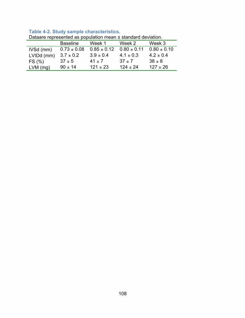
108
Table 4-2. Study sample characteristics. Dataare represented as population mean ± standard deviation. Baseline Week 1 Week 2 Week 3 IVSd (mm) 0.73 ± 0.08 0.85 ± 0.12 0.80 ± 0.11 0.80 ± 0.10 LVIDd (mm) 3.7 ± 0.2 3.9 ± 0.4 4.1 ± 0.3 4.2 ± 0.4 FS (%) 37 ± 5 41 ± 7 37 ± 7 38 ± 8 LVM (mg) 90 ± 14 121 ± 23 124 ± 24 127 ± 26

109
Table 4-3. Heritability estimates of cardiac structure and function in the HMDP.All heritability values exceed ANOVA p-value of <0.001. Control Week 1 Week 2 Week 3 IVSd 0.72 0.71 0.66 0.64 LVIDd 0.81 0.76 0.75 0.76 FS 0.79 0.74 0.71 0.76 LVM 0.84 0.77 0.72 0.79

110
Tabl
e 4-
4. G
enom
e-w
ide
sign
ifica
nt a
nd s
ugge
stiv
e ca
rdia
c re
mod
elin
g lo
ci.
A. S
igni
fican
t Loc
i A
naly
sis
Trai
t S
NP
P
-val
ue
MA
F Lo
catio
n (m
m9)
S
NP
Ty
pe
LD b
lock
(M
b)
n.
Can
dida
te G
ene(
s)
Exp
eQ
TL
Cor
S
V
Wee
k 3
Del
ta
LVM
*1 rs
4056
0913
3.17
E-0
9 0.
40
7:51
9652
67
I 51
.9-5
2.0
3 M
yh14
23
1001
6G11
Rik
Iz
umo2
†
- - -
- - -
- - -
S
- S
Wee
k 1
Del
ta
IVS
d2 rs
1348
0288
rs
2994
0243
1.15
E-0
8
0.38
9:
7724
7379
9:
7729
7835
IG
I
75.8
-80.
2 M
M
lip
5730
403I
07R
ik
Lrrc
1†
- - 6 d
- - Yes
- - Yes
MS
- S
W
eek
1 FS
rs
4879
1248
2.
18E
-07
0.10
15
:400
6535
8 IG
40
.0-4
0.2‡
0 Lr
p12
Zfpm
2 7d 8
No
No
Yes
N
o - -
B. S
ugge
stiv
e Lo
ci
Ana
lysi
s Tr
ait
SN
P
P-v
alue
M
AF
Loca
tion
(mm
9)
SN
P
Type
LD
blo
ck
(Mb)
n.
C
andi
date
G
ene(
s)
Exp
eQ
TL
Cor
S
V
Wee
k 2
LVM
3 rs
2781
6235
rs
2781
6170
2.
12E
-06
0.49
4:
4629
2922
4:
4633
1009
IG
45
.5-4
6.6
M
Xpa
Fo
xe1
5830
415F
09R
ik
Hem
gn
Anp
32b
9d 4 5 - 4
Yes
N
o N
o - N
o
Yes
N
o N
o - N
o
M
- - - - W
eek
3 D
elta
LV
M*
rs
2781
1538
1.
48E
-06
0.36
4:
5454
6700
IG
54
.2-5
5.8
4 Tm
em38
b Zf
p462
R
ad23
b K
lf4
10d
4 - 7
No
No
- Yes
No
No
- Yes
- MS
M
-
Wee
k 1
Del
ta
LVID
d rs
2785
1114
1.
75E
-06
0.06
4:
5827
7624
IG
58
.1-5
8.5
2 S
vep1
M
usk
- 4 - N
o - N
o M
S§
- W
eek
3 LV
M||
rs36
5607
6 rs
2788
0677
6.
10E
-07
0.44
4:
6378
5765
4:
6381
2861
I IG
63
.7-6
4.0
2 Tn
c A
K03
3210
† 4 -
No
- N
o -
- - W
eek
3 D
elta
IV
Sd
rs49
7248
04
2.03
E-0
6 0.
1 5:
1088
8730
9 IG
10
7.7-
109.
2 M
M
fsd7
a P
cgf3
- -
- - - -
M
S
Wee
k 3
IVS
d rs
3757
8592
1.
13E
-06
0.1
6:11
3357
105
I 11
2.7-
113.
5 M
Tt
ll3†
- -
- M
W
eek
2 LV
M4
rs37
8517
45
rs37
5359
34
rs37
8360
74
2.77
E-0
6 0.
06
9:49
0256
59
9:49
0256
82
9:49
0258
84
IG
48.9
-49.
3 0
Zw10
7
Yes
Y
es
M
Wee
k 2
Del
ta
FS*5
rs33
8966
82
2.28
E-0
6 0.
40
9:74
9040
30
I 74
.5-7
6.0
M
Arp
p19†
8 Y
es
Yes
-
Wee
k 1
Del
ta
IVS
d*
rs36
2662
87
8.62
E-0
7 0.
38
9:84
2044
00
IG
84.0
-84.
6 4
Bck
dhb
- -
- -
Wee
k 3
LVID
d6 rs
2935
0578
1.
76E
-06
0.49
10
:868
9355
1 IG
86
.0-8
7.8
M
1700
113H
08R
ik
- -
- -

111
Ana
lysi
s Tr
ait
SN
P
P-v
alue
M
AF
Loca
tion
(mm
9)
SN
P
Type
LD
blo
ck
(Mb)
n.
C
andi
date
G
ene(
s)
Exp
eQ
TL
Cor
S
V
Del
ta
rs29
3579
73
rs62
5849
0 rs
2932
8181
rs
4929
9504
10:8
6906
901
10:8
6918
146
10:8
6919
534
10:8
6938
274
Asc
l1
Pah
4 4
No
Yes
N
o N
o - -
Wee
k 3
Del
ta
LVID
d7 rs
3058
2427
rs
1348
0753
3.
75E
-06
0.42
10
:106
4755
87
10:1
0648
6522
I E
10
6.0-
107.
0 4
Acs
s3†
Lin7
a M
yf5
Myf
6
- - 4 -
- - No
-
- - No
-
S
- - - W
eek
3 D
elta
LV
IDd*
8 rs
4181
997
4.90
E-0
7 0.
43
16:4
5814
479
I 45
.0-4
6.0
1 S
lc35
a5
Cd2
00
Phl
db2†
5 5 4
Yes
Y
es
No
Yes
Y
es
No
- - - W
eek
1 FS
*9 rs
5097
7365
5.
61E
-07
0.06
19
:210
0069
2 I
20.9
-21.
5 2
Tmc1
† Zf
and5
- -
- - - -
- - D
elta
den
otes
cha
nge
from
bas
elin
e. N
umer
ical
sup
ersc
ript i
ndic
ates
repl
icat
ion
at a
diff
eren
t tim
e po
int o
r by
an in
depe
nden
tly
mea
sure
d ec
hoca
rdio
grap
hic
or w
eigh
t tra
it in
the
sam
e po
pula
tion.
MA
F de
note
s m
inor
alle
le fr
eque
ncy.
SN
P ty
pes
are
code
d I
(intro
nic)
, IG
(int
erge
nic)
, and
E (e
xoni
c). C
olum
n n.
den
otes
the
num
ber o
f gen
es in
LD
(r2 >0
.8) w
ith p
eak
SN
P, w
here
M d
enot
es
man
y ge
nes.
Can
dida
te g
enes
are
sho
rt lis
ted
by L
D c
utof
fs o
f r2 >0
.9 o
r 0.9
5, if
app
licab
le. E
xp d
enot
es a
ppro
xim
ate
aver
age
log2
in
tens
ity o
f exp
ress
ion,
ann
otat
ed b
y d fo
r up-
and
d fo
r dow
n-re
gula
tion
by is
opro
tere
nol.
eQTL
den
otes
the
pres
ence
of c
is-e
QTL
(p-
valu
e <
3.63
x 1
0-3).
Cor
den
otes
the
pres
ence
of c
orre
latio
n be
twee
n ge
ne e
xpre
ssio
n an
d cl
inic
al tr
ait (
p-va
lue
< 0.
05).
SV
den
otes
st
ruct
ural
var
iatio
ns, i
nclu
ding
M fo
r mis
sens
e (S
IFT
scor
e if
pred
icte
d de
lete
rious
) and
S fo
r spl
ice
regi
on v
aria
nts.
1 W
eek
3 V
old;
2 Wee
k 1
Del
ta IV
Ss,
Wee
k 2
Del
ta F
S/E
F, C
trl B
W/T
H/L
V/R
V/L
iver
, Iso
RV
; 3 Wee
k 1
A/E
, Wee
k 1
MN
SE
R, W
eek
1 V
cf; 4 C
trl T
H/L
V/R
V; 5 W
eek
1 D
elta
IVS
d/IV
Ss,
Wee
k 2
Del
ta E
F, C
trl B
W/T
H/L
V/R
V/L
iver
, Iso
RV
; 6 Wee
k 3
Del
ta V
old,
Iso
LV;
7 Wee
k 3
Del
ta R
WTd
; 8 Wee
k 3
Del
ta V
old,
Wee
k 3
Del
ta L
VID
s/V
ols,
Wee
k 3
FS/E
F/V
cf; 9 W
eek
1 V
cf/M
NS
ER
. *R
eplic
ated
in b
asel
ine
body
wei
ght (
BB
W) a
djus
ted
resi
dual
s; †
Gen
e co
ntai
ning
pea
k S
NP
; ‡Lo
cus
for s
udde
n ca
rdia
c ar
rest
in
coro
nary
arte
ry d
isea
se h
uman
GW
AS
(PM
ID:2
1658
281)
; §D
elet
erio
us b
y S
IFT
(rs2
7853
431,
T11
49I,
SIF
T=0.
01);
||Uni
que
for B
BW
ad
just
ed re
sidu
als
Abb
revi
atio
ns: A
/E (A
vs.
E v
eloc
ity ra
tio),
BW
(bod
y w
eigh
t), C
trl (c
ontro
l), E
F (e
ject
ion
fract
ion)
, Iso
(iso
prot
eren
ol-tr
eate
d), I
VS
s (IV
S a
t end
sys
tole
), Li
ver (
liver
wei
ght),
LV
(LV
wei
ght),
LV
IDs
(LV
ID a
t end
sys
tole
), M
NS
ER
(mea
n no
rmal
ized
sys
tolic
eje
ctio
n ra
te),
RV
(rig
ht v
entri
cula
r wei
ght),
RW
Td (r
elat
ive
wal
l thi
ckne
ss a
t end
dia
stol
e), T
H (t
otal
hea
rt w
eigh
t), V
cf (v
eloc
ity o
f ci
rcum
fere
ntia
l sho
rteni
ng),
Vol
d (L
V v
olum
e at
end
dia
stol
e) a
nd V
ols
(LV
vol
ume
at e
nd s
ysto
le).

112
Bibliography
1. Go AS, Mozaffarian D, Roger VL, Benjamin EJ, Berry JD, Borden WB, Bravata
DM, Dai S, Ford ES, Fox CS, Franco S, Fullerton HJ, Gillespie C, Hailpern SM,
Heit JA, Howard VJ, Huffman MD, Kissela BM, Kittner SJ, Lackland DT,
Lichtman JH, Lisabeth LD, Magid D, Marcus GM, Marelli A, Matchar DB,
McGuire DK, Mohler ER, Moy CS, Mussolino ME, Nichol G, Paynter NP,
Schreiner PJ, Sorlie PD, Stein J, Turan TN, Virani SS, Wong ND, Woo D, Turner
MB. Heart disease and stroke statistics--2013 update: A report from the american
heart association. Circulation. 2013;127:e6-e245
2. McMurray JJ, Petrie MC, Murdoch DR, Davie AP. Clinical epidemiology of heart
failure: Public and private health burden. Eur Heart J. 1998;19 Suppl P:P9-16
3. Kannel W, Ho K, Thom T. Changing epidemiological features of cardiac failure.
British heart journal. 1994;72:9
4. Levy D, Kenchaiah S, Larson MG, Benjamin EJ, Kupka MJ, Ho KK, Murabito JM,
Vasan RS. Long-term trends in the incidence of and survival with heart failure. N
Engl J Med. 2002;347:1397-1402
5. Taylor CJ, Roalfe AK, Iles R, Hobbs FD. Ten-year prognosis of heart failure in
the community: Follow-up data from the echocardiographic heart of england
screening (echoes) study. Eur J Heart Fail. 2012;14:176-184
6. Go AS, Mozaffarian D, Roger VL, Benjamin EJ, Berry JD, Blaha MJ, Dai S, Ford
ES, Fox CS, Franco S, Fullerton HJ, Gillespie C, Hailpern SM, Heit JA, Howard
VJ, Huffman MD, Judd SE, Kissela BM, Kittner SJ, Lackland DT, Lichtman JH,
Lisabeth LD, Mackey RH, Magid DJ, Marcus GM, Marelli A, Matchar DB,
McGuire DK, Mohler ER, 3rd, Moy CS, Mussolino ME, Neumar RW, Nichol G,
Pandey DK, Paynter NP, Reeves MJ, Sorlie PD, Stein J, Towfighi A, Turan TN,

113
Virani SS, Wong ND, Woo D, Turner MB. Heart disease and stroke statistics--
2014 update: A report from the american heart association. Circulation.
2014;129:e28-e292
7. Lymperopoulos A, Rengo G, Koch WJ. Adrenergic nervous system in heart
failure: Pathophysiology and therapy. Circ Res. 2013;113:739-753
8. Altshuler D, Daly MJ, Lander ES. Genetic mapping in human disease. Science.
2008;322:881-888
9. Smith NL, Felix JF, Morrison AC, Demissie S, Glazer NL, Loehr LR, Cupples LA,
Dehghan A, Lumley T, Rosamond WD, Lieb W, Rivadeneira F, Bis JC, Folsom
AR, Benjamin E, Aulchenko YS, Haritunians T, Couper D, Murabito J, Wang YA,
Stricker BH, Gottdiener JS, Chang PP, Wang TJ, Rice KM, Hofman A, Heckbert
SR, Fox ER, O'Donnell CJ, Uitterlinden AG, Rotter JI, Willerson JT, Levy D, van
Duijn CM, Psaty BM, Witteman JCM, Boerwinkle E, Vasan RS. Association of
genome-wide variation with the risk of incident heart failure in adults of european
and african ancestry: A prospective meta-analysis from the cohorts for heart and
aging research in genomic epidemiology (charge) consortium. Circulation:
Cardiovascular Genetics. 2010;3:256-266
10. Villard E, Perret C, Gary F, Proust C, Dilanian G, Hengstenberg C, Ruppert V,
Arbustini E, Wichter T, Germain M, Dubourg O, Tavazzi L, Aumont MC,
DeGroote P, Fauchier L, Trochu JN, Gibelin P, Aupetit JF, Stark K, Erdmann J,
Hetzer R, Roberts AM, Barton PJ, Regitz-Zagrosek V, Aslam U, Duboscq-Bidot
L, Meyborg M, Maisch B, Madeira H, Waldenstrom A, Galve E, Cleland JG,
Dorent R, Roizes G, Zeller T, Blankenberg S, Goodall AH, Cook S, Tregouet DA,
Tiret L, Isnard R, Komajda M, Charron P, Cambien F. A genome-wide
association study identifies two loci associated with heart failure due to dilated
cardiomyopathy. Eur Heart J. 2011;32:1065-1076
11. Fox ER, Musani SK, Barbalic M, Lin H, Yu B, Ogunyankin KO, Smith NL, Kutlar
A, Glazer NL, Post WS, Paltoo DN, Dries DL, Farlow DN, Duarte CW, Kardia SL,

114
Meyers KJ, Sun YV, Arnett DK, Patki AA, Sha J, Cui X, Samdarshi TE, Penman
AD, Bibbins-Domingo K, Buzkova P, Benjamin EJ, Bluemke DA, Morrison AC,
Heiss G, Carr JJ, Tracy RP, Mosley TH, Taylor HA, Psaty BM, Heckbert SR,
Cappola TP, Vasan RS. Genome-wide association study of cardiac structure and
systolic function in african americans: The candidate gene association resource
(care) study. Circ Cardiovasc Genet. 2013;6:37-46
12. Nakada K, Sato A, Hayashi J. Reverse genetic studies of mitochondrial DNA-
based diseases using a mouse model. Proc Jpn Acad Ser B Phys Biol Sci.
2008;84:155-165
13. Le Corvoisier P, Park HY, Carlson KM, Marchuk DA, Rockman HA. Multiple
quantitative trait loci modify the heart failure phenotype in murine
cardiomyopathy. Hum Mol Genet. 2003;12:3097-3107
14. Wheeler FC, Fernandez L, Carlson KM, Wolf MJ, Rockman HA, Marchuk DA. Qtl
mapping in a mouse model of cardiomyopathy reveals an ancestral modifier
allele affecting heart function and survival. Mamm Genome. 2005;16:414-423
15. Suzuki M, Carlson KM, Marchuk DA, Rockman HA. Genetic modifier loci
affecting survival and cardiac function in murine dilated cardiomyopathy.
Circulation. 2002;105:1824-1829
16. Tsujita Y, Iwai N, Tamaki S, Nakamura Y, Nishimura M, Kinoshita M. Genetic
mapping of quantitative trait loci influencing left ventricular mass in rats. Am J
Physiol Heart Circ Physiol. 2000;279:H2062-2067
17. Flint J, Valdar W, Shifman S, Mott R. Strategies for mapping and cloning
quantitative trait genes in rodents. Nat Rev Genet. 2005;6:271-286
18. Bennett BJ, Farber CR, Orozco L, Min Kang H, Ghazalpour A, Siemers N,
Neubauer M, Neuhaus I, Yordanova R, Guan B, Truong A, Yang Wp, He A,
Kayne P, Gargalovic P, Kirchgessner T, Pan C, Castellani LW, Kostem E,
Furlotte N, Drake TA, Eskin E, Lusis AJ. A high-resolution association mapping

115
panel for the dissection of complex traits in mice. Genome Research.
2010;20:281-290
19. Ghazalpour A, Rau CD, Farber CR, Bennett BJ, Orozco LD, van Nas A, Pan C,
Allayee H, Beaven SW, Civelek M. Hybrid mouse diversity panel: A panel of
inbred mouse strains suitable for analysis of complex genetic traits. Mammalian
Genome. 2012:1-13
20. Parks BW, Nam E, Org E, Kostem E, Norheim F, Hui ST, Pan C, Civelek M, Rau
CD, Bennett BJ, Mehrabian M, Ursell LK, He A, Castellani LW, Zinker B, Kirby M,
Drake TA, Drevon CA, Knight R, Gargalovic P, Kirchgessner T, Eskin E, Lusis
AJ. Genetic control of obesity and gut microbiota composition in response to
high-fat, high-sucrose diet in mice. Cell Metab. 2013;17:141-152
21. Farber CR, Bennett BJ, Orozco L, Zou W, Lira A, Kostem E, Kang HM, Furlotte
N, Berberyan A, Ghazalpour A, Suwanwela J, Drake TA, Eskin E, Wang QT,
Teitelbaum SL, Lusis AJ. Mouse genome-wide association and systems genetics
identify asxl2 as a regulator of bone mineral density and osteoclastogenesis.
PLoS Genet. 2011;7:e1002038
22. Park CC, Gale GD, de Jong S, Ghazalpour A, Bennett BJ, Farber CR, Langfelder
P, Lin A, Khan AH, Eskin E, Horvath S, Lusis AJ, Ophoff RA, Smith DJ. Gene
networks associated with conditional fear in mice identified using a systems
genetics approach. BMC Syst Biol. 2011;5:43
23. Koitabashi N, Kass D. Reverse remodeling in heart failure--mechanisms and
therapeutic opportunities. Nature reviews. Cardiology. 2012;9:147-204
24. Liu J, Rigel DF. Echocardiographic examination in rats and mice. Methods Mol
Biol. 2009;573:139-155
25. Moran CM, Thomson AJ, Rog-Zielinska E, Gray GA. High-resolution
echocardiography in the assessment of cardiac physiology and disease in
preclinical models. Exp Physiol. 2013;98:629-644

116
26. Galindo CL, Skinner MA, Errami M, Olson LD, Watson DA, Li J, McCormick JF,
McIver LJ, Kumar NM, Pham TQ, Garner HR. Transcriptional profile of
isoproterenol-induced cardiomyopathy and comparison to exercise-induced
cardiac hypertrophy and human cardiac failure. BMC Physiology. 2009;9:23
27. Molojavyi A, Lindecke A, Raupach A, Moellendorf S, Kohrer K, Godecke A.
Myoglobin-deficient mice activate a distinct cardiac gene expression program in
response to isoproterenol-induced hypertrophy. Physiol Genomics. 2010;41:137-
145
28. Smyth GK. Limma: Linear models for microarray data. In: Gentleman R, ed.
Bioinformatics and computational biology solutions using r and bioconductor.
New York: Springer Science+Business Media; 2005:xix, 473 p.
29. Langfelder P, Horvath S. Wgcna: An r package for weighted correlation network
analysis. BMC Bioinformatics. 2008;9:559
30. Smyth GK. Linear models and empirical bayes methods for assessing differential
expression in microarray experiments. Stat Appl Genet Mol Biol. 2004;3:Article3
31. Huang da W, Sherman BT, Lempicki RA. Systematic and integrative analysis of
large gene lists using david bioinformatics resources. Nat Protoc. 2009;4:44-57
32. Huang da W, Sherman BT, Lempicki RA. Bioinformatics enrichment tools: Paths
toward the comprehensive functional analysis of large gene lists. Nucleic Acids
Res. 2009;37:1-13
33. Yang H, Ding Y, Hutchins LN, Szatkiewicz J, Bell TA, Paigen BJ, Graber JH, de
Villena FP, Churchill GA. A customized and versatile high-density genotyping
array for the mouse. Nat Methods. 2009;6:663-666
34. Purcell S, Neale B, Todd-Brown K, Thomas L, Ferreira MA, Bender D, Maller J,
Sklar P, de Bakker PI, Daly MJ, Sham PC. Plink: A tool set for whole-genome
association and population-based linkage analyses. Am J Hum Genet.
2007;81:559-575

117
35. Kirby A, Kang HM, Wade CM, Cotsapas C, Kostem E, Han B, Furlotte N, Kang
EY, Rivas M, Bogue MA, Frazer KA, Johnson FM, Beilharz EJ, Cox DR, Eskin E,
Daly MJ. Fine mapping in 94 inbred mouse strains using a high-density
haplotype resource. Genetics. 2010;185:1081-1095
36. Lippert C, Listgarten J, Liu Y, Kadie CM, Davidson RI, Heckerman D. Fast linear
mixed models for genome-wide association studies. Nat Methods. 2011;8:833-
835
37. Pruim RJ, Welch RP, Sanna S, Teslovich TM, Chines PS, Gliedt TP, Boehnke M,
Abecasis GR, Willer CJ. Locuszoom: Regional visualization of genome-wide
association scan results. Bioinformatics. 2010;26:2336-2337
38. Keane TM, Goodstadt L, Danecek P, White MA, Wong K, Yalcin B, Heger A,
Agam A, Slater G, Goodson M, Furlotte NA, Eskin E, Nellaker C, Whitley H,
Cleak J, Janowitz D, Hernandez-Pliego P, Edwards A, Belgard TG, Oliver PL,
McIntyre RE, Bhomra A, Nicod J, Gan X, Yuan W, van der Weyden L, Steward
CA, Bala S, Stalker J, Mott R, Durbin R, Jackson IJ, Czechanski A, Guerra-
Assuncao JA, Donahue LR, Reinholdt LG, Payseur BA, Ponting CP, Birney E,
Flint J, Adams DJ. Mouse genomic variation and its effect on phenotypes and
gene regulation. Nature. 2011;477:289-294
39. Kumar P, Henikoff S, Ng PC. Predicting the effects of coding non-synonymous
variants on protein function using the sift algorithm. Nat Protoc. 2009;4:1073-
1081
40. Langfelder P, Horvath S. Fast r functions for robust correlations and hierarchical
clustering. J Stat Softw. 2012;46
41. Wu J, Bu L, Gong H, Jiang G, Li L, Ma H, Zhou N, Lin L, Chen Z, Ye Y, Niu Y,
Sun A, Ge J, Zou Y. Effects of heart rate and anesthetic timing on high-resolution
echocardiographic assessment under isoflurane anesthesia in mice. Journal of
ultrasound in medicine : official journal of the American Institute of Ultrasound in
Medicine. 2010;29:1771-1778

118
42. Jin Y, Kuznetsova T, Bochud M, Richart T, Thijs L, Cusi D, Fagard R, Staessen
JA. Heritability of left ventricular structure and function in caucasian families. Eur
J Echocardiogr. 2011;12:326-332
43. Clark S, Naz RK. Presence and incidence of izumo antibodies in sera of
immunoinfertile women and men. Am J Reprod Immunol. 2013;69:256-263
44. Ma X, Jana SS, Conti MA, Kawamoto S, Claycomb WC, Adelstein RS. Ablation
of nonmuscle myosin ii-b and ii-c reveals a role for nonmuscle myosin ii in
cardiac myocyte karyokinesis. Mol Biol Cell. 2010;21:3952-3962
45. Zhou B, Ma Q, Kong SW, Hu Y, Campbell PH, McGowan FX, Ackerman KG, Wu
B, Tevosian SG, Pu WT. Fog2 is critical for cardiac function and maintenance of
coronary vasculature in the adult mouse heart. J Clin Invest. 2009;119:1462-
1476
46. Qing J, Wei D, Maher VM, McCormick JJ. Cloning and characterization of a
novel gene encoding a putative transmembrane protein with altered expression
in some human transformed and tumor-derived cell lines. Oncogene.
1999;18:335-342
47. Battle MA, Maher VM, McCormick JJ. St7 is a novel low-density lipoprotein
receptor-related protein (lrp) with a cytoplasmic tail that interacts with proteins
related to signal transduction pathways. Biochemistry. 2003;42:7270-7282
48. Debette S, Visvikis-Siest S, Chen MH, Ndiaye NC, Song C, Destefano A, Safa R,
Azimi Nezhad M, Sawyer D, Marteau JB, Xanthakis V, Siest G, Sullivan L, Pfister
M, Smith H, Choi SH, Lamont J, Lind L, Yang Q, Fitzgerald P, Ingelsson E,
Vasan RS, Seshadri S. Identification of cis- and trans-acting genetic variants
explaining up to half the variation in circulating vascular endothelial growth factor
levels. Circ Res. 2011;109:554-563
49. Aouizerat BE, Vittinghoff E, Musone SL, Pawlikowska L, Kwok PY, Olgin JE,
Tseng ZH. Gwas for discovery and replication of genetic loci associated with

119
sudden cardiac arrest in patients with coronary artery disease. BMC Cardiovasc
Disord. 2011;11:29
50. Nakane H, Takeuchi S, Yuba S, Saijo M, Nakatsu Y, Murai H, Nakatsuru Y,
Ishikawa T, Hirota S, Kitamura Y, et al. High incidence of ultraviolet-b-or
chemical-carcinogen-induced skin tumours in mice lacking the xeroderma
pigmentosum group a gene. Nature. 1995;377:165-168
51. Shefer G, Benayahu D. Svep1 is a novel marker of activated pre-determined
skeletal muscle satellite cells. Stem cell reviews. 2010;6:42-51
52. Dulubova I, Horiuchi A, Snyder GL, Girault JA, Czernik AJ, Shao L,
Ramabhadran R, Greengard P, Nairn AC. Arpp-16/arpp-19: A highly conserved
family of camp-regulated phosphoproteins. J Neurochem. 2001;77:229-238
53. Haccard O, Jessus C. Greatwall kinase, arpp-19 and protein phosphatase 2a:
Shifting the mitosis paradigm. Results Probl Cell Differ. 2011;53:219-234
54. Swan L, Birnie DH, Padmanabhan S, Inglis G, Connell JM, Hillis WS. The
genetic determination of left ventricular mass in healthy adults. Eur Heart J.
2003;24:577-582
55. Post WS, Larson MG, Myers RH, Galderisi M, Levy D. Heritability of left
ventricular mass: The framingham heart study. Hypertension. 1997;30:1025-
1028
56. Faulx MD, Ernsberger P, Vatner D, Hoffman RD, Lewis W, Strachan R, Hoit BD.
Strain-dependent beta-adrenergic receptor function influences myocardial
responses to isoproterenol stimulation in mice. Am J Physiol Heart Circ Physiol.
2005;289:H30-36

120
5 Future Directions
Genetic studies have the potential to reveal insights into the mechanisms of
disease causation, progression, prognosis and potential points of therapeutic
intervention. From gene panel tests in human patients, a number of genetic variants
were implicated in known disease-causing genes with varying levels of evidence. From
exome sequencing of familial cardiomyopathy cases, we have identified a novel
candidate gene that is highly likely to be causal for disease. Genome-wide association
(GWA) in the isoproterenol-induced heart failure (HF) model in mice identified dozens of
significant and suggestive loci controlling cardiac structure, function and remodeling.
Understanding whether the identified genes and variants are indeed causal for disease
will inform the relevance of genes and variants in HF development and any future
mechanistic studies. In the case of human patients, knowing definitively that the variant
reported to be disease-causing or likely disease-causing not only helps the patient
understand his/her disease process but also inform post-genetic counseling with
patients and their family members in understanding the risk for disease. Given the
number of mouse and human candidate genes and variants to screen, we need a high-
throughput method of functional characterization protocol and candidate gene and
variant screening. We have conducted preliminary studies in a cell-based model using
neonatal rat ventricular cardiomyocytes (NRVMs) as well as in a whole organism model
the zebrafish. We will explore additional model systems for effective site mutagenesis to
fill the need for gene variant screening.

121
The choice of isolated NRVMs has several advantages, such as the ability to
sample cells from different areas of the heart including the atria, left and right ventricle.
Imaging of isolated cells is trivial and is well suited for experiments aimed at visualizing
cellular structure and the precise localization of intracellular molecules, which is
especially important for our studies aiming to observe cardiomyocyte hypertrophy.
Isolated cardiomyocytes are also routinely used for studies examining intracellular Ca2+
homeostasis, cellular mechanics, and protein biochemistry, and can be easily infected
or transfected for gene transfer studies1. We transfected NRVMs using two siRNA
oligos against the Myh14 gene, one of the top candidates for the change of left
ventricular mass, and induced hypertrophy with isoproterenol and phenylephrine for 24
hours. The Myh14 siRNA treated NRVMs failed to mount a hypertrophic response after
isoproterenol and phenylephrine stimulation as in the siRNA scramble control group
(Figure 5-1). In future experiments, cardiomyocyte cell size will be measured. The
expression levels of cardiac hypertrophy markers, atrial natriuretic peptide (Nppa), brain
natriuretic peptide (Nppb), alpha-myosin heavy chain (Myh6) and beta-myosin heavy
chain (Myh7) will be analyzed using quantitative RT-RCR. Replication-deficient
adenoviruses for each siRNA screened positive candidate gene will be constructed
using the AdEasy adenoviral system. Cell growth and marker gene induction will be
evaluated as above to establish the role of each gene in cardiomyocyte hypertrophy.
In a semi-hierarchical approach, genes screened positive in NRVMs or are
expected to have a phenotype on the whole heart level will be evaluated in the zebrafish
system to assess for their role in the intact heart. We will inactivate candidate genes by

122
injecting morpholinos into fertilized eggs, targeted to the translation initiation site or
splicing site of the genes, at the 1-2 cell stage and visualize the heart using the cardiac
myosin light chain promoter (cmlc)-drive green fluorescent protein (GFP) transgenic
zebrafish. Similar to the NRVMs, zebrafish embryo and larvae are well suited for
experiments aimed at visualizing cardiac function in the GFP tagged hearts. This is
especially important for the functional characterization of cardiac contraction. Zebrafish
are also routinely used for studies examining intracellular Ca2+ homeostasis and can be
easily injected with capped mRNA for transgene expression and anti-sense
morpholino2. We injected an anti-sense morpholino against Tnnt2, cardiac troponin T,
and demonstrated successful arrest of the heart, pericardial fluid accumulation
(congestion), and cardiac compression (Figure 5-2). We do not expect the genes that
we screen will have such a dramatic effect but rather have effect on ventricular size and
function.
To evaluate individual genetic variants, we will explore the role of
clustered regularly interspaced short palindromic repeats (CRISPR) technologies in the
zebrafish and mice, to characterize the functional effects of the candidate variant in a
high-throughput manner3, 4. We will establish cardiac functional characterization protocol
and methods to measure electrical conduction in the zebrafish through collaboration.
We will also establish detailed cardiac functional characterization protocol, including
measures of diastolic function to measure subtle changes from the candidate genetic
variants, and methods to measure electrical conduction to fully assess the status of the
heart. Finally, the role of using patient-derived iPS cells to validate candidate variant,

123
personalized therapies based on pharmacogenomic, and personalized allele-specific
knock-down technologies may be explored.
The results from genome-wide association analyses suggest that many of
the genomic region controlling susceptibility to heart failure are located in gene poor or
intergenic regions, which suggests that gene regulation rather than protein coding
sequence variations play a major role in mechanisms affecting complex traits. A number
of mechanisms have been proposed to effect non-coding sequence variations on gene
regulation, including promoter region variations, methylation changes, non-coding RNA,
and chromatin states among others. In addition to coding-sequence variations,
understanding of gene regulation in genes important in heart failure susceptibility may
provide clues to lifestyle factors that may promote heart health and prevent disease, in
spite of non-modifiable genetic backgrounds.
The future of genetics research in heart failure holds great promise for
heart failure prevention. Improved disease risk prediction informed by genomics data
will help guide counseling of patients and installment of programs to prevent disease,
such as diastolic dysfunction, for which there are no effective treatments. Improved fine
phenotyping of traits, large-scale sequencing for rare variants, and ever more
sophisticating mapping strategies will work together to improve the understanding of
heart health genetics. Improve translational research to understand how genes
predispose to disease will guide drug design to address common pathological

124
processes shared by heart failure subtypes to improve cardiac health across the
general population.

125
Figure 5-1. siRNA knock-down of Myh14 in neonatal rat ventricular cardiomyocytes abrogates hypertrophic effects of isoproterenol and phenylephrine. siMyh14#1 represents the first Myh14 siRNA. siMyh14#2 represents the second Myh14 siRNA.
!"#$%&'()*+,-.) *+/0123)42) *+/0123)45)
6789#7')
:*7;
#79(#(87
')<1
(80'(;
1#+8()

126
Figure 5-2. Transgenic zebrafish strain with cmlc2::EGFP.Control (left) and Tnnt2 (right) morpholino treated embryos at 54 hours post fertilization (hpf).

127
Bibliography
1. Louch WE, Sheehan KA, Wolska BM. Methods in cardiomyocyte isolation,
culture, and gene transfer. J Mol Cell Cardiol. 2011;51:288-298
2. Bakkers J. Zebrafish as a model to study cardiac development and human
cardiac disease. Cardiovascular research. 2011;91:279-367
3. Wang H, Yang H, Shivalila CS, Dawlaty MM, Cheng AW, Zhang F, Jaenisch R.
One-step generation of mice carrying mutations in multiple genes by crispr/cas-
mediated genome engineering. Cell. 2013;153:910-918
4. Mali P, Yang L, Esvelt KM, Aach J, Guell M, DiCarlo JE, Norville JE, Church GM.
Rna-guided human genome engineering via cas9. Science. 2013;339:823-826