Embed Size (px)
Citation preview

FA
Fa
b
c
a
ARRAA
KSMCPLATMFLf
1
elplsiIsaGrTpir
B
0d
Neuropsychologia 48 (2010) 1923–1929
Contents lists available at ScienceDirect
Neuropsychologia
journa l homepage: www.e lsev ier .com/ locate /neuropsychologia
igurative language processing after traumatic brain injury in adults:preliminary study
anpei Gloria Yanga,c,∗, Jerome Fullera,1, Navid Khodaparasta,1, Daniel C. Krawczyka,b
Center for Brain Health, University of Texas at Dallas, TX, USADepartment of Psychiatry, University of Texas Southwestern Medical Center at Dallas, TX, USADepartment of Radiology and Biomedical Imaging, University of California San Francisco, USA
r t i c l e i n f o
rticle history:eceived 16 April 2009eceived in revised form 5 March 2010ccepted 9 March 2010vailable online 15 March 2010
eywords:emanticemory
ontrol
a b s t r a c t
Figurative speech (e.g., proverb, irony, metaphor, and idiom) has been reported to be particularly sensi-tive to measurement of abstract thinking in patients who suffer from impaired abstraction and languageabilities. Metaphor processing was investigated with fMRI in adults with moderate to severe post-acute traumatic brain injury (TBI) and healthy age-matched controls using a valence-judgment task.We hypothesized that TBI patients would display decreased activation of the left inferior frontal gyrus(LIFG), which is considered central to semantic memory retrieval and abstract thought, in comparisonwith healthy controls. We also predicted that decreased activation in TBI individuals would correlate withtheir behavioral response times. A whole-brain analysis across the two participant groups revealed thatpatients did not strongly engage frontal and temporal regions related to semantic processing for novel
refrontal cortexeft inferior frontal gyrusbstractionraumatic brain injuryetaphor
metaphor comprehension, whereas control participants exhibited more intensive and concentrated acti-vation within frontal and temporal areas. A region of interest (ROI) analysis verified that the LIFG wasunderactivated in TBI patients compared to controls across all conditions. TBI patients’ impaired abstrac-tion of novel stimuli may stem from reduced prefrontal control of semantic memory as well as disrupted
ontal
igurativeanguageMRIinterconnectivity of prefr
. Introduction
Adults who have sustained traumatic brain injuries (TBI) mayxperience deficits in higher order cognitive abilities includinganguage comprehension and inferencing. Prior research has pro-osed that the linguistic competence of TBI patients is affected by
evels of cognitive functioning and that the impairment in under-tanding and use of figurative language is indicative of deficitsn overall cognitive functions (Wigg, Alexander, & Secord, 1988).mpairment in figurative language comprehension has been con-idered to result from problems in conceptual integration andbstract thought (Groher, 1983; Hagen, 1984; Levin, Benton, &rossman, 1982). Abstract thinking commonly refers to the rep-
esentation and synthesis of stimuli to deal with abstract concepts.hese prior studies used several types of figurative language (e.g.,roverb, irony, metaphor, and idiom) to measure patients’ abil-
ty to understand abstract thought. In this case abstract thoughtefers to the ability to detect that the literal interpretation of lan-
∗ Corresponding author at: University of Texas at Dallas, School of Brain andehavioral Sciences. 2200 Mockingbird, Dallas, TX 5235, USA. Tel.: +1 404 663 7481.
E-mail address: [email protected] (F.G. Yang).1 These two authors made an equal amount of contribution to this research.
028-3932/$ – see front matter © 2010 Elsevier Ltd. All rights reserved.oi:10.1016/j.neuropsychologia.2010.03.011
cortex with other regions.© 2010 Elsevier Ltd. All rights reserved.
guage is less appropriate than a symbolic interpretation whichtypically invokes similarities between prior situations and the cur-rent situation. Intact figurative language comprehension reflectsthe ability to detect a higher order meaning in language andjudge this meaning to be more informative than a narrower lit-eral interpretation. A typical example of a metaphor is “She is apeach”. A person with intact abstract thought would be able todetect the similar “sweet” property of the fruit and an agreeablepersonality and infer that the referred female is a person withan agreeable quality in interpreting this sentence. Understandingpragmatic meanings as figurative language has been suggested tobe a particularly sensitive measurement of abstract thinking inboth children with language and learning disabilities (LLD) andpatients with TBI (Blue, 1981; Donahue & Bryant, 1984; Elmore& Gorham, 1957; Wig & Semel, 1984; Winner & Gardner, 1977;Winner et al., 1980). Although there is a growing body of liter-ature on linguistic deficits, including figurative language, foundin populations who have suffered TBI, no previous studies have
employed functional neuroimaging techniques in order to inves-tigate the involvement of brain regions that subserve metaphorcomprehension in TBI populations. Therefore, no research to datehas examined the possible neural deficits associated with abstractthinking after TBI.
1 cholo
aodptbidlf1tlrgoittatcaeaufsiaSssE
ctestioaatTtboibtr2icltm2D
tMn
of water movement in white matter tracts, compared with healthy controls. The
924 F.G. Yang et al. / Neuropsy
Two theories have been proposed to understand languagend communication deficits after TBI. An early theory focusedn whether observed language impairments were categoricaleficits or the result of a global disorganization process. Sup-ort for a categorical linguistic deficit came initially from researchhat reported that language impairments following TBI resem-led Wernicke’s aphasia (Heilman, Safron, & Geschwind, 1971). An
ncreasing amount of research emerged in support of the globalisorganization theory in the 1980s. Several studies suggested that
anguage after TBI was disrupted in qualitatively different waysrom left hemisphere cerebral vascular accident aphasia (Hagen,984; Holland, 1982). Holland (1982) observed that TBI patientsalk better than they communicate, which suggests that their prob-em lies in pragmatics rather than speech production. Hagen (1984)eported that TBI language impairments are often found in lan-uage organization rather than the linguistic dataset. Hagen alsobserved that abstract thought and conceptual integration werempaired after TBI and consequently affected language formula-ion. He concluded that the language disorganization results fromhe global breakdown of the ability to structure mental processesnd to shift cognitive sets. This line of research influenced the direc-ion of research, assessment, and rehabilitation of TBI patients. Aommon consensus formed suggesting that cognitive dysfunctionffects patients’ linguistic performance and cognitive function lev-ls can be assessed by performance on measures of metalinguisticnd metacognitive measures. Wiig, Alexander, and Secord (1988)sed the Test of Language Competence (TLC) that they developedor assessing language and learning disabilities in adolescents totudy TBI patients’ impairments. A subtest of TLC specifically testsnterpretation and matching metaphoric expressions, planning fornd recreating speech acts and interpreting sentence ambiguities.ince then, studies have identified metaphoric comprehension as apecial domain of impairment for TBI patients and as a particularlyensitive measurement of abstract thinking in patients (Towne &ntwisle, 1993; Wig & Semel, 1984).
The current research tests two hypotheses. First, metaphor pro-essing is affected by deficits in abstract thought, which is part ofhe disorganization process. By abstract thought we refer to thexecutive control processes necessary to detect multiple possibleemantic interpretations of a linguistic statement and the abilityo override a literal meaning in favor of a broader symbolic mean-ng that typically conveys greater information about the subjectf the statement. Our second hypothesis is that abnormal neuralctivation in TBI patients in response to metaphors, both at globalnd local regions, will be correlated with impairment in abstracthought. Investigation of differences in patterns of activation withinBI patients may help us to identify the neural contributions tohe observed deficits in abstract thinking and also allow us toetter understand the regional contributions to semantic mem-ry control by areas typically involved in language processing. Fornstance, several neuroimaging studies have investigated the linksetween language comprehension and semantic memory leadingo the proposal of specialized roles for the frontal and tempo-al cortices in these processes (Bedny, McGill, & Thompson-Schill,008; Thompson-Schill & Gabrieli, 1999). For language process-
ng, semantic memory retrieval relies heavily upon the temporalortices (Neary, Snowden, Northen, & Goulding, 1988), while theeft inferior frontal gyrus (LIFG) is considered to be important inhe selection of appropriate interpretations when contextual infor-
ation is not sufficient (Badre & Wagner, 2007; Duncan & Owen,000; Fiez, Petersen, Cheney, & Raichle, 1992; Thompson-Schill,
’Esposito, Aguirre, & Farah, 1997).Several prior fMRI studies have reported increased activity inhe LIFG (Mashal, Faust, Hendler, & Jung-Beeman, 2007; Stringaris,
edford, Giampetro, Brammer, & David, 2007) when subjects readovel metaphors (e.g., She is a strawberry) in contrast to literal sen-
gia 48 (2010) 1923–1929
tences (e.g., She is a liar) or conventional metaphors (e.g., She is apeach). This suggests that additional semantic processing capac-ities are required in the novel metaphor comprehension relativeto the comprehension of familiar expressions such as literal state-ments and conventional metaphors. Since inferring the meaningof metaphors is a sensitive measure of patients’ abstract think-ing (Wigg et al., 1988), activations in LIFG are likely to reflect thesynthesis of abstract concepts in addition to selection of context-appropriate semantic representation.
The purpose of the present study is to identify brain regionsinvolved in impaired abstraction abilities through investigationof neural activation during figurative language processing in TBIpatients compared to healthy controls. We use a valence-judgmenttask similar to that used in several prior fMRI studies on compre-hension of metaphors in healthy individuals (Mashal et al., 2007;Rapp, Leube, Erb, Grodd, & Kircher, 2004; Stringaris et al., 2007).Valence-judgment tasks require a determination of the positive ornegative values of sentences. According to previous studies, thevalence task is able to evoke active processing of metaphors incontrast to passive reading. Using a valence task, we predict thatTBI individuals in the current study will show a behavioral deficitin abstract thinking revealed by differential performance in novelmetaphor comprehension. In addition, we predict that TBI patients’difference in neural activity will be manifested in less intensiveactivations in multiple regions globally and specifically in the LIFGcompared to healthy controls. We predicted less LIFG activationin patients due to their potentially disrupted connectivity of theprefrontal cortex (PFC) with other regions through white mat-ter injuries. We will also investigate whether additional languageregions such as areas within the temporal cortex display significantdifferences in activations using whole-brain analyses. If reductionsin activation are observed in the PFC only, this would likely reflecta compromised executive control function as primarily responsi-ble for language deficits. By contrast, reductions of activation inthe temporal region, would suggest that language deficits may bemore associated with the loss of semantic knowledge. If both PFCand temporal lobes reveal abnormal activation, it would suggestthat patients suffer from a complex impairment of the semanticmemory system that includes contributions from both semanticnetworks of the temporal lobes and frontally mediated executivefunction abilities such as selection, and retrieval of knowledge thatunderlie abstract thought.
2. Method
2.1. Participants
Twelve right handed TBI patients with age-matched thirteen healthy controls,all native English speakers, participated in the study. Mean age of patients was 30.9(SD: 10.51, range: 21–49) years and mean age of healthy controls was 27.6 (SD: 7.05,range: 25–48). The patients were at least 2 years post-trauma and were not in inten-sive care. They were able to read and speak and received an average of 14 years ofeducation (SD: 3.4, range: 12–18). The patients were assessed in terms of their levelsof functioning in physical social and psychological domains using structured inter-views of Glasgow Outcome Scale-Extended (GOS-E; Wilson, Pettigrew, & Teasdale,1998) and Functional Status Examination (FSE, Dikmen, Machamer, Miller, Doctor,& Temkin, 2001). Both are measures designed to evaluate change in activities ofeveryday life as a function of an event or illness. The causes of injuries included caraccidents, bicycle accidents, and being the victim of assault. Structural images (T1-weighted MRI, T2-weighted MRI, and Diffusion Tensor Imaging (DTI)) were acquiredin this study. All patients sustained diffused axonal injuries marked by their DTIimages showing shorter white matter fiber length, reduced fiber counts, reducedfiber density, as well as reduced functional anisotropy (FA), a measure of direction
demographic details of the TBI patients are presented in Table 1. Typical Exclusioncriteria for fMRI were applied. Subjects with past or present medical or psychiatricillness and impaired language skills were excluded. The study was approved by theinternal review boards at the University of Texas Southwestern Medical Center andthe University of Texas at Dallas. After a complete description of the study, subjectsgave their informed consent.

F.G. Yang et al. / Neuropsycholo
Table 1Patient demographics.
Patients (n = 12) Mean
Age 30.9Gender (% male) 75Education (years) 14
FunctionalityFSE 26.08GOS-E 5.67
Note: FSE: Functional Status Examination, GOS-E: Glasgow Out-come Scale-Extended.The FSE measures outcome in 10 domains, including personalcare, ambulation, work or school activities, and so on. Outcomefor each category is rated along a four-point scale with lowerscores associated with better outcome. A rating of “zero” sig-nifies no change from pre-injury; and “three” means that theindividual is completely dependent upon others or that the indi-vidual does not perform that activity at all. Ratings from eachdomain are summed to give an FSE total score of 0–30. Themean FSE of patients in the present study is 26.08. This sug-gests that patients depend on others to perform the activities in
2
etmLtsf
emidvs(tttnnagScIimiTft
2
pptposmtTcTas
a 5 mm sphere. The ROI we chose within the LIFG consisted of the pars triangularisportion and this portion was consistently the location of the local maxima across allsubjects. We extracted the ˇ values (regression co-efficient values) for each condi-tion. This procedure was repeated for each subject. We compared the mean ROI ˇvalue of the TBI patients with that of the healthy controls.
Table 2ANOVA of reaction time and percent accuracy for all conditions between TBI andcontrol groups.
Control TBI F(1, 23) p
Mean SD Mean SD
Reaction time (RT)RT literal 1867.8 396.6 2509.1 649.3 9.046 0.006*
RT conventional 2059.9 372.4 2565.4 527.5 7.762 0.011*
RT novel 2414.5 465.5 2957.4 554.5 7.071 0.014*
Percent accurate (AC)AC literal 89.8 6.9 82.1 7.4 7.250 0.013*
AC conventional 85.9 5.9 87.5 7.4 0.360 0.554AC novel 80.8 7.4 67.9 13.0 9.492 0.005*
TBI patients responded significantly slower than healthy controls for all three con-
most domains. The GOS-E employs an eight-point ordinal scalewith higher scores associated with better outcome. The meanGOS-E of patients enrolled in the study is 5.67, which suggeststhat they are moderately disabled on average.
.2. Experimental stimuli
A set of 230 short English sentences were initially created de novo for thexperiment. Sentence pairs differed only in the final one, two, or three words. Sen-ence pairs were created for three conditions: Literal sentences (LIT), conventional
etaphors (CON), and novel metaphors (NOV). For example, the sentence for theIT condition was, “She is a liar”, for the CON condition, “She is a peach”, and forhe NOV condition, “She is a strawberry”. We controlled all stimulus sentences to beimple statements of the form “X is a Y” in order to exclude possible confoundingactors such as complex syntax processing.
Prior to the study, 22 healthy young subjects, who did not take part in the fMRIxperiment, rated each sentence on a 7 point familiarity scale (1 completely unfa-iliar; 7 very familiar), and a similar imageability scale (1 least imageable; 7 highly
mageable) and judged if the sentences had valence values. Thirty sentences wereiscarded because they were regarded to be uninterpretable and without obviousalence values by at least 90% of the raters. From the remaining 200 sentences, 100entences were taken for an imageability experiment for use in a separate studyYang, Edens, Simpson, & Krawczyk, 2009). From the pool of 100 sentences, 72 sen-ences (24 for each condition) were chosen as stimuli for the valence-judgmentask for the current fMRI experiment. 63 sentences (21 for each condition) con-rolled for imageability were chosen for analysis. The numbers of positive- andegative-valenced sentences were approximately the same for all conditions. Theumber of highly imageable and non-imageable sentences was also equal acrossll conditions. All 63 stimuli analyzed in this experiment were rated as beingreater than four in imageability (NOV mean = 5.35, SD = 0.27, CON mean = 5.38,D = 0.20, LIT mean = 5.34, SD = 0.16). The mean imageability values of the threeonditions were not significantly different from each other (F(2,63) = 0.22, p > 0.05).n terms of the estimated familiarity, LIT and CON were rated as the most famil-ar, whereas NOV was rated the least familiar (NOV mean = 3.2, SD = 1.99, CON
ean = 5.83, SD = 1.75, LIT mean = 5.55, SD = 1.71). NOV was significantly less famil-ar than CON (F(1,42) = 21.669, p < 0.001) and than LIT (F(1,42) = 17.648, p < 0.001).he cutoff point for novelty was 3.5 on the familiarity scale. All stimuli were matchedor tense, number of words, word frequency of the last 3 words and positive conno-ation.
.3. Task and procedure
Subjects were given instructions and practice trials on the two tasks prior toerforming the fMRI task. During the valence task, subjects were asked to read eachresented sentence silently and decide as fast and as accurately as possible whetherhis item had a positive meaning or negative meaning, indicating their decision byressing one of three buttons. Subjects were advised to try their best to make sensef novel sentences and assign values to them even though these sentences mighteem unusual to them. Complete Sentences were visually presented via a mirrorounted above the head coil within the scanner. We used a block design. Each sen-
ence was presented on one line in silver-white letters against a black background.he task was presented in 6 blocks with each block containing 12 stimuli of oneondition (LIT, CON or NOV). Sentences were presented for a fixed duration of 6 s.he number of positive-valenced and negative-valenced sentences was balanced inll conditions. Each condition had an equal number of high- and low-imageabilityentences. Intervals between stimuli were jittered between 4 and 8 s around an aver-
gia 48 (2010) 1923–1929 1925
age inter-stimulus interval of 6 s to increase trial variance and avoid concealment ofsignal information due to overlap of the hemodynamic response. During the inter-stimulus intervals, a fixation cross was displayed on the screen. Subjects’ responsesand RTs were recorded with Eprime software (www.pstnet.com/e-prime/).
2.4. Analysis of behavioral data
Behavioral data collected during scans were averaged across each condition.Results across the three conditions were compared using ANOVA. We also comparedeach condition across the patient and control groups in order to investigate theperformance differences in the two participant groups.
2.5. Image acquisition
Imaging was performed on a 3-T Scanner (Philips MR systems Achieva Release2.5.3.0). Functional images were acquired with an echo-planar image sequence sen-sitive to BOLD-contrast (TE 30 ms, TR 2 s, ˛ flip angle 70◦). The volume covered thewhole brain with a 64 × 64 matrix and 36 transverse slices (4 mm thickness with a0 mm inter-slice gap); (voxel size 3.44 mm × 3.44 mm × 4 mm). Two runs consistingof 160 volumes were acquired during the experiment. Structural images of individ-ual subjects were acquired and serve as template images onto which the functionaldata were mapped. The structural scans include a T1-weighted Spin Echo imagesequence with 36 transverse slices and a Magnetization Prepared Rapid AccessGradient Echo image sequence with 160 sagittal slices.
2.6. Imaging data analysis
Data analysis was performed with SPM 5 (www.fil.ion.ucl.ac.uk/spm/). The func-tional images of each subject were corrected for motion and realigned in the firststage of data analysis. T1 anatomical images were co-registered to the mean of thefunctional scans and normalized to the SPM T1 template in the MNI space. The sametransformation was applied to normalize the functional data. Finally, the functionalimages were smoothed. Model time courses were calculated by defining stimulusonset asynchrony from the protocol using a function convolved with the canonicalSPM hemodynamic response function (HRF) to specify the design matrix. Condi-tion and subject effects were estimated according to the general linear model ateach voxel. Significant signal changes for each subject and condition were assessedusing t-statistics uncorrected (p = 0.001). A group map for the patients was gener-ated based on the results of the 12 subjects and a group map for the 13 healthycontrols was also generated. In order to avoid a possible imageability confound,we analyzed only 63 stimuli (21 for each condition) that have similar imageabilityratings.
2.7. Region of interest analysis
A functional region of interest (fROI) in the left inferior frontal gyrus was drawnfor each individual subject. We first located the Talaraich coordinates representingthe peak voxel activation (based on each subject’s local maxima) of the activatedvoxel cluster in the LIFG. We created a mask using the coordinates as the center of
ditions. The controls’ percent accuracy of LIT and NOV conditions were significantlyhigher than the TBI patients. However, the percent accuracy for CON conditionshowed no significant difference between the two groups.**Significance at p < 0.001.
* Significance at p < 0.05.

1 cholo
3
3
tsc
Foret
926 F.G. Yang et al. / Neuropsy
. Results
.1. Performance of metaphor comprehension
Table 2 presents mean reaction time (RT) and accuracy (AC) ofhe healthy and TBI groups for all conditions when they were in thecanner. TBI patients responded significantly slower than healthyontrols for LIT (F(1, 23) = 9.05, p = 0.006), CON (F(1,23) = 7.76,
ig. 1. Activations for metaphors. Compared to the TBI group, the control group demonstraf LIFG, precentral gyrus, supplementary motor area (SMA), superior temporal pole and lelatively weak activations in the triangularis and opercularis of LIFG and inferior tempoxhibit bilateral involvement and employ more motor regions and frontal subregions wemporal and frontal lobes.
gia 48 (2010) 1923–1929
p = 0.011) and NOV conditions (F(1.23) = 7.07, p = 0.014). The con-trols’ percent accuracy of LIT (F(1,23) = 7.25, p = 0.013) and NOVconditions (F(1,23) = 9.49, p = 0.005) were significantly better thanthe TBI patients. However, the percent accuracy for CON condition
(F(1,23) = 0.36, p = 0.554) showed no significant difference betweenthe two groups.With regard to the within-group comparison in the TBI group,RTs for LIT and CON conditions (F(1,22) = 0.054, p = 0.818) showed
ted significantly more activation in 7 clusters, including triangularis and operculariseft posterior middle temporal gyri (cf. (a)). The TBI group revealed distributed andral gyrus in addition to visual cortex (cf. (b)). For conventional metaphors, patients
hereas control subjects used left-lateralized traditional language regions in the

F.G. Yang et al. / Neuropsychologia 48 (2010) 1923–1929 1927
F p dispo uch ass cf. (b)
nccedH(d
3
oD
3
daotctsptg
3
tg
ig. 2. Activations for literal sentences. Compared to the TBI group, the control groupercularis of LIFG, superior and middle frontal gyri, as well as temporal regions showed significant activation in the superior frontal gyrus and opercularis of LIFG (
o significant difference. Comparisons of RTs for NOV and LITonditions (F(1,22) = 3.308, p = 0.083) and RTs for CON and NOVonditions (F(1,22) = 3.148, p = 0.09) did not reach significance,ither. RTs for LIT and CON conditions within the control groupid not show significant difference (F(1,24) = 1.621, p = 0.215).owever, comparisons of RTs for the LIT and NOV conditions
F(1,24) = 10.389, p = 0.004) and the RTs for the LIT and CON con-itions (F(1,24) = 4.600, p = 0.042) showed significance.
.2. Whole-brain analysis
Group maps for both subject groups were generated at a thresh-ld of 10 contiguous active voxels using an FDR correction (p = 0.05).etails of activated clusters are presented in Appendix B.
.3. Novel metaphors
The NOV condition showed more activations than the CON con-ition in both subject groups. The TBI group revealed distributednd relatively weak activations in the triangularis and opercularisf LIFG and inferior temporal gyrus in addition to visual cortex,halamus and fusiform (cf. Fig. 1b). Compared to the TBI group, theontrol group demonstrated significantly more activation in 7 clus-ers, including triangularis and opercularis of LIFG, precentral gyrus,upplementary motor area (SMA), superior temporal pole and leftosterior middle temporal gyrus (cf. Fig. 1a). This result indicateshat LIFG was most sensitive to novel metaphors across two subjectroups.
.4. Conventional metaphors
TBI activations were bilateral and distributed in frontal andemporal subregions. Six active clusters were observed in the TBIroup, including the triangularis and opercularis portions of LIFG,
layed extensive activations in the frontal lobe, including triangularis, orbitalis andthe middle temporal pole and the inferior temporal gyrus (cf. (a)). The TBI group
).
bilateral superior medial gyri, SMA and right hippocampus (cf.Fig. 1d). In contrast, the control group displayed more concentratedactivation within the pars opercularis and triangularis of LIFG,left orbitofrontal gyrus, parahippocampal gyrus, superior temporalpole, the left inferior temporal gyrus and the inferior parietal lobule(cf. Fig. 1c). The different activation patterns across the two subjectgroups suggested that the TBI patients engaged more motor regionsand frontal subregions in addition to LIFG to process conventionalmetaphors, whereas control subjects relied upon left-lateralizedtraditional language regions in the temporal and frontal lobes tocomprehend conventional metaphors.
3.5. Literal sentences
The TBI group showed significant activation in the middle frontalgyrus and pars triangularis of LIFG (cf. Fig. 2b). Compared to the TBIgroup, the control group displayed increased activation in the tri-angularis and opercularis of LIFG and the middle temporal pole (cf.Fig. 2a). The control group showed greater activation in the frontalgyri for the LIT condition compared to the CON condition. Thisindicates that literal sentences may be more difficult to processthan conventional metaphors for controls in terms of judgment.However, TBI patients engaged more cognitive resources in themotor regions in order to comprehend conventionalized figura-tive language in comparison to literal sentences. For TBI patients,conventional metaphor is a special category that requires greaterprocessing in motor regions, which can be a sign of compensatoryprocessing.
3.6. Functional ROI analysis
The LIFG ROI for the healthy controls was generally moreactive than for the TBI group across all conditions. However, spe-cific comparisons of each condition showed that the differences

1928 F.G. Yang et al. / Neuropsycholo
Table 3ANOVA of fROI values for all conditions between TBI and control groups.
ROI values for condition Control TBI F(1,23) p
Mean SD Mean SD
LIFG literal 1.509 0.818 0.773 0.724 5.636 0.026*
LIFG conventional 1.0 0.716 0.670 0.530 1.691 0.206LIFG novel 2.580 1.580 1.080 0.422 10.118 0.004*
Tai
b(dTi
4
catw
4
hpnp1smtcrfsdvTdbdlw
4
phrmgs22Tcpl
he LIFG ROI for the healthy controls was more active than for the TBI group acrossll conditions. The LIFG ROI was significantly more active in healthy controls thann the TBI group for LIT and NOV conditions but not for the CON.
* Significance at p < 0.05.
etween TBI patients and controls were only significant for the LITF(1,23) = 5.64, p = 0.026) and NOV (F(1,23) = 10.12, p = 0.004) con-itions, but not for the CON condition (F(1, 23) = 1.69, p = 0.206).he comparison of each condition across two groups is presentedn Table 3.
. Discussion
Overall, the TBI group displayed less intensive activation for allonditions in comparison with the control group. The ROI analysislso showed that LIFG was less intensively involved in TBI patientshan healthy controls. The whole-brain pattern and the ROI resultsill be explained in the following subsections.
.1. Global disorganization and metaphor comprehension
As mentioned in Section 1, the global disorganization theoryypothesized that language and communication deficits in TBIatients, like other cognitive deficits, are part of a global disorga-ization process, as communication disorders often co-occur withroblems in conceptual integration and abstract thought (Groher,983; Hagen, 1984; Levin, Benton, & Grossman, 1982). The presenttudy supports the global disorganization proposal that impairedetaphoric comprehension in TBI arises from deficits in abstract
hought, which is the ability to structure and synthesize abstractoncepts. We had hypothesized that TBI patients would showeductions in activation of the LIFG, which is an important centeror semantic memory control and abstract thought, in compari-on with healthy controls. In addition, we hypothesized that globalisorganization would be reflected in patients’ less intensive acti-ations throughout the brain in comparison with healthy controls.he results presented here support our prediction that TBI patientsisplay abnormal patterns of fMRI activation both at the whole-rain level and in specific ROIs. Specifically, we observed differentegrees of LIFG involvement and activation of language and non-
anguage regions between healthy controls and TBI patients, whichill be elaborated in the following sections.
.2. Group difference in activation
Patients exhibited a globally lower activation pattern in com-arison to controls in all conditions. This is consistent with ourypothesis that disorganized cognitive processes following TBI areeflected in less intensive activation in multiple regions. In noveletaphor comprehension, the activated regions for our control
roup were similar to prior results reported in a majority of fMRItudies on metaphors (Giora, 1997; Giora et al., 2000, 2003; Giora,007; Mashal et al., 2007, 2009; Pobric, Mashal, Faust, & Lavidor,
008; Rapp et al., 2004; Stringaris et al., 2006; Yang et al., 2009).hese regions included triangularis and opercularis of LIFG, pre-entral gyrus, supplementary motor area (SMA), superior temporalole and left middle temporal gyrus. However, TBI patients showedess intensive activation in the pars opercularis and triangularis of
gia 48 (2010) 1923–1929
LIFG and middle and inferior temporal gyri compared with controls.This finding suggests that TBI patients did not intensively engagePFC and temporal regions for semantic control to comprehendnovel metaphors as healthy controls. The reduced involvement insemantic control and retrieval may be interpreted to be a result ofglobal disorganization.
While the controls’ were more left-lateralized, the TBI groupactivations for the CON condition were bilateral. This differencesuggests that healthy controls involved the cognitive control region(LIFG), language regions (temporal gyri), and motor regions (SMAand precentral) for inference of novel abstract expressions, butthat TBI patients only activated this inference circuit for familiarfigurative language. The right hemispheric involvement may indi-cate compensatory effortful processing in the TBI group. It may bethat the patients were not capable of performing abstract thoughtof novel stimuli as actively as healthy subjects due to disruptedinterconnectivity of prefrontal cortex (PFC) with other regions rele-vant to novel metaphor processing. This interpretation is consistentwith the position proposed by Newsome et al. (2008) suggestingthat TBI compromises frontally mediated working memory abili-ties and a deficit in allocating additional neural resources to copewith increases in memory load. Since the present metaphoric stim-uli were matched on imageability with other conditions, it is likelythat TBI patients showed more extensive activation for conven-tional metaphors because these trenched expressions usually haveclear valence values that were easier to judge. TBI patients may bemore likely to engage both hemispheres in familiar metaphors thanthe novel metaphors and literal sentences.
Notably, SMA has been found to be activated for all con-ditions with higher intensity in metaphoric conditions. In anfMRI metaphor study, Yang et al. (2009) noted that the SMAwas recruited when the processing load of the target conditionwas increased compared with conditions with less cognitive loadregardless of task types. Using a valence task, they reported thatthe NOV condition evoked more activations than the LIT and theCON conditions. In an imageability task, the LIT conditions acti-vated more intensive activations than the NOV and CON conditionsand LIT was processed significantly slower than both metaphoricconditions. This suggests that SMA is sensitive to processing loadin linguistic judgments. In addition, the motor regions includingprecentral gyrus and SMA have been suggested to be parts of acommon semantic network (Chee et al., 1999) as well as a generallinguistic network that also includes phonological function (Binder,Desai, Graves, & Conant, 2009; De Carli et al., 2007; Paulesu, Frith,& Frackowiak, 1993). Therefore, the SMA involvement observed inthe current research may also indicate communication of subre-gions within the semantic network that consists both speech andmotor regions.
4.3. LIFG and abstract thought
The ROI analysis indicated that healthy subjects significantlyactivated a greater extent of cortex within LIFG for novel metaphorcomprehension than TBI patients (p < 0.05), though both groupsdisplay increased activations in this region. This finding is consis-tent with pervious studies on healthy adults suggesting a specificrole of LIFG for semantic judgment (Costafreda, Fu, Lee, Brammer,& David, 2003; McDermott, Petersen, Watson, & Ojemann, 2003).The increased demand from LIFG in novel metaphors may reflect asearch for relevant semantic information, supporting the hypoth-esis that LIFG mediates selection of semantic information when
contextual information was insufficient (Badre & Wagner, 2007;Duncan & Owen, 2000; Fiez et al., 1992; Thompson-Schill et al.,1997). Alternatively, this may be attributable to increased demandfor semantic control as proposed by Wagner, Pare-Blagoev, Clark,and Poldrack (2001). Either hypothesis can explain increased acti-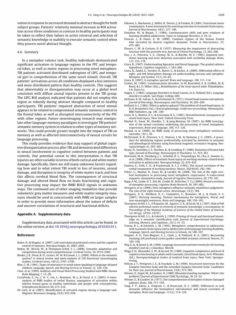
cholo
vstbst
4
srTrpatcTrpatwlswml
ticididtdtwvsia
A
t
R
B
B
B
B
C
C
D
F.G. Yang et al. / Neuropsy
ation in response to increased demand in abstract thought for bothubject groups. Patients’ relatively minimal increase in ROI activa-ion across three conditions in contrast to healthy participants maye taken to reflect their failure in active retrieval and selection ofemantic knowledge or inability to execute semantic control whenhey process novel abstract thought.
.4. Summary
In a metaphor valence task, healthy individuals demonstratedignificant activation in language regions in the PFC and tempo-al lobes, as well as motor regions in processing novel metaphors.BI patients activated distributed subregions of LIFG and tempo-al gyri in comprehension of the same novel stimuli. Overall, TBIatients’ activations across all conditions displayed a less intensivend more distributed pattern than healthy controls. This suggestshat abnormality or disorganization may occur at a global levelonsistent with diffuse axonal injuries present in the TBI group.he LIFG ROI analysis indicated that patients did not employ thisegion as robustly during abstract thought compared to healthyarticipants. TBI patients’ impaired abstraction of novel stimulippears to be related to compromised semantic memory control inhe frontal lobes as well as disrupted interconnectivity of the PFCith other regions. Future neuroimaging research may manipu-
ate other language-memory paradigms to investigate TBI patients’emantic memory system in relation to other types of memory net-orks. This could provide greater insight into the impact of TBI onemory as well as affected interconnectivity of neural circuits for
anguage processing.This study provides evidence that may support of global cogni-
ive disorganization process after TBI and demonstrated differencesn neural involvement in cognitive control between patients andontrols. One potential caveat to this interpretation is that TBInjuries are often variable in terms of both cortical and white matteramage. Specifically, there are still many unknown factors regard-
ng how and to what extent TBI result in loss of neurons, axonalamage, and disruption in integrity of white matter tracts and howhis affects cerebral blood flow. The consequences of structuralamage and altered blood flow on brain functioning and cogni-ive processing may impact the fMRI BOLD signals in unknownays. The continued use of other imaging modalities that provide
olumetric grey matter measures and white matter integrity mea-ures should be used in concurrently with fMRI on larger samplesn order to provide more information about the nature of deficitsnd uncover correlations of structural and functional deficits.
ppendix A. Supplementary data
Supplementary data associated with this article can be found, inhe online version, at doi:10.1016/j.neuropsychologia.2010.03.011.
eferences
adre, D., & Wagner, A. (2007). Left ventrolateral prefrontal cortex and the cognitivecontrol of memory. Neuropsycholgia, 45, 2883–2901.
edny, M., McGill, M., & Thompson-Schill, S. L. (2008). Semantic adaptation andcompetition during word comprehension. Cerebral Cortex, 18, 2574–2585.
inder, J. R., Desai, R. H., Graves, W. W. & Conant, L. L. (2009). Where is the semanticsystem? A critical review and meta-analysis of 120 functional neuroimagingstudies. Cerebral Cortex, 19(12), 2767–2796.
lue, C. M. (1981). Types of utterances to avoid when speaking to language-delayedchildren. Language, Speech, and Hearing Services in Schools, 12, 120–124.
hee, et al. (1999). Auditory and Visual Word Processing Studied with fMRI. HumanBrain Mapping, 7, 15–28.
ostafreda, S., Fu, C. H. Y., Lee, L., Brammer, M. J., & David, A. S. (2003). A meta-analysis of fMRI studies of verbal fluency, segregation of activation withininferior frontal gyrus in healthy individuals and people with schizophrenia.Schizophrenia Research, 60, 215–216.
e Carli, et al. (2007). Identification of activated regions during a language task.Magnetic Resonance Imaging, 25(6), 933–938.
gia 48 (2010) 1923–1929 1929
Dikmen, S., Machamer, J., Miller, B., Doctor, J., & Temkin, N. (2001). Functional statusexamination: A new instrument for assessing outcome in traumatic brain injury.Journal of Neurotrauma, 18(2), 127–140.
Donahue, M., & Bryant, T. (1984). Communicative skills and peer relations oflearning-disabled adolescents. Topics in Language Disorders, 4, 10–21.
Duncan, J., & Owen, A. M. (2000). Common regions of the human frontallobe recruited by diverse cognitive demands. Trends in Neuroscience, 23,475–483.
Elmore, C. M., & Gorham, D. R. (1957). Measuring the impairment of abstractingfunction with the proverbs test. Journal of Clinical Psychology, 13, 262–266.
Fiez, J. A., Petersen, S. E., Cheney, M. K., & Raichle, M. E. (1992). Impaired non-motor learning and error detection associated with cerebellar damage. Brain,115, 155–178.
Giora, R. (1997). Understanding figurative and literal language: The graded saliencehypothesis. Cognitive Linguistics, 7, 183–206.
Giora, R., Zaidel, E., Soroker, N., Batori, G., & Kasher, A. (2000). Differential effects ofright- and left-hemisphere damage on understanding sarcasm and metaphor.Metaphor and Symbol, 15(1), 63–83.
Giora, R. (2007). Is metaphor special? Brain and Language, 100, 111–114.Groher, M. (1983). Communication disorders. In M. Rosenthal, E. R. Griffith, M. R.
Bond, & J. D. Miller (Eds.), Rehabilitation of the head injured adult. Philadelphia:F.A. Davis Co.
Hagen, C. (1984). Langauge disorders in head trauma. In A. Holland (Ed.), Languagedisorders in adults. San Diego: College-Hill.
Heilman, K. M., Safran, A., & Geschwind, N. (1971). Closed head trauma and aphasia,Journal of Neurology. Neurosurgery, and Psychiatry, 34, 265–269.
Holland, A. L. (1982). When is aphasia aphasia? The problem of closed head injury. InR. H. Brookshire (Ed.), Clinical aphasiology: Conference proceedings. Minneapolis:BRK Publishers, pp. 345–349
Levin, H. S., Benton, A. L., & Grossman, R. G. (1982). Neurobehavioral consequences ofclosed head injury. New York: Oxford University Press.
Mashal, N., Faust, M., Hendler, T., & Jung-Beeman, M. (2007). An fMRI investiga-tion of the neural correlates underlying the processing of novel metaphoricexpressions. Brain and Language, 100, 115–126.
Mashal, et al. (2009). An fMRI study of processing novel metaphoric sentences.Laterality, 14(1), 30.
McDermott, K. B., Petersen, S. E., Watson, J. M., & Ojemann, J. G. (2003). A proce-dure for identifying regions preferentially activated by attention to semanticand phonological relations using functional magnetic resonance imaging. Neu-ropsychologia, 41, 293–303.
Neary, D., Snowden, J. S., Northen, B., & Goulding, P. (1988). Dementia of frontal lobetype. Journal of Neurology, Neurosurgery & Psychiatry, 51, 353–361.
Newsome, M. R., Steinberg, J. L., Scheibel, R. S., Troyanskaya, M., Chu, Z., Hanten, G.,et al. (2008). Effects of traumatic brain injury on working memory-related brainactivation in adolescents. Neuropsychology, 22, 419–425.
Paulesu, E., Frith, C. D., & Frackowiak, R. S. J. (1993). The neural correlates of theverbal component of working memory. Nature, 362(6418), 342–345.
Pobric, G., Mashal, N., Faust, M., & Lavidor, M. (2008). The role of the right cere-bral hemisphere in processing novel metaphoric expressions: A transcranialmagnetic stimulation study. Journal of Cognitive Neuroscience, 20, 170–181.
Rapp, A. M., Leube, D. T., Erb, M., Grodd, W., & Kircher, T. T. J. (2004). Neural correlatesof metaphor processing. Cognitive Brain Research, 20, 395–402.
Stringaris, et al. (2006). How metaphors influence semantic relatedness judgments:The role of the right frontal cortex. NeuroImage, 33(2), 784.
Stringaris, A. K., Medford, N. C., Giampetro, V., Brammer, M. J., & David, A. S.(2007). Deriving meaning: Distinct mechanisms for metaphoric, literal, andnon-meaningful sentences. Brain and Language, 100, 150–162.
Thompson-Schill, S. L., D’Esposito, M., Aguirre, G. K., & Farah, M. J. (1997). Role of leftinferior prefrontal cortex in retrieval of semantic knowledge, a reevaluation. InProceedings of the National Academy of Sciences of the United States of America,vol. 94 (pp. 14792–14797).
Thompson-Schill, S. L., & Gabrieli, J. (1999). Priming of visual and functional knowl-edge on a semantic classification task. Journal of Experimental Psychology:Learning, Memory, and Cognition., 25(1), 41–53.
Towne, R. L., & Entwisle, L. M. (1993). Metaphoric comprehension in adolescentswith traumatic brain injury and in adolescents with language learning disability.Language, Speech, and Hearing Services in Schools, 24, 100–107.
Wagner, A. D., Pare-Blagoev, E. J., Clark, J., & Poldrack, R. A. (2001). Recoveringmeaning, left prefrontal cortex guides controlled semantic retrieval. Neuron, 31,329–338.
Wig, E. H., & Semel, E. M. (1984). Language assessment and intervention for the learningdisabled (2nd ed.). Columbus: Merrill.
Wigg, E. H., Alexander, E. W., & Secord, W. (1988). Linguistic competence and level ofcognitive functioning in adults with traumatic closed head injury. In H. Whitaker(Ed.), Neuropsychological studies of nonfocal brain injury. New York: Springer-Verlag.
Wilson, J. T., Pettigrew, L. E., & Teasdale, G. M. (1998). Structured interviews for theGlasgow Outcome Scale and the extended Glasgow Outcome Scale: Guidelinesfor their use. Journal of Neurotrauma, 15(8), 573–585.
Winner, E., Engel, M., & Gardner, H. (1980). Misunderstanding metaphor: What’s the
problem? Journal of Experimental Child Psychology, 30, 22–32.Winner, E., & Gardner, H. (1977). The comprehension of metaphor in brain-damagedpatients. Brain, 100, 717–729.
Yang, F. P., Edens, J., Simpson, C., & Krawczyk, D. C. (2009). Differences in taskdemands influence the hemispheric lateralization and neural correlates ofmetaphor. Brain and Language, 111(2), 114–124.