Embed Size (px)
Citation preview

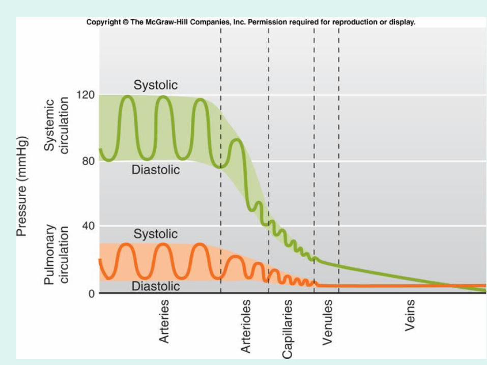
Fig. 12.01
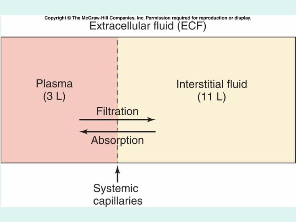

Fig. 12.47

Elephantiasis: The swelling results from blocked
lymphatic vessels
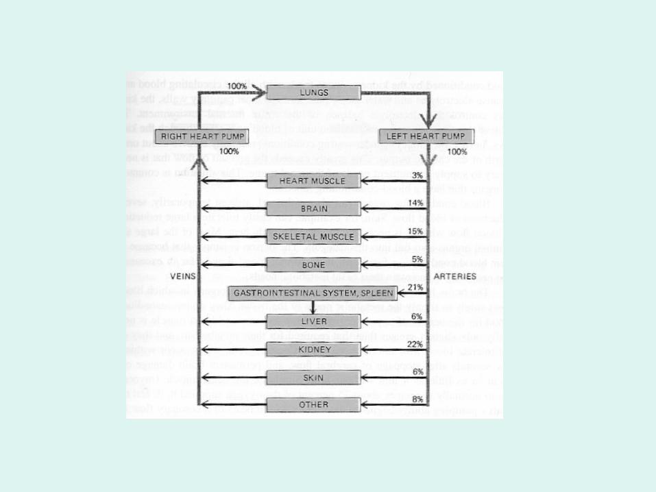
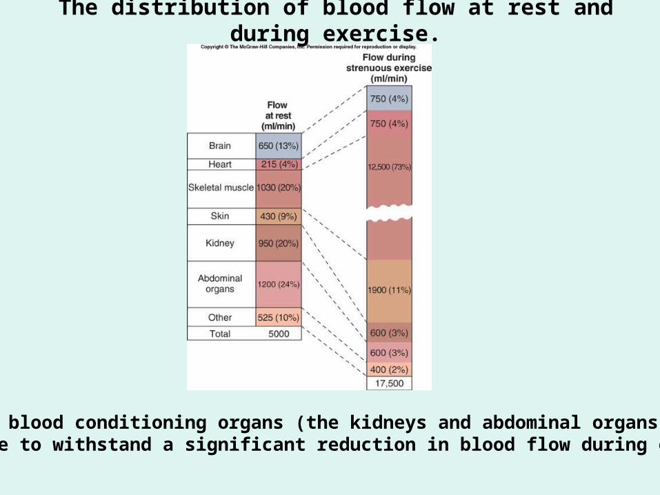
The distribution of blood flow at rest and during exercise.
Note how blood conditioning organs (the kidneys and abdominal organs) are able to withstand a significant reduction in blood flow during exercise.
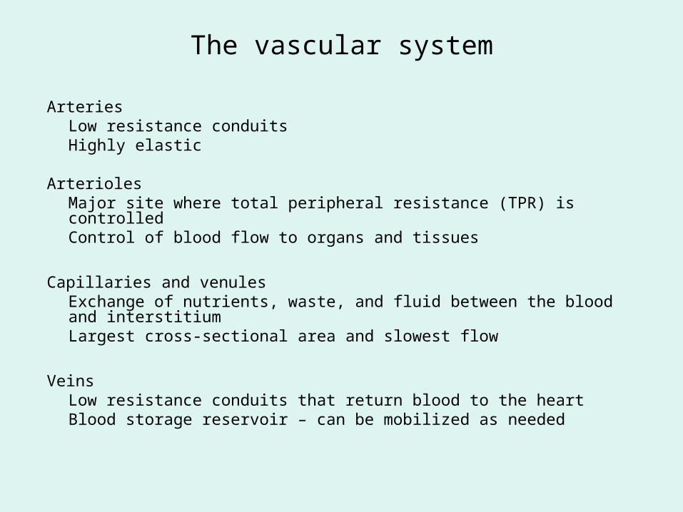
The vascular system
Arteries Low resistance conduits Highly elastic
Arterioles Major site where total peripheral resistance (TPR) is controlledControl of blood flow to organs and tissues
Capillaries and venulesExchange of nutrients, waste, and fluid between the blood and interstitiumLargest cross-sectional area and slowest flow
VeinsLow resistance conduits that return blood to the heartBlood storage reservoir – can be mobilized as needed
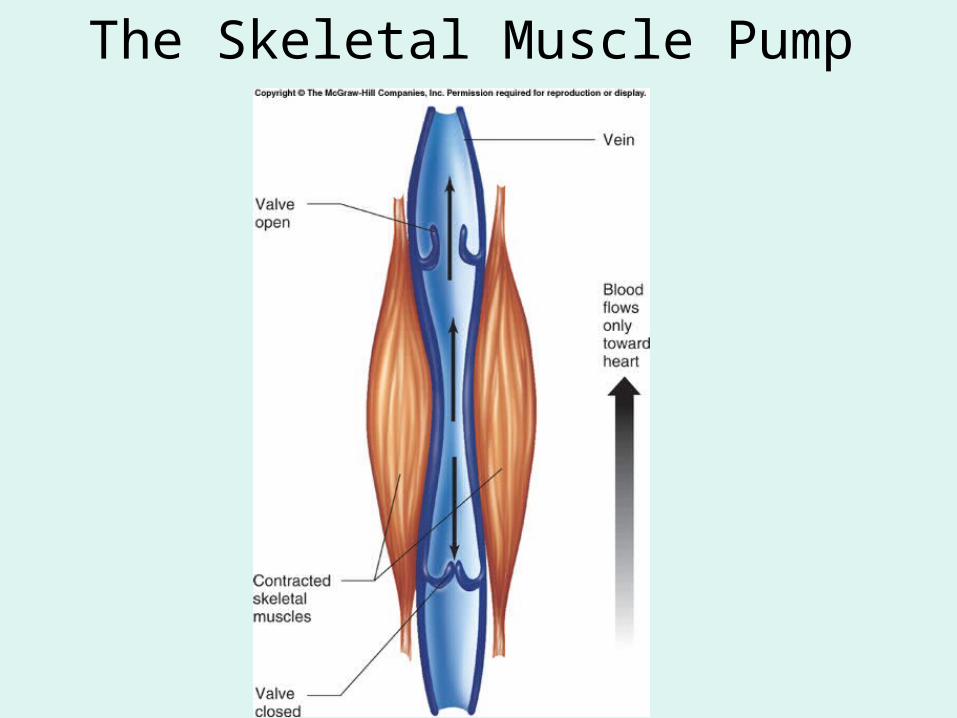
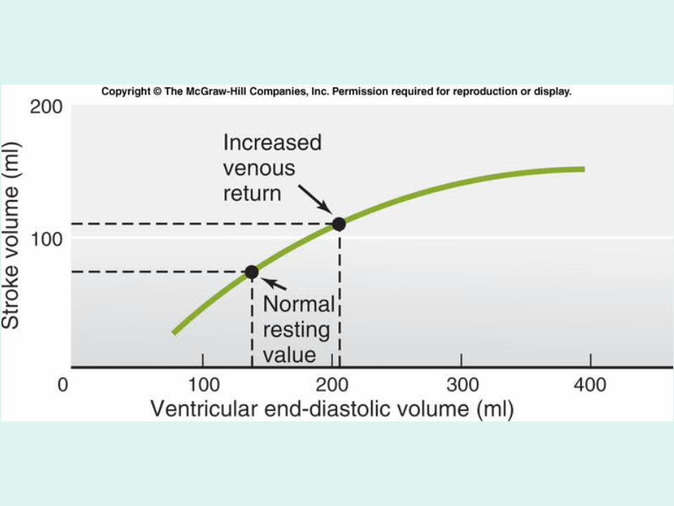
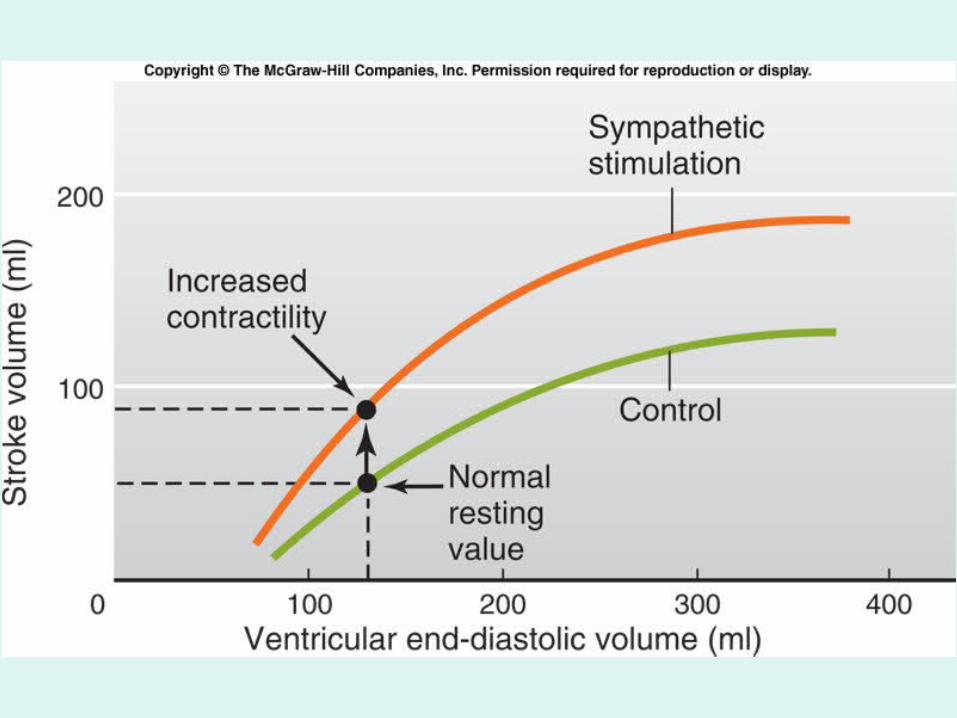
The Skeletal Muscle Pump

http://www.nlm.nih.gov/medlineplus/ency/imagepages/18072.htm
An Aortic Aneurysm

http://www.vascularweb.org/graphics/northpoint_graphics_jpg/Varicose_02_Base_300.jpg
Varicose Veins

Fig. 12.06

Cardiac Muscle
http://content.answers.com/main/content/img/oxford/Oxford_Body/019852403x.cardiac-muscle.1.jpg


Normal ECG (a), partial AV block(b) complete AV block (c)
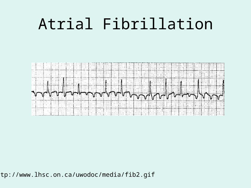
Atrial Fibrillation
http://www.lhsc.on.ca/uwodoc/media/fib2.gif
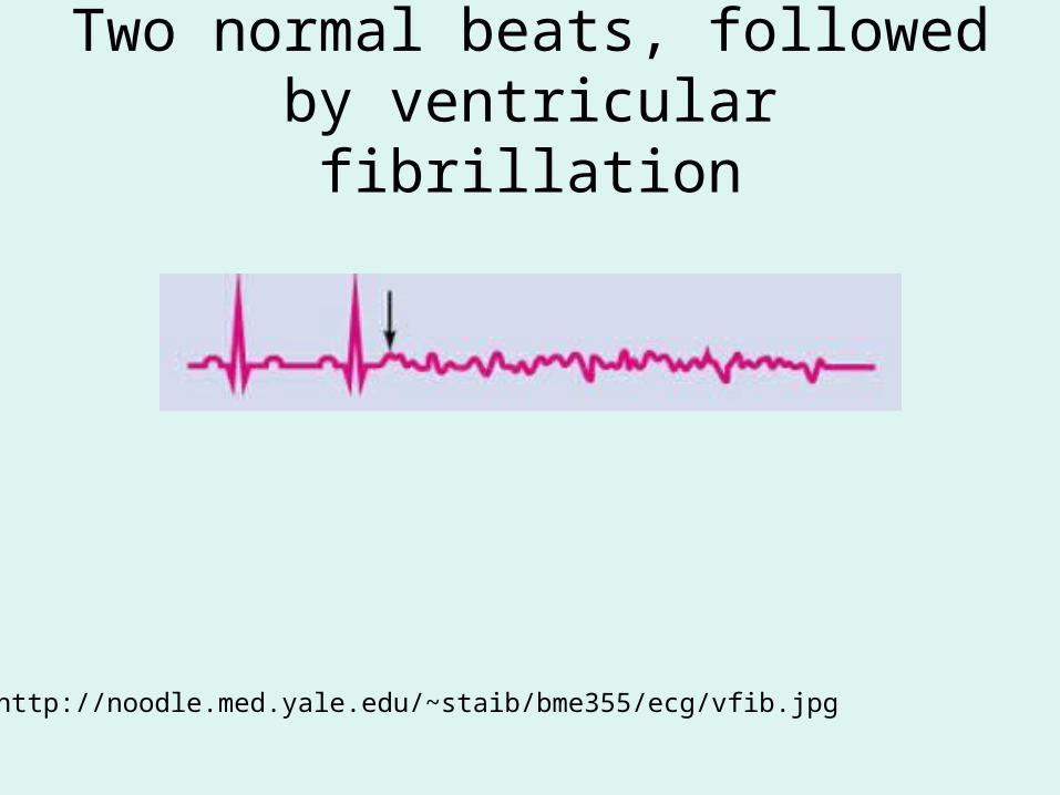
Two normal beats, followed by ventricular fibrillation
http://noodle.med.yale.edu/~staib/bme355/ecg/vfib.jpg

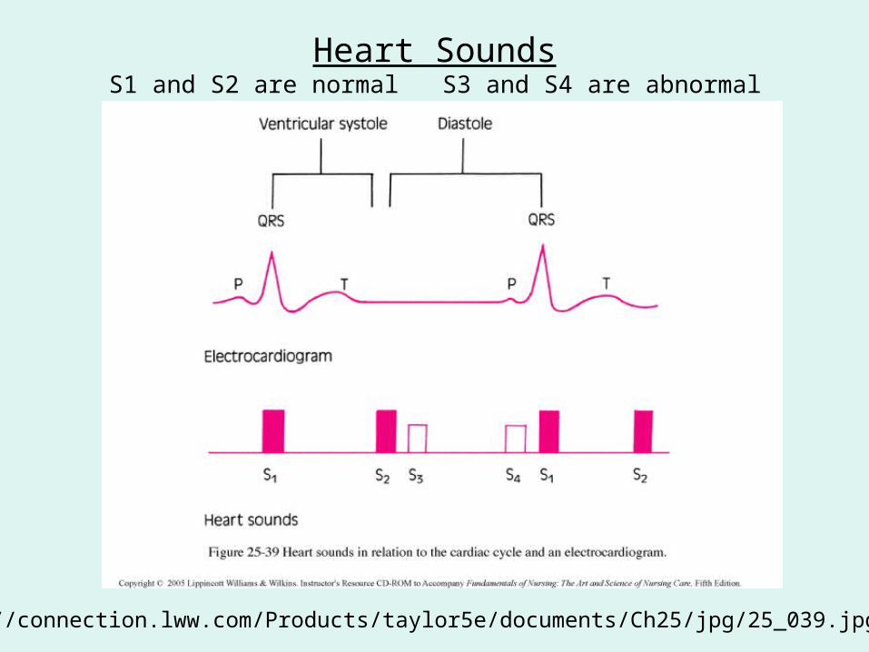
Heart Sounds
http://www.nlm.nih.gov/medlineplus/ency/images/ency/fullsize/19613.jpg
http://connection.lww.com/Products/taylor5e/documents/Ch25/jpg/25_039.jpg
Heart SoundsS1 and S2 are normal S3 and S4 are abnormal

Extra Diastolic Sounds: S3
• S3 is produced by the tensing of the chordae tendineae, which occurs during rapid filling and expansion of the ventricles.
• Common in children and young adults – the flexible ventricles of young people can expand rapidly.
• In middle-aged or older adults often indicates excessive volume in the ventricles, which usually indicates heart failure.
www.mvprolapse.com/mvp.html

Extra Diastolic Sounds: S4
• Produced by the left or right venticle contracting against a stiffened ventricle
• Usually indicates a loss of compliance of the ventricle due to ventricular hypertrophy or myocardial ischemia

The heart murmur associated with mitral valve prolapse
Mitral valve prolapse causes a late systolic murmur
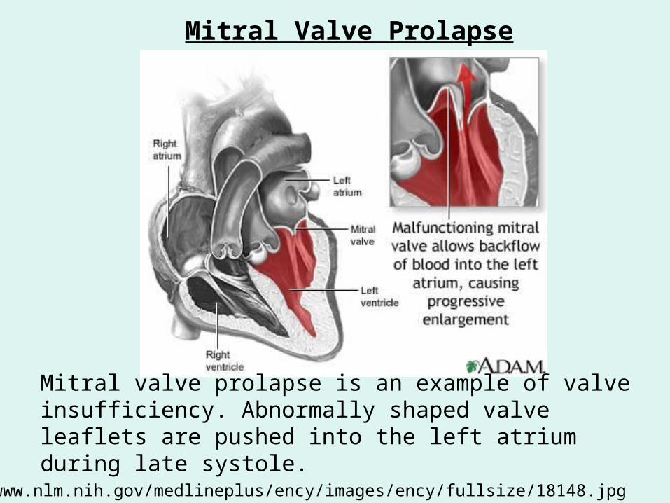
Mitral valve prolapse is an example of valve insufficiency. Abnormally shaped valve leaflets are pushed into the left atrium during late systole.
http://www.nlm.nih.gov/medlineplus/ency/images/ency/fullsize/18148.jpg
Mitral Valve Prolapse

http://www.abcbodybuilding.com/magazine03/wrench/tensionlevels2.jpg
The length-tension relationship


Children with Kwashiorkor
http://www.cs.stedwards.edu/chem/Chemistry/CHEM43/CHEM43/Leukotr/Kwashiorkor.GIF