Embed Size (px)
Citation preview
Rash appearance
Skin lesions evolve over time, but the characteristic
distribution and appearance provide important clues
to the diagnosis Macular or Maculopapular Rash
Diffuse Erythroderma
Urticarial Rash
Vesicular, Bullous, Pustular
Petechial-Purpuric
Erythema Nodosum
Differential Diagnosis of Fever and Rash
Macular or Maculopapular Rash -- virus:
Measles
Rubella
Roseola (HHV-6 or HHV-7)
Others: Erythema infectiosum (fifth disease,
parvovirus B19), Epstein-Barr virus,
Echoviruses, HBV, HIV
Differential Diagnosis of Fever and Rash
Macular or Maculopapular Rash--bacteria
Scarlet fever (group A streptococcus)
Others: Secondary syphilis, Leptospirosis,
Pseudomonas, Meningococcal infection (early),
Salmonella typhi (typhoid fever), Lyme disease
(erythema migrans), Mycoplasma pneumoniae
Measles
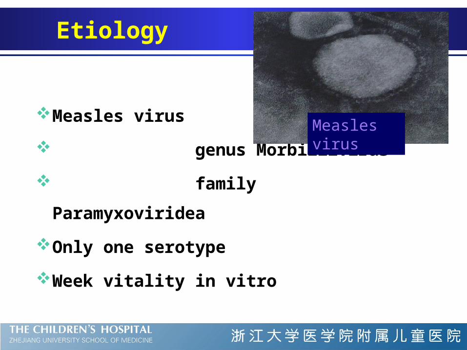
Etiology
Measles virus
genus Morbillivirus
family Paramyxoviridea
Only one serotype
Week vitality in vitro
Measles virus
Epidemiology
Source of infection
Route of transmission
Susceptible population
Source of infection
patients as the main source
infectivity from prodromal period to the
time of 5~10 days after the rash appears
respiratory secretions as the main media
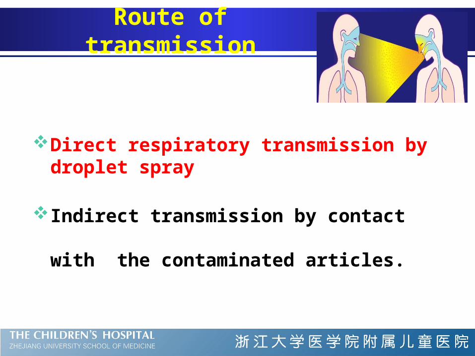
Route of transmission
Direct respiratory transmission by droplet spray
Indirect transmission by contact with the
contaminated articles.
Susceptible population
Peak incidence: 8m~5yr
Pathogenesis
Virus inhalation incubation
Local proliferation
First viremia
Proliferation in endoreticular system
Second viremia prodromal
All parts of the body eruption
Clearance of virus convalescence
Clinical Manifestation
Typical measles
Incubation stage
Prodromal stage
Eruptive stage
Convalescent stage
Stage 1: Incubation stage
6~18 days
No symptoms or low fever, malaise
Stage 2: Prodromal stage (Catarrhal stage)
Lasts 3~4 days
Low-grade to moderate fever
Mucosal catarrh: conjunctivitis,
coryza
hacking cough
Stimson line: transverse line of inflammation along the eyelid margin
Koplik spots: a pathognomonic sign
Koplik spots
—a pathognomonic sign of measles
Appear 2-3 days after fever develops
Tend to occur over the buccal mucosa opposite
the lower molars
A grayish white dots(as small as grains of
sands) with slight reddish areola
Increase and disappear 1-2d after eruption
Stage 3: Eruptive stage
Lasts 3~4 days
Characterized by the eruption of the rash
The temperature rises abruptly as rash
develops (usually >40℃)
Exacerbated mucosal catarral symptoms:
cough, vomitting and diarrhea, anorexia,
a few rales on auscultation
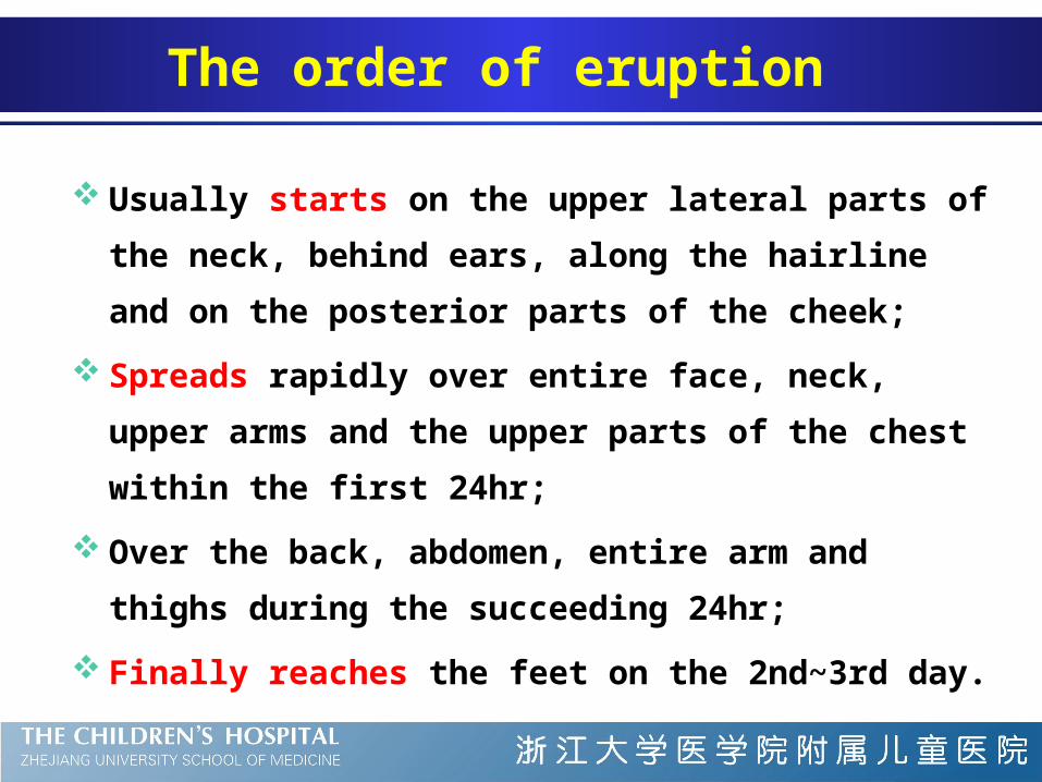
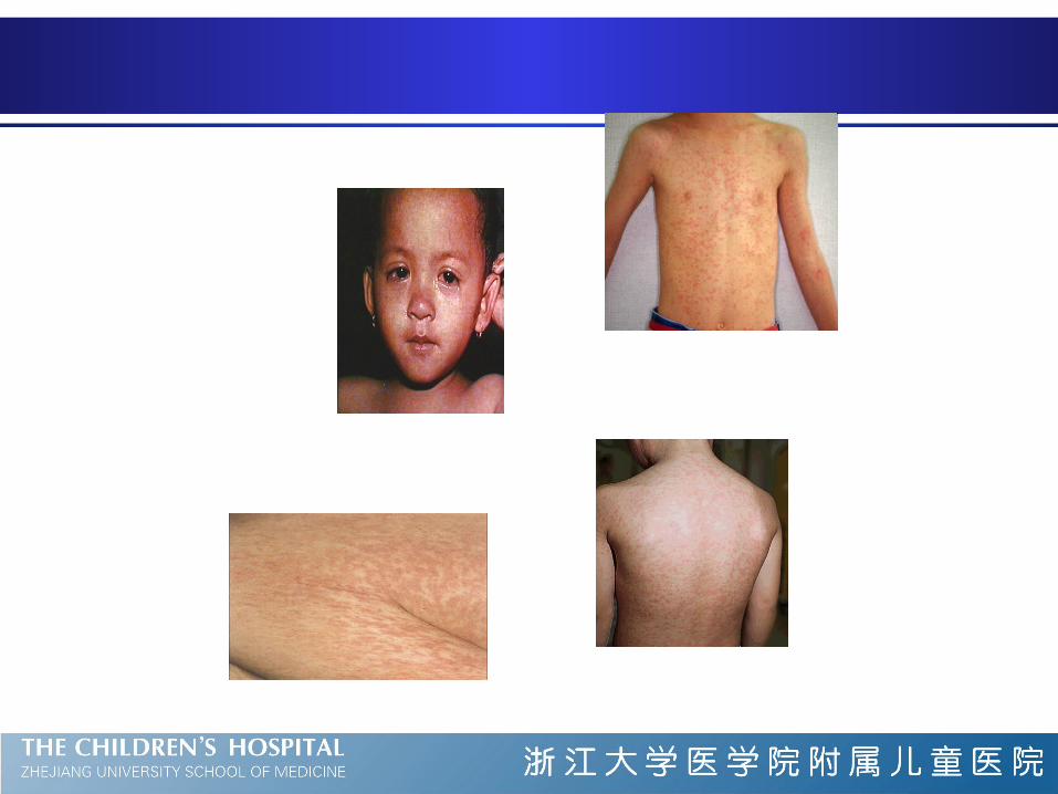
The order of eruption
Usually starts on the upper lateral parts of the neck,
behind ears, along the hairline and on the posterior parts
of the cheek;
Spreads rapidly over entire face, neck, upper arms and the
upper parts of the chest within the first 24hr;
Over the back, abdomen, entire arm and thighs during the
succeeding 24hr;
Finally reaches the feet on the 2nd~3rd day.
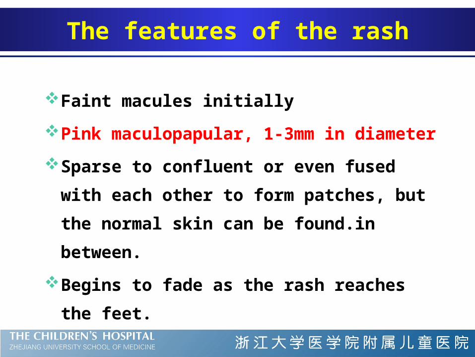
The features of the rash
Faint macules initially
Pink maculopapular, 1-3mm in diameter
Sparse to confluent or even fused with each
other to form patches, but the normal skin can
be found.in between.
Begins to fade as the rash reaches the feet.
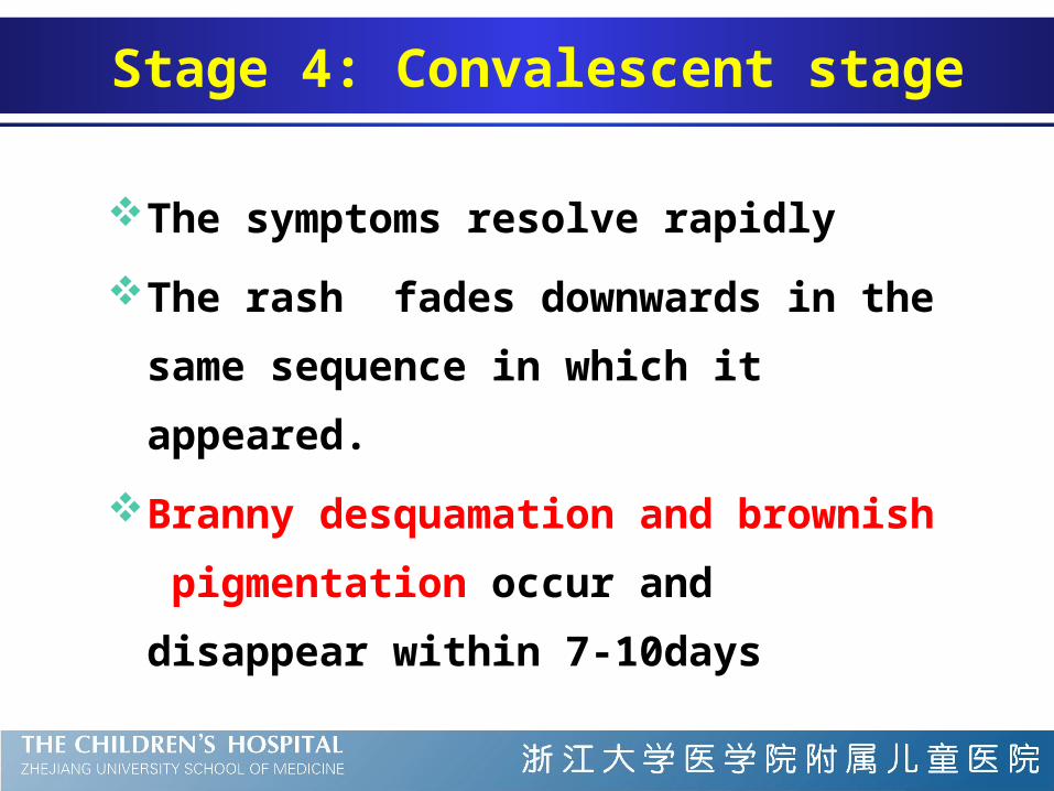
Stage 4: Convalescent stage
The symptoms resolve rapidly
The rash fades downwards in the same
sequence in which it appeared.
Branny desquamation and brownish
pigmentation occur and disappear within 7-
10days
Atypical measles
Mild measles (modified measles)
Severe measles
Measles without rash
Hemorrhagic measles (black measles)
Atypical measles syndrome
Atypical Measles
A laboratory confirmation is rarely needed
Leukopenia , with a relative lymphocytosis
Multinucleated giant cells in smear of nasal
mucosa (Warthin-Finkeldey cells)
Serum antibody(sIgM or double serum IgG)
Virus isolation or virus antigen/RNA detection
Laboratory examination
Complications
Pneumonia ( fatal giant cell (Hecht) pneumonia
in patients with impaired cell-mediated
immunity)
Laryngo-bronchitis
Myocarditis
Encephalitis
Reactive tuberculosis
Subacute sclerosing panencephalitis
Basis of diagnosis
Evidence of epidemiology
Koplik spots
The order and features of the rash
Branny desquamation and brownish
discoloration

Therapy
No specific antiviral therapy
Supportive treatment
antipyretics, bed rest,
maintenance of an adequate fluid intake
Management of complication
Vitamine A supplement
IVIG
Vitamin A and measles: evidence
Hyporetinemia is present in over 90% of measles
cases in Africa and 22-70% in USA.
There is an apparent inverse correlation between
retinol concentration and the severity of measles.
Oral Vitamin A supplement reduces the morbidity
and mortality of severe cases.
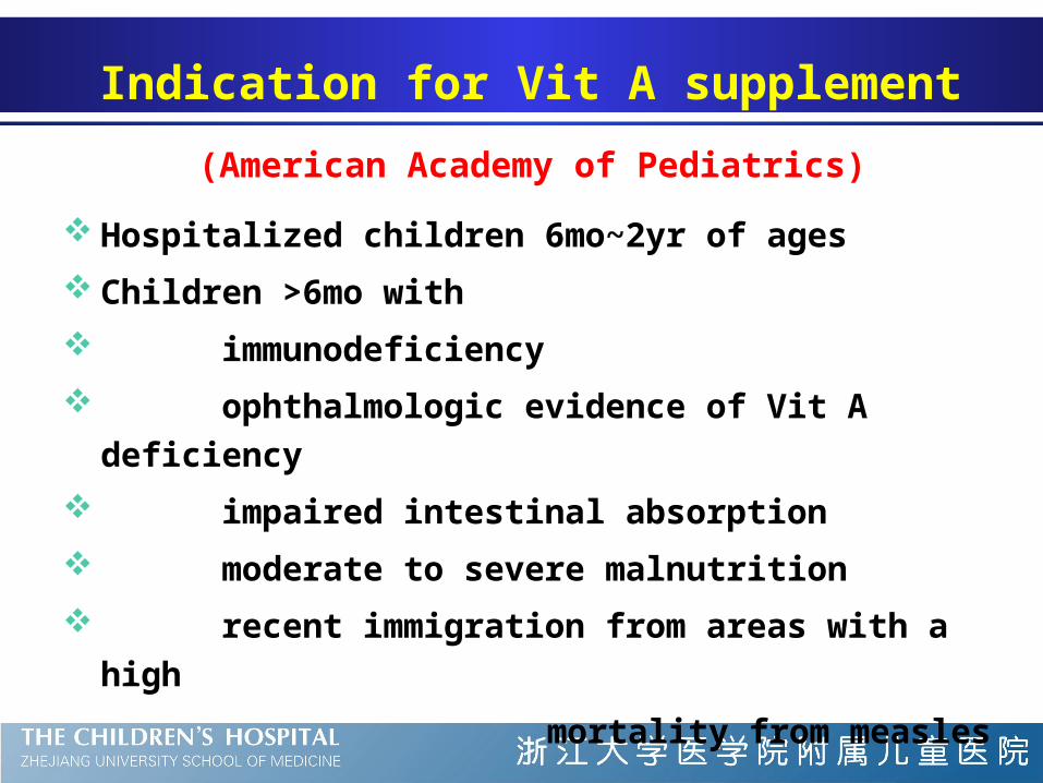
Indication for Vit A supplement
(American Academy of Pediatrics)
Hospitalized children 6mo~2yr of ages
Children >6mo with
immunodeficiency
ophthalmologic evidence of Vit A deficiency
impaired intestinal absorption
moderate to severe malnutrition
recent immigration from areas with a high
mortality from measles
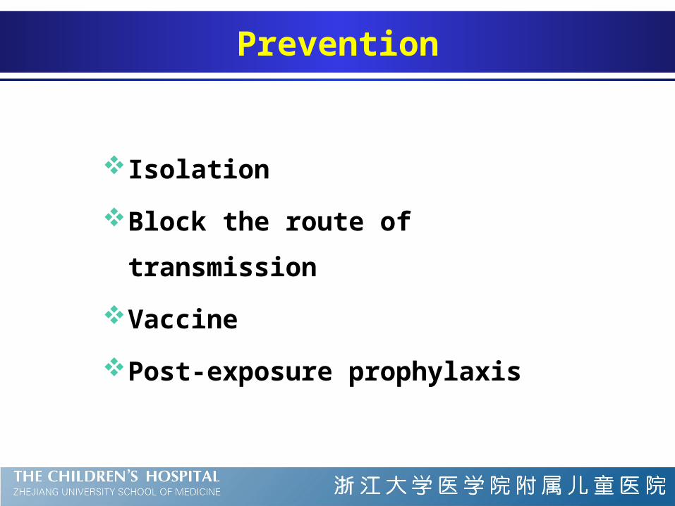
Prevention
Isolation
Block the route of transmission
Vaccine
Post-exposure prophylaxis
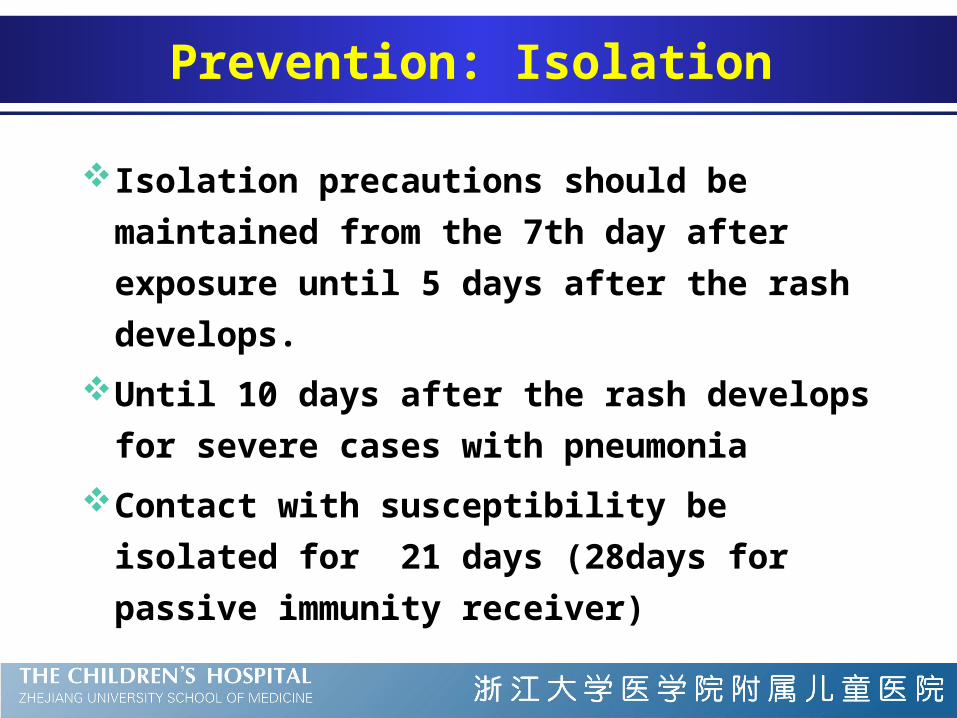
Prevention: Isolation
Isolation precautions should be maintained from
the 7th day after exposure until 5 days after the
rash develops.
Until 10 days after the rash develops for severe
cases with pneumonia
Contact with susceptibility be isolated for 21
days (28days for passive immunity receiver)
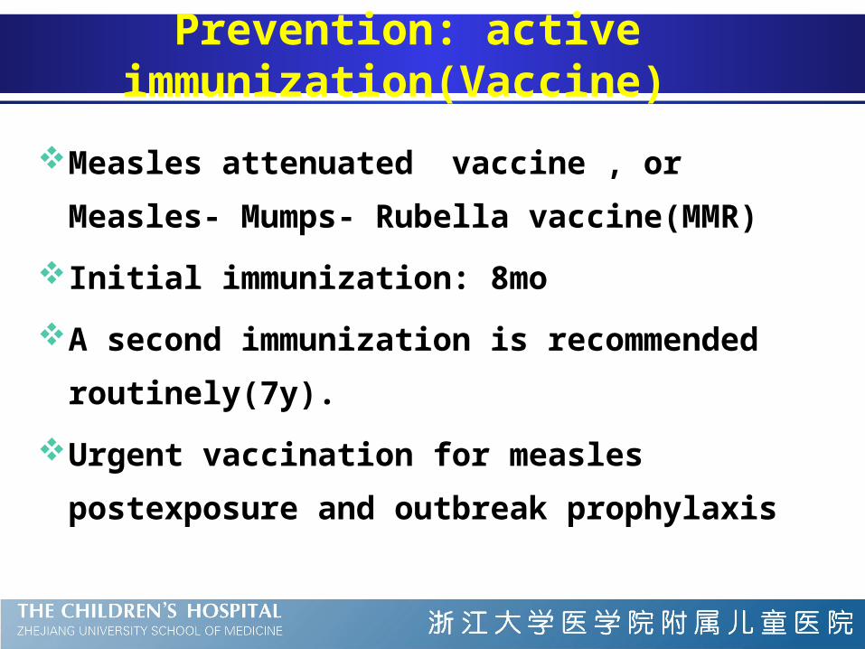
Prevention: active immunization(Vaccine)
Measles attenuated vaccine , or Measles- Mumps-
Rubella vaccine(MMR)
Initial immunization: 8mo
A second immunization is recommended
routinely(7y).
Urgent vaccination for measles postexposure and
outbreak prophylaxis
Prevention: Passive immunization(post-exposure
prophylaxis)
Passive immunization with Ig within 5 days of
exposure is effective for the prevention and
attenuation of measles.
Susceptible children <12mo should receive Ig
(0.25ml/kg, <=15ml,IM)
Differential Diagnosis of Fever and Rash
Macular or Maculopapular Rash -- virus:
Measles
Rubella
Roseola (HHV-6 or HHV-7)
Others: Erythema infectiosum (fifth disease,
parvovirus B19), Epstein-Barr virus,
Echoviruses, HBV, HIV
Rubella
also known as German measles and 3-day
measles;
congenital rubella syndrome (infection in
utero )
Etiology and epidemiology
a single-stranded, positive-sense RNA virus ( a
member of the togavirus family)
Humans as the only host
Spread either by oral droplet or transplacentally to
fetus causing congenital infection
Virus recovered from the nasopharynx 7d before
exanthem and 7-8 d after its disappearance.
Peak incidence in children 5~14 yr of age
Clinical manifestations
Incubation (14 to 21 d)
Prodromal phase
Mild catarrhal symptoms with shorter period
Low-grade fever (1~3d) with meager systemic
symptoms.
About 2/3 are subclinical.
Clinical manifestations
The most characteristic sign :
Enlarged post-occipital, retroauricular and
posterior cervical lymph nodes with tender.
Be evident 24h before rash and remain for 1
week or more
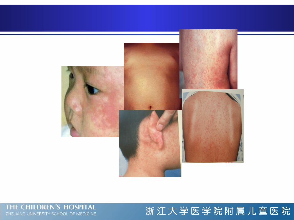
Clinical manifestations
Exanthem more variable even no rash
first appear on face
rapid evolution, usually cover the entire body in 24 h
Usually clears by the 3rd day
Discrete maculopapules with large flushing, or
pinpoint appearance, or may be confluent on face
Mild itching and minimal desquamation
Clinical manifestations
Enanthem in 20% patients
Just before rash
Discrete rose spots on the soft palate (Forchheimer
spots)
May coalesce into a red blush and extend over the
fauces
Slightly inflamed pharyngeal mucosa and conjunctivae
without photophobia
Clinical manifestations
Congenital rubella (syndrome)
Affects virtually all organ systems
The most common manifestation is intrauterine growth
retardation
Other common findings: cataracts (microphthalmia,
myocarditis, PDA); “blueberry muffin” skin lesions;
hearing loss; meningoencephalitis.
Diagnosis
Apparent diagnosis based on clinical symptoms
and signs
Laboratory findings non-specific and generally do not
aid in diagnosis
confirmed by serology or virus culture
Congenital rubella: serum sIgM or virus culture
Prenatal diagnosis: cord blood sIgM or virus
culture from amniotic fluid
Treatment and prognosis
There is no specific antiviral therapy
Entirely supportive, and antipyretics
The prognosis is excellent, but congenital rubella
syndrome may have sequalae such as intrauterine
growth retardation, cataracts, deafness, and a
patent ductus arteriosus.
Prevention
Live rubella vaccine recommended as MMR for
children( initial at 12-15m and second 4-6y)
It is important for girls to have immunity before
they reach childbearing age
Differential Diagnosis of Fever and Rash
Macular or Maculopapular Rash -- virus:
Measles
Rubella
Roseola (HHV-6 or HHV-7)
Others: Erythema infectiosum (fifth disease,
parvovirus B19), Epstein-Barr virus,
Echoviruses, HBV, HIV
Etiology
Roseola was first established as a distinct illness
at the turn of 20th century;
No causative pathogens were consistently
identified until recent 10 years;
It appears now that primary infection of human
herpesvirus type 6 (HHV-6) and less frequently
HHV-7 causes the majority of the cases of roseola.
Epidemiology
Primary HHV-6 infection occurs early in life with
peak acquisition from 6-15 months of age.
Rarely report contact with other infected children
and outbreak uncommon.
Most adults excrete HHV-6 and HHV-7 in saliva
and may serve as primary sources for virus
transmission.
HHV-6 can be transmitted in utero.
Cinical manifestations
Incubation period: 5-10d
Prodromal period:
Usually asymptomatic
Mild URT signs
Mild cervical lymphadenopathy
Cinical manifestations
Clinical illness heralded by high fever
37.9~40.0 with an average of 39℃
Persists for 3-5 days and then resolves rather abruptly
(crisis). Occasionally fever diminish over 24-36h gradually
(lysis).
May be irritable and anorexia but most behave normally
Seizures in 5~10%
Infrequent : rhinorrhea, sore throat, abdominal pain,
vomiting and diarrhea.
Cinical manifestations
Eruption and fever
A rash appears within 12~24 hours of fever
resolution
Eruption during defervescence or within a
few hours of fever resolution
Cinical manifestations
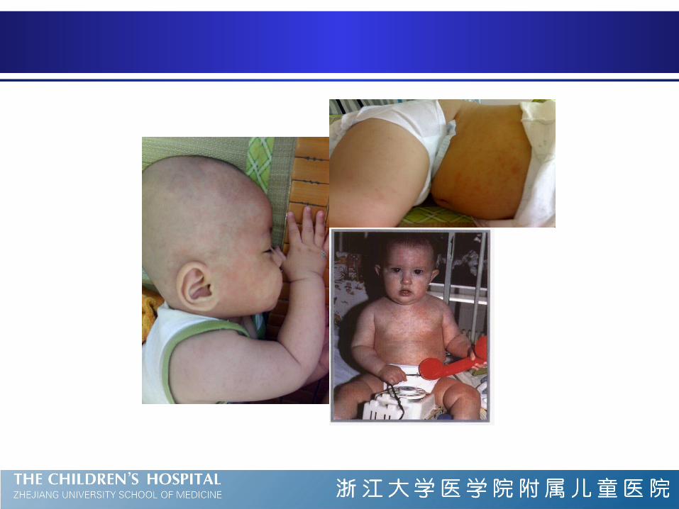
Characteristic rash
Rose colored rash ( discrete, small (2~5mm),slightly
raised pink lesions)
appears trunk , neck, behind ears, face and proximal
extremities
No pruritic, no vesicles
Fade in 1~3 days
Treatment and prevention
There is no specific therapy
HHV-6 is inhibited by ganciclovir but the
benign nature preclude consideration of
antiviral therapy.
Excellent prognosis in majority
no guidelines for prevention of roseola.
Varicella
Etiology
Chickenpox (varicella) is the manifestation of
primary infection of varicella-zoster virus (VZV).
Zoster (shingles) is the manifestation of reactivated
latent infection of endogenous VZV.
Clinical manifestation
Incubation period: 10~21d
Subclinical varicella is rare
Prodromal symptoms
Usually moderate fever
malaise, headache, anorexia and occasionally
mild abdominal pain
precede the rash by 24~48h
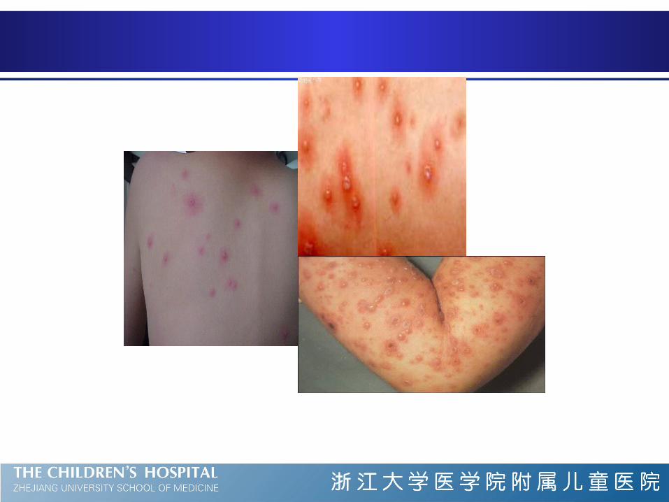
Clinical manifestation
The characteristic rash
initially as small red papules
rapidly progress to nonumbilicated, oval, "teardrop" vesicles on an erythematous base.
The fluid progresses from clear to cloudy, and the vesicles ulcerate, crust, and heal.
New crops appear for 3 to 4 days, usually beginning on the trunk followed by the head, the face, and, less commonly, the extremities.
Clinical manifestation
Pruritus, mucous membrane lesions,
lymphadenopathy
Hypopigmentation or hyperpigmentation persists
for days to weeks in some
Scarring unusual unless secondarily infected
Clinical manifestation
Progressive varicella
usually in immunocompromised children
Neonatal chickenpox
delivery within 1 week before or after the onset of maternal varicella frequently results in severe varicella in neonates
Congenital varicella
when pregnant women (especially between 8-20 weeks) contract chickenpox, 25%of the fetuses may become infected.
Complication
Complications are common
secondary infection of skin lesions by streptococci
or staphylococci, Thrombocytopenia and
hemorrhagic lesions or bleeding, pneumonia ,
myocarditis, pericarditis, orchitis, hepatitis,
ulcerative gastritis, glomerulonephritis, and
arthritis , Reye syndrome, encephalitis, cerebellar
ataxia, nystagmus, and tremor
Therapy
Symptomatic therapy
Antivirals (acyclovir, famciclovir, or valacyclovir )
are effective in preventing severe complications
the routine oral administration of acyclovir is not
recommended in otherwise healthy children because of
the marginal therapeutic benefit, the lack of difference in
complications, and the cost of acyclovir treatment.