Embed Size (px)
Citation preview

FETAL SURGERY FORMYELOMENINGOCELE

Since December 2010, fetal surgery for myelomeningocele (MMC), the most common and severe form of spina bifida, has been offered as a standard of care at Children’s Hospital of Philadelphia. This is one of the most exciting developments in the history of treatment for birth defects.
An extremely complex procedure available only to qualified candidates, fetal surgery for MMC requires significant commitment on the part of mothers who choose to go forward with it and extensive surgical experience to perform successfully. Mothers who choose fetal surgery require comprehensive counseling on the condition and the risks involved in the procedure.
This guide provides an overview of fetal MMC repair, including confirmation of the diagnosis, patient criteria for fetal surgery, the surgical procedure, delivery and follow-up care. We welcome the opportunity to discuss individual candidates for referral.
A GUIDE TO SPECIALIZED FETAL MMC SURGICAL REPAIR

TYPES OF SPINA BIFIDA• Myelomeningocele (MMC)
• Myeloschisis
• Lipomeningocele
• Myelocystocele
Open neural tube defects such as myelomeningocele and myeloschisis are treatable by fetal repair.
Closed neural tube defects such as lipomeningocele and myelocystocele are not treatable by fetal repair.
1
2
FETUS WITH MYELOMENINGOCELE 1. Part of the spinal cord and spinal nerves, usually encased in a sac, protrude through an opening in the back and are exposed to the toxic effects of amniotic fluid. 2. Arnold-Chiari II Malformation: Cerebrospinal fluid (CSF) leaks through the opening in the back, and the brain stem (hindbrain) descends, or herniates, into the spinal canal in the neck and blocks the circulation of CSF. This can cause a
damaging buildup of fluid in the brain called hydrocephalus.

FACTS ABOUT MYELOMENINGOCELE• Most common and serious form of spina bifida
• Primary failure of neural tube formation (closure)
• Genetic and micronutrient causes
• 1 per 2,000 live births, or 1,500 babies in the U.S. each year
• 14% die before age 5; 2/3 due to hindbrain herniation complications
• 85% require shunts; 45% of shunts develop complications within 1 year
• Up to 4% recurrence risk in subsequent pregnancies
LONG-TERM CONSEQUENCES • Hydrocephalus • Hindbrain herniation complications • Ventriculoperitoneal shunting • Paralysis and cognitive impairments • Orthopaedic malformations (such as club foot) • Bladder and bowel incontinence • Sexual dysfunction • Social and emotional challenges • Lifelong quality-of-life issues

1
23456781 2
34567
8
9
10
11
12
1
2
3 4
5
1 2345
Skin
Sac
Exposed nerves
Sac
Hip
Knee
Ankle (dorsiflexors)
Great Toe Ankle (plantarflexors)
Bowel & Bladder
Muscle Areas Affected
Cervical
Thoracic
Lumbar
Sacral
MMC can occur at any level in the developing spine, but occurs most often in the lumbosacral region. The higher the defect on the spine, the more severe the complications.
Areas below the level of the defect will be affected. This illustration shows a defect at the L 4-5 level.
MOTOR IMPAIRMENT RELATED TO THE LEVEL OF THE DEFECT

1. Maternal serum alpha-fetoprotein (MSAFP) test. Abnormally high AFP levels can suggest a neural tube defect.
2. Level II ultrasound to confirm spinal defect, determine level of lesion, confirm features in the brain that indicate spina bifida and assess deformities such as talipes (club foot).
3. Amniocentesis to confirm presence of elevated amniotic fluid alpha-fetoprotein (AFAFP) levels and acetylcholinesterase (AChE), which indicates open (vs. closed)neural tube defect. Amniocentesis is required to be considered for fetal surgery.
STEPS IN MAKING THE DIAGNOSIS
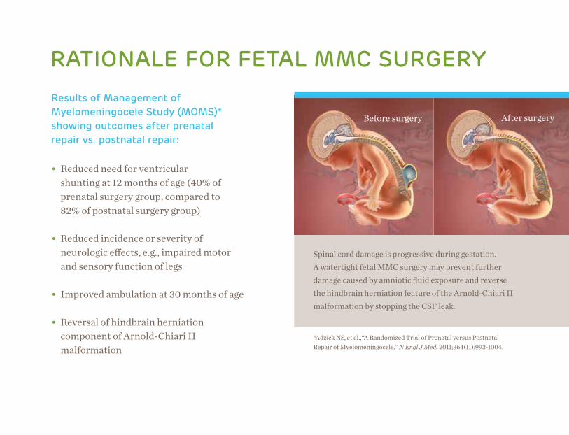
Spinal cord damage is progressive during gestation. A watertight fetal MMC surgery may prevent further damage caused by amniotic fluid exposure and reverse the hindbrain herniation feature of the Arnold-Chiari II malformation by stopping the CSF leak.
RATIONALE FOR FETAL MMC SURGERY Results of Management of Myelomeningocele Study (MOMS)* showing outcomes after prenatal repair vs. postnatal repair:
• Reduced need for ventricular shunting at 12 months of age (40% of prenatal surgery group, compared to 82% of postnatal surgery group)
• Reduced incidence or severity of neurologic effects, e.g., impaired motor and sensory function of legs
• Improved ambulation at 30 months of age
• Reversal of hindbrain herniation component of Arnold-Chiari II malformation
Before surgery After surgery
*Adzick NS, et al.,“A Randomized Trial of Prenatal versus Postnatal Repair of Myelomeningocele,” N Engl J Med. 2011;364(11):993-1004.

Key outcomes and measures of success:
• CHOP’s rate of transfusion (1.5 percent) is lower than the MOMS trial (9 percent) at the time of cesarean section delivery due to the continuity of care and experience of the delivery team members.
• Rates of preterm labor and membrane separation were similar between the CHOP cohort and the MOMS trial.
• At CHOP, more than 50 percent of babies that undergo fetal surgery deliver at or after 35 weeks, similar to the MOMS trial gestational age at delivery.
FETAL SURGERY OUTCOMES AT CHOP SINCE THE MOMS TRIAL
OUTCOMES WITH FETAL SURGERY
FOR MMC AT CHILDREN’S HOSPITAL
OF PHILADELPHIA (CHOP) ARE THE
SAME OR BETTER THAN THOSE
REPORTED IN THE MOMS TRIAL.
• CHOP had a lower rate of PPROM of 32.3 percent compared to the MOMS trial cohort of 46.2 percent.
• On neonatal MRI, hindbrain herniation was reversed in more than 90 percent of our fetal surgery patients.
• There were no CSF leaks from the fetal MMC surgery.
• The average birth weight was comparable to the MOMS trial.
• The rate of hydrocephalus therapy with a shunt or endoscopic third ventriculostomy (ETV) procedure at 12 months of age is 22 percent.
• A follow-up program is in place for improving long-term outcomes.

Partial list of inclusion criteria: • Singleton pregnancy • Maternal BMI < 40 • Confirmed presence of elevated AFAFP and AChE • Confirmed normal genetic testing • Absence of associated fetal anomalies
WHEN IS FETAL SURGERY FOR MMC AN OPTION?
To see an extensive list of inclusion and exclusion criteria
for fetal MMC repair at CHOP, visit fetalsurgery.chop.edu/spinabifida
or call 1-800-IN UTERO (468-8376).
• Myelomeningocele at level T1 through S1 • Arnold-Chiari II malformation (hindbrain herniation) • Gestational age at time of fetal surgery ≤ 25 weeks, 6 days
• Maternal age ≥ 18 years

Diagnostic testing includes:
• High-resolution level II ultrasound to confirm location of MMC and assess for other birth defects
• Ultrafast fetal MRI to confirm presence of the hindbrain herniation components of Arnold-Chiari II malformation and screen for evidence of other brain or spinal abnormalities and any other structural defects not related to MMC
• Fetal echocardiogram to evaluate heart structure and function
A COMPREHENSIVE MULTIDISCIPLINARY EVALUATION
Left: Ultrasound image showing MMC sac containing neural elements
Right: Fetal MRI showing hindbrain herniation, MMC sac containing neural elements and no extra-axial cerebrospinal fluid
UltrasoundFetal MRI
EVALUATION AT THE CENTER FOR FETAL DIAGNOSIS & TREATMENT
continued >

Patient Counseling and Education:
• Review of spina bifida and associated medical problems, and prenatal and postnatal options with high-risk obstetrician and neurosurgeon
• Physical exam of mother, review of medical history, and clearance for surgery by anesthesiologist and high-risk obstetrician
• Review of pre- and postnatal care and monitoring
• Neonatology counseling about possible preterm birth
EVALUATION AT THE CENTER FOR FETAL DIAGNOSIS & TREATMENT< continued
• Review of medications necessary before, during and after prenatal surgery
• Social work psychosocial evaluation to assess readiness for surgery, coping mechanisms and family support
• Review of surgical procedure (if opting for fetal surgery) and its risks with fetal surgeon. Risks include uterine scarring, membrane separation, infection, bleeding, prematurity and fetal demise.

• Mother and fetus receive general anesthesia
• Fetal surgeon performs laparotomy and uterus is opened with uterine stapling device that pinches off all blood vessels and keeps membranes tacked up to muscle layer of the uterus
• Sterile intraoperative ultrasound is performed by a maternal-fetal medicine specialist
• Fetus’ back is rotated into view
FETAL MMC SURGERY
continued >
laparotomyincision

FETAL MMC SURGERY
• Fetal cardiologist performs continuous fetal heart monitoring by echocardiography
• Pediatric neurosurgeon resects the MMC sac from the exposed spinal cord (neural placode) and skin edges, returns the cord to the spinal canal, and closes the surrounding tissue and skin over the defect in a watertight manner
• Uterine and abdominal incisions are closed
Adzick NS, et al. “A Randomized Trial of Prenatal versus Postnatal Repair of Myelomeningocele,” N Engl J Med. 2011;364(11):993-1004.
Heuer GG, Adzick NS, Sutton LN. “Fetal Myelomeningocele Closure: Technical Considerations.” Fetal Diagn Ther. 2015;37(3):166-171.
< continued

FROM FETAL CLOSURE TO DELIVERY• Mother remains in the hospital 3 –5 days if there are no complications
• She remains on modified bed rest for 3 – 4 weeks; restricted activity until delivery
• Weekly ultrasound monitoring
• Goal is planned cesarean delivery at 37 weeks
• Postnatal care in the Newborn/Infant Intensive Care Unit (N/IICU) using our standardized patient care protocols

WHAT TO EXPECT IN THE N/IICU
• Head ultrasounds day of life 0 and prior to discharge
• MRI of brain and spine
• Daily head circumference
• Renal and bladder ultrasound on day of life 2
• Bladder scans with handheld ultrasound device every 4 hours for the first 48 hours to estimate amount of urine in bladder
and whether baby requires catheterization
• Clean intermittent catheterization if bladder volume is greater than 50 percent of expected volume
• Video-urodynamics on day of life 2 and then at 2 months of age – special catheter measures pressure when bladder is full; soft catheter in rectum measures abdominal pressure on bladder; uroflow chair measures urine flow rate and time needed to empty bladder
• Urinary tract infection prophylaxis
• Evaluation for possible shunt
UPON ADMISSION, MULTIDISCIPLINARY CONSULT WITH:
• Neurosurgery
• Urology
• Orthopaedics
• Physical Therapy
• Spina Bifida Clinic

• Patients receiving follow-up care through CHOP are seen in our Spina Bifida Clinic, the nation’s first program to bring a multidisciplinary approach to long-term follow-up. Patients not receiving follow-up care through our Hospital will undergo follow-up at a spina bifida clinic near their home. The clinic must include experts from pediatrics, nursing, neurosurgery, orthopaedics, urology, physical therapy, social work and genetics.
• Follow-up includes visits every four to six months until age 2, then annually, with urodynamic testing and renal bladder ultrasounds to ensure kidneys function properly and bladder function is stable.
• Depending upon location of lesion and outcome after surgery, follow-up may also include: Clean intermittent catheterization; bowel management; lower extremity bracing; physical therapy evaluation and guidance to outside physical therapists and early intervention; pressure sore management; referral to appropriate psychosocial and financial resources; consultation with other subspecialties including ophthalmology, nephrology, nutrition and feeding team, and plastic surgery.
FOLLOW-UP
OUR CENTER IS COMMITTED
TO THE COMPREHENSIVE
LONG-TERM FOLLOW-UP OF
OUR PATIENTS TO PROVIDE
THE BEST CARE FOR
CHILDREN AND THEIR
FAMILIES, AND TO
CONTINUOUSLY GAINING NEW
KNOWLEDGE SO THAT WE
CAN ADVANCE CARE FOR
FUTURE GENERATIONS.

CHOP EXPERIENCE IN FETAL MMC REPAIRSuspected prenatal diagnosis of MMC since September 1995:
WE HAVE AN UNPARALLELED LEVEL OF CONTINUOUS EXPERIENCE:
• Leaders in development and research of fetal MMC repair for over 30 years
• First fetal MMC repair performed in 1998
• Performed 58 fetal MMC repairs, meeting strict inclusion criteria, before start of MOMS trial
• More than 1,500 fetal surgeries (all diagnoses) for patients from all 50 states and more than 60 countries
Adzick NS, et al. “Successful Fetal Surgery for Spina Bifida.” Lancet. 1998;352(9141):1675-1676.
Center for Fetal Diagnosis & Treatment at CHOP, data on file as of 3/2017
Patient referrals
OVER 2,150
Patient evaluations
OVER 1,180SDU deliveries
OVER 315
Fetal surgeries
OVER 300About 1 in every 3 patients we evaluate is a candidate for fetal repair.
KEY RESOURCES
CHILDREN’S HOSPITAL OF PHILADELPHIA Center for Fetal Diagnosis & Treatmentfetalsurgery.chop.edu/spinabifida1-800-IN UTERO (468-8376)
Spina Bifida Clinic215-590-2483
SPINA BIFIDA ASSOCIATIONspinabifidaassociation.org
CENTERS FOR DISEASE CONTROL AND PREVENTIONcdc.gov/ncbddd/spinabifida
SPINA BIFIDA CONNECTIONspinabifidaconnection.com
MARCH OF DIMESmarchofdimes.com

1-800-IN UTERO (468-8376) or 215-590-5190
fetalsurgery.chop.edu
CONTACT US
©2017 Children’s Hospital of Philadelphia. All Rights Reserved. • 17CFDT0195/500/4-17