Embed Size (px)
Citation preview

11Fetal Monitoring
�Antepartum Monitoring . . . . . . . . . . . . . . . . . . 163
Biophysical Profile . . . . . . . . . . . . . . . . . . . . 164Nonstress Test (NST) . . . . . . . . . . . . . . . . . . . 164Contraction Stress (Oxytocin Challenge) Test . . . . . . 165Doppler Velocimetry . . . . . . . . . . . . . . . . . . . 165Assessment of Fetal Lung Maturity . . . . . . . . . . . 165
Uterine Contraction Monitoring . . . . . . . . . . . . . . 166Fetal Heart Rate Monitoring . . . . . . . . . . . . . . . . 167
Baseline Heart Rate . . . . . . . . . . . . . . . . . . . 167Baseline Variability . . . . . . . . . . . . . . . . . . . . 169Fetal Heart Rate Pattern (Periodic Changes) . . . . . . . 170
Early Decelerations . . . . . . . . . . . . . . . . . . 170Variable Decelerations . . . . . . . . . . . . . . . . . 172Late Decelerations . . . . . . . . . . . . . . . . . . . 174
Other Modalities . . . . . . . . . . . . . . . . . . . . . . 176Fetal Scalp Blood Sampling . . . . . . . . . . . . . . . 176Fetal Pulse Oximetry . . . . . . . . . . . . . . . . . . . 176
Implications for Anesthesia Care . . . . . . . . . . . . . 176
One of the most important goals for the anesthesiologist caringfor a pregnant woman should be to maintain the uteroplacen-tal unit and fetus in optimal condition. Moreover, knowledgeof the fetal condition and activity of the uterus can have impor-tant implications for the anesthetic plan. Hence, an adequateknowledge of uterine and fetal monitoring is important.
Antepartum Monitoring
Assessment of the fetal condition prior to the onset of labor mayaffect the timing and mode of delivery.
S. Datta et al., Obstetric Anesthesia Handbook,DOI 10.1007/978-0-387-88602-2_11,C© Springer Science+Business Media, LLC 2006, 2010

164 Fetal Monitoring
Biophysical Profile
Antepartum evaluation of the high-risk fetus is possible byevaluation of five biophysical activities, most of which areassessed by ultrasound:1. Fetal movement (three gross body or limb movements in
30 min)2. Fetal tone (at least one extension of an extremity and return
to flexion)3. Fetal breathing movements (at least one episode 30 s in
30 min)4. Heart rate activity (a nonstress test, see below)5. Volume of amniotic fluid (at least 2 cm pocket of fluid
observable)The first four parameters reflect the presence of normal fetal
central nervous system activity, whereas amniotic fluid volumeis an indicator of long-term or chronic fetal condition. Theseparameters are all measured by ultrasound except for the fetalheart rate. The variables are scored 2 if normal and 0 if abnor-mal. A score of 8 or 10 is normal, and strongly predicts ahealthy newborn with a false-negative rate of less than 0.1%1
A score of 0, conversely, is strongly correlated with fetal deathor neonatal morbidity, and is considered an obstetric emer-gency.2 Intermediate scores have equivocal predictive ability,although a score of 4 or below is considered abnormal and isoften treated as indication for delivery.
Nonstress Test (NST)
This involves the detection of changes in the fetal heart rateand fetal movement in association with uterine contraction.The fetal heart rate is monitored by Doppler for 20 min (thetest can be extended for additional 20 min periods if the fetusis in a sleep cycle). The test is described as reactive (normal)if there are two fetal movements in 20 min with accelerationsof the fetal heart rate of at least 15 BPM for at least 15 s. Thetest is described as nonreactive in the absence of fetal move-ment or accelerations of the fetal heart rate. Early gestationalage, fetal sleep, and maternal smoking can cause a nonreactivetest in otherwise normal fetuses. However, a reactive NST isreassuring: stillbirth within one week of a normal test was seen

Fetal Monitoring 165
in only 0.2% of cases in a large observational study.3 On theother hand, the stillbirth rate after a nonreactive test was just2.6%, indicating a very high false-positive rate.
Contraction Stress (Oxytocin Challenge) Test
In the presence of a nonreactive NST or other concern forfetal well-being, the contraction stress test (CST), sometimescalled the oxytocin challenge test (OCT) may be employed.Intravenous oxytocin is infused to induce three adequate uter-ine contractions within a 10-min period. The oxytocin chal-lenge test is contraindicated if there is history of classicalcesarean section, placenta previa, high risk of premature labor,or preterm premature rupture of membranes. The oxytocinchallenge test is considered to be positive (nonreassuring) if per-sistent late decelerations occur in at least 50% of contractions.The test is interpreted as negative (reassuring) in the presenceof normal fetal heart rate tracings without late or significantvariable decelerations. Other patterns are graded as equiv-ocal or unsatisfactory. Positive tests predict abnormal heartrate patterns in labor and may be an indication for cesareandelivery.
Doppler Velocimetry
Ultrasound examination of blood flow through the umbilicalartery towards the placenta can detect conditions of high pla-cental vascular resistance, which correlates with uteroplacentalinsufficiency and fetal compromise. An elevation in the systolicto diastolic velocity ratio, or detection of absent or reverseddiastolic flow, is associated with a higher incidence of perina-tal death.4 Especially in cases of absent or reversed diastolicflow, prompt delivery, usually by cesarean, is indicated unlessthe fetus is very premature.
Assessment of Fetal Lung Maturity
Tests of amniotic fluid components can be used to assessmaturity of the fetal lung sufficient to avoid the neonatal respi-ratory distress syndrome. Phospholipids, the major components

166 Fetal Monitoring
of lung surfactant, are produced by fetal alveolar cells in a suf-ficient amount by 36 weeks’ gestation but may not be presentin sufficient amounts at earlier gestational ages.
Several tests are available. The lecithin/sphingomyelin ratio(L/S) was developed in the 1970 s to predict fetal lung maturityand is normal when the ratio is 2 in uncomplicated pregnan-cies. For diabetic parturients, the ratio should be at least 3.5or higher. Measurements of saturated phosphotidylglycerol areoccasionally used in normal parturients; the normal value is500 mg/dL, whereas in diabetics it is 1,000 mg/dL. Both ofthese classic tests are cumbersome, require a clean, uncontam-inated sample of amniotic fluid, and are subject to substantialinter-laboratory variation.
A simpler and faster test relies on the fluorescence generatedby polarized light when a specific probe for surfactant is mixedwith amniotic fluid. This test, the TDx-FLM (fetal lung maturity),has become much more popular. It is expressed in milligramsof surfactant per gram of albumin. A FLM value of <40 mg/gindicates immature lung, and above 55 mg/g (60 in some insti-tutions) predicts adequate lung maturity. Intermediate values,unfortunately, are considered indeterminate and have poor pre-dictive ability. Advantages of the FLM are the ability to use avaginal pool sample in patients with ruptured membranes, itssimplicity and rapidity, and its consistency across laboratories.In addition, it has proven reliable at the usual cutoff values indiabetic patients.5
Uterine Contraction Monitoring
Assessment of uterine activity is important in predicting thenormal progress of labor and also fetal well-being. Parametersdescribing uterine activity are (1) baseline uterine tone andamplitude, (2) duration of contractions, and (3) the intervalbetween contractions. Normal baseline tone varies between8 mmHg and 20 mmHg and increases to between 25 mmHgand 75 mmHg during contractions. However, the peak pressurecan rise to 130 mmHg with bearing-down efforts in the secondstage of labor. A contraction can last from 30 s to 90 s, andthe interval between contractions normally varies from 2 minto 3 min.

Fetal Monitoring 167
A tocodynamometer qualitatively measures uterine activ-ity when placed on the skin over the uterus by detecting theease of indenting the abdominal wall, which decreases dur-ing the muscular contraction of the uterus.6 However, one ofthe major limitations of external tocodynamometry is inaccu-racy from variability in positioning of the instrument, tension ofthe bands securing it, and distensibility of the abdominal wall.Internal monitoring is more accurate and reliable, as it useseither a fluid-filled catheter in the uterus connected to a pres-sure transducer, or a direct pressure sensor placed on the endof a probe placed in the uterus. Internal monitoring requires(1) engagement of the presenting part, (2) adequate cervicaldilatation, and (3) ruptured membranes. Internal measurementof uterine activity is more commonly used in high-risk cases(e.g., diabetes, postmaturity), when monitoring externally istechnically difficult (e.g., morbid obesity), or when quantitativedocumentation of adequacy of uterine contraction is neces-sary. Comparative studies, however, have not demonstrated anyclinically relevant advantage of internal monitoring.7
Fetal Heart Rate Monitoring
The fetal heart rate (FHR) can be monitored by an externalprobe that uses Doppler ultrasound to detect fetal heartbeats, orby direct application of a bipolar electrode to the fetal present-ing part. In either case, the FHR is plotted against time on a striprecorder and inspected for baseline heart rate, variability, andperiodic changes, including decelerations and accelerations.The definitions of these terms have recently been reviewed andclarified in an effort to standardize terminology and improvepredictive ability of the technique.8 FHR monitoring is verywidely used in US labor units and many other countries world-wide. Remarkably, numerous randomized controlled trials havefailed to show significant improvements in fetal condition atbirth or a reduction in cerebral palsy or perinatal death.9
Baseline Heart Rate
The normal baseline fetal heart rate varies between 110and 160 beats per minute (BPM), and it is modulated byparasympathetic and sympathetic nerve activity (Fig. 11-1). The

168 Fetal Monitoring
Figure 11–1. Normal fetal heart rate pattern.
baseline is determined by inspection and should not includeperiodic changes (accelerations and decelerations).
Fetal tachycardia is diagnosed when the baselineexceeds160 BPM.8 The major causes include:1. Fetal hypoxia2. Maternal fever, most often associated with infection3. Maternal administration of sympathomimetic drugs, e.g.,
ephedrine, β-mimetic drugs for tocolysis (terbutaline),epinephrine
4. Maternal administration of parasympatholytic drugs, e.g.,atropine, phenothiazines
5. Maternal hyperthyroidism6. Fetal anemia7. Fetal tachydysrhythmias
Fetal bradycardia is defined as a fetal heart rate less than110 BPM.8 The major causes include:1. Prolonged fetal hypoxia2. Fetal head or umbilical cord compression

Fetal Monitoring 169
3. Maternal administration of parasympathomimetic drugs,e.g., neostigmine
4. Maternal administration of β-adrenergic antagonists, e.g.,propranolol
5. Fetal congenital heart block6. Combined spinal epidural technique7. Prolonged maternal hypotension
Baseline Variability
Baseline variability is generally recognized as the singlemost important parameter for the recognition of intrauterinefetal well-being. Baseline variability is due to a constant battlebetween the fetal sympathetic (increasing the heart rate) andparasympathetic (decreasing the heart rate) systems. The pres-ence of good baseline variability is an indicator of intact centralnervous system as well as normal cardiac functions.
Variability was previously classified into short-term vari-ability, representing beat-to-beat differences of 5–15 beats,and long-term variability, fluctuations with a frequency of 3–5cycles per minute. This nomenclature is no longer consideredvalid. Variability is now classified as absent, minimal (lessthan 5 BPM), moderate (6–25 BPM), and marked (>25 BPM).8
Minimal or absent variability (Fig. 11-2) is a strong predictor ofneonatal acidosis10 but there are other causes of diminishedvariability so the false-positive rate is high. Various factors thatcan affect variability include:
1. Fetal hypoxemia2. Maternally administered opioids3. Maternally administered sedatives and hypnotics, e.g.,
barbiturates, benzodiazepines, phenothiazines4. Maternally administered parasympatholytic drugs, e.g.,
atropine or phenothiazines5. Fetal sleep6. Inhalation general anesthesia11
7. Extreme prematurity8. Fetal tachycardia, fetal heart block9. Maternally administered magnesium sulfate
10. Maternal sepsis

170 Fetal Monitoring
Figure 11–2. Decreased variability of the fetal heart rate.
Fetal Heart Rate Pattern (Periodic Changes)
Periodic changes are defined as transient accelerations ordecelerations of the fetal heart rate of short duration fol-lowed by a return to baseline levels. Recurrent decelerationsaccompany more than half of uterine contractions; intermittentchanges occur in less than half.8 There are three categories ofdecelerations: early, variable, and late.
Early Decelerations
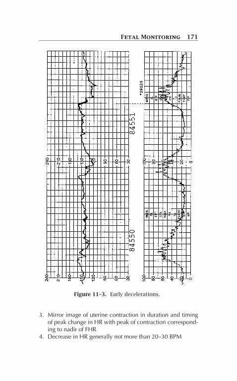
Characteristics of early decelerations (Fig. 11-3) are asfollows:1. Symmetrical, U-shaped deceleration2. Gradual onset and slow return to baseline (>30 s onset to
nadir of FHR)
Fetal Monitoring 171
Figure 11–3. Early decelerations.
3. Mirror image of uterine contraction in duration and timingof peak change in HR with peak of contraction correspond-ing to nadir of FHR
4. Decrease in HR generally not more than 20–30 BPM

172 Fetal Monitoring
Figure 11–4. Mechanism of early decelerations (FHR = fetalheart rate). (Adapted from Freeman.17)
Two mechanisms for early deceleration that have been sug-gested are (1) fetal head compression with increased intracra-nial pressure (Fig. 11-4) and (2) increased volume of bloodentering the fetal circulation during contractions, thus trigger-ing baroreceptor reflex activity. Both of these mechanisms arevagally mediated and can be prevented by atropine.12
Variable Decelerations
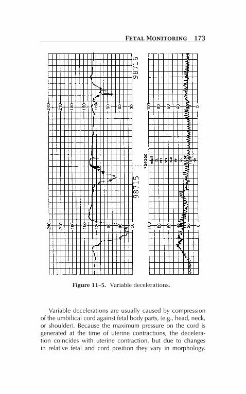
This is the most common of all fetal heart rate patterns(Fig. 11-5), and the characteristics of this pattern are as fol-lows:8
1. Abrupt onset and return to baseline (<30 s)2. Decrease ≥15 BPM, duration ≥15 s, duration <2 min3. Variability in duration, shape, size, and timing relative to
successive contractions4. May be accompanied by brief accelerations (“shoulders”)
before and after departure from baseline
Fetal Monitoring 173
Figure 11–5. Variable decelerations.
Variable decelerations are usually caused by compressionof the umbilical cord against fetal body parts, (e.g., head, neck,or shoulder). Because the maximum pressure on the cord isgenerated at the time of uterine contractions, the decelera-tion coincides with uterine contraction, but due to changesin relative fetal and cord position they vary in morphology.

174 Fetal Monitoring
They are likely mediated by vagal output. Depending upon themagnitude of the decrease in the fetal heart rate, variable decel-erations have been further subdivided into (1) mild (durationless than 30 s and deceleration not below 80 BPM), (2) mod-erate (regardless of the duration, the fetal heart rate is less than80 BPM), and (3) severe (duration greater than 60 s and a fetalheart rate less than 70 BPM). However, these classificationshave not been tested for predictive validity.8
Late Decelerations
Late decelerations (Fig. 11-6) are characterized by thefollowing:1. Symmetrical gradual onset and return to baseline (≥30 s
from onset to nadir).2. Onset and nadir lag beginning and peak of uterine contrac-
tion (typically ≥30 s)3. Return to baseline after the end of the associated uterine
contraction4. Decrease in FHR is usually mild (10–20 BPM) and rarely
more than 30–40 BPM.There is some correlation between the frequency of late
decelerations and the degree of fetal hypoxia. Moderatevariability in the setting of late decelerations is somewhatless concerning than diminished or absent variability withthe same decelerations.8 The major cause of late decelera-tions is reduced placental perfusion, as can be seen duringhypotension (e.g., following regional analgesia), aortocavalcompression, placental abruption, maternal diabetes mellitus,preeclampsia, and post-dates pregnancy.
Besides these three main patterns, the other patterns thathave been described are prolonged decelerations and asinusoidal pattern. Prolonged decelerations are decreases inHR ≥15 BPM which last≥2 min but <10 min. Longer decelera-tions are considered baseline changes. Prolonged decelerationsare considered concerning but not as ominous as recurrentlate or variable decelerations with absent variability. A sinu-soidal pattern is associated with a sine-wave pattern above andbelow the baseline with a frequency of 3–5 per min. A benign

Fetal Monitoring 175
Figure 11–6. Late decelerations. (Adapted from Martin.18)
sinusoidal pattern has been associated with opioid agonist–antagonist drugs (e.g., butorphanol). A major obstetric cause issevere fetal anemia usually associated with Rh incompatibility.In this setting, the sinusoidal pattern is considered ominous.8

176 Fetal Monitoring
Other Modalities
Fetal Scalp Blood Sampling
Fetal scalp blood pH determination is a direct way to testfor the presence of acidemia. A sample of capillary blood isobtained by a small incision in the fetal scalp and analyzedfor pH, typically in duplicate or triplicate. Normal fetal scalppH varies between 7.25 and 7.32; mild acidemia is docu-mented when the pH varies between 7.20 and 7.24; and severeacidemia is noted when the pH is less than 7.20. Unfortunately,the sensitivity and specificity of the test in predicting umbilicalblood pH at delivery is modest.13 Furthermore, scalp bloodpH laboratories are expensive and volume is not generally highenough to justify the cost for the vast majority of labor units.Thus the popularity of the technique has sharply declined overthe last decade.14
Fetal Pulse Oximetry
In theory, continuous measurement of fetal oxygenationshould provide minute-by-minute information of fetal wellbeing, because ultimately poor oxygenation is the cause ofmost fetal compromise during labor. A modified pulse oximeteruses a reflectance probe, generally placed alongside the fetalcheek or temple. Unfortunately, the technique has not provento reduce interventions for presumed fetal compromise, includ-ing cesarean delivery, nor has its use improved fetal conditionat birth.15 ACOG does not endorse its use at this time.16
Implications for Anesthesia Care
The anesthesiologist should be aware of fetal assessment forseveral reasons. First, the timing and urgency of delivery areoften dictated by the results of fetal monitoring. Awareness ofa deteriorating FHR pattern or poor biophysical profile canalert the anesthesiologist to prepare for urgent delivery, or toencourage a parturient and the obstetric caregivers to initi-ate regional anesthesia in anticipation of emergency delivery.

Fetal Monitoring 177
Second, decelerations and other nonreassuring FHR tracingsmay require obstetrical interventions for ultimate resolutionbut the anesthesiologist may be called on to seek and rem-edy reversible causes. This may include proper positioningto avoid aortocaval compression, use of supplemental oxy-gen, correction of hypotension, intravenous fluid bolus, andrarely maneuvers to reduce uterine tone (e.g., intravenousnitroglycerin). Finally, enhanced communication between theobstetrical, nursing, and anesthesiology providers is facilitatedby a common understanding of the factors influencing theformation of the obstetrical care plan.
References
1. Manning FA, Morrison I, Harman CR, Lange IR, MenticoglouS. Fetal assessment based on fetal biophysical profile scor-ing: experience in 19,221 referred high-risk pregnancies. II. Ananalysis of false-negative fetal deaths. Am J Obstet Gynecol.1987;157:880–884.
2. Manning FA, Harman CR, Morrison I, Menticoglou S. Fetal assess-ment based on fetal biophysical profile scoring. III. Positivepredictive accuracy of the very abnormal test (biophysical profilescore = 0). Am J Obstet Gynecol. 1990;162:398–402.
3. Freeman RK, Anderson G, Dorchester W. A prospective multi-institutional study of antepartum fetal heart rate monitoring. I. Riskof perinatal mortality and morbidity according to antepartum fetalheart rate test results. Am J Obstet Gynecol. 1982;143:771–777.
4. Neilson JP, Alfirevic Z. Doppler ultrasound for fetal assess-ment in high risk pregnancies. Cochrane Database Syst Rev.2000;2:CD000073.
5. Tanasijevic MJ, Winkelman JW, Wybenga DR, Richardson DK,Greene MF. Prediction of fetal lung maturity in infants of diabeticmothers using the FLM S/A and disaturated phosphatidylcholinetests. Am J Clin Pathol. 1996;105:17–22.
6. Reynolds SR, Heard OO, Bruns P. Recording uterine contractionpatterns in pregnant women: application of the strain gage in amultichannel tokodynamometer. Science. 1947;106:427–428.
7. Chua S, Kurup A, Arulkumaran S, Ratnam SS. Augmentation oflabor: does internal tocography result in better obstetric outcomethan external tocography? Obstet Gynecol. 1990;76:164–167.

178 Fetal Monitoring
8. Macones GA, Hankins GD, Spong CY, Hauth J, Moore T. The2008 National Institute of Child Health and Human Developmentworkshop report on electronic fetal monitoring: update on defi-nitions, interpretation, and research guidelines. Obstet Gynecol.2008;112:661–666.
9. Thacker SB, Stroup D, Chang M. Continuous electronic heart ratemonitoring for fetal assessment during labor. Cochrane DatabaseSyst Rev. 2001;2:CD000063.
10. Paul RH, Suidan AK, Yeh S, Schifrin BS, Hon EH. Clinicalfetal monitoring. VII. The evaluation and significance ofintrapartum baseline FHR variability. Am J Obstet Gynecol.1975;123:206–210.
11. Liu PL, Warren TM, Ostheimer GW, Weiss JB, Liu LM. Foetal mon-itoring in parturients undergoing surgery unrelated to pregnancy.Can Anaesth Soc J. 1985;32:525–532.
12. Freeman RK, Garite TJ, Neageotte MP, eds. Fetal Heart RateMonitoring. 3rd ed. Philadelphia: Lippincott, Williams andWilkins; 2003.
13. Kruger K, Hallberg B, Blennow M, Kublickas M, Westgren M.Predictive value of fetal scalp blood lactate concentration andpH as markers of neurologic disability. Am J Obstet Gynecol.1999;181:1072–1078.
14. Perkins RP. Requiem for a heavyweight: the demise of scalp bloodpH sampling. J Matern Fetal Med. 1997;6:298–300.
15. Bloom SL, Spong CY, Thom E, et al. Fetal pulse oximetry andcesarean delivery. N Engl J Med. 2006;355:2195–2202.
16. ACOG Committee Opinion. Number 258, September 2001. Fetalpulse oximetry. Obstet Gynecol. 2001;98:523–524.
17. Freeman RK, Garite TS. Fetal Heart Rate Monitoring. Baltimore:Williams & Wilkins; 1981.
18. Martin R. Prepartum and intrapartum fetal monitoring. In: DattaS, ed. Anesthetic and Obstetric Management of High RiskPregnancy. Chicago: Mosby-Year Book; 1991.