Embed Size (px)
Citation preview
30 Churchill Place ● Canary Wharf ● London E14 5EU ● United Kingdom
An agency of the European Union
Telephone +44 (0)20 3660 6000 Facsimile +44 (0)20 3660 5520
Send a question via our website www.ema.europa.eu/contact
17 December 2015 EMA/14567/2016 Committee for Medicinal Products for Human Use (CHMP)
Assessment report
Feraccru
International non-proprietary name: ferric maltol
Procedure No. EMEA/H/C/002733/0000
Note
Assessment report as adopted by the CHMP with all information of a commercially confidential nature deleted

Assessment report
EMA/14567/2016 Page 2/105
Table of contents
1. Background information on the procedure .............................................. 5
1.1. Submission of the dossier ..................................................................................... 5
1.2. Steps taken for the assessment of the product ........................................................ 6
2. Scientific discussion ................................................................................ 7
2.1. Introduction ........................................................................................................ 7
2.2. Quality aspects .................................................................................................... 8
2.2.1. Introduction...................................................................................................... 8
2.2.2. Active Substance ............................................................................................... 9
2.2.3. Finished Medicinal Product ................................................................................ 10
2.2.4. Discussion on chemical, pharmaceutical and biological aspects.............................. 13
2.2.5. Conclusions on the chemical, pharmaceutical and biological aspects ...................... 13
2.2.6. Recommendation(s) for future quality development ............................................. 13
2.3. Non-clinical aspects ............................................................................................ 13
2.3.1. Introduction.................................................................................................... 13
2.3.2. Pharmacology ................................................................................................. 14
2.3.3. Pharmacokinetics ............................................................................................ 16
2.3.4. Toxicology ...................................................................................................... 23
2.3.5. Ecotoxicity/environmental risk assessment ......................................................... 36
2.3.6. Discussion on non-clinical aspects ..................................................................... 37
2.3.7. Conclusion on the non-clinical aspects ............................................................... 39
2.4. Clinical aspects .................................................................................................. 40
2.4.1. Introduction.................................................................................................... 40
2.4.2. Pharmacokinetics ............................................................................................ 40
2.4.1. Pharmacodynamics .......................................................................................... 50
2.4.2. Discussion on clinical pharmacology ................................................................... 51
2.4.3. Conclusions on clinical pharmacology ................................................................. 54
2.5. Clinical efficacy .................................................................................................. 54
2.5.1. Dose response study(ies) ................................................................................. 54
2.5.2. Main study(ies) ............................................................................................... 54
2.5.3. Discussion on clinical efficacy ............................................................................ 78
2.5.4. Conclusions on the clinical efficacy .................................................................... 81
2.6. Clinical safety .................................................................................................... 81
2.6.1. Discussion on clinical safety .............................................................................. 91
2.6.2. Conclusions on the clinical safety ...................................................................... 93
2.7. Risk Management Plan ........................................................................................ 93
2.8. Pharmacovigilance ........................................................................................... 100
2.9. Product information .......................................................................................... 101
2.9.1. User consultation .......................................................................................... 101

Assessment report
EMA/14567/2016 Page 3/105
3. Benefit-Risk Balance ........................................................................... 101
4. Recommendations ............................................................................... 104

Assessment report
EMA/14567/2016 Page 4/105
List of abbreviations
ACVA: 4,4’-azobis(4-cyanopentanoic acid)
AE: adverse effect
API: Active Pharmaceutical Ingredient
CD: Crohn’s disease
CE: Capillary electrophoresis
DP: Drug product
DS: Drug substance
GC: Gas chromatography
GI: gastrointestinal
IBD: inflammatory bowel disease
ICP-OES: Inductively coupled plasma - optical emission spectrometry
IDA: iron deficiency anaemia
IPCs: In-process controls
KF: Karl Fischer
LoD: Detection limit
LoQ: Quantitation limit
MAA: Marketing Authorisation Application
MAA: marketing authorisation application
NTA: ferric nitriloacetic acid
OFPs: oral ferrous products
PDE: Permissible daily exposure
Ph. Eur.: European Pharmacopoeia
PSD: Particle Size Distribution
PSD: Particle Size Distribution
ROS: reactive oxygen species
SEM: Scanning electron microscopy
THF: Tetrahydrofuran
UC: Ulcetrative colitis

Assessment report
EMA/14567/2016 Page 5/105
1. Background information on the procedure
1.1. Submission of the dossier
The applicant Iron Therapeutics (UK) Ltd submitted on 1 December 2014 an application for Marketing
Authorisation to the European Medicines Agency (EMA) for Feraccru, through the centralised procedure under
Article 3 (2) (b) of Regulation (EC) No 726/2004. The eligibility to the centralised procedure was agreed upon
by the EMA/CHMP on 24 May 2012. The eligibility to the centralised procedure under Article 3(2)(b) of
Regulation (EC) No 726/2004 was based on demonstration of interest of patients at Community level.
The applicant applied for the following indication: in adults for the treatment of iron deficiency anaemia:
in patients who cannot tolerate other oral iron preparations or are non-compliant;
in inflammatory bowel disease where other oral iron preparations are ineffective;
in patients in whom treatment with intravenous iron is unsafe or not possible.
The legal basis for this application refers to:
Article 8.3 of Directive 2001/83/EC - complete and independent application. The applicant indicated that
ferric maltol was considered to be a new active substance.
The application submitted is composed of administrative information, complete quality data, non-clinical and
clinical data based on applicants’ own tests and studies and/or bibliographic literature substituting/supporting
certain tests or studies.
Information on Paediatric requirements
Pursuant to Article 7 of Regulation (EC) No 1901/2006, the application included an EMA Decision(s)
P/0228/2013 on the agreement of a paediatric investigation plan (PIP).
At the time of submission of the application, the PIP P/0228/2013 was not yet completed as some measures
were deferred.
Information relating to orphan market exclusivity
Similarity
Pursuant to Article 8 of Regulation (EC) No. 141/2000 and Article 3 of Commission Regulation (EC) No
847/2000, the applicant did not submit a critical report addressing the possible similarity with authorised
orphan medicinal products because there is no authorised orphan medicinal product for a condition related to
the proposed indication.
New active Substance status
The applicant requested the active substance ferric maltol contained in the above medicinal product to be

Assessment report
EMA/14567/2016 Page 6/105
considered as a new active substance in comparison to known ferrous active substances previously
authorised in the Union (e.g. ferrous fumarate, ferrous gluconate, ferrous sulphate) and claimed that ferric
maltol differs significantly in properties with regard to safety and efficacy from the already authorised
substances.
Scientific Advice
The applicant did not seek scientific advice at the CHMP.
Licensing status
The product was not licensed in any country at the time of submission of the application.
1.2. Steps taken for the assessment of the product
The Rapporteur and Co-Rapporteur appointed by the CHMP were:
Rapporteur: Concepcion Prieto Yerro Co-Rapporteur: Harald Enzmann
• The application was received by the EMA on 1 December 2014.
• The procedure started on 24 December 2014.
• The Rapporteur's first Assessment Report was circulated to all CHMP members on 16 March 2015. The
Co-Rapporteur's first Assessment Report was circulated to all CHMP members on 16 March 2015.
• During the meeting on 23 April 2015, the CHMP agreed on the consolidated List of Questions to be sent
to the applicant. The final consolidated List of Questions was sent to the applicant on 24 April 2015.
• The applicant submitted the responses to the CHMP consolidated List of Questions on 23 July 2015.
• The Rapporteurs circulated the Joint Assessment Report on the applicant’s responses to the List of
Questions to all CHMP members on 31 August 2015.
• During the CHMP meeting on 24 September 2015, the CHMP agreed on a list of outstanding issues to be
addressed in writing and/or in an oral explanation by the applicant.
• The applicant submitted the responses to the CHMP List of Outstanding Issues on 20 October 2015.
• During the CHMP meeting on 17 November 2015, outstanding issues were addressed by the applicant
during an oral explanation before the CHMP.
• During the meeting on 17 December 2015, the CHMP, in the light of the overall data submitted and the
scientific discussion within the Committee, issued a positive opinion for granting a Marketing
Authorisation to Feraccru.

Assessment report
EMA/14567/2016 Page 7/105
2. Scientific discussion
2.1. Introduction
Iron deficiency anaemia (IDA) occurs when iron levels are insufficient to support red blood cell production
and is defined – according to the WHO - as haemoglobin levels below 13 g/dL in men over 15 years, below
12 g/dL in non-pregnant women over 15 years, and below 11 g/dL in pregnant women.
Iron is absorbed at the apical surface of enterocytes to be transported by ferroportin, the only known iron
exporter, across the basolateral surface of the enterocyte into circulation. Inflammation from IBD interferes
with iron absorption by causing an increase in hepcidin, a peptide hormone synthesized in the liver that
inhibits ferroportin activity. Anaemia is the most common extra-intestinal complication of inflammatory bowel
disease (IBD) and although it often involves a combination of IDA and anaemia of chronic disease, IDA
remains an important contributor in this condition due to chronic intestinal bleeding and decreased iron
intake (from avoidance of foods that may exacerbate symptoms of IBD). In a variety of populations with IBD,
the prevalence of iron deficiency anaemia ranges from 36%-76%.
The serum markers of iron deficiency are low ferritin, low iron, raised total iron binding capacity, raised red
cell protoporhyrin and increased transferrin binding receptor (sTfR). Serum ferritin is the most powerful test
for iron deficiency. The cut-off level of ferritin which is diagnostic varies between 12-15 µg/L. Higher levels of
serum ferritin do not exclude the possibility of iron deficiency, and a serum ferritin level of <100 μg/L may
still be consistent with iron deficiency in patients with IBD. A transferrin saturation of <16% is indicative of
iron deficiency, either absolute or functional. Other findings on a complete blood count panel that are
suggestive of iron deficiency anaemia, but are not considered diagnostic, include microcytosis, hypochromia,
and elevation of red cell distribution width.
A deficiency of iron can have a significant impact on a patient’s quality of life. Appropriate diagnosis and
treatment of iron deficiency anaemia are important to improve or maintain the quality of life of patients.
The goals of treatment are to treat the underlying cause, limit further blood loss or malabsorption, avoid
blood transfusions in haemodynamically stable patients, relieve symptoms, and improve quality of life. More
specifically, therapeutic goals of treatment include normalizing haemoglobin levels within 4 weeks (or
achieving an increase of >2 g/dL) and replenishing iron stores (transferrin saturation >30%). Oral iron
supplementation has been considered standard treatment because of an established safety profile, lower
cost, and ease of administration. It has been shown to be effective in correcting anaemia and repleting iron
stores. One concern with higher doses of daily oral iron is intolerance due to GI side effects. Symptoms
include nausea, vomiting, diarrhea, abdominal pain, constipation, and melena-like stools.
Guidelines on the Diagnosis and Management of Iron Deficiency and Anaemia in Inflammatory Bowel
Diseases recommend IV iron therapy over oral iron supplementation in the treatment of iron deficiency
anaemia in patients with IBD, citing faster and prolonged response to treatment, decreased irritation of
existing GI inflammation, improved patient tolerance, and improved quality of life. Patients with severe
anaemia (haemoglobin level of <10 g/dL), failure to respond or intolerance to oral iron therapy, severe
intestinal disease or patients receiving concomitant erythropoietin are recommended indications for IV iron
therapy. Other conditions where patients should be considered for first-line IV therapy over oral therapy
include congestive heart failure, upper GI bleeding, and in situations where rapid correction of anaemia may
be required.

Assessment report
EMA/14567/2016 Page 8/105
Across EU there are several iron (Fe+2) oral preparations as ferrous fumarate, ferrous gluconate, ferrous
sulphate and ferrous glycine sulfate, formulated as tablet, solution or gastroresistent capsules. All ferrous
compounds are oxidised in the lumen of the gut or within the mucosa with release of activated hydroxyl
radicals, which may attack the gut wall and can effect a range of gastrointestinal symptoms and discomfort.
Ferric preparations also exist but with less bioavailability. Across EU there are also several IV products on the
market: iron (III) hydroxide dextran complex, iron sucrose, ferric carboxymaltose, iron isomaltoside. IV iron
therapy, however, is inconvenient, invasive and associated with the risk of rare but serious hypersensitivity-
reactions; it is used in those situations when oral preparations cannot be used or when there is a need to
deliver iron rapidly.
Feraccru is a trivalent iron, oral iron replacement preparation. The active substance of Feraccru is ferric
maltol (also known as 3-hydroxy-2-methyl-4H-pyrane-4-one iron (III) complex, or ST10, or ferric trimaltol
or ferric maltol) an oral ferric iron/maltol complex. It is presented as red hard gelatine capsules containing
30 mg iron (ferric iron).
Maltol is a sugar derivative that strongly chelates iron in the ferric form (FeIII) rendering the iron stable and
available for absorption. Upon dissociation of the ferric maltol complex, the maltol molecules are absorbed
and glucuronidated in the intestinal wall, and within the liver during first pass metabolism, and subsequently
eliminated from the body in the urine. The iron is absorbed via the endogenous dietary iron uptake system.
The indication finally agreed with the CHMP was: Feraccru is indicated in adults for the treatment of iron
deficiency anaemia (IDA) in patients with inflammatory bowel disease (IBD) (see section 5.1).
The proposed dosage is one 30 mg capsule twice daily on an empty stomach, corresponding to 60 mg ferric
iron per day.
There was agreement in the paediatric investigation plan to grant a deferral and a waiver for iron as iron
(III)-maltol complex (EMEA-001195-PIP01-11).The PDCO granted a waiver in infants under 6 months of age
and a referral for the completion of the planned paediatric studies (ST10-021 PK-PED/ST10-01-102, an open
label, randomised, multiple-dose, parallel PK study and ST10-01-303, a randomised, open label comparative
safety and efficacy study of ST10 and oral ferrous sulphate as comparator) until the adult studies are
completed.
2.2. Quality aspects
2.2.1. Introduction
The finished product is presented as hard capsules containing 30 mg of iron (III) (as ferric maltol) as active
substance.
Other ingredients are:
Capsule contents: lactose monohydrate, sodium lauril sulphate, magnesium stearate, colloidal anhydrous
silica, and crospovidone (Type A) Capsule shell: gelatin, brilliant blue (E133), allura red (E129), titanium dioxide (E171), and sunset yellow FCF (E110).
The product is available in HDPE bottles with plastic closure.

Assessment report
EMA/14567/2016 Page 9/105
2.2.2. Active Substance
General information
The chemical name of ferric maltol is 3-hydroxy-2-methyl-4H-pyrane-4-one iron (III) complex (3:1) and has
the following structure:
Scanning electron microscopy (SEM) confirmed that ferric maltol powder is polycrystalline in character i.e. it
is a material composed of aggregates of individual crystalsStandard physical techniques were used to
elucidate and confirm the structure of ferric maltol were IR, MS, UV/VIS, ESR, elemental analysis, XRD, DSC
and TGA, GVS, and NMR.
Theoretically the active substance may exist as a mixture of four isomers, two cis and two trans.
Enantiomeric purity is controlled routinely by chiral HPLC/specific optical rotation. Polymorphism has been
observed for ferric maltol. Two different crystalline forms can be isolated under aqueous synthetic conditions
used to manufacture ferric maltol: Form A and Form C. Consistent formation of Form C within the
manufacturing process is ensured during the manufacturing process, and it is controlled by XRD analysis.
Manufacture, characterisation and process controls
Ferric maltol is synthesized from iron salts and maltolin seven main steps: dissolution of ferric citrate in
purified water, mixing of maltol with a sodium hydroxide solution , filtration of the sodium maltol solution,
addition of the ferric citrate solution to the sodium maltol solution under controlled temperature to and
crystallisation, drying and milling using well defined starting materials with acceptable specifications.
Adequate in-process controls (IPCs) are applied during the synthesis. The specifications and control methods
for intermediate products, starting materials and reagents have been presented.
The characterisation of the active substance and its impurities are in accordance with the EU guideline on
chemistry of new active substances. Potential and actual impurities were well discussed with regards to their
origin and characterised.
The active substance is packed in double polyethylene lined bags in fibre drums

Assessment report
EMA/14567/2016 Page 10/105
Specification
The active substance specification includes tests for appearance, identification (UV, IR, Iron), pH (Ph Eur),
water content (KF), ferrous II iron content (redox titration), iron (III) content (HPLC), maltol content
(HPLC), assay (UV), related substances (HPLC), elemental impurities (ICP-OES), particle size distribution (Ph
Eur), polymorphic form (XRD) and microbial quality (Ph. Eur.).
The analytical methods used have been adequately described and (non-compendial methods) appropriately
validated in accordance with the ICH guidelines.
Batch analysis data of eleven batches (five pilot scale batches and six commercial scale batches) of the active
substance are provided. The results are within the specifications and consistent from batch to batch.
Stability
Stability data on six pilot scale batches of active substance from the proposed manufacturer stored in the
intended commercial package for 36 months under long term conditions at 25 ºC / 60% RH and for up to 6
months under accelerated conditions at 40 ºC / 75% RH, according to the ICH guidelines, were provided. A
forced degradation study under the following degradation conditions: acid (0.1N HCl), base (0.1N NaOH),
hydrogen peroxide (1%), 4,4’-azobis(4-cyanopentanoic acid) (ACVA) (0.01M), and heat (105°C) was also
performed.
The following parameters were tested: appearance, pH, water content (KF), iron content (HPLC), maltol
content (HPLC), assay ferric maltol (UV), and related substances (HPLC).
No significant changes or trends were observed for any of the parameters tested in the study. The active
substance is very stable both under accelerated and long-term conditions. All data are in compliance with the
proposed specification.
Forced degradation studies indicate that solid ferric maltol is very stable to heat and light. Ferric maltol in
solution is slightly labile to acid and base, whilst being highly labile to oxidative stress.
The stability results indicate that the active substance manufactured by the proposed suppliers is sufficiently
stable. The stability results justify the proposed retest period in the proposed container.
2.2.3. Finished Medicinal Product
Description of the product and Pharmaceutical development
Feraccru capsules are conventional pharmaceutical grade hard gelatin capsules containing 30 mg of ferric
iron, in the form of ferric maltol in a conventional excipient base, comprising pharmacopoeial grade lactose,
sodium laurilsulfate, crospovidone, colloidal silica and magnesium stearate.Considering the dosage form
(hard capsules) and the manufacturing process (conventional standard dry blending and capsule filling), the
dissolution of the finished product is almost exclusively dependent of the properties of the active substance.
As indicated in the active substance section, two polymorphic forms of the active substance were identified
during development. It was demonstrated that the polymorphic form has no impact on the quality of the
finished product; in particular, finished product formulated using the two different forms had virtually
identical dissolution profiles. Furthermore data that confirm the stability of three pilot batches of finished

Assessment report
EMA/14567/2016 Page 11/105
product manufactured from Form C after 6 months stored under accelerated and long term conditions was
provided. These results are comparable to the results obtained for batches of drug product manufactured
using Form A.
Data were provided demonstrating that the dissolution method was able to differentiate batches
manufactured with active substances having different PSD. As indicated above, the particle size distribution is
controlled in the active substance specification.
The disintegration time, content uniformity and dissolution rate were investigated to show that the
formulation developed is in line with the requirements of the Ph. Eur. monograph on hard capsules.
Eleven batches of different scales of the active substance have been manufactured up to date.
All excipients are well known pharmaceutical ingredients and their quality is compliant with Ph. Eur standards
with the exception of colouring agents. The colouring agents are in compliance with the EU Directive
2008/128/EC. There are no novel excipients used in the finished product formulation. The list of excipients is
included in section 6.1 of the SmPC. The excipients were selected to provide the functionality, and each has
been used in approved, oral capsule formulations. All excipients are presented at levels well below the
maximum used in oral solid dosage forms.
The proposed commercial formulation was used in the pivotal Phase 1 and Phase 3 trials. The formulation
development and scale-up programme was built on what was known about the previous clinical presentations
used in the pharmacokinetic and efficacy/safety studies. Initial scale-up formulation investigations focused on
determining if a powder blend would be suitable or if granulation would be needed. The studies conducted
showed that the active substance was suitable for encapsulation in a powder blend and did not require
granulation. The proposed commercial manufacturing process is identical to that used to produce the Phase 3
clinical batches.
The applicant initially applied for two primary packages: high density polyethylene bottles and
polyvinylchloride (PVC)/Aluminium (Alu) blisters. However, during the assessment, the applicant withdrew
the primary packaging polyvinylchloride (PVC)/Aluminium (Alu) blisters due to stability issues.
As a result, the finished product is packed in high density polyethylene bottles with child-proof white
polypropylene push-lock closures. The bottle and cap specifications were provided. The containers comply
with EU Directives 2002/72/EC, 1935/2004/EC, 2023/2006/EC and their subsequent amendments regulating
products that come in contact with pharmaceuticals and foods and with USA FDA Regulation CFR21.177.1520
for olefin polymers intended to come into contact with food.
Manufacture of the product and process controls
Feraccru capsules are manufactured using a simple and conventional standard dry blending and capsule
filling process. The manufacturing process consists of seven main steps: dispensing ingredients in a blender,
blending, adding magnesium stearate, blending, capsules filling, collecting capsules and packaging. The
active substance and excipients are all sieved prior to blending to ensure blend consistency. The process is
considered to be a standard manufacturing process.
Major steps of the manufacturing process have been validated by a number of studiesIt has been
demonstrated that the manufacturing process is capable of producing the finished product of intended quality
in a reproducible manner. The in-process controls are adequate for this type of manufacturing process.

Assessment report
EMA/14567/2016 Page 12/105
Product specification
The finished product release specifications include appropriate tests for this kind of dosage form:
appearance/description, identification (UV, Iron), water content (KF), Ferrous (II) Iron Content
(potentiometric titration), Ferric (III) Iron Content (ICP-OES), content uniformity (Ph Eur), dissolution (Ph
Eur), microbial quality (Ph Eur), maltol (HPLC), related substances (HPLC). With regards to the validation of
HPLC method used for the evaluation of related substances, furylethanol has been used to determine the
detection limit at 210 nm instead of 275 nm. The CHMP recommended providing additional validation data
confirming the limits of detection and quantification of the related substances HPLC assay Method 3, using a
suitable reference standard as maltol. The applicant committed to submit it to the Regulatory Authorities
before the product is placed on the marketBatch analysis results are provided for seven production and one
pilot scale batches confirming the consistency of the manufacturing process and its ability to manufacture to
the intended product specification. The CHMP recommended providing certificates of analysis, including HPLC-
chromatograms and spectra, for the 3 commercial scale validation batches.
Stability of the product
Stability data of three pilot scale batches of finished product stored under long term conditions for 12
months at 25 ºC / 60% RH and 30 ºC / 75% RH and for up to 6 months under accelerated conditions at 40
ºC / 75% RH according to the ICH guidelines were provided. The batches of the medicinal product are
identical to those proposed for marketing and were packed in the primary packaging proposed for marketing.
Samples were tested for appearance , water content (KF), dissolution (Ph Eur), microbial quality, ferrous (II)
iron content, ferric (III) iron content, maltol (HPLC) and related substances (HPLC) . The analytical
procedures used are stability indicating.
There were no changes in appearance or other parameters tested; the assay results, maltol content and
dissolution remained consistent. Related substances were below the limit of detection at all-time points.
Moreover, supportive stability data for 36 months, from two representative clinical batches stored in the
same primary container system stored under the same ICH long-term conditions mentioned above were
provided. The results indicate that the product is very stable. There were no changes in appearance or other
parameter tested. Related substances were below the level of detection.
In addition, 12 month long-term stability data have been provided on three pilot scale batches, which were
placed on stability in the initially proposed blister packaging (PVC/foil blisters). The stability data generated
with regard to dissolution suggest that this packaging material is not considered appropriate for Feraccru
capsules
An in-use stability study was conducted for 45 days on two pilot scale batches. Bottles were opened twice
each working day for a minimum of one minute. Capsules were removed for testing on days 0, 14, 28 and
45. There were no changes in appearance or other parameters tested. Related substances were below the
limit of detection and no microbial growth was shown.
The CHMP recommended conducting stability tests on bulk regarding specified bulk storage conditions.
In conclusion, based on available stability data, the shelf-life of 15 months stored below 25ºC and an in-use
shelf life of 45 days after first opening container as stated sections 6.3 and 6.4 of the SmPC are acceptable.

Assessment report
EMA/14567/2016 Page 13/105
Adventitious agents
Lactose is manufactured from cow’s milk sourced from healthy animals in the same conditions as milk for
human consumption and rennet used for the production of whey in accordance with Public Statement
EMEA/CPMP/571/02 and Note for Guidance EMEA/410/01 rev.3.
Gelatine obtained from bovine sources is used in the product. Valid TSE CEP from the suppliers of the
gelatine used in the manufacture is provided.
2.2.4. Discussion on chemical, pharmaceutical and biological aspects
Feraccru is a chemically stable complex (chelate) of ferric iron and maltol. Information on development,
manufacture and control of the active substance and finished product has been presented in a satisfactory
manner. The results of tests carried out indicate consistency and uniformity of important product quality
characteristics, and these in turn lead to the conclusion that the product should have a satisfactory and
uniform performance in clinical use.
At the time of the CHMP opinion, there were minor unresolved quality issues having no impact on the
Benefit/Risk ratio of the product.
2.2.5. Conclusions on the chemical, pharmaceutical and biological aspects
The quality of this product is considered to be acceptable when used in accordance with the conditions
defined in the SmPC. Physicochemical and biological aspects relevant to the uniform clinical performance of
the product have been investigated and are controlled in a satisfactory way. Data has been presented to give
reassurance on viral/TSE safety.
2.2.6. Recommendation(s) for future quality development
In the context of the obligation of the MAHs to take due account of technical and scientific progress, the
CHMP recommends the following points for investigation:
- To provide additional validation data confirming the limits of detection and quantification of the related
substances HPLC assay Method 3, using a suitable reference standard as maltol. The applicant committed to
submit it to the Regulatory Authorities before the product is placed on the market.
- To conduct stability tests on bulk regarding specified bulk storage conditions
- To provide certificates of analysis, including HPLC-chromatograms and spectra, for the 3 commercial scale
validation batches.
2.3. Non-clinical aspects
2.3.1. Introduction
The pharmacology of the present application is based entirely on data published in the scientific literature.
Assessment report
EMA/14567/2016 Page 14/105
ST10 is a chelate complex of maltol and iron. Given that both parts of the complex are only systemically
available as separate entities and do not occur in systemic circulation as a complex, existing nonclinical data
for the individual components of the complex were bridged. Bridging studies were conducted with ST10 (14
day non-GLP study in rats, 28 day sub-chronic GLP study in dogs and in vitro Ames test).
2.3.2. Pharmacology
Primary pharmacodynamic studies
ST10 is a soluble, chemically stable complex of ferric iron and maltol acting orally as an iron delivery system
to duodenal enterocytes. Iron enters the duodenal enterocytes by a saturable process. The iron in the cell is
bound to transferrin and ferritin and the maltol dissociates from the ST10 complex, which is absorbed,
glucuronidated and/or sulphated and excreted in the urine. The transferrin-bound iron is subsequently
absorbed into the bloodstream (Barrand et al, 1991; Barrand and Callingham, 1991). Therefore, once in the
enterocyte, iron and ligand will be able to enter the tissues by separate pathways. ST10 is not absorbed
intact into the systemic circulation.
ST10 - In vitro
The absorption of radiolabeled iron from ST10 and from ferric nitrilotriacetic acid (NTA) by isolated fragments
of duodenum and ileum from iron deficient and iron replete rats was investigated (Callingham 1987). The
absorption of iron from ST10 by the duodenum in intestinal fragments of iron deficient rats was 6.94 ± 0.49
pmol/min/mg wet tissue and in intestinal fragments of iron replete rats was similar at 7.53 ± 0.84
pmol/min/mg wet tissue, while in the ileum in iron deficient rats absorption was 4.38 ± 0.86 pmol/min/mg
wet tissue and in iron replete rats was much higher at 7.77 ± 081 pmol/min/mg wet tissue. The absorption of
iron from ferric nitrilotriacetic acid was generally much lower and ranged between 0.5 and 2.0 pmol/min/mg
wet tissues, while in iron deficient rat duodenal fragments the absorption was higher than in duodenal
fragments from iron replete animals. The intestinal iron uptake from ST10 is strictly regulated and saturable.
No increase of iron absorption after ST10 administration following bile salt treatment of the intestine was
seen, indicating no relevant diffusion through the intestinal wall.
ST10 - In vivo
The effect of mucosal damage on intestinal absorption of iron from ST10 was investigated in rats (Barrand et
al, 1991). The bile salt, chenodeoxycholate, was administered by intestinal perfusion at a concentration of
5mM to induce structural damage to the GI tract, confirmed on electron micrograph to be similar to that seen
with high doses of ferrous sulphate (Nayfield 1976). After perfusion of the intestine with bile salt for 45
minutes, rats were administered oral doses 7mg of elemental iron via either ferric maltol (FeCl3 and maltol
powder at a molar ratio of 1:4 in sufficient saline to provide 7 mg of elemental iron in a 500μl dose) or FeSO4
containing 5 μCi of 59Fe directly into the intestine. A significant difference was noted in the way the iron was
absorbed from the two compounds - the rate of uptake was slightly increased for ST10 and markedly
increased for ferrous sulphate compared with controls. The uptake from ST10 was still a saturable process
with a small diffusional component; whereas iron uptake from ferrous sulphate was highly enhanced by a
diffusional process, which indicated loss of physiological control. In these studies with bile acid damaged
intestines, iron absorption from ST10 was still subject to regulatory control, while enhanced (potentially
dangerous) absorption of ferrous sulphate was seen.
In a 14 day oral (non-GLP) study in the rat (male Wistar 150-200g), which was carried out to examine the
absorption and distribution of iron in tissues of the GI tract after two weeks of treatment with ST10, the
ultrastructure, enzyme activities, and physiological function of the GI tract through glucose uptake was

Assessment report
EMA/14567/2016 Page 15/105
examined in detail (Barrand et al, 1991). Groups of 6 male Wistar rats were either pre-treated with ST10
(7mg iron twice daily) by gavage for two weeks or were given saline only. On the last day of the study, all
animals received a final oral dose of 7mg radiolabeled 59Fe as ST10. Animals were killed at 2 hours after drug
administration and the 59Fe content of blood, urine, femoral bone marrow, liver, spleen, kidneys and the
unabsorbed contents and washed segments of small intestine was determined. The results are shown in the
table below.
Table 1: 59Fe content in control and ST10-pre-treated rat tissues at 2 hours after oral administration of a final dose of 7mg 59Fe as ST10.
ST10-pre-treated animals absorbed significantly less iron than control animals. The small intestines of test
and control groups were examined for overt signs of cellular damage and biochemical abnormalities. Portions
of duodenum were fixed overnight in 4% glutaraldehyde and processed for electron microscopy. No
differences in the morphology of intestinal epithelium were observed in the treated animals when compared
with controls. There were no obvious signs of damage to the mitochondria or to the microvilli along the brush
border of the epithelium, nor were there any gaps between the cells suggesting loss of intercellular contact
(Barrand 1991a).
Absorption of 59Fe from ST10 or ferrous sulphate was also examined following intraduodenal administration to
rats (Barrand et al, 1991). In this study, the absorption of iron from ST10 was much higher than that from
ferrous sulphate. The authors suggested that these results could be explained on the basis of the lower
bioavailability of the ferrous iron. Precipitates of iron were adherent to the mucosal lining in duodenal
sections exposed to 59Fe ferrous sulphate at the end of the 2h exposure. Since the intestines were tied off,
movement of intestinal contents, including iron, along the gut was prevented, thus accentuating the
difference between ferric maltol which holds iron in a soluble complex and ferrous sulphate from which iron
rapidly precipitates at neutral pHs.
Table 2: Absorption of 59Fe from ST10 or ferrous sulphate
For the pharmacology of the iron component of ST10, the Applicant is relying on the published literature.

Assessment report
EMA/14567/2016 Page 16/105
Iron is essential in the diet in animals and man since it has an irreplaceable role as a catalyst for many intra-
and extra-cellular reactions and is an essential component of the blood. However, iron may exist in
chemically redox reactive forms causing significant tissue and systemic toxicity both in man and other
mammalian species. In order to negate toxicity, iron in the body is bound to high molecular weight transport
and storage proteins; these proteins hold iron in the less reactive ferric form and behave as protective
complexes. In normal health, the bodily content of iron is physiologically regulated through recirculation of
iron from senescent red cells involving phagocytosis with the daily negative balance being corrected through
gastro-intestinal absorption. In iron deficiency anaemia, usually caused by blood loss, the absorption of iron
is up regulated but the forms of iron found in the diet are usually relatively inefficient in correcting the iron
deficit. The normal diet contains many ligands which bind to iron and are either enhancing or inhibitory on
iron uptake (Hazell 1988). The mechanisms which control oral iron uptake are 1) the chemical form of the
iron within the gastrointestinal tract 2) binding and transport mediators in the duodenal enterocytes and 3)
the relative iron saturation and turnover rate of transferrin, a plasma protein involved in the systemic
transport of iron (Nathanson 1984).
The transport of dietary iron from the duodenal absorption site to either bodily storage sites or to sites of
biosynthesis of physiological substances, such as haemoglobin, is accomplished by transferrin which has two
binding sites for iron. The iron receptor recognition on the haemoglobin progenitor cells is the transferrin-
iron complex. This transferrin behaves as a siderophore. The iron saturation of transferrin in the blood
regulates haemopoiesis: below 20% transferrin saturation, apoiesis is markedly impaired with a progression
to iron deficiency anaemia (Bothwell 1979).
The disposition of iron in the body is divided between compounds involved in physiological functions for which
iron is essential (predominantly haemoglobin and myoglobin but also haem enzymes such as cytochromes,
catalase, peroxidase and the metaloflavoprotein enzymes including xanthine oxidase and alpha-
glycerophosphate oxidase) and the iron storage compounds ferritin and haemosiderin, which are located
mainly in the reticulo-endothelial system and in hepatocytes.
Maltol, a simple sugar derivative and dehydration product of glucose, forms a suitable ferric iron (III)
complex for the delivery of iron in the duodenum.
Secondary pharmacodynamic studies
Data on secondary pharmacodynamics were not submitted.
Safety pharmacology programme
No data available from safety pharmacology studies to evaluate the cardiovascular or renal safety of ST10.
Pharmacodynamic drug interactions
Data on pharmacodynamic drug interactions were not submitted.
2.3.3. Pharmacokinetics
Absorption of ST10
ST10 labeled with iron-59 was added to isolated intestinal fragments (Levey 1988) or administered directly
into the stomach or duodenum of anesthetized rats and iron absorption measured by blood sampling at
intervals (Barrand 1987, 1991a & b).

Assessment report
EMA/14567/2016 Page 17/105
The mechanisms involved in the intestinal absorption of iron from ST10 as compared to ferric ethyl maltol
and iron NTA complex were examined (Levey 1988). Isolated pieces of rat small intestine (duodenum and
jejunum) were incubated with the three complexes labelled with radioactive iron in vitro under different
conditions. The uptake of iron into the intestinal tissue was highest from maltol at the lower concentration
and from ethyl maltol at the higher concentration, while the uptake from NTA complex was only about one
tenth of that from either maltol and ethyl maltol complex. When the concentration was increased with a fixed
incubation time of 10 minutes, the iron uptake appeared to be saturable over a range of 10-6 - 10-4 M, while
at higher concentrations a nonsaturable uptake was apparent. The iron uptake from tri-maltol complex was
not sensitive to metabolic inhibition but was sensitive to lowering of temperature. Within the intestinal tissue,
at low concentrations 35-40% of the iron was bound to the iron transport proteins ferritin and transferrin in
the mucosa cells, while the serosa did not contain relevant amounts. The characteristics of iron uptake from
the iron complexes suggest that the ligands donate their iron to the endogenous iron uptake system, and
high ligand concentrations (excess maltol/ ratio iron:maltol 1:10) will compete with the binding to these
proteins. However, at high concentrations of the 1:3 complex (ST10) saturable iron kinetics were still evident
(Rennhard 1971).
The absorption of ST10 (7μg iron radiolabeled) and several other iron salts (sulphate, fumarate, gluconate, or
EDTA complex) was examined in male Wistar rats following intraduodenal administration (Barrand, 1987).
The total amount absorbed was determined at 1, 2, 4, or 6 hours after administration in whole body, bone
marrow, liver, and total blood. The highest absorption was observed after ST10 administration with a total
amount of about 3μg in whole body, 1.8μg in bone marrow, 0.6μg in liver, and 1.8μg in total blood
representing a total absorption of about 43%. The highest blood concentration was observed at one hour
after administration followed by redistribution from blood into bone marrow, where highest concentrations
were seen after 6 hours. All other compounds provided iron absorption lower than ST10 with total body
amounts between 1.2 and 2.4 μg. When increasing dose levels of 0.7, 7.0, 70, or 700 μg per animal were
administered, the total absorption decreased with dose from about 30 % of dose at 0.7 μg to about 10% of
the dose at 700 μg indicating saturable absorption mechanisms. The difference between absorption after
intragastric and intraduodenal administration was also studied in groups of 4 rats at dose levels of 0.7 or
70μg. Only small differences were observed with 45.0% absorption after intragastric administration and
40.3% after intraduodenal administration at the low dose and 21.1% and 23.8%, respectively, at the high
dose. In further experiments, the administration of 7μg iron as ST10 to normal and iron-deficient rats was
compared. The total body uptake of iron from ST10 at 4 hours after administration was considerably
increased in iron-deficient animals with a mean of 64.2% in 8 rats when compared to 21.8% in 9 normal
control rats.
It was also demonstrated that 59Fe taken up from ST10 into intestinal fragments from normal iron-replete
rats is largely sequestered by ferritin in the duodenal enterocytes whereas in isolated intestinal fragments
from iron-deficient rats the 59Fe is passed on to the mucosal transferrin (Barrand & Callingham, 1987).
In another study, the absorption of radiolabeled ST10 (59Fe and tritiated maltol) after intraduodenal
administration to groups of 4 male Wistar rats was investigated at dose levels corresponding to 100μg or
7mg iron (Barrand 1987). Blood samples were obtained at 5 to 10 minute intervals. At the low dose, maltol
entered the circulation rapidly with peak values being attained within 10 minutes, while iron levels rose
slowly reaching a plateau after about 60 minutes. At the high dose, maltol levels rose gradually over the
course of 60 minutes while the iron blood level was highest initially and then declined. At both dose levels,
iron and maltol entered the circulation at different rates, whereby maltol appeared in blood exclusively in the
conjugated form, mainly as glucuronide conjugate. No ST10 was detected in plasma indicating a complete
dissociation of the ST10 complex into iron and maltol upon intestinal absorption. On the other hand, maltol

Assessment report
EMA/14567/2016 Page 18/105
absorption showed similar saturable kinetics as iron absorption from ST10, indicating no absorption of maltol
independent of iron. During absorption of ST10 from the duodenal lumen, iron and maltol entered the
circulation at different rates, the pattern of entry depending on the dose of ST10 given. This strongly
suggests that dissociation of metal and ligand takes place before reaching the blood, and presumably prior to
uptake in the duodenum.
The ability of the ST10 complex to cross the intestinal wall without prior dissociation was investigated in rats
(Barrand 1991b). When the passage of orally administered ST10 through the intestinal wall was traced,
unabsorbed ST10 remained in the undissociated form throughout intestinal passage until excretion in faeces -
no dissociated iron or maltol was detected. This stability in the gastro-intestinal tract is not unexpected since
few ligands can displace maltol from iron at the pH values found in the small and large intestine. Phosphate
was found to slowly form a complex ternary structure. Phytate did not displace the iron from ST10.
The in vitro studies showed that iron uptake was regulated by saturable kinetics and inhibitable by metabolic
inhibitors and reduced temperature. The iron was associated with ferritin, transferrin and the glycocalyx, and
only at concentrations above 10-4M was there any discernible association with low molecular weight fractions.
In tissues from iron-replete animals there were no significant differences in uptake between the different
regions of the small intestine, duodenum, jejunum or ileum. Pre-treatment of rats with ST10 at a dose of
560mg/kg, equivalent to 2g/70kg man, for two weeks, inhibited in vitro uptake by the duodenum relative to
the other regions of the gastro-intestinal tract. Studies on the uptake of ST10 in iron-deficient animals
showed significantly increased relative uptake of iron from the duodenum.
Comparative absorption of iron (III) from ST10 and standard iron (II) compounds
As described above, (Barrand 1987), there was a significantly higher absorption of iron-59 from ST10 at 35%
of the dose administered, than from ferrous salts or ferric EDTA (circa 20%). The blood levels of iron peaked
at one hour after administration with all compounds, whereas the concentrations in the bone marrow and
spleen rose throughout the duration (6 hours) of the study. Some of the iron was detected in the liver but
there was no radioactivity in the urine with any compound. The sum of the iron found in the aforementioned
tissues accounted for the total body radioactivity of iron. At doses of up to 70mg of iron, iron from ST10 was
twice as effectively absorbed as ferrous sulfate but at the higher dose of 700mg (≅ 200mg to man) there was
no difference in the percentage of dose absorbed; at this dose only 10% absorption being measured for both
compounds.
Absorption of iron
Iron administered orally is absorbed predominantly in the duodenum via a complex process that involves high
affinity binding proteins (Barrand 1991, Ganz 2011). The process at the gastro-intestinal level has the
characteristics of an active transport system that is saturable (Barrand 1991 & Barrand and Callingham
1991). The iron-regulatory hormone hepcidin and iron channel ferroportin control the dietary absorption,
storage and tissue distribution of iron. Hepcidin causes ferroportin internalisation and degradation, thereby
decreasing iron transfer into blood plasma from the duodenum from macrophages involved in the recycling of
senescent erythrocytes and from iron-storing hepatocytes. Hepcidin is under feedback control by iron
concentrations in the plasma and liver and by erythropoietic demand for iron (Ganz 2011).
Absorption of maltol
[3H] maltol rapidly diffuses into intestinal fragments, but when presented to tissues as part of the ST10
complex, diffusion into cells was much slower and the kinetics of its diffusion were saturable and similar to
those associated with iron uptake (Barrrand & Callingham 1991).

Assessment report
EMA/14567/2016 Page 19/105
Following administration of ST10, the maltol was detected in the blood in the form of the glucuronide. The
appearance of [3H]maltol conjugates and iron-59 in the blood after administration of ST10 followed very
different kinetics. The iron-59 and tritium were found associated with different subcellular fractions: iron-59
was found associated with the high molecular weight fraction, suggesting it was protein-bound; tritium was
found in the soluble fraction. Thus, once absorbed into the intestinal tissue, maltol appears to be cross
quickly into the systemic circulation, whilst iron is processed by the duodenal mucosal iron transfer systems,
prior to being transported into the circulation attached to high molecular weight proteins.
Distribution of iron
Rapid dissociation of the ST10 complex occurs in the presence of apotransferrin and apoferritin when mixed
with plasma in vitro; the t½ for this exchange was 30-60 seconds and the equivalent value for FeEDTA was
24 h. Similar rates of exchange were seen in both the rat and the dog (Barrand et al, 1987).
Following intraduodenal administration in anaesthetised rats the distribution of iron from ST10 was similar to
that seen with other iron preparations, the majority of the absorbed iron being detected in bone marrow,
spleen and liver (Barrand et al, 1987). Plasma iron levels reached a plateau after approximately 1 h, then
decreased rapidly for all preparations (t½ for all preparations 133 min) suggesting that the form of iron in the
blood is the same for each complex. If the binding capacity of transferrin was exceeded in iron-deficient
animals the t½ appeared to be approximately 44 min which correlates with the expected enhanced utilisation
of iron in anaemia. Similar variations have been observed in the dog (Nathanson, 1984).
ST10 labelled with 59Fe was administered IV in the rat at a doses of 100 μg and 1 mg iron in order to
investigate the dissociation of the complex. Binding of 59Fe occurred almost immediately even at dose levels
at which the amount of iron greatly exceeded the iron-binding capacity of transferrin. Non-tranferrin-bound 59Fe in the plasma is complexed to low molecular weight compounds such as amino acids or to citrate, but to
a lesser extent attachment to albumin may occur. No 59Fe was found in the low molecular weight fraction
(Barrand & Callingham 1987). Only trace amounts were found in the urine after IV injection but significant
amounts of radioactivity were found in the liver, bone marrow and skeletal muscle. 59Fe and [3H] maltol could
not at any time be detected together in plasma, even with ST10 dosage levels as high as 1 mg of elemental
iron.
Distribution of maltol
Maltol rapidly crosses the brush border membrane in vitro and enters the intestinal cells in a concentration
dependent manner at concentrations up to 5mM (Levey 1988, Callingham 1987) and this is reflected in vivo
in the rat in which, after intraduodenal administration of ST10, maltol metabolites are found in the blood
within two minutes (Barrand & Callingham 1987).
Extensive and rapid absorption is found in the dog (Rennhard 1971) and the metabolites are largely cleared
within 6 hours, 88% of the total excretion occurring within this time.
Metabolism of iron
Iron metabolism in all species, especially man, is essentially conservative in that most iron is re-utilized from
red cell destruction. In normal health, gastro-intestinal absorption of 1-4 mg per day in man, maintains iron
balance (Finch 1984). The slightest dis-equilibrium in iron homeostasis can lead to deficiency or overload.
Sucrose gradient separation techniques demonstrated that after both in vitro incubation with intestinal
fragments and after in vivo oral administration of ST10, 59Fe within the intestinal tissues of rats was
Assessment report
EMA/14567/2016 Page 20/105
associated with the membrane, the transport protein (transferrin) and the storage protein (ferritin) (Barrand
et al, 1987; Barrand & Callingham, 1991; Barrand et al, 1991).
Metabolism of maltol
Following absorption, maltol is metabolised by glucuronidation and/or sulphation in the intestinal cells and
the liver, moves quickly into the systemic circulation and is renally excreted. The metabolism of maltol is
catalyzed by UDP glucuronyl transferase.
Reverse phase HPLC techniques have been used to resolve maltol from closely related molecules in vitro
(Barrand et al 1987; Barrand & Callingham, 1991; Barrand et al, 1991). Even at concentrations of 10- 3M,
maltol is almost completely metabolised when incubated in vitro with rat liver homogenate at 37°C in
physiological saline. Similar results were obtained from dual labeled studies with ST10 (59Fe and [3H] maltol).
Maltol was found exclusively in the soluble fraction of the homogenated mucosa as a compound of slightly
larger molecular weight than maltol itself. Incubation of fractions with glucuronidase resulted in the
identification of maltol.
These findings were confirmed in vivo in respect to maltol in ST10 (Barrand & Callingham, 1991). Following
IV administration of 59Fe-ferric [3H]-maltol to groups of 4 male Wistar rats at dose levels corresponding to
100 μg and 1 mg of elemental iron, tritiated maltol could be detected in the plasma at 2 minutes. After 20
and 60 minutes all tritiated material was in the conjugated form. After a 7 times higher ST-10 dose
intraduodenally no unchanged maltol could be found in the plasma even at 5 minutes after administration.
The presence of two radioactive peaks near the origin suggests that two separate conjugates may have been
formed.
Detailed studies on the metabolism of maltol and ST10 have been published (Barrand & Callingham, 1991;
Barrand et al, 1991) and iron from ST10 appears to be absorbed by a similar mechanism to other iron
compounds. The in vivo results in the rat using ST10 are in agreement with the work of Rennhard (1971)
using maltol in the dog following acute and chronic PO and IV administration. Glucuronide and sulphate
conjugates of maltol were detected in the plasma. Metabolism appeared to be more extensive following oral
administration since trace quantities of free maltol were detected in the urine after IV dosing.
Distribution of iron
Rapid dissociation of the ST10 complex occurs in the presence of apotransferrin and apoferritin when mixed
with plasma in vitro; the t½ for this exchange was 30-60 seconds and the equivalent value for FeEDTA was
24 h. Similar rates of exchange were seen in both the rat and the dog (Barrand et al, 1987).
Following intraduodenal administration in anaesthetised rats the distribution of iron from ST10 was similar to
that seen with other iron preparations, the majority of the absorbed iron being detected in bone marrow,
spleen and liver (Barrand et al, 1987). Plasma iron levels reached a plateau after approximately 1 h, then
decreased rapidly for all preparations (t½ for all preparations 133 min) suggesting that the form of iron in the
blood is the same for each complex. If the binding capacity of transferrin was exceeded in iron-deficient
animals the t½ appeared to be approximately 44 min which correlates with the expected enhanced utilisation
of iron in anaemia. Similar variations have been observed in the dog (Nathanson, 1984).
ST10 labelled with 59Fe was administered IV in the rat at a doses of 100 μg and 1 mg iron in order to
investigate the dissociation of the complex. Binding of 59Fe occurred almost immediately even at dose levels
at which the amount of iron greatly exceeded the iron-binding capacity of transferrin. Non-tranferrin-bound 59Fe in the plasma is complexed to low molecular weight compounds such as amino acids or to citrate, but to
a lesser extent attachment to albumin may occur. No 59Fe was found in the low molecular weight fraction

Assessment report
EMA/14567/2016 Page 21/105
(Barrand & Callingham 1987). Only trace amounts were found in the urine after IV injection but significant
amounts of radioactivity were found in the liver, bone marrow and skeletal muscle. 59Fe and [3H] maltol could
not at any time be detected together in plasma, even with ST10 dosage levels as high as 1 mg of elemental
iron.
Distribution of maltol
Maltol rapidly crosses the brush border membrane in vitro and enters the intestinal cells in a concentration
dependent manner at concentrations up to 5mM (Levey 1988, Callingham 1987) and this is reflected in vivo
in the rat in which, after intraduodenal administration of ST10, maltol metabolites are found in the blood
within two minutes (Barrand & Callingham 1987).
Extensive and rapid absorption is found in the dog (Rennhard 1971) and the metabolites are largely cleared
within 6 hours, 88% of the total excretion occurring within this time.
Metabolism of iron
Iron metabolism in all species, especially man, is essentially conservative in that most iron is re-utilized from
red cell destruction. In normal health, gastro-intestinal absorption of 1-4 mg per day in man, maintains iron
balance (Finch 1984). The slightest dis-equilibrium in iron homeostasis can lead to deficiency or overload.
Sucrose gradient separation techniques demonstrated that after both in vitro incubation with intestinal
fragments and after in vivo oral administration of ST10, 59Fe within the intestinal tissues of rats was
associated with the membrane, the transport protein (transferrin) and the storage protein (ferritin) (Barrand
et al, 1987; Barrand & Callingham, 1991; Barrand et al, 1991).
Metabolism of maltol
Following absorption, maltol is metabolised by glucuronidation and/or sulphation in the intestinal cells and
the liver, moves quickly into the systemic circulation and is renally excreted. The metabolism of maltol is
catalyzed by UDP glucuronyl transferase.
Reverse phase HPLC techniques have been used to resolve maltol from closely related molecules in vitro
(Barrand et al 1987; Barrand & Callingham, 1991; Barrand et al, 1991). Even at concentrations of 10- 3M,
maltol is almost completely metabolised when incubated in vitro with rat liver homogenate at 37°C in
physiological saline. Similar results were obtained from dual labeled studies with ST10 (59Fe and [3H] maltol).
Maltol was found exclusively in the soluble fraction of the homogenated mucosa as a compound of slightly
larger molecular weight than maltol itself. Incubation of fractions with glucuronidase resulted in the
identification of maltol.
These findings were confirmed in vivo in respect to maltol in ST10 (Barrand & Callingham, 1991). Following
IV administration of 59Fe-ferric [3H]-maltol to groups of 4 male Wistar rats at dose levels corresponding to
100 μg and 1 mg of elemental iron, tritiated maltol could be detected in the plasma at 2 minutes. After 20
and 60 minutes all tritiated material was in the conjugated form. After a 7 times higher ST-10 dose
intraduodenally no unchanged maltol could be found in the plasma even at 5 minutes after administration.
The presence of two radioactive peaks near the origin suggests that two separate conjugates may have been
formed.
Detailed studies on the metabolism of maltol and ST10 have been published (Barrand & Callingham, 1991;
Barrand et al, 1991) and iron from ST10 appears to be absorbed by a similar mechanism to other iron
compounds. The in vivo results in the rat using ST10 are in agreement with the work of Rennhard (1971)
using maltol in the dog following acute and chronic PO and IV administration. Glucuronide and sulphate

Assessment report
EMA/14567/2016 Page 22/105
conjugates of maltol were detected in the plasma. Metabolism appeared to be more extensive following oral
administration since trace quantities of free maltol were detected in the urine after IV dosing.
Excretion of ST10
No analytical methodologies are available for the direct detection of ST10 in biological matrices. In dog
toxicology studies faecal examinations indicated that the faeces were colored dark red, probably reflecting
the unabsorbed portion of the administered dose of ST10. ST10 in the faeces suggests that the iron is being
retained in its chelated form if not absorbed and this may contribute to a reduction of irritancy associated
with the presence of free iron within the gastro- intestinal tract. No ST10 was found in the urine in these
studies.
Excretion of iron
There is virtually no excretion of iron in the urine or the bile due to its tight complexion with high molecular
weight species in tissues and in the circulation. In the human male, iron loss mainly occurs through cell loss
following desquamation of skin, exfoliation and possibly minor extravasation within the gastrointestinal tract;
additionally to these losses menstruation is a significant factor in the human female. Pharmacologically it is
possible to cause biliary or urinary excretion of iron if low molecular weight water soluble compounds with a
high affinity for iron can be introduced into the blood (Porter 1994).
Elimination of 59Fe from the blood plasma after IV injection of 100 μg or 1 mg of iron as ST10 in rats was
investigated by analysis of blood samples taken at intervals of 10 to 20 minutes post dose. Elimination of 59Fe
appeared to obey single compartment first order kinetics with a half life of approximately 70 minutes. Sixty
minutes after injection of 100 μg of 59Fe ferric maltol, the tissues with the highest 59Fe content were bone
marrow (11 ± 4% of administered dose, n=4 rats) and liver (18 ± 1%). Urine 59Fe content was 2.6 ±1%),
probably reflecting elimination during the finite time for iron to exchange from maltol to transferrin (Barrand
& Callingham 1991). In addition, no 59Fe was detected in the urine up to 6 hours after intraduodenal or
intragastric administration of labeled ST10 to anaesthetised rats over the dosage range 0.7 to 700 μg Fe/rat
or during a sub-acute study of two weeks duration (Barrand 1987).
These findings are consistent with a rapid transfer of iron from ST10 to the physiological iron transporting
system. Urinary excretion is not usually a significant pathway for the elimination of iron. Most iron loss
normally occurs by faecal elimination but this is essentially by failure of absorption of dietary or therapeutic
iron rather than loss from the bile.
Excretion of maltol
Maltol is excreted rapidly in the urine, mainly in the form of conjugated metabolites (Barrand & Callingham
1991, Barrand et al 1991) following IV or GI administration in the rat. After IV injection of rats with 59Fe-
ferric [3H] maltol, equivalent to 1mg elemental iron, 30-40% of the 59Fe and 5-10% of the [3H], respectively,
was detected in the urine at 60 minutes after injection (Barrand & Callingham 1991). Four dogs received 10
mg/kg maltol intravenously; 57% of the intravenously administered dose was recovered from the urine in 24
hours; 88% of the total dose was excreted in the first 6 hours after injection (Rennhard 1971).
Pharmacokinetic drug interactions
The ST10 complex is considered to be a new chemical entity; however the sole function of the complex is the
delivery of ferric iron to the intestinal mucosa as a means of mitigating the adverse effects of IBD. Iron is not
a new chemical entity and hence does not strictly require evaluation for its potential to induce

Assessment report
EMA/14567/2016 Page 23/105
pharmacokinetic drug interactions. Evidence from toxicological and pharmacokinetic studies supports the
safety of this means of delivery of iron in a variety of species including man.
Maltol is widely used as a flavour ingredient, has GRAS status, is rapidly absorbed, glucuronidated or
sulphated and rapidly excreted in the urine after delivery of ferric iron.
Other pharmacokinetic studies
No other pharmacokinetic studies or identified relevant published references were submitted.
2.3.4. Toxicology
Following oral administration, ST10 donates iron to the endogenous iron uptake process in duodenal
enterocytes and thus behave as a pro-drug for the delivery of iron. Maltol is rapidly metabolised and is
excreted in the urine, mainly in the form of glucuronides and sulphates. The Applicant states that the
toxicology of iron and maltol can therefore be considered separately from a systemic point of view. With the
exception of bridging studies that have been performed with ST10 (14 day study (non-GLP), 28 day
subchronic GLP study in dogs and in vitro Ames test), the Applicant is relying primarily on the published
literature for toxicology of the iron and maltol component of ST10.
Single dose toxicity
ST10
No acute toxicity data are available for ST10.
The acute toxicity of ferrous sulphate (FS), iron amino chelate (AC) and iron polymaltose complex (IPC) was
compared in groups of 6 male and 6 female SD rats when administered by intragastric intubation (Toblli
2008). Iron (III) polymaltose is a macromolecular complex consisting of nanoparticulate (ferric) iron
hydroxide surrounded by a carbohydrate polymaltose shell. FS was the most toxic (LD50 255 mg Fe/kg). AC
and IPC were substantially less toxic than FS such that the dose volume for both AC and IPC exceeded the
stomach volume of the rat and the LD50 was considered to be greater than the highest dose tested (2,800
mg/kg). There were no sex differences in acute toxicity.
Iron
The acute toxicity of ferrous and ferric salts and elemental iron is shown in the following table:
Table 3: Acute toxicity of ferric and ferrous salts and elemental iron

Assessment report
EMA/14567/2016 Page 24/105
Toxic effects include rapid, shallow respiration, coma, convulsion, respiratory failure and cardiac arrest.
Diarrhoea and vomiting also occur. Congestion and haemorrhagic areas in the GI tract occur or erosion and
sloughing of the mucosa if death is delayed one or two days.
Maltol
Maltol has a relatively low acute toxicity ranging from 550-848 mg/kg in female and male mice, respectively,
1440 mg/kg in male rats and 1410 mg/kg in male guinea pigs (WHO 2006).
Repeat dose toxicity
Repeated dose toxicity studies reported in the published literature are summarised below:
Table 4: Repeated dose toxicity studies Reference/ GLP (Y/N)
Species/ Strain
Route Drug/Doses (mg/kg/day)
Relevant Findings Duration of
dosing
Barrand & Callingham 1991;
Barrand et at, 1991 Non-GLP
Rat Wistar
Oral, gavage
ST10 500
No gross differences intestines of treated animals vs.controls.
Iron distribution confined to RES
14 days
Toblli 2008 Rat SD Oral FS/25
AC &IPC /280 4 weeks
Toblli 2008
Rat SD Oral FS/AC/IPC 14 weeks
HUK Project No. 6148-148/40,1990 Hazelton Labs, UK
GLP
Dog, Beagle Oral
ST10 0,250,500,1,000 0,250,500,750 0,125,250,500
NOAEL at 125mg/kg ST10; reduced bw
and anaemic changes at higher doses; mortality at
1,000mg/kg; MTD=500mg/kg
28 days
WHO 1980, Pfizer Report No. 72029,1980
GLP
Mouse, CD-1 Oral in diet Maltol
0,100,200,400 NOAEL at 400mg/kg 6 months
WHO 1980, Pfizer Report No. 79031,
1980 GLP
Rat,Charles River
Oral in diet Maltol
0,100,200,400
NOAEL at 400mg/kg; slight increase of liver weight, blood
cholesterol and creatinine, slight
reduction of bw at 400mg/kg not
considered toxicologically
significant.
6 months
WHO 2006; Gralla, 1969
Unknown
Rat, Charles River
Oral Maltol/ Ethyl maltol ≤1000
Reduced bw gain, kidney lesions with albuminuria, death from
kidney failure
90 days
Bertholf, 1989 Non-GLP
New Zealand White Rabbit
Intravenous
0,225 mmol Al/rabbit/week 0,675 mmol
Al/rabbit/week
Lymphocytic infiltration in
lung, pyelonephritis
17-29 week, 3 times per week
WHO 1980; Gralla, 1969
Non-GLP Dog, Beagle
Maltol 0,125,250,500
NOAEL at 125 mg/kg;
mortality, anaemia, focal
hepatic
90 days

Assessment report
EMA/14567/2016 Page 25/105
Oral in
capsules
necrosis, fatty degeneration of myocardium,
adrenal necrosis, testicular
degeneration at high dose
WHO, 1980, Pfizer Report No.79032,
1980 GLP
Dog, Beagle
Oral in
capsules Maltol
0,100,200,300 NOAEL at
300mg/kg/day 90 days
ST10
Repeated dose toxicity studies have been conducted with ST10 in rats (14 days) and dogs (28 days).
Barrand et al 1991: 14 days study in male rats: (non-GLP)
Male rats were treated with ST10 twice daily for 14 days. This study was carried out to examine the
absorption and distribution of iron in tissues after ST10 oral administration and examine in detail the
ultrastructure, enzyme activities and physiological function of the gastrointestinal tract through glucose
uptake (Barrand 1991). A daily dose of ST10 od 500mg/kg (as 250mg/kg twice daily), which is equivalent to
70 mg/kg of iron, was chosen in the light of the maximum tolerated dose in the dog and the known acute
toxicity in the rat of ferrous sulfate to the gastrointestinal tract. On the last day of the study, 59Fe-ST10 was
administered to both ST10-treated and saline control groups for tissue distribution studies. No gross
differences in the morphology of the intestinal epithelium were observed in the treated animals when
compared with controls and the unabsorbed fraction of the test compound remained as un-dissociated ST10
complex throughout the small intestine as evidenced by the characteristic dark red colouration of the
intestinal contents which is associated with the presence of the ferric maltol complex. The tissue distribution
of iron was confined to the liver and bone marrow and the results suggested that physiological control of
uptake of iron was intact since the test group iron uptake was inhibited when compared to that of the
controls.
Toblli (2008): A comparative 4-week toxicity study in rats
Groups received ferrous sulphate (FS), iron amino chelate (AC) or iron polymaltose complex (IPC) in drinking
water. IPC, in common with ST10, consists of ferric iron with a carbohydrate shell. Dose levels were FS, 25
mg/kg iron, AC and IPC 280 mg/kg. Statistically significant reductions in bodyweight and food consumption
were recorded in rats receiving FS or AC compared with rats receiving IPC or the controls (tap water).
Reduced faecal output in rats receiving FS suggested a degree of constipation. Serum iron and % transferrin
saturation values for groups treated with FS or AC were significantly higher than those of groups treated with
IPC or the control. All three liver enzymes evaluated had significantly higher values in rats treated with FS
than in all other groups. Gastric mucosal erosions evident as inflammatory cell infiltration in the submucosa
were present in the FS group. Changes ranging from mucosal oedema and congestion to submucosal
haemorrhages were present in the colon and rectum in rats treated with FS and AC. The villi: crypt ratio was
statistically significantly lower in the FS and AC groups when compared with IPC and controls. Lesion scores
were higher in the colon and rectum in rats treated with FS and, to a lesser extent, AC than in rats receiving
IPC or the control. These data indicate that, although chelation of ferrous iron (AC) appears to reduce toxicity

Assessment report
EMA/14567/2016 Page 26/105
compared with ferrous sulphate, the ferrous iron delivered by the chelate continues to induce some oxidative
stress when compared to IPC. TBARS values for IPC and the controls were similar in both tissues. IPC had no
inflammatory effects in the intestine, was similar to the controls in terms of haematological parameters and
did not cause increases in liver enzymes, transferrin saturation values or serum iron.
Table5: Haematological and liver enzyme data at week 4
Toblli 2008: 14 weeks study in rats.
The animals received ferrous sulphate (FS), iron amino chelate (AC) or iron polymaltose complex (IPC) in
drinking water at the doses employed in the 4 week designed to evaluate the possible occurrence of late
toxicity following prolonged exposure to the test substances. Rats were killed after 4 months of treatment,
the liver and intestines were removed and oxidative stress parameters were evaluated in the fresh tissues.
Analyses of stress parameters gave similar results in small intestinal mucosa and liver throughout. Rats from
the FS and AC groups showed a statistically significant increase in TBARS (lipoperoxidation by thiobarbituric
acid-reactive substances) in both tissues when compared with rats treated with IPC and the controls and
values in rats treated with FS were also significantly greater (p<0.01) that those in rats receiving AC. These
data indicate that, although chelation of ferrous iron (AC) appears to reduce toxicity compared with ferrous
sulphate, the ferrous iron delivered by the chelate continues to induce some oxidative stress when compared
to IPC. TBARS values for IPC and the controls were similar in both tissues. The antioxidant enzymes, catalase
and CuZn-SOD were increased in both intestinal mucosa and liver in rats from the FS and AC groups and
there was a marked and statistically significant decrease in GSH (p<0.01) compared with rats treated with
IPC or the controls suggesting a high level of oxidative stress in rats receiving FS or AC. GPx activity, which is
associated with removal of H2O2 via GSH, was statistically significantly elevated in rats treated with FS or AC
compared with the IPC and control groups.

Assessment report
EMA/14567/2016 Page 27/105
Table6: Oxidative stress parameters in small intestine mucosa at Month 4
Table7: Oxidative stress parameters in liver at Month 4
Dog
28 day oral study in beagle dogs (GLP)
Groups of 6 (3 of each dose) dosage levels of ST10 of 0,250,500, 1000 mg/kg/day, administered as a
suspension by gavage (Hazelton Labs, UK). The high dose level of 1000mg/kg was reduced to 750mg/kg
after day 1 since one of the females convulsed after dosing and was killed in extremis. There were two other
deaths at day 5 in the 750mg/kg group due to asphyxiation after inhaling vomit as evidenced by macroscopic
and microscopic examination. Consequently, the dosage form was changed to a powder filled into a capsule
for the remainder of the study. The dosage groups for days 6-30 were 0, 125, 250, and 500mg/kg. The dogs
assigned to the low dose group were those previously given 750mg/kg for days 2-5. The incidence of post
dose vomiting was reduced to less than 10 days per animal in the medium and high dose and the low dose
group exhibited no post dose vomiting for the duration of the study. There were no further mortalities at any
dose level for the remainder of the study.
As an iron preparation was the subject of the evaluation, particular attention was given to indices and organs
which may be susceptible to iron toxicity, such as the gastrointestinal tract and liver and other parts of the
reticuloendothelial system and the hematological indices.
There were no treatment-related ophthalmoscopy, haematology or urinalysis findings. Females receiving 500
mg/kg/day had an increased mean total bilirubin concentration compared to respective controls and baseline
concentrations. Dose-related increases in relative liver weights were apparent in males and females and may

Assessment report
EMA/14567/2016 Page 28/105
reflect the increases in iron accumulation in the cytoplasm of hepatocytes and staining of Kupffer cells in the
liver noted histologically. Hepatocellular hypertrophy was also evident in the liver of high dose females. There
were no treatment related findings in other tissues, although traces of stainable iron were observed in the
proximal epithelial cells in the kidney of the high dose females. No iron was found in the kidneys of the low
and intermediate dose group animals. In view of the known toxic effects of ferrous salts on the gastro-
intestinal tract, complete transverse sections adjacent to those taken for light microscopy for the esophagus,
stomach, duodenum, jejunum, ileum, cecum, and colon were taken into Karnowskii's fixative for electron
microscopy. There were no treatment related findings.
The report concluded that the no effect level in beagle dogs could be regarded as 125mg/kg. Above this
level, the only effects seen were associated with iron deposition. Even at the maximum tolerated dose of
500mg/kg (66mg/kg as iron) there were no effects on the gastro-intestinal tract. Furthermore, at this dose
the feces were dark red, the color of ST10, whereas it is usual for feces to be black after iron administration
due to the formation of insoluble iron complexes such as oxides and sulphides. At the higher doses initially
used (1000mg/kg for 1 day and 750 mg/kg for days 2-5), post dose vomiting and inhalation of vomit
resulted in convulsions and asphyxiation.
The proposed therapeutic dose for ST10 is 60mg per day, equivalent to 1 mg/kg/day. These compare with
the no effect level in the dog of 125mg/kg, a level of 250mg/kg where the compound was essentially free of
measurable changes other than some liver deposition of iron and the maximum tolerated dose of 500mg/kg
where changes were seen in some indices secondary to increased iron uptake.
Iron
JECFA cited a study in which ten dogs were fed from 1 to 9 years on diets containing iron oxide at 570
mg/pound body weight. Daily consumption was estimated at 428 mg/dog of iron oxide. Two Labradors fed for
one year had loose faeces; no other effects were reported (Food Additive Series 571. Iron, WHO).
In another study, dogs were injected with iron oxide i.v. weekly for 6-10 weeks until a total of 0.5 or 1.0
g/kg had been administered to each of two dogs. The four dogs were observed for 7 years.
Haemochromatosis was not induced, but blindness, with lesions similar to that of retinitis pigmentosa,
developed in all dogs. No control group was included in this study (Food Additive Series 571. Iron, WHO).
No adverse effects were reported in cats maintained on a diet containing 0.19% of iron (equivalent to 0.27%
of iron oxide) for 2-9 years. Similarly, mink fed iron oxide as 0.75% of their diet showed similar reproduction
whelping and lactation to controls. Ten of the pups were similarly treated with dietary iron oxide for 165 days
and although growth was normal acute nephrosis and hepatosis were noted at pelting (Food Additive Series
571. Iron, WHO).
Maltol
6-month dietary toxicity in mice with maltol (WHO 1980, Pfizer Report No. 72029)
Groups of 50 male and 50 female Charles River CD-1 mice received maltol by dietary inclusion at dose levels
of 0, 100, 200 or 400 mg/kg/day for 3 or 6 months in this GLP-compliant study (in accordance with the
standards in place at the time). Male bodyweights in rats of the intermediate and high dosage groups were
reduced at 3 months compared with controls. No treatment-related changes were seen at either 3 or 6
months in clinical chemistry, organ weights or macroscopic pathology. The NOAEL was considered to be 400
mg/kg/day.

Assessment report
EMA/14567/2016 Page 29/105
6-month dietary toxicity in rats with maltol (WHO 1980, Pfizer Report No. 79031)
Groups of 25 male and 25 female Charles River rats received maltol by dietary inclusion at dose levels of 0,
100, 200 or 400 mg/kg/day for 6 months. Maltol was well tolerated and there were no clinical signs, changes
in body weight or food consumption attributable to treatment. Slight but statistically significant increases in
cholesterol and creatinine were recorded at both 3 and 6 months in males compared with controls.
Furthermore, increases relative in liver weight were recorded in the intermediate and high dose animals of
both sexes. The authors suggest a “no-untoward effect” level for maltol of about 400 mg/kg/day for this GLP-
compliant study (in accordance with the standards in place at the time). A further 6-month toxicity study in
rats (strain not specified) was reported (WHO, 1980) which indicated a NOEL of 500 mg/kg/day.
90-day oral toxicity in rats with maltol (Gralla, 1969, WHO 2006)
Maltol and ethyl maltol, were compared at dose levels of ≤1000 mg/kg/day by dietary administration for 90
days in groups of Charles River rats. The control group was untreated. Three mortalities occurred in the
group receiving maltol and reduced food intake and weight gain were observed. Evidence of an induced
haemolytic anaemia with decreased haemoglobin levels, jaundice, haematuria and evidence of renal damage
which appeared to be the cause of two of the mortalities were observed in rats receiving maltol. Similar
effects were observed with ethyl maltol. Interpretation of the findings of this study were in line as for the dog
study where the high dose had increased iron uptake from the gastro-intestinal tract, but at the same time as
the metabolic inactivation of maltol has been overwhelmed, free maltol in the blood has disturbed the
distribution of iron within the body, particularly stressing the hematopoietic system.
17- 29-week intravenous toxicity study in the rabbit with maltol (Bertholf, 1989).
New Zealand White rabbits received either aluminium maltol 0.225 mmol Al / rabbit / week or maltol, 0.675
mmol / rabbit / week (the molar equivalent) as a control I.V., three times per week. A further group of
untreated rabbits was included as a control. Injections of maltol were well-tolerated and the mean weight
gain of maltol-treated rabbits of this group over the study period was 0.62 ± 0.50 kg (mean duration of
treatment 17.1 ± 5.7 weeks). Blood chemistry results for the maltol-treated rabbits appeared to be normal
and no changes in organ pathology were reported.
90-day oral toxicity study in dogs with maltol (Gralla, 1969) Non-GLP
Maltol was administered orally (capsule) at daily dosage levels of 0, 125, 250 or 500 mg/kg/day to groups of
4 dogs not necessarily distributed according to sex. After 30 days treatment, elevated serum bilirubin was
observed at 500 mg/kg/day and 250 mg/kg/day. Histologically, a moderate number of Kupffer cells (laden
with haemosiderin and small amounts of bilirubin) were seen at 250mg/kg/day. By Day 41, three animals
receiving 500 mg/kg/day maltol had died exhibiting signs of liver damage, red cell destruction, emesis,
ataxia, and prostration and the fourth was killed in extremis. Histopathology revealed pulmonary oedema,
pericentral and midzonal hepatic necrosis, fatty degeneration of the myocardium, adrenal cortical and
medullary necrosis and testicular degeneration. The finding of testicular degeneration warrants further
discussion. The author comments that histopathological examination of tissues from these dogs was
hampered by post-mortem autolysis and it is possible that the findings cited as treatment related were a
consequence of autolytic changes. The time interval between death and autopsy was not stated, nor was
there any indication of the storage temperature of the carcasses. No similar testicular changes were observed
in dogs treated with ST10 during 28 days or in a 90-day toxicity study in dogs with maltol at dose levels up
to 300 mg/kg/day. A finding of an increased incidence of testicular atrophy was reported in mice receiving
maltol at 400 mg/kg/day after 18 months of treatment but did not occur in rats which received an identical
dose level for 24 months (non-GLP).

Assessment report
EMA/14567/2016 Page 30/105
90-day oral toxicity study in dogs with maltol (WHO, 1980, Pfizer Report No. 79032). GLP
In a later GLP-compliant study (in accordance with the standards in place at the time), maltol was
administered orally (capsule) in daily doses of 0, 100, 200 or 300 mg/kg/day to groups of 8 (4 of each sex)
beagle dogs for 90 days. No treatment-related effects were observed on gross or microscopic pathology,
clinical chemistry, haematology or clinical signs. Compared to the controls, the treated females exhibited
lower body weights throughout the study. In this study when the dogs were first screened they were
diagnosed as being iron deficient, from haematological indices, and were given a high iron diet for 72 days
prior to entry into the trial. The average intake of iron per day was 40 mg and with the addition of maltol it
would be in a bioavailable form. The animals were clearly iron replete based on the iron replacement
treatment administered. The prior iron status and the lower maximum dose of 300 mg/kg/day employed
could explain the difference between this and the previously reported study.
Genotoxicity
Table8 of the overview of genotoxicity studies: Type of study/GLP
Test System
Species and Strain
Method of Admin.
Duration of dosing
Doses (mg/kg/day)
Relevant Findings
Reference
ST10
Gene mutation in bacteria GLP
S.typhimurium TA98,TA100, TA1535,TA1537,TA1538
In vitro - 50-5000 µg/plate +/- S9-mix
Weak positive TA1535 and TA100 at 5000 µg/plate
HCR Report No. ETP 2A/921557, 1992
ST10
Gene mutation in bacteria GLP
S.typhimurium TA1535
In vitro -
1500 -7500 μg/plate in the absence of S9 and in the presence of 5%, 10% or 20% S9, with and without pre-incubation.
Increases in revertant colony counts were observed, mainly at dose levels at or in excess of 2500 μg/plate.
HCR Report No. ETP 2A/921557, 1992
ST10, Ferric citrate,Ferric sulphate
Gene mutation
S.typhimurium TA1535 on citrate rich minimal agar
In vitro - 150 - 7500 μg/plate -S9-mix
Increases in revertant colony counts observed with ST10 at 5000 and 7500 μg/plate
ETP 2A/921557
ST10, maltol, maltol β D glucoside, 3-0 methyl maltol
Gene mutation in bacteria Non-GLP
S.typhimurium TA1535
In vitro -
Maltol:1.6 to 5000 μg/plate +/-S9 Maltol: 1000 to 5000 μg/plate +S9(with and without pre-incubation) ST10:5.476 to 4363.3 μg/plate +/- S9 ST10: 855.56 to 4277.9 +S9(with and without pre-
Both maltol and ST10 induced mutations in TA1535 at the higher doses; ST10 gave positive responses in +/- S9 at dose levels in excess of 1700 μg/plate
Hazleton Report No. VPL 1/SX, 1994

Assessment report
EMA/14567/2016 Page 31/105
incubation)
Maltol
Gene mutation in bacteria GLP
S.typhimurium TA98,TA100, TA1535, TA1537
In vitro 24-48 hours
50-10,000µg/plate - S9-mix
No increase of mutant frecuency
King et al 1980
Maltol
Gene mutation in bacteria GLP
TA98, TA100, TA1535, TA1537, E.coli WP2 pKM101, E.coli WP2 uvrA- pKM101
In vitro - 8-5000 μg/plate – S9-mix
Non-reproducible increased mutant frequency in TA100 (and possibly TA1535) ±S9-mix at high doses only.
Microtest Report RJG 1/S,1990
Maltol DNA damage (SOS Chromotest)
E.coli PQ37 In vitro NA 631 µg/ml Negative WHO 2006
Maltol Sister Chromatid Exchange
CHO cells In vitro NA 12.6-189µg/ml Positive WHO 2006
Maltol Sister Chromatid Exchange
Human lymphocytes
In vitro 88-90 hours
Up to 1.0mM
Small increase in SCE frequency
WHO 2006
Maltol
Micronucleus test in vivo GLP
Mouse /CD-1 i.p Single dose
193.5, 387, 774 mg/kg
Reduced PCE/NCE ratios at 72 h sampling time at 774 mg/kg/day only
Microtest Report RJG 1/MNT, 1990
Maltol
Micronucleus test in vivo Unknown
Mouse, ddy i.p Single dose
0,125,250,500 mg/kg
Increased frequency of micronuclei at 2 highest doses.
Hayashi et al 1988
In vitro
Genotoxicity in vitro, ferric maltol
In 1992 a ST10 mutation assay was carried out by Huntingdon Research Ltd in accordance with GLP
standards (Report No. ETP/2A/921557). ST10 was tested in the standard Ames plate incorporation test in
Salmonella typhimurium TA 98, TA 100, TA 1535, TA 1537, and 1538. In two experiments concentrations of
50, 150, 500, 1,500, and 5,000μg/plate were used and in an additional experiment with preincubation, these
concentrations and an additional high concentration of 7,500μg/plate were tested in TA 1537 only with and
without metabolic activation by rat liver S9-mix. Two additional experiments with TA 1535 only were
performed using a similar concentration range. Each concentration was tested in triplicate plates.
Concentration-dependent increases of the mutant frequency were observed in TA 100 in experiment 1 and in
TA 1535 in all experiments, with and without metabolic activation, at concentrations above 1,500μg/plate. In

Assessment report
EMA/14567/2016 Page 32/105
an additional experiment the same concentrations of ST10 were tested in parallel with ferrous sulfate in TA
1535. Again, ST10 caused a clear and concentration-dependent increase of the mutant frequency, while
ferrous sulfate was nonmutagenic.
The conclusion arising from the study was that ST10 showed evidence of weak mutagenic activity. However,
as iron is a bacterial growth factor the situation is complicated and other interpretations may be put forward.
The marginal results were only obtained with TA 100 and TA 1535 and, as with maltol itself, at very high
concentrations of 5mg per plate which may represent concentrations near to 10mM. There was no dose
related increase in mutant frequency above these concentrations. Such concentrations with the control iron
compounds ferrous sulfate and ferric citrate could not be attained, due to precipitation and poor solubility.
Bacterial mutation screening study results maltol, maltol β D glucoside, ferric maltol, 3-0 methyl
maltol.
A non-GLP-compliant study conducted by Hazleton Europe in 1994. Both maltol and ferric maltol (ST10)
induced mutations in TA1535 at the higher doses evaluated; maltol was positive for mutagenic activity at
>5000 μg/plate only in the absence of metabolic activation, whereas ferric maltol (ST10) gave positive
responses in both the absence and presence of metabolic activation at dose levels in excess of 1700 μg/plate.
Two other related compounds were evaluated in this study, maltol β D glucoside and 3-0 methyl maltol,
neither of which induced mutations in TA1535 when tested up to doses which were equimolar with maltol at
5000 μg/plate.
The positive effects observed in both studies has been attributed to the maltol component and was shown to
be marginal and exerted only at concentrations at which maltol was likely to interfere with iron metabolism
and hence bacterial growth and replication.
Genetic Toxicology Report P-3442 (Kojic Acid) GS-4978 (Maltol).
In a further in vitro reverse mutation study, using 50-10,000μg/plate, maltol did not cause any increase in
mutant frequency in S. typhimurium (strains TA98, TA100, TA1535, TA1537).
Study to determine the ability of maltol to induce mutation in four histidine requiring strains of
Salmonella typhimurium and two tryptophan-requiring strains of Eschericia coli.
An GLP-compliant Ames test with maltol was also conducted by Microtest Research Ltd using four strains of
S. typhimurium (TA98, TA100, TA1535 and TA1537) and two strains of E. coli (WP2 pKM101 and WP2 uvrA-
pKM101), using 8-5000μg/plate, maltol was reported to have a weak mutagenic action in S. typhimurium
strain TA100 with equivocal results using TA1535, in both the absence and presence of S-9 mix, and negative
results in strain TA98 and 1537.
Review of the genotoxicity of maltol carried out by The Joint FAO/WHO Expert Committee on Food
Additives (JECFA) (WHO 2006).
Inconsistent results were obtained in six reverse mutation assays (two positive and four negatives studies).
No evidence of DNA damage was reported when maltol was incubated with E. coli strain PQ37 at a
concentration of 5 mM for 2 hours at 37°C. Maltol did, however, induce sister chromatid exchange in CHO
cells and in human lymphocytes.
DNA unwinding assay using isolated human placental DNA (Lunec, unpublished data, 1993).

Assessment report
EMA/14567/2016 Page 33/105
The possible genotoxic effects of maltol, ST10, ethyl maltol, and ferric ethyl maltol were tested. No significant
difference in the rate of DNA unwinding was detected for any of the compounds as compared to the negative
control. Neither ST10 nor maltol exerted any genotoxic effect in this test.
In vivo
The potential of maltol to induce micronuclei in the polychromatic erythrocytes (PCE) of CD-1 mice
was investigated.
The study was conducted by Microtest Research Ltd, York, UK in compliance with GLP and was reported on
11 May 1990. Groups of 10 mice (5 of each sex) were given intraperitoneal doses of maltol of 193.5, 387 or
774mg/kg and sacrificed 24, 48 and 72 hours after treatment. Mice treated with maltol exhibited ratios of
polychromatic erythrocytes (PCE) to normochromatic erythrocytes (NCE) which were similar to vehicle control
at the 24 and 48 hour sampling times but which were depressed, indicative of inhibition of bone marrow
proliferation, at 72 hours. Increased frequencies of micronucleated PCE were observed at the top dose level
in female animal group sampled at 24 hours, but not at either 48 or 72 hours.
Hayashi (1988) reported positive effects in the micronucleus test 24 hours after IP maltol
administration in olive oil at dose levels of 250 and 500 mg/kg in groups of 6 mice (sex not stated).
Equivocal results were obtained for induction of sex-linked recessive lethal mutation in Drosophila
melanogaster larvae fed up to 6000 mg/kg. Two further lethal mutation studies were however
negative.
JECFA concluded that the weakly positive results with maltol observed in vitro and in vivo (very high doses
via the intraperitoneal route) in some genotoxicity studies are not relevant to human oral intake and this
conclusion is supported by the results of dietary carcinogenicity studies in rats and mice in which no
carcinogenic effects were apparent in either species (World Health Organisation, WHO Food Additives Series,
1980, No. 16).
Carcinogenicity
No carcinogenicity studies have been conducted with ST10.
An investigation (non-GLP) of an IBD AOM/DSS mouse model intended to compare the local carcinogenic
potential of ST10 and ferrous sulphate and is ongoing at the time of this assessment); preliminary results
suggest that ST10 and ferrous sulphate at dietary dose levels of 450 mg elemental iron / kg diet do not
increase colorectal cancer compared to that observed in mice treated at a dietary dose level of ferrous
sulphate, 45 mg elemental iron / kg diet (negative control), when assessed on the bases of mean tumour
number/mouse, mean tumour size /mouse or mean tumour burden/mouse (sum of the area of all tumours).
Groups were compared using ANOVA with Dunnett post hoc analysis. The positive control, EDTA Fe+++ Na at
a dietary dose level of 450 mg elemental iron / kg diet statistically significantly increased mean tumour
number/mouse (p=0.001), mean tumour size /mouse (p=0.023) and mean tumour burden/mouse (p=0.001)
compared with the negative control.
Iron
No data from long term feeding studies are available. Studies are reported in which injection site tumours
occurred in rats and mice, but not in monkeys (species not stated), when the animals were repeatedly
injected I.M. with iron dextran preparations. No tumours occurred at distant sites. Dextran alone failed to

Assessment report
EMA/14567/2016 Page 34/105
elicit injection site tumours. Mice and rats injected with iron-sorbitol citric acid complex or saccharated iron
oxide developed few if any injection site tumours. No details of the study designs or dose levels employed are
available (Food Additive Series 571. Iron, WHO).
Maltol
The most relevant studies for assessing human carcinogenic risk from in vivo exposure to maltol are the 18-
month dietary mouse and 24-month dietary rat carcinogenicity studies reported by WHO, as summarized
below:
Type
of
study
Test System Species
and
Strain
Method
of
Admin.
Duration
of
dosing
Doses
(mg/kg/day)
Relevant Findings Reference
Maltol Long-term
carcinogenicity
Non-GLP
Mouse
CD-1
Oral in
diet
18
months
0,100,200,400
mg/kg/day
(Groups of 100
equally divided
by sex)
NOAEL: 200mg/kg; trend towards ↓ potassium, ↑
urea and chloride plasma levels in all treated groups. Plasma enzyme activities (AP, GOT,GTP) slightly ↑.
Significant ↓ to relative
weights of testes and kidneys of top dose males. Testicular atrophy was marked in top dose males and slight in mid dose group. Slight ↑ in incidence of
subcutaneous nodules in pubic and inguinal areas of treated male animals. Only four malignant tumours were found in these cases.
WHO
1980,
Pfizer
Report
No.75-
009, 1977
Long-term
carcinogenicity
Non-GLP
Rat,
Charles
River
Oral in
diet
24
months
0,100,200,400
mg/kg/day
(Groups of 100
equally divided
by sex)
No carcinogenic effect, no toxic effect NOAEL at400mg/kg; no significant adverse effects at highest dose; slight increase in plasma K+, urea (both sexes), Cl- and bilirubin (M only) not considered toxicologically significant.
WHO
1980,
Pfizer
Report
No.74107,
1978
Dietary carcinogenicity studies have been reported in Charles River CD-1 mice (WHO Food Additives Series,
1980, No. 16; King T O et al, 1978a) and Charles River Crl: COBS-CD (SD) BR rats ( WHO Food Additives
Series, 1980, No. 16; King T O et al, 1978) of 18 months and 24 months’ duration, respectively. These
studies were conducted prior to the implementation of GLP regulations and were, therefore, not GLP-
compliant. Both studies employed dose levels of 0, 100, 200 and 400 mg/kg/day. An increased incidence of
focal testicular atrophy was reported in mice at a dose level of 400 mg/kg/day. It is noted that a similar
finding was reported in dogs following oral administration of maltol at 500 mg/kg/day for up to 41 days. The
author of the report describing testicular atrophy in dogs comments that histopathological examination of
tissues from these dogs was hampered by post-mortem autolysis and it is possible that the findings cited as

Assessment report
EMA/14567/2016 Page 35/105
treatment related were a consequence of autolytic changes. No similar testicular changes were observed in
dogs treated with ST10 or in a 90 day toxicity study in dogs with maltol at dose levels up to 300 mg/kg/day.
No toxicity attributable to treatment with maltol was reported in rats and no carcinogenic effects were
apparent in either species. The rats used in this study were derived from parents which had been exposed to
maltol at 0, 100, 200 and 400 mg/kg/day. These animals were maintained on the same dietary exposure to
maltol and mated at days 189 and 245 respectively to produce the F2a and F2b generations of a three
generation reproduction study.
Reproduction Toxicity
No reproductive and developmental toxicity studies have been conducted with ST10.
Iron
The Expert Group on Vitamins and Minerals (2003) reported that a multigeneration study in rats showed no
adverse effects of 20mg/kg/week maternal iron supplementation (by intramuscular injection, but not during
pregnancy) on the numbers of offspring produced or their growth weights, with no significant evidence of
excess iron transfer across the placenta. A study of maternal iron poisoning in an ovine model also showed
that extremely elevated maternal serum iron concentrations were not accompanied by corresponding
increases in foetal serum iron levels.
Developmental toxicity studies have been reported for ferrous sulphate, and ferric sodium pyrophosphate in
pregnant CD-1 mice and Wistar-derived rats (24/group). Mice and rats were treated P.O. from days 6 to 16
and 6 to 15 of gestation, respectively. All dams were subjected to Caesarean section and the number of
implantation sites, resorption sites, live and dead fetuses and the body weights of live fetuses were recorded.
The urogenital tract of each dam was examined in detail for abnormality. Ferrous sulphate showed no
maternal toxicity or developmental effects at dose levels up to 160 mg/kg body weight in mice and up to 200
mg/kg body weight in rats. Ferric sodium orthophosphate similarly showed no maternal toxicity or
developmental effects at dose levels up to 160 mg/kg body weight in mice or rats (Food Additive Series 571.
Iron, WHO).
Maltol
A 3-generation study was performed to examine the effects of maltol on reproductive capability (World
Health Organisation, WHO Food Additives Series, 1980, No. 16 and King T O et al 1978, Pfizer Report No.
74107). Groups of 40 (20 of each sex) rats were used from those in the 24-month carcinogenicity study and
these formed the F0 generation. The F0 generation received dietary maltol at dosage levels of 0,100,200 or
400 mg/kg/day from weaning onwards. Males and females from the same dosage groups were mated at
about day 70 to produce the F1 generation. The F1 generation, therefore, received maltol initially in utero
and subsequently in the diet for 2 years.
The F1 generation were mated twice to produce the F2A group of which 20/group were used to produce the
F3 generation which were sacrificed after weaning. The second generation from the F1 (F2B) were all killed
after weaning. There were no treatment-related deaths or clinical signs, and food intake and bodyweight gain
were unaffected. Maltol treatment did not affect copulation rate, mating behavior (both sexes), mating index,
fertility index, gestation or parturition. Pup numbers in the F1 and F2 generations were unaffected by
treatment and although a slight reduction in pups/litter in the F3 generation occurred, the numbers were still
within the historical range for the laboratory. The still-birth rate was low and showed no association with
treatment and the survival rates of pups to days 1, 4 and 21 respectively were unaffected by treatment.

Assessment report
EMA/14567/2016 Page 36/105
There was no evidence of either dysgalactia in the dams or of teratological effects in the offspring at any
dosage level of maltol.
No juvenile animal studies of ST10 or maltol have been identified.
Toxicokinetic data
N/A
Local Tolerance
In a non-GLP study, ST10 (equivalent to 7mg elemental iron b.i.d.) was given to Wistar rats by gavage for 14
days (Barrand et al 1991). No signs of damage to intestinal epithelium were apparent under light or electron
microscopy. There were no obvious signs of damage to the mitochondria or to the microvilli along the brush
border of the intestinal epithelium, nor were there any gaps between the cells. In comparison, in rats treated
with ferrous sulphate at a dose level of 25 mg/kg iron via the drinking water for 4 weeks gastric mucosal
erosions evident as inflammatory cell infiltration in the submucosa were present. Histopathological changes
ranging from mucosal oedema and congestion to submucosal haemorrhages were present in the colon and
rectum and the villi: crypt ratio was statistically significantly lower when compared with controls (Toblii,
2008).
In a 28-day oral toxicity study in dogs ST10 treatment did not result in any macroscopic or histopathological
findings in the gastro-intestinal tract at dose levels up to 500 mg/kg/day (as ST10 ≡ 66 mg/kg as iron).
Dose-related increases in relative liver weights were apparent in males and females and may reflect the
increases in iron accumulation in the cytoplasm of hepatocytes and staining of Kupffer cells in the liver noted
histologically. Hepatocellular hypertrophy was also evident in the liver of high dose females. These findings
are in marked contrast to those reported for ferrous sulphate which, at an oral dose of 0.75 g Fe++ /kg,
caused extensive ulceration and necrosis of the gastric or intestinal mucosa and even at a low dose of 0.02g
Fe++ /kg caused isolated patches of ulceration in dogs (D’Arcy, 1962).
Other toxicity studies
No other relevant toxicity studies of ST10 or maltol have been identified.
2.3.5. Ecotoxicity/environmental risk assessment
No Environmental Risk Assessment (ERA) was submitted.
As detailed in the CHMP Guideline on the Environmental Risk Assessment of Medicinal Products for Human
Use (EMEA/CHMP/SWP/4447/00 corr 1*, 1 June 2006), vitamins and electrolytes are exempted from the
need for a complete environmental risk assessment, as they are unlikely to result in significant risk to the
environment. Likewise, the small quantities of the active moiety, iron, in ST10 Capsules provide negligible
risk to the environment, given the nature of iron found ubiquitously in the environment and since it is
administered to replace iron in a specific group of anaemic patients. Maltol is a simple sugar and a
dehydration product of glucose.

Assessment report
EMA/14567/2016 Page 37/105
2.3.6. Discussion on non-clinical aspects
The nonclinical dossier of the present application is based on published literature for ST10 and the individual
iron and maltol components of ST10 and original nonclinical study reports conducted with ST10. In general,
the key pivotal nonclinical studies conducted with both maltol and ST10 were conducted in accordance with
GLP.
ST10 is a chelate complex of maltol and iron. Given that both parts of the complex are only systemically
available as separate entities and do not occur in systemic circulation as a complex, the argument to bridge
from the existing nonclinical data for the individual components of the complex can be acceptable. Bridging
studies were conducted with ST10 (14 day non-GLP study in rats, 28 day sub-chronic GLP study in dogs and
in vitro Ames test).
As ST10 delivers iron using well-established physiological pathways, it is not considered necessary to show
that the iron absorbed results in increased haemoglobin levels (see clinical efficacy). The iron in the cell is
bound to transferrin and ferritin and the maltol dissociates from the ST10 complex. The transferrin-bound
iron is subsequently absorbed into the bloodstream. As such, it would be important to evaluate the uptake of
iron from ST10 in the hypotransferrinemic (hpx) mouse model. This is a model of inherited transferrin
deficiency [Blood, 1999, 94(9):3185-92], and the results may be of utmost importance to predict the
absorption in congenital atransferrinemia/hypotransferrinemia, inflammatory state in depression, chronic
alcoholism, chronic haemodialysis, nephrotic syndrome, critically ill patients, GRACILE syndrome, as well as
in congenital disorders of glycosylation [Biometals, 2012, 25(4):677-86]. In a 14 day oral (non-GLP) study in
the rat (male Wistar 150-200 g), it was observed that ST10-pre-treated animals absorbed significantly less
iron than control animals (Barrand et al 1991). Pre-treatment of the animals with ST10 would ensure that
they were iron replete and they would therefore be expected to absorb less iron than animals that were iron
deficient. The normal physiological adaptation in response to normal iron levels is well described (Frazer et al
2005). When iron levels are normal/high there is a down-regulation of haem (HCP-1) and non-haem (e.g.
DMT- 1) uptake proteins at the enterocyte apical membrane; reduced efflux of ferritin via ferroportin, and
reduced macrophage release of Fe from breakdown of red blood cells.The long-term safety and efficacy data
for study ST10-01-301/302 confirms that there is no indication of resistance other than the normal
physiological control of iron uptake.
The absence of data from secondary pharmacodynamics, safety pharmacology, and pharmacodynamic drug
interaction studies of ST10 is considered to be justified since there is evidence that ST10 is not systemically
absorbed and is simply a different way of delivering the active moiety, namely iron, to the primary transport
and distribution proteins in the blood and tissues, transferrin and ferritin. Maltol is rapidly glucuronidated and
is excreted in the urine.
The absorption of iron from ST10 was demonstrated in vitro using isolated tissues from SD and Wistar rats,
in vivo by intra-duodenal and intra-gastric administration and via oral single and repeated administration in
Wistar rats. It was demonstrated that iron enters the duodenal enterocytes by a saturable process. The iron
in the cell is bound to transferrin and ferritin and the maltol dissociates from the ST10 complex. The
transferrin-bound iron is subsequently absorbed into the bloodstream.
Rapid dissociation of the ST10 complex occurs in the presence of apotransferrin and apoferritin when mixed
with plasma in vitro. Distribution of iron from ST10 was similar to that seen with other iron preparations, the
majority of the absorbed iron being detected in bone marrow, spleen and liver. Maltol is rapidly absorbed,
glucuronidated or sulphated and rapidly excreted in the urine.

Assessment report
EMA/14567/2016 Page 38/105
Following absorption, maltol undergoes rapid and complete first pass metabolism in the intestinal cells
themselves and in the liver by conjugation with glucuronic and sulphuric acids, with the metabolites rapidly
excreted in the urine due to its high hydrophilicity. The metabolism of maltol is catalysed by UDP glucuronyl
transferase (UGT). The metabolites themselves do not chelate iron since conjugation occurs at the active
chelation site. The glucuronide conjugate is the predominant metabolite produced from maltol and excreted
in the urine of human subjects.
Given that glucuronidation is a predominant pathway of maltol, in vitro studies with UGT recombinant
enzymes to identify which UGT isoforms are responsible for metabolism are required. Main UGTs
recommended to be studied: UGT1A1, 1A3, 1A4, 1A6, 1A9, and 2B15. The Applicant commits to identifying
which UGT enzymes are responsible for metabolism of maltol. These studies in conjunction with the DDI
studies that the applicant has committed to conduct will help to identify any potential drug interactions. (See
RMP). In the meantime, the SmPC instructs the patient to avoid taking Feraccru within in 2 hours of taking
any other medication.
ST10 in the faeces suggests that the iron is being retained in its chelated form if not absorbed and this may
contribute to a reduction of irritancy associated with the presence of free iron within the gastro- intestinal
tract. No ST10 was found in the urine. Maltol is excreted rapidly in the urine, mainly in the form of
conjugated metabolites.
The toxicology of iron and maltol have been considered separately from a systemic point of view. Bridging
studies assessing the safety of the iron and maltol complex during its exposure to the GI tract have
additionally been conducted.
Repeated dose toxicity studies have been conducted with ST10 in rats (14 days, non-GLP) and dogs (28
days, GLP).
In the 28 day sub-chronic GLP study in dogs, the ST10 NOAEL was 125mg/kg/day. At a projected clinical
dose level of 463mg ST10 per day, equivalent to 7.7 mg/kg/day (or 1mg/kg/day iron), the safety margin is
approximately 9. Changes observed at dose levels in excess of the NOAEL included reductions in body weight
and anaemia. The stainable iron was found only in the reticuloendothelial system and no iron was found in
parenchymal tissue of other organs such as heart, reaffirming that even in excessive doses up to
500mg/kg/daily in otherwise animals there is no suggestion of an induced haemochromatosis.
Maltol has a long history of use as a flavouring agent and dietary toxicity studies ranging in duration from 90
days in dogs, 6 months in both rats and mice to 24 months in rats and 18 months in mice. Maltol up to 250
mg/kg/day could chelate the dietary iron, increasing iron uptake and this could easily account for the tissue
toxicity observed as indicated by an increase in iron deposition in the liver Kupffer cells. Higher dose levels
(500mg/kg/day) with maltol caused reduced bodyweight gain and increased mortality and signs of iron
deficiency as anaemic changes. Testicular degeneration was observed in dogs receiving 500 mg/kg/day of
maltol for 41 days (non-GLP study). However, it is possible that this finding cited as treatment related were a
consequence of autolytic changes since histopathological examination of tissues from these dogs were
hampered by post-mortem autolysis and no similar findings were apparent in any of the other GLP studies
conducted (in 28-day toxicity study in dogs treated with ST10 and in a 90 day toxicity study in dogs with
maltol at dose levels up to 300mg/kg/day).
A GLP-compliant Ames test with Ferric maltol (ST10) indicated evidence of weak mutagenic activity at high
dose levels. This apparent mutagenic activity may be attributable to maltol.

Assessment report
EMA/14567/2016 Page 39/105
A recent review by EFSA (EFSA Journal 2014; 12(5):3661) has concluded that the concern for genotoxicity
for maltol could not be ruled out. In this publication some more recent literature studies have been reviewed
that would not have been considered in the JECFA review (2006).
No carcinogenicity studies have been conducted with ST10 as ferric maltol is not systemically available and is
not intended for chronic use. The most relevant non-GLP studies for assessing human carcinogenic risk from
in vivo exposure to maltol are the 18-month dietary mouse and 24-month dietary rat carcinogenicity studies
reported by WHO. There is no indication that maltol had an effect on tumour incidence in either species at
doses of up to 400mg/kg/day.
It is considered unnecessary to perform additional ST10 iron toxicology studies, since nonclinical
pharmacokinetic studies have indicated that iron from ST10 follows the same biochemical pathways as does
iron ingested from other sources in either ferrous or ferric states. Moreover, the toxicological profile in man of
excess iron is well known.
The most important toxic effect produced by 18-month oral administration of maltol to mice was on the testis
(at 400mg/kg/day). However, the finding of an increased incidence of testicular atrophy did not occur in rats
which received and identical dose level for 24 months. It seems to be an exacerbation of the ageing process
normally observed on this organ in mice. Thus, the highest dose of maltol (400mg/kg) increases the extent
of this ageing phenomenon but do not accelerate its onset.
No reproductive and developmental toxicity studies have been conducted with ST10 as ferric maltol is not
systemically available. In the non-GLP 3-generation study in rats with maltol no effects on fertility were
apparent, development of the foetuses and the offspring was unaffected by treatment and there was no
evidence of developmental toxicity. Due to the limitations of the available reprotoxicity studies with maltol
(non-GLP and only one species used for segment II), the following wording has been included in the SmPC:
“As a precautionary measure, it is preferable to avoid the use of Feraccru during pregnancy”. The local effects
of ST10 on the intestinal tract were investigated in a 14-day study in rats and in a 28-day oral toxicity study
in dogs ST10. No signs of damage to the intestinal mucosa were observed. The data indicate ST10 offers
significant improvements in gastro-intestinal tolerance in comparison to ferrous sulphate.
The Applicant justifies the ERA omission based on the fact that both components of this medicinal product
(iron and maltol) are unlikely to result in significant risk to the environment. Iron is ubiquitously found
throughout the environment and maltol a simple sugar and a dehydration product of glucose. The justification
for the absence of a complete ERA for the active moiety, iron, is also in line with the current guidance
(EMEA/CHMP/SWP/4447/00 corr 1*, 1 June 2006).
2.3.7. Conclusion on the non-clinical aspects
The non-clinical documentation to support a marketing authorisation for Feraccru is considered sufficient. An
ERA is not required based on the fact that both components of this medicinal product (iron and maltol) are
unlikely to result in significant risk to the environment.
The data provided are reflected appropriately in an allowed an update of sections 4.6 and 5.3 of the SmPC
that now reflect the state of the art after introduction of the corrections proposed.
The Applicant will investigate which UGT enzymes are responsible for metabolism of maltol as part of studies
to be conducted post authorisation to identify potential drug interactions. (See RMP).

Assessment report
EMA/14567/2016 Page 40/105
2.4. Clinical aspects
2.4.1. Introduction
GCP
The Clinical trials were performed in accordance with GCP as claimed by the applicant.
The applicant has provided a statement to the effect that clinical trials conducted outside the community
were carried out in accordance with the ethical standards of Directive 2001/20/EC.
Tabular overview of clinical studies
2.4.2. Pharmacokinetics
Analytical methods
Plasma NTBI was measured as a potential marker of intact ST10 in the pivotal PK studies (ST10-01-101 and
ST10-01-102) and was proposed as a surrogate for undissolved ST10 complex.

Assessment report
EMA/14567/2016 Page 41/105
The LC-MS/MS method for the determination of maltol and maltol glucuronide in human plasma and urine
met the validation criteria.
The results from calibration standards and QCs for maltol and maltol glucuronide in both, human plasma and
urine, demonstrated acceptable performance of the method.
Absorption
The absorption of iron from ST10 has been evaluated in several clinical pharmacology studies (GCP non-
compliant and GCP-compliant studies) in healthy subjects and patients and these have been summarised
below.
Iron absorption profile from ST10 (ferric maltol) tablets in iron-deficient subjects (Kelsey et al, 1991)
In this study, absorption from ST10 was compared to equivalent doses of ferrous sulphate; two different
formulations (aqueous solution and tablets) and two dose levels (10 mg and 60 mg) were also examined.
Twenty-one subjects were included in a three-stage sequential study (20 female, one male). Mean age was
53 years (range 28-80 years). All patients were iron deficient by laboratory criteria (serum ferritin < 15 pg/L)
except two subjects, who had high ferritins of 26 and 46 μg/L, respectively, in the absence of
haemoglobinopathy or evidence of inflammatory disease. Subjects with active acute or chronic inflammation,
or known neoplasic disease, were excluded.
Subjects were fasted overnight and randomized to receive a test dose of oral iron as either ferric maltol or
ferrous sulphate and were studied sequentially, according to the following groups:
Nine subjects received 10 mg iron in 20 ml as aqueous solution (6 ST10, 3 ferrous sulphate);
Six subjects received 10 mg iron as a single tablet (3 ST10, 3 ferrous sulphate);
The remaining 6 subjects received 60 mg iron as two 30 mg tablets (3 ST10, 3 ferrous sulphate).
Serum iron was measured pre-dose and at 1 and 2 h post-dose using ferrichrome-based colorimetry;
previous studies have shown that this is the period during which serum iron peaks after a low dose of oral
iron in almost all iron deficient subjects.
Maximal rise in serum iron over this period was then calculated and the mean rises in serum iron were
compared by unpaired t-test.
Results
For the 10 mg dose, the results are presented in the following table:
Dose: 10 mg Aqueous ferric
maltol
Aqueous ferrous
sulphate
Ferric maltol
tablets
Ferrous sulphate
tablets
Serum iron (µmol/L)
Mean
5.1 to 19.4 (± 9)
14 (± 6)
8.7 to 19.0 (± 8)
11 (± 7)
5.0 to 15.0 (± 9)
10 (± 9)
3.0 to 17.0 (± 5)
14.3 (± 5.5)
For the 60 mg dose, the results are presented in the following table:
Dose: 60 mg Ferric maltol
tablets
Ferrous sulphate
tablets
Serum iron (µmol/L)
Mean time 0
Mean 1 h
Mean 2 h
6.3 ± 0.6
52.0 ± 29
62.0 ± 27
7.0 ± 1.0
39.0 ± 13.0
47 ± 4.0

Assessment report
EMA/14567/2016 Page 42/105
Following a 10 mg dose of oral iron, as either ferric maltol or ferrous sulphate in liquid or tablet form,
maximal rise in serum iron was seen at 1 hour post-test dose in 50% cases. In the other cases the 2-h level
was only marginally higher than that seen at 1 hour indicating considerable plateau of the absorption curve
by this time.
A similar plateau after 1 h was seen with the higher dose 60 mg tablets. Proportionate increases in serum
iron were also observed in patients receiving the 60 mg doses. For the patients who received ST10, serum
iron increased by approximately 56 μmol/l (equivalent to 14% of the administered dose), to a mean serum
iron concentration after dosing of 62 ±27 μmol/l. Patients who took ferrous sulphate experienced a serum
iron increase of approximately 41 μmol/l (equivalent to 10% of the administered dose), to a mean serum iron
concentration after dosing of 47 ± 4 μmol/l.
There is no statistically significant difference between the results obtained for the two preparations (P > 0·4,
unpaired I-test).
A further study compared the absorption of iron from an enteric-coated capsule formulation versus liquid
formulations of ST10 and ferrous sulphate.
Absorption of iron from an enteric coated capsule formulation in patients and comparison with liquid
presentations (Maxton et al, 1994)
The absorption of 59Fe from preparations of FeSO4 and the ferric hydroxypyranone complexes maltol and
ethyl maltol was studied by whole-body counting in normal subjects and patients with Fe deficiency. All
percentage absorption values are given as means and standard deviations.
When FeSO4 and ferric maltol were taken with milk or soup, absorption from the two preparations was
similar. Thus, the percentage Fe absorption in the Fe-deficient subjects taking soup was 35.4% (SD 22.1) vs.
20.6% (SD 9.0) (p = 0.06) for sulphate and maltol respectively. The efficiency of absorption from water was
higher, the mean percentage Fe absorption for FeSO4 and ferric ethyl maltol reaching 52.0% (SD 17.7) vs.
28.7% (SD 11.3) (p < 0.05) respectively.
In normal subjects the absorption of Fe from solutions of ferric maltol given as the 1:2 complex and from
FeSO4, solutions was similar.
Iron absorption profile of ST10 in healthy volunteers (Thompson & Hider; Study 1) sub-therapeutic dose
(equivalent to 10 mg of iron)
The systemic uptake and excretion of iron was investigated in 9 healthy Caucasian subjects (3 females aged
22-31 years and 6 males aged 41-47 years) following single-dose oral administration of enteric coated
capsules containing 59Fe radiolabelled ST10 (equivalent to 10 mg of iron) after an overnight fast.
The percentage absorption of 59Fe 7 days after the oral dose in 9 normal subjects is presented in the table
below. These values are significantly different (p <0.05) but are based on a small number of observations.
Mean absorption of 59Fe in normal subject (mean whole body count
corrected for background and isotope decay)
% of dose for all subjects (n=9) 14.7±1.8%
% of dose for female subjects (n=3) 19.2±2.9%
% of dose for male subjects (n=6) 12.4±1.9%
% Range 6.8% to 23.2%

Assessment report
EMA/14567/2016 Page 43/105
Distribution of iron and maltol after administration of ST10 (ferric maltol) (Thompson & Hider, Study 2)
The objective of the study was to determine the systemic uptake of iron and maltol after the administration
of a single dose of ST10 (equivalent to 10 mg iron) in two healthy male subjects. The ST10 used in this study
was radiolabelled with 59Fe and 3H maltol. The subjects’ baseline Hb values were within normal limits (14.9
and 15.2 g/dl, respectively).
Blood samples were obtained pre-dose and at 1 and 2 hours post-dose. A 48-hour urine sample was also
collected. Plasma proteins were analysed to determine the distribution of radioactivity in high and low
molecular weight fractions.
In both subjects, the absorption of iron was low. In the first subject, 0.08% and 0.11% of the administered
dose was detectable in plasma at 1 and 2 hours after dosing, respectively. In the second subject, the
corresponding values were 0.5% and 0.4% of the administered dose at 1 and 2 hours, respectively. The level
of iron absorption was similar to that observed in normal subjects in prior studies, i.e., approximately 1μm/l.
The absorption of maltol was higher than that of iron. In the first subject, 7.6% and 3.3% of the
administered dose was detectable at 1 and 2 hours post-dose, respectively. In the second subject, the values
were 2.8% and 1.8% of the administered dose.
82% and 71 % of the maltol dose was eliminated in the urine, of which 95% was as the glucuronide
conjugate. No ferric maltol, maltol or iron was found in the urine.
Studies conducted with the proposed dose (30 mg iron) studies
A further study reported on the PK of high-dose ferric 3H-tri-maltol in healthy male volunteer (Hb>13 g/dL
following single dose oral administration of four different formulations of ST10 in the proposed dose (30 mg
iron).
Iron absorption from ferric trimaltol (Reffitt et al, 2000)
Iron absorption was investigated in 12 healthy male volunteers (Hb > 13g/dl; aged 19-26 years) after an
overnight fast following single dose oral administration of four different formulations of ST10 – all containing
30 mg iron with 205 mg maltol- in a capsule in a double-blind, cross-over, randomised study with 100 mL of
water. The subjects returned to the study site once a week for four weeks and received a randomised single
dose of one of the four formulations at each visit. Blood samples were taken pre-dose and at 15, 30, 45, 60,
90, 120, 180, 240, and 300 minutes post-dose.
The serum ferritin values of the volunteers decreased significantly over the 4-week period, but there were no
differences in the rate of decrease of these ferritin values between the individual formulations even when
adjusted for differences between the subjects (see figure below).
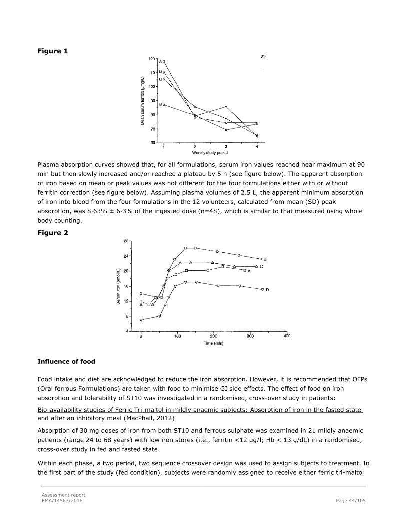
Assessment report
EMA/14567/2016 Page 44/105
Figure 1
Plasma absorption curves showed that, for all formulations, serum iron values reached near maximum at 90
min but then slowly increased and/or reached a plateau by 5 h (see figure below). The apparent absorption
of iron based on mean or peak values was not different for the four formulations either with or without
ferritin correction (see figure below). Assuming plasma volumes of 2.5 L, the apparent minimum absorption
of iron into blood from the four formulations in the 12 volunteers, calculated from mean (SD) peak
absorption, was 8·63% ± 6·3% of the ingested dose (n=48), which is similar to that measured using whole
body counting.
Figure 2
Influence of food
Food intake and diet are acknowledged to reduce the iron absorption. However, it is recommended that OFPs
(Oral ferrous Formulations) are taken with food to minimise GI side effects. The effect of food on iron
absorption and tolerability of ST10 was investigated in a randomised, cross-over study in patients:
Bio-availability studies of Ferric Tri-maltol in mildly anaemic subjects: Absorption of iron in the fasted state
and after an inhibitory meal (MacPhail, 2012)
Absorption of 30 mg doses of iron from both ST10 and ferrous sulphate was examined in 21 mildly anaemic
patients (range 24 to 68 years) with low iron stores (i.e., ferritin <12 μg/l; Hb < 13 g/dL) in a randomised,
cross-over study in fed and fasted state.
Within each phase, a two period, two sequence crossover design was used to assign subjects to treatment. In
the first part of the study (fed condition), subjects were randomly assigned to receive either ferric tri-maltol

Assessment report
EMA/14567/2016 Page 45/105
on Day 1, followed by ferrous sulphate on day 2 (Test – Reference) or vice versa, with a high phytate meal.
On day 14, the same subjects entered into the fasted phase of the study, receiving either ferric tri-maltol on
Day 14, followed by ferrous sulphate on day 15 (Test – Reference) or vice versa. The subjects were
instructed not to have any food or drink for 10 hours prior to the study and for 3 hours afterwards for this
phase.
The meal fed on days 1 and 2 consisted of a breakfast of maize meal porridge (150 g) and served with milk
and sugar ad lib plus a slice of toast with margarine. The total iron content of the meal was 3.9 mg.
The haematological variables and absorption response data (absolute and percentage) was summarized by
treatment using descriptive statistics (minimum, maximum, mean, median, standard deviation).
Results are presented in the following table [Patient Data and Absorption (% of dose administered) of Iron
from a 30 mg Dose of Ferrous Sulfate or ST10 (Ferric Maltol)].
Subject Hb
g/dl
Tf sat
%
Serum
ferritin
μg/l
FeSO4
with
food†
FeM
with
food†
FeSO4
Fasting
FeM
Fasting
Ratio (%)
FeM(fasting)/
FeSO4(fasting
1 13.9 23 1 3.5 2.0 12.2 8.8 72.1
2 16.0 10 5 5.9 1.3 12.0 14.7 122.5
3 12.4 16 1 5.0 0.5 14.5 14.2 97.9
4 15.7 45 1 1.3 0.4 8.7 1.7 19.5
5 13.0 12 1 26.1 4.0 24.1 25.4 105.3
6 13.1 18 9 4.3 1.5 10.8 12.6 116.6
7 13.0 26 17 18.5 4.1 - 24.2 -
8 11.6 9 6 16.8 2.7 14.4 12.2 84.7
11 13.5 14 1 16.6 2.3 29.1 28.5 97.9
12 15.7 32 200 4.5 2.2 14.9 10.8 72.4
13 13.8 37 11 3.6 0.9 11.3 11.0 97.3
14 10.1 5 1 16.5 4.3 25.3 23.9 94.4
15 10.1 8 1 23.4 4.7 22.0 8.0 36.4
16 12.5 8 1 19.1 13.3 42.5 34.8 81.9
17 13.0 9 1 12.1 3.3 25.5 23.4 95.5
18 11.6 17 1 17.2 7.8 22.7 13.4 59.0
19 14.2 34 95 2.6 1.3 8.6 16.4 190.6
20 12.8 16 1 12.9 2.5 23.2 18.7 80.6
21 14.2 17 6 5.4 1.1 16.6 8.7 52.4
22 13.1 12 1 17.6 14.8 31.6 29.9 94.6
23 14.2 25 1 31.3 4.7 22.2 31.9 143.6
Meana - - - 9.3 2.5 17.9 15.1 -
Meanb
(SD)
13.2
(1.55)
18.7(1
9.6) 17.2
12.5
(8.67)
3.8
(3.85)
19.7
(8.68)
17.7
(9.06) -
Median 13.1 16.0 1 12.9 2.5 19.3 14.2 -
Range 10.1-
16.0 5-45 1-200
1.3-
31.3
0.4-
14.8
8.6-
42.5
8.0-
34.8 -
a Geometric mean;
b Arithmetic mean

Assessment report
EMA/14567/2016 Page 46/105
Fasting conditions
The unadjusted geometric mean for the % absorption of iron from 30 mg was 17.9% (range 8.6-42.5) and
15.1% (range 8.0–34.8) for ferrous sulfate and ferric trimaltol, respectively in the fasting conditions.
Summary of statistical analysis of the absolute iron (mg) absorbed (above) and anti-logged ratios (%; below)
after both iron treatments in the fasted state are presenting below:
According to the Applicant, both tables indicate that across the range of subjects studied, ferric tri-maltol
shows similar absorptions compared with ferrous sulphate.
Fed conditions
The geometric mean absorption of iron from ferrous sulphate (9.3%, range 1.3-31.3) was significantly better
than from ferric tri-maltol (2.5 %, range 0.4-14.8) in the presence of a high phytate food. The presence of a
meal appears to reduce iron absorption in both cases, relative to the amount absorbed on an empty stomach.
Despite the inhibition by food a higher absorption of iron from both iron compounds was seen in the more
anaemic subjects.
Summary of statistical analysis of the absolute iron (mg) absorbed (above) and anti-logged ratios (%; below)
after both iron treatments in the fed state are presenting below:
Results from the statistical analysis, show a highly significant treatment difference between Ferric Trimaltol
and Ferrous Sulphate, both administered after a high phytate meal. On average, there is 2.48 mg difference
of iron absorbed with Ferrous Sulphate compared with Ferric Trimaltol when both treatments are
administered after a high phytate meal.
Fasted versus Fed Results
The following tables show the effect of food on the absorption of iron by Ferric Trimaltol.

Assessment report
EMA/14567/2016 Page 47/105
Summary of Statistical Analysis of the absolute Iron (mg) absorbed by ferric trimaltol and anti-logged ratios
(%; below): fed versus fasted.
Results from the statistical analyses, show a highly significant treatment difference between Ferric Trimaltol
(Fed) compared with Ferric Trimaltol (Fasted). The effect of food on ferric trimaltol yields, on average, a
reduction of 4.03 mg of iron compared with when it is administered on an empty stomach. Administering
Ferric Trimaltol on an empty stomach will increase the iron absorbed 5-fold (ratio of Fed over fasted=18.5%).
The tables below present the adjusted treatment mean effect of ferric trimaltol in fasted state over ferrous
sulphate in fed state.
Results from the statistical analyses show a highly significant treatment difference between ferrous sulphate
(Fed) compared with Ferric Trimaltol (Fasted). On average, there is an increase of 1.55 mg iron absorbed
after administration of Ferric trimaltol on a fasted stomach, compared with Ferrous Sulphate administered
after a high phytate meal.
Distribution
Thompson & Hider, Study 2 showed that all 59Fe-associated radioactivities were associated with the high
molecular weight protein fractions of the blood. Nearly all 3H radioactivity was associated with the low
molecular weight plasma protein fraction
In study ST10-01-101 the PK profiles of both maltol and maltol glucuronide were comparable between Day 1
and Day 8. For both analytes, mean Day 8/Day 1 ratios suggest that no accumulation of maltol and maltol
glucuronide occurred after 1 week of bid administration, as Cmax and AUC0-t were comparable between Day 1
and Day 8 for all three dosing regimens. In addition, exposure to maltol glucuronide was approximately dose
proportional on both Day 1 and Day 8 in this study.

Assessment report
EMA/14567/2016 Page 48/105
Elimination
The preliminary studies in healthy volunteers and subjects with IDA conducted by Thompson & Hider showed
that maltol itself was not detected in the systemic circulation, suggesting that following absorption, maltol
undergoes rapid and complete first pass metabolism and is bio-transformed to maltol-glucuronide (Thompson
& Hider, Study 4). In addition, the majority of the maltol dose (approx. 80 %) was eliminated in the urine,
primary as glucuronide conjugate (approx. 95%). No ST10, maltol or iron was detected in the urine of either
subject (Thompson & Hider, Study 2).
In study ST10-01-102, exposure to maltol glucuronide was considerably higher than exposure to maltol, and
only 0.266% of the maltol dose administered was excreted unchanged in the urine, compared to an
equivalent of 41.6% as maltol glucuronide. These findings are consistent with maltol undergoing extensive
first-pass metabolism, being rapidly glucuronidated and renally excreted, as observed in early clinical and
nonclinical studies.
Although the liver is the major site of UDP-glucuronosyltransferase expression (Ohno, 2009), a non-clinical
study using radiolabelled ferric maltol complex indicated that maltol is also extensively glucuronidated at the
site of absorption in the intestinal mucosa (Barrand, 1991b).
In study ST10-01-101, as in the previous PK sub-study (ST10-01-102), the profiles of maltol and maltol
glucuronide were similar, although exposure to maltol glucuronide was considerably higher compared to
maltol and most of the ingested maltol dose was excreted as maltol glucuronide in the urine. Values for Ae3-6h
were below the quantification limit for maltol for subjects in the 30 mg and 60 mg dosing groups on both Day
1 and Day 8. For subjects receiving the 90 mg dosing regimen, mean values for Ae0-3h were higher than Ae3-
6h (0.402 mg compared to 0.0821 mg for Day 1, and 0.263 mg compared to 0.169 mg for Day 8) indicating
that most of the unchanged maltol is excreted within 3 hours. Values of Durine0-6h on Day 1 and Day 8 for all
dosing regimens ranged between 0.0377% and 0.0800%. These data indicate that a very low proportion of
the ingested maltol is excreted unchanged in the urine after dosing with ST10. As with maltol, the mean
amount of maltol glucuronide excreted in the urine was highest in the first 3 hours post-dose. Overall,
arithmetic mean values of Durine0-6h ranged between 39.8% and 60.0%, indicating that much more of the
maltol ingested from ST10 was excreted as maltol glucuronide compared to unchanged maltol. Mean CLR for
maltol and maltol glucuronide was comparable for all dose groups on both Day 1 and Day 8, with arithmetic
mean values between 20.1 and 27.1 L/h.
In study ST10-01-101, the apparent maltol and maltol glucuronide t1/2 values were comparable for all dosing
regimens and were also consistently short ranged between 0.5 and 1.2 hours for matol and between 0.838
and 1.05 hours for maltol glucuronide (for several subjects receiving the 30 mg dosing regimen, plasma
maltol concentrations were close to the lower limit of quantification for maltol and t1/2 could not be
calculated), indicating rapid elimination of maltol glucuronide.
These t1/2 values were consistent with study ST10-01-102 in which, both maltol and maltol glucuronide had a
short t½ 0.717 and 1.22 hours, respectively (as individual values of AUC0-∞, λz and t½ for maltol could not be
accurately determined for all subjects, summary statistics for this parameter is therefore based on a limited
number of subjects.
Dose proportionality and time dependencies
In study ST-1001-101, dose-normalised parameter for Cmax, AUC0-t, and AUC0-∞ were graphically displayed
for maltol and maltol glucuronide as function of the dose, to explore dose-proportionality.

Assessment report
EMA/14567/2016 Page 49/105
Dose-normalised parameter plots for Cmax, AUC0-t, and AUC0-∞ were consistent with dose-proportional
increases in maltol exposure across the 30 mg to 90 mg bid dosing range, although for Subject 101-101-005
in the 60 mg dosing regimen Cmax, AUC0-t, and AUC0-∞ were considerably higher on both days. These values
had a considerable impact on the corresponding mean values for this group (please see below, Dose-
Normalized Pharmacokinetic Parameter Plots of Maltol).
Mean values for Cmax, AUC0-t, and AUC0-∞ increased with higher doses. Dose-normalised PK parameter plots
for maltol glucuronide indicate that exposure to maltol glucuronide was dose proportional across the 30 to 90
mg bid dose range.
Special populations
Impaired renal function and Impaired hepatic function
No specific studies have been performed.
Gender
There were twice as many women as men who participated in studies ST10-01-101 and ST10-01-102,
however, subjects were generally well matched across treatment sequences for demographic characteristics
including gender. No gender specific PK data on ST10 were noted.
Race
All participants in studies ST10-01-101 and ST10-01-102 were white and mainly Caucasian.
Elderly
There are limited PK data on ST10 in the elderly; the oldest subject in either of the prospective GCP-
compliant studies was 57 years old. In a single-dose pilot study, iron absorption from ST10 (administered as
a 10 mg iron dose in aqueous solution) was investigated in three elderly patients with anaemia and three
elderly healthy subjects (Murray, Blake & Kelsey).
Children
No children specific PK data on ST10 are available.

Assessment report
EMA/14567/2016 Page 50/105
Pilot study in iron-deficient and normal subjects (Murray, Blake & Kelsey)
In this single-dose pilot study, iron absorption from ST10 (administered as a 10 mg iron dose in a 20 mL
aqueous solution) was investigated in the elderly: three patients with anaemia (74 ± 7 years) and three
healthy subjects (84 ± 7 years). One of these patients with anaemia was administered 10 mg ST10 and an
equivalent dose of ferrous sulphate (10 mg as iron) on consecutive days. Iron formulations were
administered after an overnight fast. Serum iron was assayed at baseline and at 1 and 2 hours post-dose.
In the iron deficient patients, mean serum iron rose from 7.9 ± 5.1 μmol/L to 23.8 ± 7.0 μmol/L at 2 hours
after intake. In the healthy controls, mean serum iron rose from 12.3 ± 4.4 μmol/L to 14.9 ± 7.0 μmol/L at
the same time point. Iron from ST10 was thus absorbed to a greater extent in iron-deficient subjects. Mean
maximum rise in the normal subjects group was 2.96 ± 2.4 μmol/L compared with 18.0 ± 1.5 μmol/L for the
iron deficient group.
For the iron deficient patient who received both formulations on consecutive days. Rise in serum iron at one
hour, and maximum rise in serum iron was almost similar for an identical oral dose of elemental iron,
suggesting similar gastrointestinal absorption (please see the table below).
Dose: 10 mg (n=1) Ferric maltol Ferrous sulphate
Serum iron (µmol/L)
Baseline
1 h
2 h
Maximum rise
4.2
23.8
17.6
19.6
4.0
23.3
14.3
19.3
Overall, the results of this study were not substantially different from other studies that examined iron
absorption from ST10 in younger healthy volunteers or younger patients with IDA.
Pharmacokinetic interaction studies
No pharmacokinetic drug interactions studies were submitted.
Whilst ST10, as a new complex of an existing substance that differs in safety and efficacy to those existing
substances, can be considered to be a new chemical entity, the iron that is released and delivered to the
intestinal enterocytes is a known active ingredient with well established PD drug interactions.
Pharmacokinetics using human biomaterials
N/A
2.4.1. Pharmacodynamics
Mechanism of action
The iron in ST10 is unable to bind to maltol and transferrin simultaneously. Intracellularly ferric trimaltol can
rapidly exchange its iron onto high affinity binding proteins, with the same elution profiles as ferritin and
transferrin (Barrand et al 1987). The iron from ferric trimaltol would require a specific active mechanism
since its molecular size (M Wt 470) would preclude direct absorption through tight channels. Uptake of the
iron from ferric trimaltol onto a high affinity binding protein in the duodenal enterocyte was shown in

Assessment report
EMA/14567/2016 Page 51/105
nonclinical study in the small intestine of the rat (Barrand & Callingham 1991) and the uptake was saturable.
These findings were consistent with the observations of Teichmann & Stremmel 1990 who observed, using
human microvillous membrane vesicles that ferric ions could be taken up by a vesicular pathway as a
facilitated but saturable mechanism involving a membrane bound high affinity binding protein.
Primary and Secondary pharmacology
Dedicated studies were not submitted.
2.4.2. Discussion on clinical pharmacology
ST10 (Feraccru) is a novel complex of ferric iron and maltol developed for the treatment of iron deficiency
anaemia (IDA).
Dietary iron is taken up by enterocytes. The mechanism of uptake is dependent on the form of iron (haem,
Fe2+ or Fe3+). After uptake into the enterocyte, the iron either enters the labile iron pool (LIP) or is diverted
into ferritin. Iron efflux across the basolateral surface of the enterocyte is affected by the iron transporter
protein, ferroportin, which delivers iron to transferrin for entry into the circulation (Sharp and Srai, 2007).
Under normal physiological circumstances, free iron does not enter the circulation. In ST10, iron binds to
maltol using the same non-covalent bonding mechanism that it uses to bind to transferrin.
All evidence from both human and animal studies suggest that the ST10 complex is not absorbed “per se”
into the systemic circulation since complete dissociation seem to occur at the surface of, or within the
enterocyte but ST10 delivers iron via the endogenous iron uptake system in the proximal duodenum. This is
supported by the absence of ST10 in blood and ST10, maltol and iron in urine and. Thus, ST10 is simply a
pro-drug vehicle to deliver iron.
The absorption has been evaluated in patients in studies ST-10-01-101 and ST-10-01-102. The PK and iron
uptake in iron deficient patients has been described in study ST-10-01-101.The pharmacokinetic properties of
Feraccru was assessed through measurement of plasma and urine concentrations of maltol and maltol
glucuronide, together with serum iron parameters after a single dose and at steady state (after 1 week) in 24
subjects with iron deficiency, randomised to receive 30 mg, 60 mg or 90 mg Feraccru twice daily. Blood and
urine samples were assayed for maltol and maltol glucuronide. Serum samples were assayed for iron
parameters. On Day 1 and Day 8, plasma concentrations of both maltol and maltol glucuronide increased
rapidly after initial lag phase, reaching Cmax in around 1 to 1.5 hours post-dose, before declining to baseline
levels within between 3 and 6 hours. Both total serum iron concentration and TSAT values were generally
higher with increasing ST10 dose, with maximum values between 2 and 3 hours post-dose, and then declined
gradually after 3 hours on Day 1 and were comparable on Day 8.
The PK and iron uptake in patients with IBD and anaemia has been described in study ST-10-01-102. As
similar to the study ST-10-01-101, the plasma concentrations of both maltol and maltol glucuronide
increased rapidly after initial lag phase reaching Cmax in around 1 hour post-dose, before declining to baseline
levels at around 4 hours. Both maltol and maltol glucuronide had a short median tmax (0.98 and 1.00 hours,
respectively) and mean t½ (0.717 and 1.22 hours, respectively).
Maltol was transiently measured in plasma with a Cmax between 0.022 and 0.205 h.µg/mL across all dosing
regimens and both study days. The maltol appeared to be rapidly metabolised to maltol glucuronide (Cmax
between 9.83 and 30.9 h.µg/mL across all dosage regimens). Maximum maltol and maltol glucuronide
concentrations were reached 1 to 1.5 hours after oral administration of Feraccru. Exposure to maltol

Assessment report
EMA/14567/2016 Page 52/105
glucuronide increased dose proportionally over the Feraccru 30 to 90 mg twice daily dosing range and there
was no significant accumulation of either after 7 days treatment with Feraccru. Of the total maltol ingested, a
mean of between 39.8 % and 60.0 % was excreted as maltol glucuronide. Peak transferrin saturation (TSAT)
and total serum iron values were reached 1.5 to 3 hours after oral administration of Feraccru. Total serum
iron concentrations and TSAT values were generally higher with increasing Feraccru doses. TSAT and total
serum iron profiles were comparable between Day 1 and Day 8.
The pharmacokinetic properties of Feraccru were also investigated at steady state in 15 subjects who were
already participating in the AEGIS1/2 study described above and who had been in the open-label treatment
phase for at least 7 days (Feraccru 30 mg twice daily). Maltol was again transiently measured in plasma with
a half-life of 0.7 hours, with a Cmax of 67.3 + 28.3 ng/mL. The maltol appeared to be rapidly metabolised to
maltol glucuronide (Cmax = 4677 + 1613 ng/mL). Maximum maltol and maltol glucuronide concentrations
were reached approximately 1 hour after oral administration of Feraccru. Maximum total iron serum
concentrations were measured 1-2 hours after administration. The pharmacokinetic profiles of maltol/maltol
glucuronide and iron parameters were independent of one another.
In addition, a series of early clinical studies with ST10 in healthy subjects and in anaemic patients (with and
without IBD), has provided information on the absorption and tolerability of a range of doses of ST10 (Kelsey
et al, 1991; Maxton et al, 1994; Thompson & Hide; Reffitt et al, 2000) in support of the submitted results.
The effect of food on iron absorption and tolerability of ST10 was investigated in a randomised, cross-over
study in patients (MacPhail, 2012). The unadjusted geometric mean for the % absorption of iron from 30 mg
was 17.9% (range 8.6-42.5) and 15.1% (range 8.0–34.8) for ferrous sulfate and ferric trimaltol, respectively
in the fasting conditions being 9.3% (range 1.3-31.3) for ferrous sulfate and 2.5 % (range 0.4-14.8) for
ferric tri-maltol in the presence of food. The absorption of iron from both ST10 and ferrous sulphate was
decreased when the formulations were administered with a meal as compared to the fasted state. The
presence of the inhibitory meal appeared to have a slightly greater effect on the uptake of iron from ST10 as
compared to ferrous sulphate. Absorption of iron was similar from ST10 or ferrous sulphate in each state
(fasted or fed), respectively. When rates of absorption were compared for ST10 (fasted) and ferrous sulphate
(after a meal, which is how ferrous sulphate must be given clinically), almost twice the iron was absorbed (on
average) with the administration of ST10. The results of this study confirm that, when administered clinically,
ST10 should be given on an empty stomach to maximize iron absorption.
The SmPC therefore advises that food has been shown to inhibit uptake of ST10, so ST10 should be taken on
an empty stomach.
No tissue distribution studies were considered necessary since iron is absorbed using the well-defined
physiological iron uptake and storage mechanisms. In addition, there is no accumulation of maltol and maltol
glucuronide after 1 week of bid administration.
The data on excretion and elimination supported the conclusion that following absorption, maltol undergoes
rapid and complete first pass metabolism being rapidly bio-transformed to maltol-glucuronide, before renally
excreted. In addition, the majority of the maltol dose (approx. 80 %) was eliminated in the urine, primary as
glucuronide conjugate (approx. 95%). No ST10, maltol or iron was detected in the urine of either subject.
The iron active moiety that is released from ST10 in the proximal duodenum and delivered to the intestinal
enterocytes is a well-established physiological substance and, therefore, no specific studies have been
conducted to assess the effect of intrinsic factors, including populations, on the PK of the drug.

Assessment report
EMA/14567/2016 Page 53/105
Iron is not metabolised and excreted and maltol is rapidly metabolised to maltol glucuronide in the intestinal
mucosa prior to being renally excreted. Glucuronides form a routine route of excretion and there are no data
to indicate any toxicity of maltol glucuronide in the renal and hepatic impaired. There are no known
polymorphisms of the iron transport proteins in the enterocyte at a frequency that would justify specific in
vitro or clinical studies of ST10.
The SmPC lists the following general class drug interactions :
Iron-drug interactions of clinical significance have been reported to occur with a large number of concomitant
therapies. Concurrent ingestion of iron causes marked decrease in the bioavailability of a number of drugs
due to the formation of iron-drug complexes (chelation or binding of iron by the second drug). Examples of
affected drugs are quinolone, bisphosphonates, angiotensin-converting enzyme inhibitors, folic acid,
methyldopa, levodopa, carbidopa, levothyroxine, and mycophenolate mofetil.
Absorption of both iron and the antibiotic may be reduced if oral iron is given with tetracycline.
Absorption of oral iron may be reduced by calcium and magnesium salts (as magnesium trisilicate).
Chloramphenicol delays plasma iron clearance, incorporation of iron into red blood cells and interferes with
erythropoiesis. Conventional iron preparations interfere with the uptake of zinc from the diet (Meadows,
1983), although ST10 has been shown not to inhibit zinc uptake (Barker, Data on File). Iron and dimercaprol
should not be used together because the dimercaprol-iron complex is more toxic, especially to the kidneys,
than the metal alone.
In addition, pharmacokinetic data indicate that the maltol component of ST10, is rapidly absorbed,
glucuronidated and excreted in the urine. The potential for pharmacodynamic drug interaction with maltol
would, therefore, appear to be very limited.
ST10 is a chemically very stable form of iron complex. There are no further data in man on ST10 drug
interactions at present. Therefore, to minimise the potential for drug interactions, as well as optimising
absorption, ST10 should be given on an empty stomach at least 2 hours prior to other medications or
supplements.
ST10 may display reduced interaction characteristics with other drugs given concomitantly, compared to
when such products are given concomitantly with oral ferrous products, potentially leading to an increased
risk of overdose of the concomitantly administered drugs if prescribers inadvertently ‘compensate’ for a
potential DDI with ST10 and administered higher doses of the concomitant medication. Therefore the CHMP
requested that the applicant conducts DDI studies, initially with a small number of drug classes known to
interact with ferrous products to establish whether the interaction profile of ST10 is different from these
existing oral iron products (See RMP). The need for further studies will be evaluated once the results of the
ongoing DDI study are available.
Mean TIBC, transferrin and soluble transferrin receptor concentrations remained relatively constant
throughout PK sampling, and were comparable between dosing regimens, and between Day 1 and Day 8.
Ferritin concentrations remained relatively constant throughout PK sampling, although higher mean values
were recorded on Day 8 compared to Day 1 for all dosing regimens. Hb levels at Day 8 were comparable
between dosing groups. Overall, positive NTBI values were determined for 25 samples (10.6%). Mean pre-
dose values were close to 0.0 eLPI units on both Day 1 and Day 8. Positive NTBI values were more common
with the 60 and 90 mg dosing regimens. Most of the positive NTBI values were associated with TSAT and
serum iron concentrations that were above the normal range for these parameters.
Assessment report
EMA/14567/2016 Page 54/105
Maximum values for total serum iron concentration and TSAT were reached shortly after dosing (1 to 2 hours
post-dose for total iron and 1 to 3 hours post-dose for TSAT). Plots comparing either total iron or TSAT with
exposure to maltol and maltol glucuronide showed no clear relationship. Serum values for TIBC and
concentrations of transferrin, soluble transferrin receptor and ferritin remained constant throughout the 8-
hour sampling period. The time difference for first appearance and Cmax of maltol/maltol glucuronide and iron
is consistent with separate mechanisms of uptake and/or distribution. Serum ferritin concentrations for most
subjects remained <30 μg/L throughout the sampling period. Total serum iron concentrations, TSAT values
and pre-dose Hb concentrations in this sub-study were consistent with ST10 dosing improving iron deficiency
anaemia in these subjects. All but one sample had serum NTBI values that indicated there was no ‘free’ redox
active iron at the corresponding time points. This is consistent with there being no intact ST10 present in
serum.
No significant differences in the absorption of iron from ST10 in older healthy subjects or older patients with
IDA were observed as compared to younger healthy subjects or younger patients with IDA.
2.4.3. Conclusions on clinical pharmacology
Clinical pharmacology data obtained from clinical trials and extensive information from scientific literature
provide sufficient basis to support the marketing authorisation of Feraccru.
The CHMP considers the following measures necessary to address the issues related to pharmacology:
The Applicant will conduct DDI studies recommended by CHMP. The need for further studies will be evaluated
once the results of the ongoing DDI study are available.
In the context of the obligation of the MAHs to take due account of technical and scientific progress, the
CHMP recommends the applicant to explore the feasibility of including prospective measurement of hepcidin
and markers of inflammation (e.g. CRP), in future studies of Feraccru in inflammatory conditions.
2.5. Clinical efficacy
2.5.1. Dose response study(ies)
No formal dose-response studies have been conducted
2.5.2. Main study(ies)
ST10-01-301 (AEGIS 1)/ST10-01-302 (AEGIS 2)
A prospective, multicentre, randomised, double-blind, placebo controlled, phase III study with oral ST10 for
the treatment of iron deficiency anaemia in subjects with inflammatory bowel disease where oral ferrous
preparations have failed or cannot be used was conducted through two separate protocols:
AEGIS 1 (Protocol ST10-01-301 for subjects with quiescent ulcerative colitis [UC])
AEGIS 2 (Protocol ST10-01-302 for subjects with quiescent Crohn’s disease [CD]).

Assessment report
EMA/14567/2016 Page 55/105
Methods
Figure 3: Study design schematic
Study Participants
Inclusion Criteria were:
1. Subject must sign and date the informed consent prior to any study mandated procedure
2. Subject must be willing and able to comply with study requirements
3. Subject must be at least 18 years of age
4. Subject must have a current diagnosis of IBD and IDA, as defined by the following criteria:
a) Subject must have IBD, either quiescent UC defined by a SCCAI score of <4 as measured at the Screening
Visit and at the Randomisation Visit, or b) quiescent CD defined by a CDAI score of <220 as measured at the
Randomisation Visit
Subject must have anaemia defined by Hb ≥9.5 g/dL and <12.0 g/dL (5.9 to 7.5 mmol/L) for females and
≥9.5 g/dL and <13.0 g/dL (5.9 to 8.1 mmol/L) for males as measured at the Screening Visit.
Subject must have iron deficiency defined by ferritin <30 μg/L as measured at the Screening Visit.
5. Subject must have failed oral ferreous product (OFP) in the past. Failure defined by:
Adverse drug effects that led to withdrawal from OFP (at least one of the following: nausea, diarrhoea,
constipation, abdominal pain, flatulence) and/or
b. Deterioration of the primary disease caused by OFP and/or

Assessment report
EMA/14567/2016 Page 56/105
c. Lack of efficacy and/or
d. Other signs of failure to OFP or reasons why OFP cannot be used as documented by the Investigator.
6. Subject receiving protocol-allowed immunosuppressants at screening must have been
on stable dose for at least 4 weeks prior to randomisation. Allowed immunosuppressants included
azathioprine, 6-mercaptopurine, tumour necrosis factor (TNF)-alpha antagonists and corticosteroid doses less
than or equal to the equivalent of 25 mg/day prednisolone.
7. Female subjects of childbearing potential (including perimenopausal females who had had a menstrual
period within 1 year prior to screening) must agree to use a reliable method of contraception until study
completion and for at least 4 weeks following their final study visit. Reliable contraception was defined as a
method which results in a low failure rate, i.e., less than 1% per year when used consistently and correctly,
such as implants, injectables, some intrauterine devices (IUDs), sexual abstinence, or a vasectomised
partner. Oral contraceptive medications are allowed in this study.
Female subjects who were surgically sterile (bilateral tubal ligation, bilateral oophorectomy or hysterectomy)
or postmenopausal (defined as no menstrual period within 1 year of screening) were also allowed to
participate.
Exclusion Criteria were:
1. Subject with anaemia due to any cause other than iron deficiency, including, but not limited to:
a. Untreated or untreatable severe malabsorption syndrome
b. Immunosuppressant use.
2. Subject who had received within 12 weeks prior to randomisation:
a. Intramuscular or IV injection or administration of depot iron preparation
b. Blood transfusion
c. Erythropoietin.
3. Subject who had received within 4 weeks prior to randomisation:
a. Oral iron supplementation
b. Immunosuppressant with known effect of anaemia induction, including, but not limited to methotrexate,
cyclosporin A or tacrolimus.
4. Subject who had received vitamin B12 injections/infusions within 4 weeks prior to randomisation
5. Subject who had received a folic acid injection/infusion within 4 weeks prior to randomisation
6. Subject with vitamin B12 concentration below the lower limit of normal (LLN) as measured at the
Screening Visit
7. Subject with folic acid deficiency as assessed at the Screening Visit
8. Subject with known hypersensitivity or allergy to ST10 or components of the study medication

Assessment report
EMA/14567/2016 Page 57/105
9. Subject with contraindication for treatment with iron preparations, e.g., haemochromatosis, chronic
haemolytic disease, sideroblastic anaemia, thalassaemia, or lead intoxication induced anaemia
10. Subject with creatinine >2.0 mg/dL (176 μmol/L) as measured at the Screening Visit
11. Subject with alanine aminotransferase (ALT) or aspartate aminotransferase (AST) levels ≥5 times the
upper limit of normal (ULN) as measured at the Screening Visit 12. Subject with cardiovascular, liver, renal,
haematologic, gastrointestinal, immunologic, endocrine, metabolic, or central nervous system disease that, in
the opinion of the Investigator, may have adversely affected the safety of the subject and/or efficacy of the
study drug or severely limit the lifespan of the subject
13. Subject with history of malignancy within the past 5 years with the exception of in situ removal of basal
cell carcinoma
14. Subject with significant neurologic or psychiatric symptoms resulting in disorientation, memory
impairment, or inability to report accurately that might interfere with treatment compliance, study conduct or
interpretation of the results (e.g., Alzheimer’s disease, schizophrenia or other psychosis, alcohol or drug
abuse)
15. Participation in another interventional clinical study within 4 weeks prior to randomisation or during the
study
16. Subject who was an inmate of a psychiatric ward, prison, or other state institution
17. Subject who was an Investigator or any other team member involved directly or indirectly in the conduct
of the clinical study
18. Subject with scheduled or expected hospitalisation and/or surgery during the course of the study
19. Female subject who was pregnant or lactating.
Treatments
The investigational product was ST10 orange hard gelatine capsules (231.5 mg of ST10 [ferric maltol]
equivalent to 30 mg iron per capsule). The capsules were taken orally first thing in the morning before
breakfast and last thing at night before bed. Capsules were taken on an empty stomach with water only.
Objectives
To demonstrate the efficacy effect of oral ST10 over placebo in the treatment of IDA, as measured by change
in Hb concentration from Baseline to Week 12, in subjects with IBD where OFP have failed or cannot be used.
Secondary Objectives
-To evaluate the safety and tolerability of ST10 in subjects with IDA and IBD where OFP have failed or cannot
be used over a treatment duration of up to 12 weeks
-To evaluate long-term safety and tolerability of ST10 in subjects with IDA and IBD where OFP have failed or
cannot be used over a treatment duration of up to 64 weeks
-To evaluate long-term efficacy of ST10 in subjects with IDA and IBD, where OFP have failed or cannot be
used, over a treatment duration of up to 64 weeks.

Assessment report
EMA/14567/2016 Page 58/105
Outcomes/endpoints
The primary efficacy endpoint was defined as the change in Hb concentration from Baseline to Week 12.
Secondary Efficacy Endpoints
-Proportion of subjects that achieved an increase from baseline in Hb concentration of ≥1 g/dL at Week 12
-Proportion of subjects that achieved an increase from baseline in Hb concentration of ≥2 g/dL at Week 12
-Proportion of subjects that achieved Hb concentration within normal range at Week 12 (‘normal’ was defined
as ≥12 g/dL (7.5 mmol/L) for females or ≥13 g/dL (8.1 mmol/L) for males).
- Time to normalisation of Hb concentration during the double-blind phase.
- Change in Hb concentration from Baseline to Week 8
- Change in Hb concentration from Baseline to Week 4.
Other Efficacy Endpoints
Iron Indices
-Ferritin, serum iron, transferrin and transferrin receptor were collected at the same clinic visits as described
for Hb concentrations. Total iron-binding capacity and TSAT were derived from other iron parameters by the
central laboratory.
-SF-36 Response over Time
The SF-36 was collected at clinic visits: Weeks 0, 12, 24, 36, 48 and 64. Subject responses to each of the
questions were listed.
-IBDQ Response over Time
The IBDQ was collected at clinic visits: Weeks 0, 12, 24, 36, 48 and 64. Subject responses to each of the
questions were listed.
Safety Analyses
-Study Drug Exposure
Duration on treatment and total dose of ST10 received was summarised for both phases together.
-Simple Clinical Colitis Activity Index
The SCCAI Score was evaluated at every clinic visit. Change from baseline of SCCAI was evaluated and
summarised with appropriate descriptive statistics
-Crohn’s Disease Activity Index
The CDAI Score was evaluated at every clinic visit. Change from baseline of CDAI was evaluated and
summarised with appropriate descriptive statistics.
Sample size
The sample size calculation was based on results from previous exploratory studies (Noether, 1987). A
sample size of 49 subjects in each group would have 95% power to detect a probability of 0.711 P (X<Y) that
an observation in the Control group (Placebo) would show a lower increase of Hb concentration than in the
Test group (ST10) using the Wilcoxon (Mann-Whitney) rank-sum test (the method planned to be used for the
evaluation of the primary efficacy criterion in the original protocol) with a 0.025 one-sided significance level.

Assessment report
EMA/14567/2016 Page 59/105
To allow for non-evaluable subjects (e.g., drop-outs), a total of 120 subjects were to be recruited across
studies AEGIS 1 and AEGIS 2; approximately 60 subjects in each study.
Randomisation
Randomisation was done by the Interactive Voice Response System (IXRS) which assigned a study drug kit
(uniquely numbered) to each subject.
Blinding (masking)
This study was performed in a double-blind fashion. The Investigator and site staff, subjects, monitors,
Sponsor and Contract Research Organisation (CRO) staff remained blinded to the treatment assignment
during the conduct of the randomised phase of the study.
Statistical methods
Population Analysis Sets were:
Safety Analysis Set (Double-blind Phase)
This analysis set was defined as all subjects who had at least one dose of study drug and one subsequent
contact with the Investigator in the double-blind phase. Subjects who received study drug other than what
they were randomised to, were reported under the actual treatment received.
Safety Analysis Set (Open-label Phase)
This analysis set was defined as all subjects who had at least one dose of study drug and one subsequent
contact with the Investigator in the open-label phase.
Safety Analysis Set (Cumulative)
This analysis set was defined as all subjects who had at least one dose of study drug and one subsequent
contact with the Investigator in either the double-blind phase or open label part
Randomised Set
This analysis set was defined as all subjects who were randomised. The Randomised Set was used for the
summary of demographic data.
Full Analysis Set (FAS)
All subjects who had at least one dose of study drug were included in the Intent to Treat (ITT) analysis. The
ITT analysis was used in the primary efficacy analysis.
Per Protocol Analysis Set (PPAS)
Subjects who fulfilled at least one of the criteria defined in table 1, were not eligible for the PPAS. The
determination of eligibility of subjects for the PPAS was described in a separate document to the SAP. Prior to
unblinding of the database, and when data were considered clean, listings of protocol deviations were
reviewed by the study clinician to determine the final PPAS. The study statistician and study clinician signed
off this final list prior to the unblinding of the database.
The null and alternative hypothesis for the comparison of the Test group (ST10) to the Control group
(Placebo) was formulated as follows (superiority):

Assessment report
EMA/14567/2016 Page 60/105
H0:μx =μy
HA:μx <μy
Where H0 = Null-hypothesis; HA = Alternative Hypothesis; Y = Test group; X = Control group.
The primary efficacy endpoint was defined as the change in Hb concentration from Baseline to Week 12.
Baseline was defined as the pre-dose Hb concentration measured at the Randomisation Visit (Week 0)
Results
Participant flow
Figure 4: Disposition of subjects in the double-blind phase
Recruitment
A total of 128 subjects were randomised into both studies ST10-01-301 and ST10-01-302. The majority of
protocol deviations were minor in nature. Nine ST10 and 7 Placebo subjects were excluded from the PPAS
due to deviations of a more important nature; these are presented as subjects excluded from the efficacy
analysis (i.e., from the PPAS).
Conduct of the study
The original AEGIS 1 and 2 protocols versions 1.0 respectively dated 21-Apr-2011 and 03-May-2011 were
each amended five times as follows: (1); The study design of ST10-01-301 was modified to include a 52-
week open-label extension to further evaluate the safety and efficacy of ST10 in all randomised subjects.
There were changes to subject discontinuation criteria, changes to study endpoints/statistics, changes to

Assessment report
EMA/14567/2016 Page 61/105
inclusion/exclusion criteria, changes to study objectives and changes to study design/visit schedule (2);
Changes to inclusion criteria (3); There were changes to exclusion criteria (4); Changes to Emergency
Unblinding (5) Changes to the analysis of the study
Baseline data
Across both treatment groups, subjects were well matched for age, race, ethnicity and height. Almost twice
the number of females compared to males participated in the study.
The proportion of females was slightly higher in the Placebo group i.e., 40 (66.7%) compared to 37 (61.7%)
in the ST10 group. The proportion of males was slightly higher in the ST10 group i.e., 23 (38.3%) compared
to 20 (33.3%) in the Placebo group. The mean (SD) age of subjects was 40.4 (13.71) years in the ST10 and
38.9 (12.31) years in the Placebo group. The majority of subjects, 58 (96.7%) in the ST10 and 56 (93.3%)
in the Placebo group, were white.
The only medical history recorded in at least 10% of subjects in either treatment group was osteoporosis in
the Placebo group (6 subjects, 10.0%)
Table 9: Demographic characteristics
Median duration of UC was 6.85 years in the ST10 compared to 7.82 years in the Placebo group. Median
duration of CD was also similar in both groups (10.12 years in the ST10 compared to 11.27 years in the
Placebo group). Median duration since last disease flare was comparable for both groups (7.36 months in the
ST10 compared to 6.89 months in the Placebo group)
Fewer than 10% of subjects in both groups were taking oral supplemental vitamin B12 or folic acid. All
subjects, with the exception of ST10 Subject 302-305-503, whose folic acid dosing was unstable, were on
stable dosing of their supplement from 3 months before randomisation through study Week 12.

Assessment report
EMA/14567/2016 Page 62/105
Table10: Baseline disease characteristics and concomitant therapy status (safety
analysis set)
Common prior OFPs used by at least 10% of subjects included ferrous glycine sulphate in
25 (41.7%) ST10 and 23 (38.3%) Placebo subjects, and ferrous sulphate in 19 (31.7%) ST10 and 12
(20.0%) placebo subjects. All other prior OFPs were used by no more than 5 (8.3%) subjects in either
treatment group.
Mean and median length of treatment with prior OFPs was lower in the ST10 (mean 97.51 [SD 179.549],
median 38.05 days) compared to the Placebo (mean 130.49 [SD 276.787], median 60.88 days) group. Mean
and median time since last dose of prior OFP was also lower in the ST10 (mean 35.11 [SD 39.682], median
21.13 days) compared to the Placebo (mean 33.56 [SD 45.044], median 16.04 days) group. 70.0% of ST10
and 61.7% of Placebo subjects stopped taking prior OFPs due to adverse drugs effects. In at least one
quarter of the subjects withdrawing from prior OFP use, the adverse drug effects experienced were nausea,
diarrhoea or abdominal pain. Prior OFPs were not efficacious for 21 (35.0%) ST10 and 24 (40.0%) Placebo
subjects. One ST10 and two Placebo subjects experienced a deterioration of their primary disease due to OFP
use.

Assessment report
EMA/14567/2016 Page 63/105
Table 11: Prior oral ferrous preparations – treatment and failure (safety analysis set)
Numbers analysed
A total of 128 subjects were randomised into both studies ST10-01-301 and ST10-01-302.
Of these, 120 subjects, 60 in each treatment group, were included in the double-blind analysis. All 120
subjects were included in the Full Analysis Set and Safety Set. A total of 51 (85.0%) ST10 and 53 (88.3%)
Placebo subjects were included in the Per Protocol Analysis Set.
Table 12: Subject evaluation groups (randomized analysis set)
Outcomes and estimation
Primary Efficacy Endpoint: Change in Haemoglobin Concentration From Baseline to Week 12
Mean Hb levels improved in ST10 subjects from baseline (mean Hb 10.98 g/dL [SD 1.047]) to Week 12
(mean Hb 13.19 g/dL [SD 1.061]), i.e., a mean overall improvement of 2.25 g/dL. Mean Hb levels in Placebo
subjects were similar at 11.10 g/dL (SD 0.793) at baseline and 11.13 g/dL (SD 0.970) at Week 12. The mean

Assessment report
EMA/14567/2016 Page 64/105
improvement in Hb levels delivered by ST10 was statistically significantly different (p<0.0001) compared to
Placebo. ST10 therefore met the primary efficacy endpoint of change in Hb concentration after 12 weeks of
treatment compared to Placebo.
Table 13: Primary efficacy endpoint: change in haemoglobin concentration from baseline
to week 12 (full analysis set)
Table 14: summary of statistical analysis of primary efficacy endpoint: change in
haemoglobin concentration from baseline to week 12: multiple imutation (full analysis
set)
The robustness of the primary efficacy analysis on the FAS was confirmed (p<0.0001) by all applied
sensitivity analyses including analysis of the PPAS; analysis of the FAS using an LOCF approach; analysis of
complete cases (subjects with both baseline and Week 12 Hb concentrations) in the FAS; analysis of the FAS
using an MMRM approach and analysis of the FAS excluding the non-GCP Subjects 301-106-001, 301-106-
002, 302-106-501 and 302-106-504 from Site 106 . As described in the SAP a sensitivity analysis on the
primary endpoint was performed on all 128 subjects randomised and the results from this analysis were
entirely consistent with the results from the primary efficacy analysis performed on the first 120 subjects.
Secondary Efficacy Endpoints
Total of 47 (78.3%) ST10 subjects and six (10.0%) Placebo subjects achieved at least an increase of 1 g/dL
from baseline Hb concentration at Week 12. Logistic regression analysis confirmed that the odds of achieving
a 1 g/dL increase over baseline with ST10 were significantly greater than with Placebo (OR 43.499; 95% CI
13.505, 140.111).
Baseline Hb concentration also significantly affected the odds of achieving a 1 g/dL increase over baseline
with higher baseline concentrations being associated with lower odds of response (OR 0.478; 95% CI 0.263,

Assessment report
EMA/14567/2016 Page 65/105
0.868). Disease type did not significantly affect the odds of achieving a 1 g/dL increase over baseline (OR
2.009; 95% CI 0.670, 6.026).
Fewer (34 [56.7%]) ST10 subjects and no Placebo subjects achieved at least an increase of 2 g/dL from
baseline Hb concentration at Week 12. Logistic regression analysis confirmed that the odds of achieving a 2
g/dL increase over baseline with ST10 were significantly greater than with Placebo although the OR was not
estimable as there were no Placebo subjects with at least a change of 2 g/dL.
Baseline Hb concentration also significantly affected the odds of achieving a 2 g/dL increase over baseline
with higher baseline concentrations being associated with lower odds of response (OR 0.324;
95% CI 0.162,0.646). Disease type did not significantly affect the odds of achieving a 2 g/dL increase over
baseline (OR 0.678; 95% CI 0.200,2.293).
A total of 39 (65.0%) ST10 subjects and six (10.0%) Placebo subjects achieved normalised Hb concentration
at Week 12. Logistic regression analysis confirmed that the odds of achieving a normalised Hb concentration
with ST10 were significantly greater than with Placebo (OR 18.452; 95% CI 6.597,51.614). Baseline Hb
concentration did not significantly affect the odds of achieving a normalised Hb concentration although higher
baseline concentrations were associated with greater odds of response (OR 1.360; 95% CI 0.842,2.195).
Disease type did not significantly affect the odds of achieving a normalised Hb concentration (OR 1.656; 95%
CI 0.651,4.213)
Table 15: Secondary efficacy endpoints: proportion of subjects achieving increases of
haemoglobin concentration of >1g/dL and >2g/dL at week 12 and proportion of
subjects wih haemoglobin within normal range at week 12 (full analysis set)
Time to normalisation of Hb concentration for ST10 subjects took a median of 57.0 days (n = 60; Q1 29.0,
Q3 85.0; 12 subjects censored due to non-normalisation of Hb concentration by Week 12). Time to
normalisation was not derived for the Placebo group (n = 60; 47 subjects censored due to non-normalisation
of Hb concentration by Week 12).
Change from baseline to week 4 and week 8 Mean Hb levels improved in ST10 subjects from baseline (mean
Hb 10.98 g/dL [SD 1.047]) to Week 4 (mean Hb 12.03 g/dL [SD 0.805]), i.e., a mean overall improvement
of 1.08 g/dL. Mean Hb levels in Placebo subjects was the same (11.10 g/dL) at baseline as at Week 4. The
mean improvement in Hb levels delivered by ST10 was statistically significantly different (p<0.0001)
compared to Placebo.
Mean Hb levels improved in ST10 subjects from baseline (mean Hb 10.98 g/dL [SD 1.047]) to Week 8 (mean
Hb 12.72 g/dL [SD 0.965]), i.e., a mean overall improvement of 1.76 g/dL. Mean Hb levels in Placebo

Assessment report
EMA/14567/2016 Page 66/105
subjects were similar (11.16 g/dL) at Week 8 compared to baseline. The mean improvement in Hb levels
delivered by ST10 was statistically significantly different (p<0.0001) compared to Placebo.
Table 16: secondary efficacy endpoint: change in haemoglobin concentration from
basline to week 4 and week8 (full analysis set)
Table 17: summary of statistical analysis of secondary efficacy endpoint: change in
haemoglobin concentration from baseline to week 4 to week 8: multiple imputation (full
analysis set)

Assessment report
EMA/14567/2016 Page 67/105
Other Efficacy Endpoints: Iron Indices
Table 18: Other Efficacy Endpoints: Change in Iron Indices Over Time to Week 12 (Full
Analysis Set)

Assessment report
EMA/14567/2016 Page 68/105

Assessment report
EMA/14567/2016 Page 69/105

Assessment report
EMA/14567/2016 Page 70/105
Other Efficacy Endpoints: SF-36 and IBDQ
Limited physical and mental improvement was evident in the SF-36 questionnaire results from randomisation
to Week 12 for ST10 subjects. Limited mental improvement was evident for Placebo subjects.
In ST10 subjects completing the SF-36 questionnaire, the randomisation mean physical component score of
48.43 (SD 9.061) was 50.78 (SD 6.845) by Week 12. General physical improvement was most evident in the
individual components of physical functioning; role limitations due to physical health problems; general
health and vitality.
In Placebo subjects, the randomisation mean physical component score (48.35 [SD 8.260]) was unchanged
by Week 12 (48.23 [SD 7.441]), although score improvements were evident in general health and vitality.
In ST10 subjects, the randomisation mean mental component score of 44.80 (SD 12.055) was 46.10 (SD
12.512) by Week 12. General mental improvement was most evident in the individual component of social
functioning. In Placebo subjects, the randomisation mean mental component score was 44.67 [SD 11.419])
and the Week 12 score was 45.49 (SD 11.432).
There were no meaningful changes in either group’s IBDQ scores from randomisation to Week 12. In ST10
subjects mean total IBDQ score at randomisation was 175.6 (SD 31.43) and 179.7 (SD 32.57) by Week 12.
In Placebo subjects mean total IBDQ score at randomisation was 171.0 (SD 33.83) and 176.0 (SD 32.18) by
Week 12.

Assessment report
EMA/14567/2016 Page 71/105
Examination of Subgroups
Plot of the primary endpoint by gender.
Figure 5: change from baseline in haemoglobin at week 12 by gender

Assessment report
EMA/14567/2016 Page 72/105
Plot of the primary endpoint by age (continuous)
Figure 6: change from baseline in haemoglobin at week 12 by age
Plot of the primary endpoint by baseline Hb.
Subjects starting with a lower baseline Hb tended towards a greater increase in Hb than subjects starting
with a higher baseline Hb, by Week 12.

Assessment report
EMA/14567/2016 Page 73/105
Figure 7: change from baseline in haemoglobin at week 12 by baseline haemoglobin
Ancillary analyses
Post hoc analysis by disease group (CD/ UC) and by demographic and disease-specific factors
The key demographic and treatment group data is presented below for the two IBD disease subgroups.

Assessment report
EMA/14567/2016 Page 74/105
Table 19: Key demographic and treatment data from AEGIS 1 and 2 populations
Table 20: primary efficacy analysis for AEGIS 1 and 2 studies, by FAS and PP datasets
Summary of main studies
The following tables summarise the efficacy results from the main studies supporting the present application.
These summaries should be read in conjunction with the discussion on clinical efficacy as well as the benefit
risk assessment (see later sections).

Assessment report
EMA/14567/2016 Page 75/105
Summary of Efficacy for trials ST10-01-301 (AEGIS 1) and ST10-01-302 (AEGIS 2)
Title:
A prospective, multicentre, randomised, double-blind, placebo controlled study with oral 1ST10-021
for the treatment of iron deficiency anaemia in subjects with inflammatory bowel disease where oral
ferrous preparations have failed or cannot be used ST10-021 is henceforth referred to as ST10.
Study identifier NUMBER ST10-01-301 (AEGIS 1) and ST10-01-302 (AEGIS 2)
Design Prospective, multicentre, randomised, double-blind, placebo controlled study
with oral ST10 for the treatment of iron deficiency anaemia in subjects with
inflammatory bowel disease where oral ferrous preparations have failed
Duration of main phase: 12 weeks
Duration of Run-in phase: screeining 14 days for AGIS 1 and 7 days for
AEGIS 2
Duration of Extension phase: 52 weeks
Hypothesis Superiority
Treatments groups
ST10group
30 mg capsule bid
Placebo capsule bid
Endpoints and
definitions
Primary
endpoint
Change in Hb concentration from Baseline to
Week 12.
Secondary
endpoint
Proportion of subjects that achieved an
increase in Hb
concentration of ≥1 g/dL at Week 12
• Proportion of subjects that achieved an
increase in Hb
concentration of ≥2 g/dL at Week 12
• Proportion of subjects that achieved Hb
concentration
within normal range at Week 12
• Change in Hb concentration from Baseline
to Week 8
• Change in Hb concentration from Baseline
to Week 4
(early response).
Secondary
endpoint
Quality of life parameters
Iron indice (ferritin, transferrin etc) and
safety endpoints
Database lock Last Subject Last Visit: 11-October-2013
Interim analysis: 31 st of March 2014
Results and Analysis
Analysis description Primary Analysis

Assessment report
EMA/14567/2016 Page 76/105
Analysis population
and time point
description
Intent to treat
Screening: day -14 (in UC) or day -7(in CD)
Baseline: day 1
Study assessment: Week 4, 8 and 12 (week 16,20,24,36,48 and 64)
Descriptive statistics
and estimate
variability
Treatment group ST10
Placebo
Number of
subject
N=64 N=64
Baseline Hb
(g/dl)
Mean (SD)
10.98( 1.047)
Mean (SD)
11.10 (0.793)
Mean change in
Hb (gr/dl) from
BS to week 12
Mean (SD)
13.19
[1.061]
mean overall
improvement of
2.25 g/dL.
mean (SD)
11.13 ( 0.970)
mean overall improvement of
-0.02g/dL
Secondary
endpoints
Proportion of
subjects achiving
an Hb increase >
2 g/dl
N(%)
34 (56.7%)
N/A
Proportion of
subjects achiving
Hb normalization
from BS to week
12
N(%)
39 (65.0%)
N(%)
6 (10.0%)
Proportion of
subjects achiving
Hb > 1 gr/dl
N(%)
47 (78.3%)
N(%)
6 (10.0%)
Change in Hb
concentration
from Baseline to
Week 4
mean (SD)
12.03
g/dL (0.805)
mean overall
improvement of
1.08 g/dL
Mean(SD)
11.10 g/dL(0.831)
mean overall improvement of 0.00
g/dL
Change in Hb
concentration
from Baseline to
Week 8
mean (SD)
12.72
g/dL (0.965)
mean overall
improvement of
1.76 g/dL
Mean (SD)
11.16 g/dL(0.920)
mean overall improvement of 0.00
g/dL
Effect estimate per
comparison
Primary endpoin
Comparison groups ST10 vs placebo

Assessment report
EMA/14567/2016 Page 77/105
Mean change in
Hb from BS to
week 12
treatment difference
(1-sided lower 97.5% CI)
2.25 (1.88)
p-value
p<0.0001.
Secondary
endpoints
Proportion of
subjects achiving
an Hb increased
>2 gr/dl
Proportion of
subjects achieving
Hb normalization
Proportion of
subjects achiving
an Hb increased 1
gr/dl
Comparison groups ST10 vs placebo
treatment difference (95%IC)
N/A
treatment difference
(95% CI)
treatment difference
(95% CI)
18.452;
(6.597,51.614)
43.499 ( 13.505,140.111)
Change in Hb
concentration
from Baseline to
Week 4
Change in Hb
concentration
from Baseline to
Week 8
difference between mean
(1-sided lower95% CI)
difference between mean
(1-sided lower95% CI)
1.04 (0.83,)
p<0.0001
1.76 (1.47,)
P<0.00
Analysis performed across trials (pooled analyses and meta-analysis)
N/A
Supportive studies
Published studies were submitted by the applicant (Blake and Kelsey; Green and Thompson 1995) or reports
from individual patients were not considered supportive by the CHMP due to heterogeneity of populations,
inconsistent information and difficulties to draw conclusions.
Harvey 1998
Assessment report
EMA/14567/2016 Page 78/105
This is an open study in which patens with iron deficiency anaemia and severe intolerance to ferrous
preparations were recruited from gastroenterology clinics.
Inclusion criteria
Documented intolerance to 200 mg ferrous sulphate (65 mg elemental iron daily) precluding its use and with
refusal to try ferrous iron again; blood Hb < 130 g/dL (>120g/dL in females); ferritin <15 micgrog/L ; -
normal serum C reactive
Treatment
Ferric trimaltol (30 mg iron) was given twice daily before breakfast and the evening meal, for three months.
Results
24 patients were recruited, one was excluded with coeliac disease , 13 had Crohn´s disease, two ulcerative
colitis, two partial gastrectomy, one caecal angiodysplasia and five idiopathic iron deficiency. Two were
withdrawn during the first week and two patients were lost. Data were presented on the remaining 19
patients. At 3 months, there was a significant increase in haemoglobin (106.15 to 126 . 16 g/L, mean . s.d.;
P < 0.001, paired t-test) (Figure 1) and in 14 of the 19 patients (74%) the Hb value was within the normal
range. Similarly, there was a significant increase in ferritin from pre-treatment levels (8.1 to 17.4 µg/L ; P <
0.001) and 11 out of 19 patients (58%) were in the normal range (> 15 µg/L). Of the five patients still with a
low Hb at 3 months, two had nevertheless greatly improved (76±118 g/L and 84±106 g/l, respectively), and
the remaining three were actively bleeding and yet maintained their haemoglobin levels with treatment: one
caecal angiodysplasia (before treatment Hb on average decreased by 10±20 g/L per month) and the two with
idiopathic gastrointestinal bleeding (before treatment Hb decreased by 0±10 and 10±20 g/L per month).
2.5.3. Discussion on clinical efficacy
Design and conduct of clinical studies
The ferric maltol clinical development program in adults with anaemia and IBD consists of is based on a
prospective, multicentre, randomized, double-blind, placebo controlled trial with oral ST10 for the treatment
of iron deficiency anaemia in subjects with IBD. The pivotal trial integrated studies AEGIS 1 (for subjects with
quiescent ulcerative colitis [UC]) and AEGIS 2 (for subjects with quiescent Crohn’s disease [CD]). Both were
essentially identical in design and therefore, such integration is considered acceptable. Following the
randomized phase all subjects received open-label ST10 for up to an additional 52 weeks (i.e., up to a
maximum of 64 weeks exposure).
Patients with quiescent IBD were enrolled - excluding those with moderate to severe UC or CD in order to
avoid the confounding effects of active inflammation on iron stores and the potential for oral iron to
exacerbate GI symptoms. The definition of "quiescent" was vague and a combination of patients with no-
active and active diseases was finally enrolled in the studies. To define the severity of CD the Applicant used
the validated CDAI score but the colitis activity index (SCCAI) used by the Applicant to define the severity of
UC is not validated. The population enrolled had iron deficiency anaemia but it seemed to be heterogenous
with regard to the severity of IBD as time since the last flare was large with a wide range (mean around 34
months;max-min: 0.0-45), 38.7% of patients received TNF inhibitors and 30.6% were on azathioprine. These
data are reflecting the heterogenous population suffering from a diseases caracterised by remission and
active disease periods.

Assessment report
EMA/14567/2016 Page 79/105
A protocol amendment widened the anaemia definition moving down the limit for Hb level from Hb ≥10.0
g/dL to Hb ≥9.5 g/dL. It is recognized that this modified criteria allowed recruiting more patients but iv iron
is the recommended treatment in clinical practice when Hb decreases below 10 g/dl. The Applicant has
clarified that most patients had mild to moderate anaemia at baseline being candidate to be treated with oral
iron compounds. Subjects with Hb concentrations ≤8.5 g/dL (5.3 mmol/L) and/or flare-up of IBD were
withdrawn from the study to receive the best standard medical care.
Patients had to have failed previous oral ferrous treatment in such a way that ferric maltol was offered as a
second line treatment. However, it is not well documented that patients were intolerant to previous OFP. The
last pre-study oral iron treatment took place over 2 years (mean time) before entering the pivotal study and
no information was provided with regard to the administered pre-study iron doses. A post-hoc survey with
responses from 13 German and Hungarian sites has been presented by the Applicant. The percentage of
subjects who had previously received IV iron ranged from 100% to 11% (mean: 55%). The average number
of days since last IV iron treatment was 285 (range 84-567). Specific information/data is lacking and the
submitted information on the conducted post-hoc survey is considered insufficient to clarify the issue related
to the previously received treatments.
The design of the study is considered, in general, acceptable. However, the lack of an active comparator is
regarded as a drawback given that there are several oral iron preparations on the market that could have
been appropriate for comparison (particularly gastro-resistant formulations). Indirect comparisons with
ferrous compounds coming from published literature showed that ST10 has a similar effect to those standard
preparations, however external comparisons were not considered as valid alternatives.
Efficacy data and additional analyses
Ferric maltol has shown to increase the Hb concentration correcting the anaemia at week 12(change of 2.25
gr/dl from baseline). The effect is observed at week 4 and it is maintained until de end of the study. No
changes from baseline were observed in the placebo group. It can be concluded that the Hb change achieved
in the patients treated with ferric maltol is clearly superior to that seen in the placebo arm in the population
studied. The internal validity of the result is supported by the analysis in ITT and PP populations and several
sensitivity analyses. The Hb recuperation was progressive but the largest effect was seen within the first 4
weeks (change of 1.08 gr/dl from baseline). Feraccru normalized Hb concentration in the 65% of the patients
with a median time to normalization of around 2 months. 78.3% of patients got an increase of 1 gr/dl and
56.7% achieved an increase of 2 gr/dl. In addition, Feraccru treatment has shown to achieve substantial
mean increases in iron indices (mainly ferritin and transferrin saturation) although values were highly
variable.
The patients included in the study seemed to be relatively stable as indicated by the absence of meaningful
changes in the SF-36 questionnaire results and IBDQ scores. The results of the subgroups analyses by
gender, ages or underlying conditions (UC and CD) are consistent with the overall results in terms of the
primary endpoint. Nevertheless, the positive effect of ferric maltol has been demonstrated in a population
selected under strict inclusion and exclusion criteria (mild or moderate anaemia and quiescent UC and CD).
The Applicant has tried to clarify the uncertainties related to the oral iron intolerance status to previous OFP,
however information provided from a conducted post-hoc survey is considered insufficient. Therefore, this
part of the indication "patients who cannot tolerate other oral iron preparations or are non-compliant" is not
acceptable based on the data provided.

Assessment report
EMA/14567/2016 Page 80/105
The baseline status of IBD is not well characterised either. It seems that patients with active disease were
also included in the trials but it is not clear if they were mildly or moderately active. The inclusion of a
reference to the disease status in the indication is not considered appropriate.
Patients were given two capsules of ferric maltol (30 mg ferric iron) equivalent to 60 mg daily or placebo.
Previous investigations support this regimen although a dose finding study has not been conducted. Posology
and duration of the treatment according to the results of the clinical trials and in line with that of other oral
iron products on the market are appropriately reflected in section 4.2. of the SmPC.
The superiority of ferric maltol over placebo seems clear and clinically relevant both regarding the increment
of Hb and the proportion of responders. However, an important inter-patient variability is observed in the
different analyses submitted which could be explained by the wide range of baseline Hb concentration rather
than by the underlying disease. Additional analysis were required to the Applicant to better understand this
variability and if there are potential predictive factor of a positive response. The Applicant justified that
screening laboratory values were used for inclusion into the study and that these values can change at
randomization (14 days later in UC and 7 days later in CD). As a result some subjects had only iron levels
within normal range at baseline whereas other patients had also other values within normal ranges (5
patients had normal ferritin, iron and transferring saturation at baseline). Per protocol results and other
sensitivity analysis are in the same line than those of ITT analysis and the magnitude of the effect suggest
efficacy of Feraccru in all the analyses.
The Applicant has provided subgroup analyses by gender, age and baseline haemoglobin from the pivotal trial
showing consistent results. Data from AEGIS1 and AEGIS2 studies given separately also show that the
baseline characteristics are consistent with those of the overall population for the same subgroups. The
analysis of the primary endpoint showed statistically significant increase in Hb concentrations in the ferric
maltol group (both in the full analysis and the PP analysis) for both the UC and the CD populations. The mean
increase from baseline was very similar in both diseases: 2.18 in UC and 2.16 in CD. These results are
consistent with the overall result observed. The possibility of imbalances in iron resistant patients was
investigated and the Applicant has provided the responder rate for increase of Hb 1 gr/dl, 2gr/dl and
normalization of Hb in UC and CD showing a relationship between Hb level and the likelihood of achieving Hb
increase of 1 gr/dl or 2 gr/dl in Hb, as expected.
Indirect comparisons with other available products used as second line have been provided by the Applicant
in order to gain additional information on the comparative efficacy and safety of ferric iron versus already
known products. The most important evidence comes from the paper by Pereira, 2015 where data on Hb
change for ST10 were compared with those for ferrous sulphate compounds collected in a recent systematic
review and meta-analysis (Tolkien et al 2015). Results show that the Feraccru efficacy is in line with
expected for this kind of products considering the different amount of iron administrated in each study.
As discussed, a definition of "quiescent" was lacking and a combination of patients with no-active and active
diseases was finally enrolled in the studies. The use of Feraccru in the proposed populations ("inflammatory
bowel disease where other oral iron preparations are ineffective”, "in patients who cannot tolerate other oral
iron preparations or are non-compliant" and "in patients in whom treatment with intravenous iron is unsafe
or not possible") is not supported by the data provided. IBD patients are expected to be a more difficult
population to treat (“representing the worst case scenario”) and possibility to extrapolate the efficacy data
from IBD patients to other underlying diseases causing IDA was explored, however, it was considered that
the absorption of iron from ferric maltol may be affected by IBD pathology, and the rate or extent of iron
absorption may also be different in non-GI diseased subjects.

Assessment report
EMA/14567/2016 Page 81/105
2.5.4. Conclusions on the clinical efficacy
The submitted data support the efficacy of Feraccru in adults for the treatment of iron deficiency anaemia
(IDA) in patients with inflammatory bowel disease (IBD). Information from the submitted clinical trials is
included in section 5.1. of the SmPC.
2.6. Clinical safety
Patient exposure
Clinical safety data derive from the pivotal phase III study (ST10-01-301 and ST10-01-302), from both the
12-week double-blind phase and the 52 week open-phase. The safety data from these study protocols were
combined according to a pre-specified analysis.
Additionally, this pivotal study included a PK sub-study (ST10-01-102) in which some safety endpoints were
measured. Some safety endpoints were also measured in the PK study (ST10-01-101). Extent of exposure to
ST10 in this PK study was n=24. All subjects in the safety analysis set were exposed to study drug for a total
of 8 days. Subjects received ST10 at dose levels of 2x30 mg (n=9), 2x60 mg (n=8) or 2x90 mg (n=7).
During the double-blind phase, 64 subjects were treated with ferric maltol and 64 with placebo; 64 and 47
subjects, respectively, entered the open-label phase and received treatment with ferric maltol. The total
number of ferric-maltol-treated subjects included in this interim safety update is thus 111.
Safety endpoints were also measured in the PK study ST10-01-101 in subjects with iron deficiency (with or
without anaemia).

Assessment report
EMA/14567/2016 Page 82/105
Description of Clinical Safety Studies
Study Ref. No.
Study Start
Enrollme
nt status and date Total enrollment/
Enrollment
goal
Design
Control
type
Study and
Control Drugs Dose, route and regime
Study Objective
No. of Subjects
by Arm Entered/ completed
Duration
Gender
M/F
Mean age (range)
Diagnosis
Inclusion
criteria
Primary
Safety
Endpoint
ST10-01-101
24 Planned: 24 (with no fewer than 25% of one gender)
Open-label, randomised, single and repeat dose parallel group
ST10 30, 60 or 90 mg bid
Primary objective -to evaluate the PK and iron uptake of ST10 in blood and urine. Secondary objectives were to evaluate: • The effect on NTBI, TIBC, ferritin, soluble transferrin receptor, routine haematology parameters and reticulocyte Hb concentration • Safety and tolerability
9 (30mg bid), 8 (60 mg bid)
7 (90 mg bid)
8 days 2M/7F 40.0±10.86y (30mg bid)
2M/6F 39.0±12.93y (60mg bid) 4M/3F 38.0±4.58 (90mg bid)
Aged 18 years or older with iron deficiency, defined by ferritin <30 μg/L, or ferritin <50 μg/L and TSAT <20%. Subjects with anaemia were permitted providing their blood Hb concentration was ≥8.5 g/dL.
Safety and Tolerability (vital signs, AEs, concomitant medicines)
ST10-01-301/302
128/128 Multicentre,
randomised, double blind, placebo controlled
Oral ST10 30 mg capsule bid vs placebo
Safety and efficacy
128 12 weeks
plus up to a further year open label
23M/37F
40.4±13.71y (ST10) 20M/40F 38.9±12.31y
(placebo)
Over 18 years with current IBD or IDA: - Either quiescent UC (SCCAI score of <4) - Or quiescent CD (CDAI score of <220) - Anaemia (Hb≤9.5g/dL & ≥12.0g/dL for females and ≤9.5g/dL & ≥13.0g/dL for males- Iron deficiency (ferritin <30µg/L) - Past OFP failure or reasons OFP cannot be used
Safety and tolerability (vital signs, AEs, concomitant medicines)
Adverse events
ST10-01-101
Treatment-emergent AEs reported in this study are summarised in Table 12. A total of 10 subjects (41.7%)
experienced 14 treatment-emergent AEs in this study.

Assessment report
EMA/14567/2016 Page 83/105
Table 21: summary of treatment emergent adverse events (safety analysis set)
A smaller proportion of subjects receiving the ST10 30 mg bid dosing regimen (22.2%) experienced
treatment-emergent AEs compared to those receiving ST10 60 or 90 mg bid (50.0% and 57.1%,
respectively). A single subject was discontinued due to an SAE that was not considered related to study drug.
PK data indicate a higher rate of TEAEs following a higher doses of ST 10, but numbers are too small to draw
any valid conclusions. The Applicant states that it is possible that increased doses of ferric maltol to a level
that results in an excess of unabsorbed iron passing down the gut, will cause more adverse events. Given
this uncertain about the safety profile of higher doses in other IDA, the final indication claimed by the
Applicant is anaemia caused by IBD.
ST10-01-301/ST10-01-302
In the pivotal trial the safety profile of the ferric maltol group was worse than that of the placebo arm. There
were more AEs related to treatment (25.0% vs 11.7%, respectively) , moderate to severe related TEAEs
were also more frequently with ST10 (17% vs 7% placebo) and more patients discontinued the treatment
(13.3% vs 8.3%, respectively
Table 22: summary of treatment-emergent adverse events in the double-blind pahse –
all casualities including identification of treatment-related events (safety analysis set)
The most common AEs in ST10 subjects were abdominal pain (7 [11.7%] subjects) followed by constipation
and diarrhoea (5 [8.3%] subjects); flatulence and nasopharyngitis (4 [6.7%] subjects) and rectal
haemorrhage and arthralgia (3 [5.0%] subjects). The most common AEs in placebo subjects were
nasopharyngitis (7 [11.7%] subjects) and diarrhea (6 [10.0%] subjects) followed by Crohn’s disease (5
[8.3%] subjects); abdominal pain (4 [6.7%] subjects) and abdominal pain upper, fatigue, Hb decreased,

Assessment report
EMA/14567/2016 Page 84/105
headache and oropharyngeal pain (3 [5.0%] subjects). All other AEs occurred in 1 (1.7%) or 2 (3.3%)
subjects in either treatment group.
According to the new CSR2 in the doble blind phase more placebo-patients than ST-10 patients. had any
TEAE: 71.9% vs. 60.9% (N=64 each group). However, severe AEs occurred more often in the ST-10-group.
(10.9% vs. 4.7% with placebo; moderate 29.7% vs. 35.9% respectively). Numbers for the GI TEAEs were
(ST-10 vs. plac.): any GI TEAE: 43.8% vs. 37.5%, and severe GI-TEAEs: 10.9% vs. 3.1% (moderate GI-
TEAEs 17.2% in both groups). Abdominal pain (including upper abdom. pain and abdom. discomfort was
noted for 12 (18.8%) vs. 8 patients (12.5%; St-10 vs. Plac.). Severe pain was experienced by 7.8% and
1.6% of pat. respectively (moderate 9.4% vs. 4.7%).About 73% of any GI AEs occurred in the first 12 weeks
of treatment, 58% of the moderate, and 60% of the severe GI-TEAEs.
Cumulatively ulcerative colitis was expressed as TEAE in 9.9% of patients and Crohn’s disease in 7.2%
adding up to 17.1% of patients with TEAE indicating an aggravation of the chronic disease. 16/111 of these
(14.4%) with moderate to severe intensity (4.5% in the first 12 weeks). In general a lot of the registered
TEAEs seemed to be related to the chronic disease of the study subjects.
Table 23:- Incidence and Severity of Treatment-Related Treatment Emergent Adverse
Events in the Double-Blind Phase (Safety Analysis Set)
Open-label phase (interim data-cut-off: 31 st March 2014)
The cumulative data showed a total of 80 (73.4%) ST10-treated s. with TEAEs and related TEAEs in 25 s.
(22.9%). In the first 12 weeks of ST10 treatment, 61s. (56.0%) experienced TEAEs, which were reported as
related in 20 s. (18.3%). See table below.

Assessment report
EMA/14567/2016 Page 85/105
Table 24: summary of treatment-emergent adverse events: open-label phase and
cumulative data – all casualties including identification of treatment-related events
(safety analysis set)
The most common (>5%) events in the cumulative data set were abdominal pain (17 subjects [15.6%)]),
abdominal pain upper (6 [5.5%]), colitis ulcerative (8 [7.3%]), constipation (7 [6.4%]), Crohn’s disease (6
[5.5%]), diarrhoea (15 [13.8%]) flatulence (9 [8.3%]), nasopharyngitis (17 [15.6%]) and arthralgia (9
[8.3%]). Overall, events of the Gastrointestinal Disorders SOC were recorded for 55 (50.5%) ST10-treated
subjects. Several of the above events were also more common (>5%) in the first 12 weeks of ST10
treatment. Overall, 43 (39.4%) subjects experienced events of the Gastrointestinal Disorders SOC over the
first 12 weeks of ST10 treatment.
The majority of subjects experienced TEAEs that were mild or moderate in intensity. In total, 20 (18.3%)
subjects overall and 10 (9.2%) over the first 12 weeks of ST10 treatment experienced one or
more events that were reported as being severe. Most of these events were of the Gastrointestinal Disorders
SOC, with the most common severe event being abdominal pain (9 [8.3%] subjects overall and 6 [5.5%]
within the first 12 weeks.

Assessment report
EMA/14567/2016 Page 86/105
Table 25: incidence and severity of treatment-related treatment emergent adverse
events, cumulative data (safety analysis set)
When the AE of the gastrointestinal tract are assessed in detail in the double-blind part, it is observed that
more patients experimented abdominal discomfort (5.0% vs 0%), abdominal distension (3.3% vs 0%),
abdominal pain (11.7 % vs 6.7%), constipation (8.3% vs 1.7%), flatulence (6.7% vs 0%), gastrointestinal
disease (3.3% vs 0%) and rectal haemorrhage (5% vs 1.7%) in the treatment arm compared to placebo.
This imbalance suggests that, although treatment seems to be tolerated (with most of patients completing
the study) adverse events known for other oral iron containing products (ferrous) are also observed with
ferric maltol. The data from the 109 patients studied in the accumulated analysis show similar results.
As 111 patients are included in the cumulative ST10 exposure 18% of patients had gastrointestinal disorders
treatment-related (5.5% severe), 7.3% abdominal pain, 4.6% flatulence, and 3.7% constipation, 2.8%
diarrhea and 1.8% nauseas. Cumulatively Ulcerative colitis was expressed as TEAE in 9.9% of patients and
Crohn’s disease in 7.2% adding up to 17.1% of patients with TEAE indicating an aggravation of the chronic

Assessment report
EMA/14567/2016 Page 87/105
disease. 16/111 of these (14.4%) with moderate to severe intensity (4.5% in the first 12 weeks). In general
a lot of the registered TEAEs seemed to be related to the chronic disease of the study subjects.
The number of patients treated with ferrous sulphate who withdrew (5 out 6) due to TEAEs connected to the
IBD (UC and CD) were reported for 13.7% of patients in the cumulative set (UC 7,3% plus CD 5.5%).
Serious adverse event/deaths/other significant events
ST10-01-101
A single subject experienced an SAE of abscess (verbatim term: sacral abscess), which led to study drug
withdrawal, due to unplanned hospitalization.
ST10-01-301/ST10-01-302
There were 3 SAEs in one patient of the ST10-group (severe abdominal pain, severe diarrhoea and severe
exacerbation of CD, all in one subject) and 2 SAEs in the Plac-group (1s. each with rectal abscess and
moderate exacerbation of CD); all SAEs were considered unrelated to the study treatment.
Open-label phase (interim data-cut-off: 31 st March 2014)
There were 8 SAEs in 8 subjects in the group previously treated with ST10 in the double-blind phase and 2
SAEs in one subject previously treated with placebo. Only one event (severe abdominal pain in a subject
previously treated with ST10) was reported as related to treatment.
Deaths
There were no deaths reported in the PK studies ST10-01-101 and ST10-01-102, or the prospective pivotal
study (ST10-01-301/302) up to the interim safety analysis cut-off date or in any of the supportive studies.
There were not deaths throught the clinical development
Laboratory findings
PK study ST10-01-101
No laboratory findings were reported during this study. Mean values for Day 8 haematology assessments
were comparable to baseline. At the highest dose (90 mg bid) the TSAT levels exceeded normal values, but
dropped to normal limits within 1-2 hours.
Non-transferrin bound iron (NTBI) was also measured as a safety parameter. Positive NTBI values were
detected in all dose groups, most frequently when TSAT was at its peak, and more frequently in the higher
dose groups. The levels seen were generally just above the lower limit of quantification (LLQ).
Subjects with positive NTBI values (≥ 0.2 eLPI units) - according to the applicant - did not have a higher rate
of total or GI-AEs compared to other subjects with negative NTBI values in the same dosing group.
Pivotal study ST10-01-301/302
Increase of C reactive protein reported in one ST10-subject was the only changed lab.-parameter reported as
TEAE in this group.
In the Plac-group there were 5 lab-AEs in 3 patients. All were connected to the main outcome parameters
(decrease in Hb and TSAT).

Assessment report
EMA/14567/2016 Page 88/105
The laboratory evaluations showed irrelevant changes in some parameters like folic acid or vitamin B12 that
were similar in both the experimental and the placebo groups. However discrepancies were observed in the
hematological parameters between arms, as expected. These changes reflect the efficacy results. Other
changes affecting WC or platelet are irrelevant, particularly considering that these patients were treated with
concomitant medication (some of them with immunosuppressant drugs).
The percentages of patients having low values at baseline and normal values at week 12 in the experimental
group compared to placebo were as follows: ferritin: 50% vs 6.7%, respectively, iron: 48.3% vs 13.3%, and
transferrin saturation: 40% vs 5%. The percentage of patients with high values at baseline and normal at
week 12 in experimental and placebo group were: total iron capacity and trasferrin 20% vs 3.3%, transferrin
receptor: 35.0% vs 3.3%. These values reassure the efficacy of the product, however the ranges used to
define normal values of ferritin and transferrin saturation should be supplied, the normalization of patients to
theses parameters seem higher than the expected observing the mean(SD) values at week 12 and mean
overall improvement (ferritin at week 12; 26.4 (17.5) µg/L mean overall improvement of ferritin 17.5 µg/L,
trasferrin saturation at week 12; 28.4(17.8)% mean the overall improvement 17.8%)
Based on the laboratory evaluations it seems that some patients were enrolled without having met the
inclusion criteria since 12 patients on ST10 and 11 patients on placebo had normal ferritin values at baseline.
Moreover, for other values not required in the inclusion criteria some patients had also normal values at
baseline in both arms: iron normal values for 14 and 13 patients, respectively, and transferrin saturation
normal values for 5 and 4 patients. The Applicant has justified that screening laboratory values were used for
inclusion into the study and that these values can change at randomization ( 14 days later in UC and 7 days
later in CD). As a result some subjects had only iron levels within normal range at baseline whereas other
patients had also other values within normal ranges (5 patients had normal ferritin, iron and transferring
saturation at baseline). The assessor acknowledges the biologic variability of some parameters both in
healthy subjects and IBD patients. However, the fact of that some patients started treatment with ferric
maltol when they were not anemic can have an impact on the results. Per protocol results and other
sensitivity analysis are in the same line than those of ITT analysis and the magnitude of the effect suggest
efficacy of Feraccru in all the analyses.
The cumulative data for ST10 include the following PTs; those classed as treatment related by the
investigator are shown in bold: anaemia (2 [1.8%] subjects), hypothyroidism (1 [0.9%]), blood alkaline
phosphatase increased (1 [0.9%]), blood creatinine increased (1 [0.9%]), blood folate decreased (1
[0.9%]), blood iron increased (1 [0.9%]), blood thyroid stimulating hormone increased (1 [0.9%]), C-
reactive protein (1 [0.9%]), C-reactive protein increased (1 [0.9%]), faecal calprotectin increased (1
[0.9%]), gamma-glutamyltransferase increased (1 [0.9%]), haemoglobin decreased (1 [0.9%]), serum
ferritin decreased (1 [0.9%]), serum ferritin increased (1 [0.9%]), transferrin saturation decreased (2
[1.8%]), Vitamin B12 decreased (1 [0.9%]), hypercholesterolaemia (1 [0.9%]), hypoglycaemia (1 [0.9%]),
hypoproteinaemia (1 [0.9%]).
Safety in special populations
MedDRA Terms Age <65
N=106
(95.4%)
Age 65-
74
N=2
(1.8%)
Age 75-
84
N=3
(2.7%)
Age
85+
0
(0%)
Total AEs 84
(79.2%
)
2
(100.0
%)
3
(100.0
%)
0

Assessment report
EMA/14567/2016 Page 89/105
Serious AEs – Total 9 (8.5%) 1 (50.0%) 1 (33.3%) 0
- Fatal 0 0 0 0
- Hospitalization/prolong existing hospitalization 11
(10.4%
)
1 (50.0%) 1 (33.3%) 0
- Life-threatening N/I N/I N/I 0
- Disability/incapacity N/I N/I N/I 0
- Other (medically significant) N/I N/I N/I 0
AE leading to drop-out 17
(16.0%
)
0 1 (33.3%) 0
Psychiatric disorders 5 (4.7%) 0 1 (33.3%) 0
Nervous system disorders 12
(11.3%
)
0
1 (33.3%) 0
Accidents and injuries1 6 (5.7%) 0 0 0
Cardiac disorders 1 (0.9%) 1 (50.0%) 0 0
Vascular disorders 2 (1.9%) 1 (50.0%) 0 0
Cerebrovascular disorders N/I N/I N/I 0
Infections and infestations 41
(38.7%
)
1 (50.0%) 1 (33.3%) 0
Anticholinergic syndrome N/I N/I N/I 0
Quality of life decreased N/A N/A N/A 0
Sum of postural hypotension, falls, black outs, syncope, dizziness,
ataxia, fractures
2 (1.9%) 0 0 0
<other AE appearing more frequently in older patients> N/I N/I N/I 0
N/I = none identified; N/A = not applicable 1Listed as Injury, poisoning and procedural complications: D120 Post-Hoc Analysis PH14.3 Q17.5
Life-threatening: there are no reports in the data base of life-threatening AEs.
Disability/incapacity: there were no reports of disability/capacity in any subject during the clinical
development programme.
Other (medically significant): there were no other clinically significant events not captured in the table
above.
Cerebrovascular disorders: The PTs recorded under vascular disorders are epistaxis (1), flushing (1) and
hypertension (1); those recorded under Nervous system disorders are dizziness(1), drooling, (1) headache
(5), hypoaesthesia (1), lethargy(1), migraine(1), restless legs syndrome(1), restlessness(1), sciatica(1),
syncope(1) and vertigo(1). None of these PTs would be considered a cerebrovascular disorder.
Anticholinergic syndrome: No events recorded in data base
Quality of life decreased: Quality of life as measured by the SCCAI and CDAI is discussed:
Mean total SCCAI score increased in both ST10 (mean change 0.4) and Placebo (mean change 0.5) subjects
by Week 12 compared to Baseline. Median total SCCAI score did not change significantly over the course of

Assessment report
EMA/14567/2016 Page 90/105
double-blind treatment in ST10 or Placebo subjects. There were no marked changes in SCCAI over open-
label treatment or over time during ST10 treatment.
Over the double-blind phase, an improvement (decreased score) in quality of life was discernible with the
CDAI instrument for CD study participants receiving ST10. Mean (mean change -20.0, SD of change 43.47)
and median (median change -24.0) total CDAI sum decreased in ST10 subjects by Week 12 compared to
Baseline. The scores increased on average in the placebo group: mean change of 13.7 (SD 45.17), median
change of 12.5. During the open-label phase a decrease from baseline (expressed as median change) was
apparent at all assessment times to Week 64. The mean change was also a decrease for those previously
treated with ST10 and overall, but was not consistently the case for those previously treated with Placebo.
The SCCAI and CDAI demonstrated that ST10 did not exacerbate IB symptoms over the 12-week double-
blind treatment period or during open-label treatment.
Other AE appearing more frequently in older patients:
The age of the patients enrolled ranged between 18 and 76 years. The analysis of subgroups by age did not
show a different response for older patients
Pregnancy
There were two cases of pregnancy during the course of the two ST-10 studies (one in study 302 and one in
the PK study 101) despite hormonal contraception, however, based on the additional information submitted,
a potential drug-drug-interaction with the investigational treatment can be ruled out.
Immunological events
No information was related to hypersensitivity reactions/immunological events. An analysis of immunological
events is lacking
Safety related to drug-drug interactions and other interactions
The patient population received several medicinal products to treat their baseline condition. However, no
information about DDI studies has been provided in this dossier, given that it is unknown if new interactions
different to the well identified in other iron molecules can exist.
Discontinuation due to AES
A total of 8 (13.3%) ST10 arm and 5 (8.3%) in the placebo arm were discontinued due to AEs, of which 4
and 3 respectively were considered related. One patient in each treatment group was discontinued due to
SAE (exacerbation of CD in 1 Plac-patient and 1 ST10-patient who also had abdominal pain-SAE and
diarrhoea-SAE). AEs leading to withdrawal from ST10 treatment were abdominal pain, diarrhoea, flatulence
and constipation were considered related (one case for each AE).
In the open-label phase (interim data-cut-off: 31 st March 2014) a total of 12 subjects discontinued
treatment in the open-label phase because of AEs. Three were due to SAEs: hospitalisation, abdominal pain
and rectal haemorrhage. The abdominal pain was reported as related to treatment. Overall, 22 patients
discontinued during the double blind and open label phases but only information for the double-blind phase
has been submitted. All patients discontinued due to gastrointestinal disorders.

Assessment report
EMA/14567/2016 Page 91/105
A more conservative assessment of discontinuations requested by the CHMP showed 6/64 (9.4%) for ST10
and 5/64 (7.8%) for placebo withdrawing from the double-blind phase because of adverse events, with 3/64
(4.7%) and 2/64 (3.1%), respectively, withdrawing for the double-blind phase because of treatment-related
adverse events. In the open-label phase, 14 subjects discontinued because of adverse events (all causalities)
and 5 because of treatment-related adverse events. Overall, 20/111 (18.0%) discontinued because of
adverse events during ST10 treatment and 8/111 (7.2%) discontinued because of ST10-related adverse
events.
Post marketing experience
N/A
2.6.1. Discussion on clinical safety
The main safety data come from study ST10-01-101 where occurrence of adverse events was assessed as to
whether it is related to the dose; from the integrated analysis of ST10-01-301 and ST10-01-302 studies that
assessed the safety profile of ST10 compared to placebo, and the open label phase that evaluated the long-
term safety of ST10. In the pivotal study (ST10-01-301/302) men and women, including women of child-
bearing potential, older than 18 years of age were included; In this study, efficacy and safety of ST10 was
evaluated in 128 subjects (age range 18-76 years; 43 males and 77 females) with IBD (53 with UC and 67
with CD).
The data of exposure are limited both in number of patients and in duration of exposure. The exposure to
ST-10 in the pivotal study was of 64 patients in the double blind phase and additional 47 patients (placebo to
ST-10) in the open label phase, in all 111 patients. 98 out of these patients had an exposure time of (at
least) 12 weeks, 78 of (at least) ½ year and 65 of (at least) 1 year. Including the PK-study, 134 patients
were exposed to ST-10 (60 – 180 mg) for at least 1 week (135 to ≥ one dose).
This limited exposure does not allow identification of rare/very rare adverse events. Some adverse events
related with oral iron molecules as hypersensitivity reaction1 have not been described, however, they cannot
be ruled out. Number/frequency of SAEs in the double blind phase was low. Due to the low exposure valid
safety conclusions can be drawn for ADRs with a frequency of ≥ 2.3%. Moreover, without an active
controlled study the safety profile cannot be compared directly to established standard iron treatments on the
market. In principle, the safety profile of ferric maltol is not expected to be different from that of other oral
iron containing products that are safely used in daily practice and whose management is well known.
The Applicant has submitted a comprehensive comparison of the safety of Ferraccru with data published on
existing oral iron products (including extended release praparations) (Tolkien 2015 and Pereira 2015). Due to
differences between the ST-10 study and published studies (definitions and study population) the external
comparison is inconclusive as to whether or not ST-10 conveys a higher / lower risk of discontinuations due
to AEs.
1 -Canadian Adverse Reaction Newsletter, volume 20, issue 3-july 2010
(http://www.hc- sc.gc.ca/dhp-mps/medeff/bulletin/carn-bcei_v20n3-eng.php.)
-Ann Allergy Asthma Immunol. 2000 Jan;84(1):43-5,
-J Investig Allergol Clin Immunol. 2008;18(4):305-8
Zoe Tolkien 2015. Ferrous Sulfate Supplementation Causes
Significant Gastrointestinal Side-Effects in Adults: A Systematic Review and Meta- Analysis. PLOS ONE | DOI:10.1371/journal.pone.0117383
Assessment report
EMA/14567/2016 Page 92/105
Long-term use of iron in chronic diseases should not be problematic from the safety point of view as the risk
of iron overload is expected to be low in a population with continuous blood loss. It has been described that
the persistent gastrointestinal exposure to iron can be associated with an increased risk of colon-rectal
cancer2. A relevant warning that - “Treatment duration will depend on the severity of iron deficiency but
generally at least 12-week treatment is required. The treatment should be continued as long as necessary to
replenish the body iron stores according to blood tests -has been included in the SmPC.
The presence of trimaltol ligand in the formulation is new but its safety is not, in principle, of concern as
maltol is widely used in the alimentary industry. Nevertheless, the accepted daily intake of maltol is set by
the WHO on 1 mg/kg/day. The estimated intake of maltol through e.g. diet is around 0.16 mg/kg/day
(InChem). The amount of ST10 in a tablet is 231.5 mg, composed of 30 mg Iron III and 201.5 mg maltol.
The prescription is twice daily one tablet. This results in a daily intake of >400 mg maltol. An average person
weighing 65 kg will receive 6 mg/kg/day maltol. This will exceed the accepted daily intake and substantially
increase the estimated daily intake. Clinical consequences for exceeding this acceptable daily Intake has been
addressed by the applicant and a justification for exceeding this ADI has been provided.
Severe AEs occurred more often in the ST-10-group. (10.9% vs. 4.7% with placebo; moderate 29.7% vs.
35.9% respectively). Numbers for the GI TEAEs were (ST-10 vs. plac.): any GI TEAE: 43.8% vs. 37.5%, and
severe GI-TEAEs: 10.9% vs. 3.1% (moderate GI-TEAEs 17.2% in both groups). Abdominal pain (including
upper abdom. pain and abdom. discomfort was noted for 12 (18.8%) vs. 8 patients (12.5%; St-10 vs. Plac.).
Severe pain was experienced by 7.8% and 1.6% of pat. respectively (moderate 9.4% vs. 4.7%). About 73%
of any GI AEs occurred in the first 12 weeks of treatment, 58% of the moderate, and 60% of the severe GI-
TEAEs.
The more conservative assessment of discontinuations gives 6/64 (9.4%) for ST10 and 5/64 (7.8%) for
placebo withdrawing from the double-blind phase because of adverse events, with 3/64 (4.7%) and 2/64
(3.1%), respectively, withdrawing for the double-blind phase because of treatment-related adverse events.
In the open-label phase, 14 subjects discontinued because of adverse events (all causalities) and 5 because
of treatment-related adverse events. Overall, 20/111 (18.0%) discontinued because of adverse events during
ST10 treatment and 8/111 (7.2%) discontinued because of ST10-related adverse events.
Data from the PK study (ST 10-01-101) show that the percentage of patients with adverse events was
directly related with the dose: 57.1% of patients with AEs received 180 mg daily, 50.0% received 120 mg
daily and 22.2% received 60 mg daily. All AEs that occurred with the 180 mg/daily dose were related to ST10
being gastrointestinal disorders the most frequently reported. The relevance of these events is hampered by
the fact that a comparator arm was not included in this study. These data would support the dose proposed
for treatment of IBD was 60 mg a day at least from the safety point of view. The safety profile of an
intermediate dose of 90mg daily is unknown.
The pivotal trial shows that the safety profile of the ferric maltol is reassuring. In general, the product is well
tolerated and the profile of adverse events is as expected since adverse events were similar to those
described for other iron containing compounds and their incidence is low. Numbers for the GI TEAEs were
(ST-10 vs. plac.): any GI TEAE: 43.8% vs. 37.5%, and severe GI-TEAEs: 10.9% vs. 3.1% (moderate GI-
TEAEs 17.2% in both groups). Abdominal pain (including upper abdominal pain and abdom. discomfort was
noted for 12 (18.8%) vs. 8 patients (12.5%; St-10 vs. Placebo). Severe pain was experienced by 7.8% and
1.6% of patients respectively (moderate 9.4% vs. 4.7%).
2 Fonseca-Nunes A, Jakszyn P, Agudo A. Iron and cancer risk--a systematic review and meta-analysis of the epidemiologicalevidence. Cancer Epidemiol Biomarkers Prev 2014;23(1):12-31.

Assessment report
EMA/14567/2016 Page 93/105
About 73% of any GI AEs occurred in the first 12 weeks of treatment, 58% of the moderate, and 60% of the
severe GI-TEAEs.
In the analysis of accumulated once the open label of the study was completed showed that , 18% of patients
had gastrointestinal disorders treatment-related (5.5% severe), 7.3% abdominal pain, 4.6% flatulence, and
3.7% constipation, 2.8% diarrhea and 1.8% nauseas. Cumulatively Ulcerative colitis was expressed as TEAE
in 9.9% of patients and Crohn’s disease in 7.2% adding up to 17.1% of patients with TEAE indicating an
aggravation of the chronic disease. 16/111 of these (14.4%) with moderate to severe intensity (4.5% in the
first 12 weeks). In general a lot of the registered TEAEs seemed to be related to the chronic disease of the
study subjects
During the open-label phase 10 subjects had SAE: one peritonitis, one worsening of UC, two abdominal pain
(one of them the only SAE related to treatment) who withdrew from treatment, one rectal haemorrhae and
one cholesteatoma, among others.
There were no AEs that appeared more frequently in older patients, however the number of older patients
included in the Clinical Data Base is small. The lack of information in the elderly and in hepatic and renal
impariment are reflected in the SmPC. The most frequently reported adverse reactions were gastrointestinal
symptoms (abdominal pain [8%], flatulence [4%], constipation [4%], abdominal discomfort [2%]/distension
[2%] and diarrhoea [3%]) and these were mainly mild to moderate in severity. Reported severe adverse
reactions were abdominal pain [4%], constipation [0.9%] and diarrhoea [0.9%].
Laboratory abnormalities (blood alkaline phosphatase increased; blood thyroid stimulating hormone increased
and Gamma-glutamyltransferase increased) were assessed by the investigator as related to the treatment
and have been listed in the SmPC reflects.
From the safety database all the adverse reactions reported in clinical trials have been included in the
Summary of Product Characteristics.
2.6.2. Conclusions on the clinical safety
Treatment with ferric maltol is associated with an acceptable safety profile although 18% of the patients
discontinued treatment; the AE-related discontinuation rate of ST10 is unexpectedly high when compared to
some published studies. The occurrence of these AE has been described with other oral iron products,
although the incidence could be lower with ST10. The long term safety profile of ST10 has been fully
characterised as the extension phase of the pivotal trial has been completed.
The CHMP considers the following measures necessary to address issues related to safety:
The applicant will perform drug – drug interaction studies. (see clinical pharmacology and RMP)
2.7. Risk Management Plan
The CHMP received the following PRAC Advice on the submitted Risk Management Plan:
The PRAC considered that the risk management plan version 3 is acceptable. The PRAC endorsed PRAC
Rapporteur assessment report is attached.
The CHMP endorsed this advice without changes.
The CHMP endorsed the Risk Management Plan version 3 with the following content:

Assessment report
EMA/14567/2016 Page 94/105
Safety concerns
Summary of safety concerns
Important identified risks Gastrointestinal (GI) effects
Important potential risks Interactions (drugs)
Worsening of IBD symptoms
Hypersensitivity and allergic reactions
Missing information Use in pregnancy and lactation
Use in children
Pharmacovigilance plan
Study/activity
Type, title and
category (1-3)
Objectives Safety concerns
addressed
Status
(planned,
started)
Date for
submission
of interim or
final reports
(planned or
actual)
Drug-drug
interaction study
(clinical, 3)
To investigate drug-
drug interactions with
Feraccru
Drug-drug interactions Ongoing May 2016 for
Final Report
Drug-drug
interaction study
(clinical, 3)
Identification of UGT
isoenzymes that are
responsible for
metabolism of maltol.
Drug-drug interactions Started May 2016 for
Final Report
*Category 1 are imposed activities considered key to the benefit risk of the product.
Category 2 are specific obligations Category 3 are required additional PhV activity (to address specific safety concerns or to measure effectiveness of risk minimisation measures)
The PRAC Rapporteur also considered that routine PhV remains sufficient to monitor the effectiveness of the
risk minimisation measures.
Risk minimisation measures
Safety concern Routine risk minimisation measures Additional risk
minimisation
measures
Gastrointestinal effects SmPC
Section 4.8 Undesirable Effects
The most frequently reported adverse reactions were
None
Assessment report
EMA/14567/2016 Page 95/105
Safety concern Routine risk minimisation measures Additional risk
minimisation
measures
gastrointestinal symptoms (flatulence [4%] and
constipation [4%] and abdominal discomfort
[3%]/distension [3%]) and these were mainly mild to
moderate in severity.
Table 1 presents all adverse reactions occurring during
the 12 week controlled phase with a high er frequency
than occurred in the placebo group and during the 52
week extension phase in subjects treated with Feraccru.
Adverse reaction frequencies are defined as: very
common (≥1/10), common (≥1/100, <1/10),
uncommon (≥1/1,000, <1/100), rare (≥1/10,000,
<1/1,000) or very rare (<1/10000).
Summary of Table 1 data in SmPC: Common adverse
reactions (i.e. those occurring ≥1/100 to < 1/10)
reported during the double-blind and openlabel phases
(up to data cut-off) of the pivotal studies (ST10-01-
301/302) in more than one patient that were considered
to be Gastrointestinal disorders are: flatulence,
constipation, abdominal discomfort/ distension and
nausea.
PIL
Section 4 Possible side effects
Like all medicines, this medicine can cause side
effects, although not everybody gets them.
The most common (may affect up to 1 in 10
people) side effects reported for Feraccru are:
Flatulence (wind)
Constipation
Discomfort or bloating in the stomach
Nausea (feeling sick)
Interactions (drugs) SmPC
Section 4.5 Interactions with other medicinal products
and other forms of interaction
No drug interaction studies have been performed with
None

Assessment report
EMA/14567/2016 Page 96/105
Safety concern Routine risk minimisation measures Additional risk
minimisation
measures
Feraccru. Intravenous administration of iron salts
Concomitant administration Feraccru and IV iron should
be avoided as the combination may induce hypotension
or even collapse due to the fast release of iron resulting
from saturation of transferrin caused by IV iron. Impact
of Feraccru on absorption of other medicinal products
Oral iron is known to reduce the absorption of
penicillamine, bisphosphonates, ciprofloxacin,
entacapone, levodopa, levofloxacin, levothyroxine
(thyroxine) moxifloxacin, mycophenolate, norfloxacin
and ofloxacin (give at least 2 hours apart). Feraccru
does not inhibit zinc uptake. Absorption of both iron and
antibiotic may be reduced if oral iron is given with
tetracycline. Administration of iron preparations and
tetracyclines should be separated by 2 to 3 hours.
Medicinal products that may impact absorption and
distribution of iron from Feraccru Absorption of oral iron
may be reduced by calcium and magnesium salts (such
as magnesium trisilicate). Administration of iron
preparations with such compounds should be separated
by at least 2 hours.
Pharmacodynamic interactions Avoid concomitant use of
iron with dimercaprol:
the combination of dimercaprol and iron is nephrotoxic.
Chloramphenicol delays plasma iron clearance,
incorporation of iron into red blood cells and interferes
with erythropoiesis. Oral iron may antagonise the
hypotensive effect of methyldopa.
PIL
Section 2 What you need to know before you take
Feraccru capsules
Other medicines and Feraccru
Before taking Feraccru capsules tell your doctor or
pharmacist if you:
Are taking other medications that contain iron
Are taking supplements, herbal remedies or
other products that contain iron

Assessment report
EMA/14567/2016 Page 97/105
Safety concern Routine risk minimisation measures Additional risk
minimisation
measures
Are taking any other medicines.
Worsening of IBD
symptoms
SmPC
Section 4.8 Undesirable Effects
The most frequently reported adverse reactions were
gastrointestinal symptoms (flatulence [4%] and
constipation [4%] and abdominal discomfort
[3%]/distension [3%]) and these were mainly mild to
moderate in severity.
Table 1 presents all adverse reactions occurring during
the 12 week controlled phase with a higher frequency
than occurred in the placebo group and during the 52
week extension phase in subjects treated with Feraccru.
Adverse reaction frequencies are defined as: very
common (≥1/10), common (≥1/100, <1/10),
uncommon (≥1/1,000, <1/100), rare (≥1/10,000,
<1/1,000) or very rare (<1/10000).
Summary of Table 1 data in SmPC: Common adverse
reactions (i.e. those occurring ≥1/100 to <1/10)
reported during the double-blind and openlabel phases
(up to data cut-off) of the pivotal studies (ST10-01-
301/302) in more than one patient that were considered
to be Gastrointestinal disorders are: flatulence,
constipation, abdominal discomfort/ distension and
nausea.
PIL
Section 4 Possible side effects
Like all medicines, this medicine can cause side effects,
although not everybody gets them. The most common
(may affect up to 1 in 10 people) side effects reported
for Feraccru are:
Flatulence (wind)
Constipation
Discomfort or bloating in the stomach
Nausea (feeling sick)
Uncommon (may affect up to 1 in 100 people) side
None

Assessment report
EMA/14567/2016 Page 98/105
Safety concern Routine risk minimisation measures Additional risk
minimisation
measures
effects are: thirst, increased enzyme levels, increased
levels of thyroid stimulating hormone, stiff joints, pain in
fingers/toes, headache, acne, skin redness.
Hypersensitivity and
allergic reactions
SmPC
Section 4.3 Contraindications
Hypersensitivity to the active substance or to any of the
excipients listed in section 6.1.
PIL
Feraccru capsules contain lactose, E110 (Sunset Yellow)
and E129 (Allura Red). Lactose: if you have been told by
your doctor that you have an intolerance to some
sugars, contact your doctor before taking this medicinal
product Section 4 Possible side effects
Reporting of side effects
If you get any side effects, talk to your doctor or
pharmacist. This includes any possible side effects not
listed in this leaflet. You can also report side effects
directly via the national reporting system listed in
Appendix V. By reporting side effects you can help
provide more information on the safety of this medicine.
None
Use during pregnancy and
lactation
SmPC
Section 4.6 Fertility, pregnancy and lactation
Pregnancy
There are no data from the use of Feraccru in pregnant
women. Ferric maltol is not systemically available. Oral
iron preparations are routinely administered during the
second and third trimesters. Pre-natal development was
unaffected following maltol treatment in rats, however
definitive animal studies are not available for maltol with
respect to reproductive toxicity (see section 5.3). As a
precautionary measure therefore, it is currently
preferable to avoid the use of Feraccru during
pregnancy.
Breastfeeding
Ferric maltol is not available systemically and is
therefore unlikely to pass into the mother's milk. No
None

Assessment report
EMA/14567/2016 Page 99/105
Safety concern Routine risk minimisation measures Additional risk
minimisation
measures
clinical studies are available to date. It is currently
preferable to avoid the use of Feraccru during breast-
feeding.
Fertility
There are no data on the effect of ferric maltol on
human fertility. Ferric maltol is not systemically
available. Fertility was unaffected following maltol
treatment in animal studies.
However, the conducted repoductive toxicity studies are
insufficient to discard any risk In humans (see section
5.3).
PIL
Section 2 What you need to know before you take
Feraccru capsules
Pregnancy and breast-feeding There is no information
available on the use of Feraccru in women who are
either pregnant or breastfeeding. If you are pregnant or
breastfeeding, think you may be pregnant or are
planning to have a baby, ask your doctor or pharmacist
for advice before taking this medicine.
Other routine measures: Close monitoring of any
reported use during pregnancy or lactation as an event
of special interest.
Use in children SmPC
Section 4.1 Therapeutic indications
Feraccru is indicated in adults for the treatment of iron
deficiency anaemia:
Section 4.2 Posology and method of administration
Posology
Paediatric population
The safety and efficacy of Feraccru in children (17 years
and under) has not yet been established. No data are
available.
Section 4.4 Special warnings and precautions for use
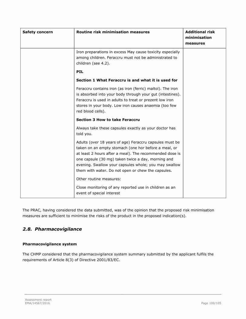
Assessment report
EMA/14567/2016 Page 100/105
Safety concern Routine risk minimisation measures Additional risk
minimisation
measures
Iron preparations in excess May cause toxicity especially
among children. Feraccru must not be administrated to
children (see 4.2).
PIL
Section 1 What Feraccru is and what it is used for
Feraccru contains iron (as iron (ferric) maltol). The iron
is absorbed into your body through your gut (intestines).
Feraccru is used in adults to treat or prezent low iron
stores in your body. Low iron causes anaemia (too few
red blood cells).
Section 3 How to take Feraccru
Always take these capsules exactly as your doctor has
told you.
Adults (over 18 years of age) Feraccru capsules must be
taken on an empty stomach (one hor before a meal, or
at least 2 hours after a meal). The recommended dose is
one capsule (30 mg) taken twice a day, morning and
evening. Swallow your capsules whole; you may swallow
them with water. Do not open or chew the capsules.
Other routine measures:
Close monitoring of any reported use in children as an
event of special interest
The PRAC, having considered the data submitted, was of the opinion that the proposed risk minimisation
measures are sufficient to minimise the risks of the product in the proposed indication(s).
2.8. Pharmacovigilance
Pharmacovigilance system
The CHMP considered that the pharmacovigilance system summary submitted by the applicant fulfils the
requirements of Article 8(3) of Directive 2001/83/EC.

Assessment report
EMA/14567/2016 Page 101/105
2.9. Product information
2.9.1. User consultation
The results of the user consultation with target patient groups on the package leaflet submitted by the
applicant show that the package leaflet meets the criteria for readability as set out in the Guideline on the
readability of the label and package leaflet of medicinal products for human use.
3. Benefit-Risk Balance
Benefits
Beneficial effects
In the pivotal trial Feraccru has shown to significantly improve the Hb concentration (change of 2.25 gr/dl
from baseline) over 12 weeks of treatment in IBD patients compared to placebo. The internal validity of the
result is supported by the analysis in ITT and PP populations and several sensitivity analyses.
The Hb recuperation was progressive but the largest effect was seen within the first 4 weeks (change of 1.08
gr/dl from baseline). Feraccru normalized Hb concentration in the 65% of the patients with a median time to
normalization of around 2 months. 78.3% of patients got an increase of 1 gr/dl and 56.7% achieved an
increase of 2 gr/dl. In addition, Feraccru treatment has shown to achieve substantial mean increases in iron
indices (mainly ferritin and transferrin saturation) although values were highly variable.
The results of the subgroups analyses by gender, ages or underlying conditions (UC and CD) are consistent
with the overall results in terms of the primary endpoint.
The treatment with Feraccru did not cause either a beneficial or detrimental impact on QoL scores in SF 63
and IBDQ scores.
Uncertainty in the knowledge about the beneficial effects
Uncertainties were related to the characteristics of the enrolled population; patients with either quiescent UC
(SCCAI score of <4) or quiescent CD (CDAI score of <220) and anemia exclusively due to IDA with HB levels
>9.5 gr/l, who failed to oral ferrous products and without previous iv or oral iron; patients suffered from
mild or moderate anaemia which neither required rapid iron supplement nor affect significantly the patients
QOL, however, the population studied could not be established as "quiescent”. In addition, it is not well
documented that patients were intolerant to previous OFP. Therefore “intolerance to other oral iron
preparations” could not be part of the final indication.
The indication applied for by the applicant is restricted to patients with IBD and did not include patients with
other types of IDA. The applied indication reflects the population selected for the pivotal trial. The possibility
to extend the indication to include patients with other types IDA has not been assessed in view of the
restricted indication applied for by the applicant.

Assessment report
EMA/14567/2016 Page 102/105
Risks
Unfavourable effects
The safety profile of Feraccru seems acceptable. The incidence of adverse reactions is low and from a
qualitative point of view it is in line with the already known profile of other oral iron preparations.
Nevertheless, related TEAES occurred more frequently with ST10 compared to placebo-treatment (25% of
subjects vs. 12%). Severe AEs occurred more often in the ST-10-group (10.9% vs. 4.7% with placebo;
moderate 29.7% vs. 35.9% respectively). Numbers for the GI TEAEs were (ST-10 vs. placebo): any GI TEAE:
43.8% vs. 37.5%, and severe GI-TEAEs: 10.9% vs. 3.1% (moderate GI-TEAEs 17.2% in both groups).
Abdominal pain (including upper abdominal pain and abdominal discomfort was noted for 12 (18.8%) vs. 8
patients (12.5%; St-10 vs. placebo). Severe pain was experienced by 7.8% and 1.6% of patients
respectively (moderate 9.4% vs. 4.7%). About 73% of any GI AEs occurred in the first 12 weeks of
treatment, 58% of the moderate, and 60% of the severe GI-TEAEs.
There were 3 SAEs in one ST10-patient (severe abdominal pain, severe diarrhoea and severe exacerbation of
CD, all in one subject) and 2 SAEs in the placebo group (1subjects each with rectal abscess and moderate
exacerbation of CD; all were considered unrelated by the investigators).
In the open-label phase, 14 subjects discontinued because of adverse events (all causalities) and 5 because
of treatment-related adverse events. Overall, 20/111 (18.0%) discontinued because of adverse events during
ST10 treatment and 8/111 (7.2%) discontinued because of ST10-related adverse events.18% of patients had
gastrointestinal disorders treatment-related (5.5% severe), 7.3% abdominal pain, 4.6% flatulence, and 3.7%
constipation, 2.8% diarrhea and 1.8% nauseas. Cumulatively Ulcerative colitis was expressed as TEAE in
9.9% of patients and Crohn’s disease in 7.2% adding up to 17.1% of patients with TEAE indicating an
aggravation of the chronic disease. 16/111 of these (14.4%) with moderate to severe intensity (4.5% in the
first 12 weeks). In general a lot of the registered TEAEs seemed to be related to the chronic disease of the
study subjects.
Uncertainty in the knowledge about the unfavourable effects
As patients included in the trial suffered from IBD it is difficult to distinguish the AEs due to the underlying
disease from those caused by the treatment.
The database is currently limited in number and exposure. Adverse events related to oral iron molecules as
hypersensitivity reaction have not described in this dossier but it is impossible to rule out them completely.
Comparative rates of TEAEs, related TEAEs, SAEs and withdrawal rates in comparison to standard oral iron
preparations and IV iron treatment are unknown (due to lack of active comparator). Due to the lack of a
direct comparison with standard treatment (no active control) only external comparisons are possible. A
comprehensive overview on published data was not submitted. Due to differences between the ST-10 study
and published studies (definitions and study population) the external comparison was inconclusive.
It has been described in the published literature that continuous exposure to iron can be associated with an
increased risk of colon-rectal cancer although for the time being this is only a theoretical risk; however,
unnecessary exposure should be avoided and a relevant statement on treatment duration based on
haematological parameters has been included in the PI.
Ongoing studies will provide knowledge in potential drug – drug interactions (see RMP).

Assessment report
EMA/14567/2016 Page 103/105
Effects table
Effects Table
Effect Short
Description
Unit Treatment Control Uncertainties/
Strength of evidence
References
Favourable Effects
Hb Changes in Hb
concentration
from baseline
to week 12
gr/dl 2.25 -0.02 Difference between adjusted
means (SE) 2.25 (0.19), 97.5%CI
1.88
(1)
Unfavourable Effects
Abdominal
pain
Overall
incidence
% 11.7(a) 6.7(b) Incidence of treatment -
related AEs: abdominal
pain
Treatment vs control (6.7
vs 5.0)
(1)
Constipation Overall
incidence
% 8.3 (a) 1.7 Incidence of treatment
related AEs:
Constipation. Treatment
vs control (6.7 vs 1.7)
(1)
Flatulence Overall
incidence
% 6.7(a) 0 Incidence of treatment
related AEs: Flatulence.
Treatment vs control (6.7
vs 0)
(1)
Abdominal
discomfort
Overall
incidence
% 5.0 0 Incidence of treatment
related AEs: Abdominal
discomfort. Treatment vs
control (3.3 vs 0)
(1)
Abdominal
distension
Overall
incidence
% 3.3 0 Incidence of treatment
related AEs: Distension.
Treatment vs control (3.3
vs 0)
(1)
Gastrointesti
nal disease
Overall
incidence
% 3.3 0 (1)
Rectal
hemorrhage
Overall
incidence
% 5 (b) 1.7 (1)
Abbreviations: AEs: Adverse events BS: baseline
Notes: (1) Integrated analysis AEGIS1 and AEGIS2
(a) One case leading to discontinuation of study drug
(b) Two cases leading to discontinuation of study drug

Assessment report
EMA/14567/2016 Page 104/105
Benefit-risk balance
Importance of favourable and unfavourable effects
The results showed superiority of Feraccru versus to placebo with an increase of 2.25gr/dl in Hb
concentration from baseline over 12 weeks of treatment with a responder rate of 78.3% of patients achieving
an increase of 1 gr/dl, around 50% of them achieved an increase of 2 gr/dl and 65% normalized Hb
concentration.
The safety profile seems to be in line with that of other iron products.
Benefit-risk balance
The efficacy and safety of Feraccru in iron deficiency anaemia has been studied in a very specific population
sufferring IBD; Therefore the benefit –risk balance is established as positive in the following indication:
"Feraccru is indicated in adults for the treatment of iron deficiency anaemia in patients with inflammatory
bowel diseases (see 5.1)
Discussion on the benefit-risk balance
The efficacy and safety of Feraccru have been studied in a very specific population sufferring IBD, therefore
the indication is established as: "Feraccru is indicated in adults for the treatment of iron deficiency anaemia
in patients with inflammatory bowel diseases (see 5.1)
4. Recommendations
Outcome
Based on the CHMP review of data on quality, safety and efficacy, the CHMP considers by consensus that the
risk-benefit balance of Feraccru in the treatment of iron deficiency anaemia (IDA) in patients with
inflammatory bowel disease (IBD) is favourable and therefore recommends the granting of the marketing
authorisation subject to the following conditions:
Conditions or restrictions regarding supply and use
Medicinal product subject to medical prescription.
Conditions and requirements of the Marketing Authorisation
Periodic Safety Update Reports
The requirements for submission of periodic safety update reports for this medicinal product are set out in
the list of Union reference dates (EURD list) provided for under Article 107c(7) of Directive 2001/83/EC and
any subsequent updates published on the European medicines web-portal.

Assessment report
EMA/14567/2016 Page 105/105
Conditions or restrictions with regard to the safe and effective use of the medicinal product
Risk Management Plan (RMP)
The MAH shall perform the required pharmacovigilance activities and interventions detailed in the agreed
RMP presented in Module 1.8.2 of the Marketing Authorisation and any agreed subsequent updates of the
RMP.
An updated RMP should be submitted:
At the request of the European Medicines Agency;
Whenever the risk management system is modified, especially as the result of new information
being received that may lead to a significant change to the benefit/risk profile or as the result of an
important (pharmacovigilance or risk minimisation) milestone being reached.
Additional risk minimisation measures
Not applicable.
Conditions or restrictions with regard to the safe and effective use of the medicinal product to be implemented by the Member States.
Not applicable.
These conditions fully reflect the advice received from the PRAC.
New Active Substance Status
Based on the CHMP review of data on the quality, non-clinical and clinical properties of the active substance,
the CHMP considers that ferric maltol which is a complex of iron is not qualified as a new active substance as
it does not differ significantly in properties with regard to safety and efficacy from the previously authorised
substance.