Embed Size (px)
Citation preview

Feasibility of HPV16, HPV18, and p16 Expression as Biomarkers for Distinguishing Normal Oral Epithelium From Oral Epithelial Dysplasia and Oral Intraepithelial Neoplasia
Koji TSUNEMATSU1,2¶), Eiji NAKATANI3¶), Teruaki IWAHASHI1¶), Katsumi HIDESHIMA1¶), Masaaki KARINO1¶), Yoshiki NARIAI4¶), Takahiro KANNO1¶), Tatsuo KAGIMURA3¶)and Joji SEKINE1)
1)Department of Oral and Maxillofacial Surgery, Shimane University Faculty of Medicine, Izumo, 693-8501, Japan2)Division of Oral and Maxillofacial Surgery, National Hospital Organization Hamada Medical Center, Hamada, 697-8511, Japan3)Translational Research Informatics Center, the Foundation for Biomedical Research and Innovation, Kobe, 650-0047, Japan4)Department of Oral and Maxillofacial Surgery, Matsue City Hospital, Matsue, 690-8509, Japan(Received December 25, 2015; Accepted December 28, 2015)
This preliminary study evaluated the expression of human papilloma virus (HPV) 16, HPV18, and p16 as possible biomarkers to distinguish normal oral epithelium (NOE) from oral epithelial dyspla-sia (OED) and oral intraepithelial neoplasia (OIN). Subjects comprised 150 cases including 41 with OED, 30 with OIN, and 67 with oral squamous cell carcinoma (OSCC). NOE was also taken from 12 healthy participants. Staining indices of HPV16, HPV18, and p16 were immunohistochemically ex-amined using paraffin-embedded specimens. A sta-tistically significant difference was seen in the ex-pression of HPV18 and p16 in OED/OIN compared with NOE and OSCC. Furthermore, regression tree analysis of independent variables, including p16 and HPV18 expression and the participants’ age, was performed to distinguish NOE from OED/OIN. In conclusion, this preliminary study demonstrated that p16 and HPV18 expression and participants’ age (60 years) can be used to distinguish NOE from OED/OIN.
Key words: HPV16, HPV18, p16, oral epithelial dysplasia, oral intraepithelial neoplasia, oral squa-mous cell carcinoma
INTRODUCTION
Many oral squamous cell carcinoma (OSCC) le-sions develop from potentially malignant disorders
(PMDs) [1]. The term “PMD” was defined by the World Health Organization (WHO) as the risk of malignancy being present in a lesion or condition either during the time of initial diagnosis or at a fu-ture date. The WHO also classified PMDs into two subgroups: a) precancerous lesions, benign lesions with morphologically altered tissue, which have a greater than normal risk of malignant transformation; and b) precancerous conditions, which are diseases or patients’ habits that do not necessarily alter the clinical appearance of local tissue, but are associated with a greater than normal risk of precancerous le-sions or cancer development in that tissue [2, 3].
Numerous criteria exist for the diagnosis of oral epithelial dysplasia (OED), and there is not always a clear-cut distinction of what represents mild dys-plasia— consisting of only focal atypia, moderate dysplasia, and severe dysplasia —which may present as carcinoma in situ (CIS) [4]. According to the general rules for clinical and pathological studies on oral cancer [5], mild and moderate dysplasia are defined as OED, while severe dysplasia is defined
Address for Corresponding author: Joji Sekine, D.D.S., Ph.D., FIBCSOMSDepartment of Oral and Maxillofacial Surgery, Shimane University Faculty of Medicine, Izumo, 693-8501, JapanTel: +81-853-20-2301Fax: +81-853-20-2299E-mail: [email protected] ¶ These authors contributed equally to this work
69Shimane J. Med. Sci., Vol.32 pp.69-79, 2016

as oral intraepithelial neoplasia (OIN). As for CIS and OIN, however, a definitive distinction cannot always be drawn between mild and moderate dys-plasias and CIS/OIN. Dysplasia that encompasses an area greater than what could be considered “focal”, when extremely severe in degree or when exhibiting a “top to bottom” change, particularly with respect to basilar hyperplasia, must be diagnosed as CIS, provided of course that it has not progressed to the point of true invasion of the connective tissue [4].
Although OED and CIS are defined by the pres-ence of dysplastic cells in the epithelium, accurate clinical and histopathological diagnoses have been controversial [6]. The most important research focus must be on the development of molecular or histo-logical markers that allow a stricter differentiation to be made between the diagnosis of normal tissue and OED/OIN, because histopathological diagnosis of OSCC has been established in routine paraffin-embedded specimens [3-5]. Recently, cytokeratin 13 and 17 are reportedly suitable for such a distinction between non-neoplastic tissue and a dysplastic or neoplastic (malignant) oral lesion; however, a clear distinction between OED/OIN and CIS is currently unavailable [7]. Furthermore, nucleus accumbens-associated protein 1 (NAC1) was reported to be a potential biomarker for distinguishing OED from CIS/OSCC, using a cut-off value of 50% with the NAC1 labeling index (LI) [8]. However, Ohira et al. [9] reported that NAC1 was not a definitive biomarker for distinguishing oral malignancies from non-malignancies.
The natural history of the human papilloma virus (HPV) has been characterized extensively in the
uterine cervix [10-14], whereas much less data are available on the difference phases of HPV infection and oncogenesis in the oral, head, and neck regions
[14]. The identification of HPV in oropharyngeal carcinoma has prognostic significance, with longer survival and a higher rate of response to therapy in cases positive for HPV [14-16]. In the field of OSCC, HPV infection was found to be present in 26.0% of cases, and high risk (HR)-HPV-positive cases have similar clinical characteristics as HR-HPV-negative cases, but had a significantly worse prognosis [17-20]. However the detail of HPV identification and the roles of these infections in
terms of the prognosis and carcinogenesis still re-main unclear especially in OSCC [19-21].
Also, detection of HPV DNA alone is not strin-gent proof of HPV-induced carcinogenesis [19-21]. Only transcriptionally active HPV DNA is biologically and clinically relevant for carcinogenesis
[22]. HPV integration in the infected cell results in the deletion of the viral E2 gene promoter caus-ing transcription of E6 and E7. Binding of the E7 oncoprotein to the Rb protein leads to Rb protein degradation and presumably to the compensatory overexpression of both cytoplasmic and nuclear p16 protein in HPV infected cells; hence, p16 is used widely as a surrogate biomarker for HPV infection
[22, 23]. In this preliminary study, we thus evaluated
the association between the expression of HPV16, HPV18, and p16 and various lesions derived from the oral epithelium, immunohistochemically, test-ing the hypothesis that the expression of HPV16, HPV18, and p16 could be feasible biomarkers to distinguish PMDs in the oral cavity. Furthermore, we evaluated the expressions of HPV16, HPV18, and p16 in OSCCs as an index of carcinogenesis to compare these with those found in PMDs.
METHODS
Participants and samplesAll participants with clinically diagnosed OIN,
OED, and OSCC underwent a preoperative biopsy at the Department of Oral and Maxillofacial Surgery, Shimane University Hospital, Japan from April 1980 to January 2014. Normal oral epithelium (NOE) was taken from healthy volunteers with no symp-toms or medical history of any oral mucous disorder who provided consent for their samples to be used as standard controls.
Biopsy specimens taken from the margin of the oral mucosal lesions located on the tongue, gingiva, buccal mucosa, lip, and palate were fixed with 10% neutral buffered formalin for 24 h, processed as routine paraffin-embedded sections, stained with he-matoxylin and eosin, and diagnosed by pathologists of the Department of Pathology. All cases were also diagnosed according to the WHO classification [5]. NOE samples taken from normal oral mucosa were
70 Tsunematsu et al.

also processed as paraffin-embedded sections.All participants provided informed consent to par-
ticipate, following approval of the study protocol (approval no. 1676; November 12, 2014) by the ethics committee of Shimane University Hospital.
HPV16, HPV18, and p16 immunohistochemistryHPV16 and HPV18 expression was determined
immunohistochemically using an anti-HPV16 E1+E4 antibody (Abcam, Cambridge, UK; diluted at 1:100) and an anti-HPV18 E6 antibody (Abcam, Cambridge, UK; diluted at 1:500). As a surrogate marker of HPV presence, p16INK4a (VENTANA, AZ, USA, ready to use) was also used.
After deparaffinization and rehydration, antigen retrieval was performed by autoclaving at 120°C for 20 min in citrate buffer solution (pH6.0). After autoclaving, the slides were allowed to cool to room temperature and the sections were incubated for 30 min in 0.3% hydrogen peroxide in methanol to quench endogenous peroxidase activity. The sections were incubated sequentially with 10% rabbit block-ing serum to block nonspecific reactions. Sequen-tially, the sections were incubated with each primary antibody overnight at 4°C. Immunoperoxidase stain-ing was performed using an EnVision™+ Kit (Dako, CA, USA). Counterstaining was done with Mayer’s hematoxylin (MUTO PURE CHEMICALS Co., Ltd., Tokyo, Japan). Negative controls for immunohisto-chemistry were incubated with phosphate-buffered saline instead of the primary antibodies and showed no positive reaction.
HPV16, HPV18, and p16 SIs All sections were examined using a standard light
microscope with a ×40 objective lens. An attached digital camera was used to capture images and es-timate the number of HPV16-, HPV18-, and p16-positive cells (at least 100 cells/field) [24]. In OSCC, three high-power (×40 objective) fields in full sections were selected to represent the spectrum of staining seen on the initial overview of the whole section. The SI (stained cells / total cells counted ×100 [%]) was expressed as the percentage of positive cells among the total number of cells in the area scored [24]. In NOE, OED, and OIN, at least three high-power (×40 objective) fields were
selected (which were always selected from whole epithelial layers to avoid errors for scoring a partial dense area), and the percentage of positive epithelial cells among the total number of epithelial cells was scored.
Statistical analysisThe participants were stratified according to a
pathological classification with four levels: NOE, OED, OIN, and OSCC. In addition to analysis by all participants, subgroup analysis in participants with NOE, OED, and OIN was performed. Continu-ous and categorical variables were summarized as the mean ± standard deviation (SD) and frequency
(percentage), respectively. To explore an increasing or decreasing trend of variable distribution in the order of NOE, OED, OIN, and OSCC (NOE, OED and OIN), the Jonckheere-Terpstra test and Cochran-Armitage test were used for continuous and categor-ical variables, respectively. In addition, to construct a clinically useful decision tool for the diagnosis of NOE or OED/OIN, regression tree analysis was performed using a conditional inference method with a splitting criterion of p < 0.05. Age, sex, posi-tive/negative, and SIs for HPV16, HPV18, and p16 were used as candidate predictors in the regression tree analysis. A p-value < 0.05 was considered significant. All statistical analyses were performed using SAS™ version 9.3 (Cary, NC, USA) and R version 3.2.2 (R Foundation, Vienna, Austria) with “ctree” in “party” library.
RESULTS
Participants’ backgroundsThe participants comprised 150 cases (age range:
29-91 years), 12 with NOE (48-76 years), 41 with OED (39-86 years), 30 with OIN (29-91 years), and 67 with OSCC (30-90 years). Sex and age distributions are shown in Table 1. The participants’ age increased in the order of NOE (mean ± SD: 60.7 ± 10.1 years), OED (65.2 ± 12.6 years), and OIN (71.7 ± 10.8 years; trend test: p = 0.002, Table 1). However, there was no significant trend when the OSCC group (63.8 ± 15.4 years) was included in the analysis (p = 0.823). Sex distribu-tion of the lesions had no significant trend in the
71HPV16, HPV18 and p16 in oral lesions

Abbreviations: NOE: normal oral epithelium, OED: oral epithelial dysplasia, OIN: oral intraepithelial neoplasia, OSCC: oral squamous cell carcinoma,
CIS: carcinoma in situ, SCC: squamous cell carcinoma, WHO: World Health Organization, NE: not evaluated
Table 1. Participants’ information
72
Fig. 1. Expression of HPV16, HPV18, and p16 in oral squamous cell carcinoma (OSCC). Abbreviations are those used in Table 1. In OSCC, staining for HPV16-positive cells was distributed in the nucleus of dysplastic or tumor cells (A, ×40). Staining for HPV18-positive cells was distributed predominantly in the nucleus of dysplastic or tumor cells (B, ×40), and staining for p16-positive cells was distributed predominantly in the nucleus and/or cytoplasm of dysplastic or tumor cells (C, ×40)
Tsunematsu et al.
order of NOE, OED, and OIN (p = 0.755), but had a significant trend when the OSCC group was included (p = 0.010).
According to the general rules for clinical and pathological studies on oral cancer, cases with mild dysplasia were classified into OED (31 cases), and cases with severe dysplasia and CIS were classified into OIN (severe dysplasia: 12 cases; CIS: 10 cas-es). In the cases with moderate dysplasia, 10 were classified into OED and 8 were classified into OIN
(Table 1).
Immunohistochemical findings for HPV16, HPV18, and p16
Staining in HPV16-positive cells in OED and OSCC was distributed in the nucleus of dysplas-tic or tumor cells (Fig. 1A). In addition, in NOE, staining in HPV18-positive cells was distributed predominantly in the nucleus of parabasal cells and prickle cells. Staining in HPV18-positive cells in OED, OIN, and OSCC was distributed predomi-nantly in the nucleus of dysplastic or tumor cells
(Fig. 1B). Staining in p16-positive cells in OED, OIN, and OSCC was distributed predominantly in the nucleus and/or cytoplasm of dysplastic or tumor cells (Fig. 1C).
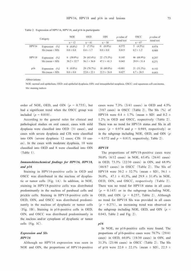
Expression and SIs HPV16
Although no HPV16 expression was seen in NOE and OIN, the proportions of HPV16-positive
cases were 7.3% (3/41 cases) in OED and 4.5% (3/67 cases) in OSCC (Table 2). The SIs (%) of HPV16 were 0.4 ± 1.7% (mean ± SD) and 0.2 ± 1.2% in OED and OSCC, respectively (Table 2). There was no trend for HPV16 status and SIs in all cases (p = 0.974 and p = 0.989, respectively) or in the subgroup including NOE, OED, and OIN (p = 0.572 and p = 0.815, respectively; Table 2).
HPV18 The proportions of HPV18-positive cases were
50.0% (6/12 cases) in NOE, 63.4% (26/41 cases) in OED, 73.3% (22/30 cases) in OIN, and 68.9%
(46/67 cases) in OSCC (Table 2). The SIs of HPV18 were 30.2 ± 32.7% (mean ± SD), 56.1 ± 36.0%, 47.1 ± 41.3%, and 29.9 ± 31.4% in NOE, OED, OIN, and OSCC, respectively (Table 2). There was no trend for HPV18 status in all cases
(p = 0.145) or in the subgroup including NOE, OED, and OIN (p = 0.257, Table 2). Although no trend for HPV18 SIs was provided in all cases
(p = 0.271), an increasing trend was observed in the subgroup including NOE, OED, and OIN (p = 0.043, Table 2 and Fig. 2).
p16In NOE, no p16-positive cells were found. The
proportions of p16-positive cases were 70.7% (29/41 cases) in OED, 60.0% (18/30 cases) in OIN, and 31.3% (21/46 cases) in OSCC (Table 2). The SIs of p16 were 22.0 ± 22.1% (mean ± SD), 22.5 ±
Abbreviations:
NOE: normal oral epithelium, OED: oral epithelial dysplasia, OIN: oral intraepithelial neoplasia, OSCC: oral squamous cell carcinoma,
SIs: staining indices
<
n = 12 n = 41 n = 30 n = 67HPV16 Expression (%) 0 (0.0%) 3 (7.3%) 0 (0.0%) 0.572 3 (4.5%) 0.974
SIs (mean ± SD) 0.0 ± 0.0 0.4 ± 1.7 0.0 ± 0.0 0.815 0.2 ± 1.2 0.989
HPV18 Expression (%) 6 (50.0%) 26 (63.4%) 22 (73.3%) 0.145 46 (68.9%) 0.257SIs (mean ± SD) 30.2 ± 32.7 56.1 ± 36.0 47.1 ± 41.3 0.043 29.9 ± 31.4 0.271
p16 Expression (%) 0 (0.0%) 29 (70.7%) 18 (60.0%) 0.001 21 (31.3%) 0.152SIs (mean ± SD) 0.0 ± 0.0 22.0 ± 22.1 22.5 ± 26.8 0.027 8.7 ± 20.5 0.053
Categoryp-value oftrend test
p-value oftrend test
NOE OED OIN OSCC
Table 2. Expression of HPV16, HPV18, and p16 in participants
73HPV16, HPV18 and p16 in oral lesions

26.8%, and 8.7 ± 20.5% in OED, OIN, and OSCC, respectively (Table 2). Although there was no trend for p16 status and SIs in all cases (p = 0.152 and p = 0.053, respectively), significant trends were found in the subgroup including NOE, OED, and OIN (p < 0.001 and p = 0.027, respectively; Table 2 and Fig. 2).
Discrimination between NOE and OED/OINAge, the SIs of HPV18, and the status and SIs
of p16 had increasing trends in the order of NOE, OED and OIN (Tables 1, 2 and Fig. 3); therefore, age and the variables related to p16 and HPV18 were used further as covariates in regression tree analysis for predicting OED/OIN against NOE. As a result, 4 stratified groups (100.0%, 93.8%, 60.0%, and 30.0% of OED/OIN) by age, p16 status, and HPV18 status were provided. The p16-positive group included all OED/OIN cases; the p16-nega-tive, age > 60 years, and HPV18-positive group in-cluded 93.8% of OED/OIN cases; the p16-negative, age > 60 years, and HPV18-negative group included 60.0% of OED/OIN cases; and the p16-negative and age ≤ 60 years group included 30.0% of OED/OIN cases (Fig. 3).
DISCUSSION
HVP detection strategies vary not just in design, but in their detection targets. These targets have in-cluded HPV DNA, HPV RNA, viral oncoproteins, cellular proteins and HPV-specific serum antibodies
[25]. Recently, multimodality methods of HPV de-tection using p16 immunohistochemistry and HPV DNA detection such as DNA in situ hybridization and PCR based technique is recommended [25].
There are a few limitations of this study that merit consideration. This study was retrospective, some of the materials were preserved for a long pe-riod of time, and the expression of HPV16, HPV18, and p16 was evaluated in formalin-fixed and paraf-fin-embedded tissue. Formalin fixation now appears to have a less damaging effect on the quality of the proteomic analysis obtained from formalin-fixed and paraffin-embedded tissue than had been assumed
[26]. The time in storage of formalin-fixed and paraffin-embedded tissue was initially also thought to be a potential variable factor in protein extraction yield [27, 28]. Several studies have reported that storage of unstained slides long term may lead to false-negative immunostaining for antigens [29]. Re-cent studies have however suggested that there is no significant difference in the number of proteins iden-
74
Fig. 2. Boxplot stratified by pathological classification of staining indices of HPV18 and p16SIs: staining indices; other abbreviations are those used in Table 1. SIs of A: HPV18 and B: p16. NOE showed a lower value than OED/OIN for expression of HPV18 and p16 (p = 0.043, and 0.027, respectively, Jonckheere-Terpstra test)
Tsunematsu et al.

tified from fixed tissues even with prolonged storage [30, 31]. And the antigen retrieval (AR) could
obtain the highly accurate results in this study. The AR technique is a non-enzymatic antigen unmasking method that is utilized prior to immunohistochemi-cal staining of formalin-fixed, paraffin-, celloidin- or plastic-embedded tissue sections [25, 32]. In this study, a non-enzymatic pretreatment based on high-temperature heating was applied.
Although our study did not contain cases with oropharyngeal carcinoma, limited to OSCC lesions, HPV16 was detected in 4.5% of OSCC cases and HPV18 was observed in 68.8% of OSCC cases in formalin-fixed and paraffin-embedded sections of OSCC. Krüger M et al. described in his up-to-date review of 136 literatures of HPV infection in OSCC that the prevalence of OSCC related HPV infection including low risk (LR)-HPV to HR-HPV varies from 0% to 100% and possible reasons for the different prevalence rates could be the detection method used for HPV infection, ethnos, geographical site and collective size [33].
In this study, HPV16 was also detected in OED
(7.3%), while HPV1 8 was observed in OED (63.4%) and OIN (73.3%) in the oral cavity, in-cluding tongue, gingiva, buccal mucosa, lip, and palate samples. HPV causes a range of epithelial lesions from common warts to neoplasia and cancer
[19]. The main criticism of studies that concentrate on the possible premalignant characteristics of OED and OIN is the lack of sufficient clinical and his-tological data to support the initial diagnosis in pa-tients who eventually develop OSCC [5]. Agrawal et al. [34] reported that the SI of HPV16 was 10% in OED, and 60% of HPV16-positive OED were positive for p16INK4a, suggesting a role for HPV16 in the pathogenesis of OED. As for the association of the expression of HPV and PMDs, although LR-HPV-positive lesions result in papillary and atypical proliferation [35], HR-HPV is not always associated with atypical oral lesions [36] and/or OSCC [37].
Conversely, our study showed the expression of HPV18 in NOE (50.0%). According to the detec-tion of HPV by sensitive PCR-based sequencing analysis, HPV18 was detected in 86.7% of adult NOE cases, suggesting that subclinical and latent
0
80
60
40
20
n = 10
Normal
OED/OIN (30.0%)
n = 10
Normal
OED/OIN
n = 16 Normal
OED/OIN
n = 47
OED/OIN
HPV18 p = 0.037
Age p = 0.042
p16 p<0.001
positive
negative
≤60 years
>60 years
positive negative
100
(%) 0
80
60
40
20
100
(%) 0
80
60
40
20
100
(%) 0
80
60
40
20
100
(%) (60.0%) (93.8%) (100.0%)
75
Fig. 3. Result of regression tree analysis for discrimination between NOE and OED/OINAbbreviations are those used in Table 1. The independent variables were p16, HPV16, HPV18, and age. To predict OED/OIN, the primary variable for the hierarchical tree was p16 status, the secondary variable was age (60 years), and the tertiary variable was HPV18 status
HPV16, HPV18 and p16 in oral lesions

HPV infection of NOE is common and HPV18 would be the predominant genotype [38]. Further-more, a low level HPV infection would also suggest that the oral cavity could be a reservoir of HPV, and this infection in combination with other factors is associated with the later development of PMDs, including OED/OIN [34].
To the best of our knowledge, these preliminary results obtained here might shed light on the first attempt in terms of HPV infection to distinguish NOE from OED/OIN in consideration of carcinogen-esis of OSCC [8, 9].
In this study, p16 was used as a surrogate marker for the expression of HPV, as p16 is a surrogate marker for HPV infection in the cervix [39]. It has been observed that HPV16 disrupts the regulation of p16INK4a suppressor protein and its overexpression can be used as surrogate marker for the detection of HPV16 in association with oral SCC [34]. Simi-larly, the value of immunostaining for p16INK4a was shown for the identification of oral dysplastic le-sions [34]. Combined p16 and HPV testing is also reported to be useful for the prediction of head and neck cancer survival [40, 41] as well in a cohort study on oropharyngeal tumors [42].
There have been no reports so far comparing the prevalence of HPV infection in PMDs with that in OSCC by the same detection methods. In our study, we thus compared the expressions of HPV16, HPV18, and p16 in OSCCs with those in PMDs, obtained from the identical immunohistochemistry study as described here. There were no significant trends for HPV16, HPV18, and p16 status and SIs in all study groups. These results might suggest that HPV infections would be related to the initial phase of HPV-induced carcinogenesis.
In this study, a statistical trend test of each vari-able was first performed, then followed by the man-ifest confirmation of statistically significant values, which revealed putative feasible candidates for bio-markers or factors, namely, age, p16, and HPV18, to distinguish NOE from OED/OIN. Further regres-sion tree analysis considering the participants’ age revealed that p16 and HPV18 expression and the participants’ age (60 years) are feasible biomarkers to distinguish NOE and OED/OIN, as summarized in Table 2 and Fig. 3. With respect to the biomark-
ers and factors of aging in this study, their role in the carcinogenesis of oral, head, and neck squamous cell carcinoma remains unclear. In cancer of the cer-vix uteri, HPV infection, such as HR-HPV, together with the senescence programs mediated by HPV infections, repeatedly cause genetic changes in cervi-cal cells that play important roles in carcinogenesis from normal cervical epithelium or precancerous cervical lesions, such as cervical intraepithelial neo-plasia, to SCC transformation, if the virus is not cleared [43]. Further, it has been proposed recently that epigenetic variations and environmental factors, including chronic inflammation and immune evasion of infected cells, are related to persistent HPV infec-tion. These factors could contribute to the high risk of complex genetic diseases such as carcinogenesis
[44-46]. Therefore, the aging variable identified in this study is a possible factor for analysis. In other words, detailed investigation of cases of persistent HPV infection using regression tree analysis is rea-sonable, although further investigations, both in vivo and in vitro, are necessary to determine its role in carcinogenesis of oral, head, and neck squamous cell carcinoma.
In conclusion, the expression of HPV18 and p16 and participants’ age are factors that distinguish NOE from OED/OIN.
ACKNOWLEDGMENTS
The authors would like to thank Prof. Riruke Maruyama (Department of Organ Pathology, Shi-mane University Faculty of Medicine) for providing his expert technical and valuable advice regarding pathological diagnosis.
The staff in the Translational Research Informat-ics Center analyzed the dataset that the principal in-vestigator collected and guarantee the quality of the data.
Conflicts of Interest: None
REFERENCES
1)Mortazavi H, Baharvand M and Mehdipour M (2014) Oral potentially malignant disorders: an
overview of more than 20 entities. J Dent Res
76 Tsunematsu et al.

Dent Clin Dent Prospects 8: 6-14.2)Amagasa T (2011) Oral premalignant lesions.
Int J Clin Oncol 16: 1-4.3)Warnakulasuriya S, Johnson NW and van der
Waal I (2007) Nomenclature and classification of potentially malignant disorders of the oral mucosa. J Oral Pathol Med 36: 575-580.
4)Shafer WG (1983) Benign and malignant tu-mors of the oral cavity. In: A Textbook of Oral Pathology. 4th ed. pp. 98-110, W. B. Saunders Company, Philadelphia.
5)Japan Society for Oral Tumors (2010) Oral in-traepithelial neoplasia (OIN) / (CIS). In: General rules for clinical and pathological studies on oral cancer. pp. 80-81, KANEHARA & CO., LTD., Tokyo.
6)Casparis S, Borm JM, Tektas S, et al. (2015) Oral lichen planus (OLP), oral lichenoid lesions
(OLL), oral dysplasia, and oral cancer: retro-spective analysis of clinicopathological data from 2002-2011. Oral Maxillofac Surg 19: 149-156.
7)Farrukh S, Syed S and Pervez S (2015) Dif-ferential expression of cytokeratin 13 in non-neoplastic, dysplastic and neoplastic oral mucosa in a high risk Pakistani population. Asian Pac J Cancer Prev 16: 5489-5492.
8)Sekine J, Nakatani E, Ohira K, et al. (2015) Nucleus accumbens-associated protein 1 expression has potential as a marker for distinguishing oral epithelial dysplasia and squamous cell carcinoma. PLoS One 10: e0131752. doi: 10.1371/journal.pone.0131752.
9)Ohira K, Hideshima K, Urano T and Sekine J (2015) Is nucleus accumbens-associated protein 1 a feasible marker for distinguishing oral malignan-cies from non-malignancies? First investigation of nucleus accumbens-associated protein 1 expression in oral lesions. Shimane J Med Sci 31: 29-41.
10)Reid R, Stanhope CR, Herschman BR, Booth E, Phibbs GD and Smith JP (1982) Genital warts and cervical cancer. I. Evidence of an association between subclinical papillomavirus infection and cervical malignancy. Cancer 50: 377-387.
11)Barbosa MS, Vass WC, Lowy DR and Schiller JT (1991) In vitro biological activities of the E6 and E7 genes vary among human papillomaviruses of different oncogenic potential. J Virol 65: 292-
298.12)Stubenrauch F and Laimins LA (1999) Human
papillomavirus life cycle: active and latent phases. Semin Cancer Biol 9: 379-386.
13)Doobar J, Quint W, Banks L, et al. (2012) The biology and life-cycle of human papillomavi-ruses. Vaccine 30(Suppl.5): F55-70.
14)Morbini P, Dal Bello B, Alberizzi P, et al. (2013) Oral HPV infection and persistence in patients with head and neck cancer. Oral Surg Oral Med Pathol Oral Radiol 116: 474-484.
15)Fakhry C, Westra WH, Li S, et al. (2008) Improved survival of patients with human papil-lomavirus-positive head and neck squamous cell carcinoma in a prospective clinical trial. J Natl Cancer Inst 100: 261-269.
16) St Guily JL, Jacuard AC, Pétet JL, et al. (2011) Human papillomavirus genotype distri-
bution in oropharynx and oral cavity cancer in France - The EDiTH VI study. J Clin Virol 51: 100-104.
17)Jayasooriya PR, Kurose K, Terai M, et al. (2003) Human papillomavirus in oral cancer from Sri Lanka: prevalence and relationship with clinico-pathological parameters. Oral Med Pathol 8: 45-50.
18)García-de Marcos JA, Pérez-Zafrilla B, Arriaga Á, Arroyo-Rodríguez S and Poblet E (2013) Hu-man papillomavirus in carcinomas of the tongue: clinical and prognostic implications. Int J Oral Maxillofac Surg 43: 274-280.
19)Wong GR, Ha KO, Himratul-Aznita WH, et al. (2014) Seropositivity of HPV 16 E6 and E7 and the risk of oral cancer. Oral Dis 20: 762-767.
20)Ramqvist T, Mints M, Tertipis N, Näsman A, Romanitan M and Dalianis T (2015) Studies on human papillomavirus (HPV) 16 E2, E5 and E7 mRNA in HPV-positive tonsillar and base of tongue cancer in relation to clinical outcome and immunological parameters. Oral Oncol 51: 1126-1131.
21)Sritippho T, Chotjumlong P and Iamaroon A (2015) Roles of human papillomaviruses and p16 in oral cancer. Asian Pac J Cancer Prev 16: 6193-6200.
22)Psyrri A, Prezas L and Burtness B (2008) Oropharyngeal cancer. Clin Adv Hematol Oncol 6:
77HPV16, HPV18 and p16 in oral lesions

604-612. 23)Rautava J and Syrjanen S (2012) Biology of
human papillomavirus infections in head and neck carcinogenesis. Head Neck Pathol 6(Suppl.1): S3–15.
24) Dowsett M, Nielsen TO, A'Hern R, et al. (2011) Assessment of Ki67 in breast cancer: recommendations from the International Ki67 in Breast Cancer Working Group. J Natl Cancer Inst 103: 1656-1664.
25)Westra WH (2014) Detection of human papil-lomavirus (HPV) in clinical samples: evolving methods and strategies for the accurate determina-tion of HPV status of head and neck carcinomas. Oral Oncol 50: 771-779.
26)Balgley BM, Guo T, Zhao K, Fang X, Tavas-soli FA and Lee CS (2009) Evaluation of archi-val time on shotgun proteomicsof formalin-fixed and paraffin-embedded tissues. J Proteome Res 8: 917-925.
27)Gnanapragasam VJ (2010) Unlocking the mo-lecular archive: the emerging use of formalin-fixed paraffin-embedded tissue for biomarker research in urological cancer. BJU Int 105: 274-278.
28)Nirmalan NJ, Harnden P, Selby PJ and Banks RE (2009) Development and validation of a nov-el protein extraction methodology for quantitation of protein expression in formalin-fixed paraffin-embedded tissues using western blotting. J Pathol 217: 497-506.
29)Jacobs TW, Prioleau JE, Stillman IE and Schnitt SJ (1996) Loss of tumor marker-immu-nostaining intensity on stored paraffin slides of breast cancer. J Natl Cancer Inst 88: 1054-1059.
30)Xu H, Yang L, Wang W, et al. (2008) An-tigen retrieval for proteomic characterization of formalin-fixed and paraffin-embedded tissues. J Proteome Res 7: 1098-1108.
31)Hood BL, Conrads TP and Veenstra TD (2006) Mass spectrometric analysis of formalin-fixed paraffin-embedded tissue: unlocking the proteome within. Proteomics 6: 4106-4114.
32)Ralton LD and Murray GI (2011) The use of formalin fixed wax embedded tissue for proteomic analysis. J Clin Pathol 64: 297-302.
33)Krüger M, Pabst AM, Walter C, et al. (2014) The prevalence of human papilloma virus (HPV)
infections in oral squamous cell carcinomas: a retrospective analysis of 88 patients and litera-ture overview. J Craniomaxillofac Surg 42: 1506-1514.
34)Agrawal GP, Joshi PS and Agrawal A (2013) Role of HPV-16 in pathogenesis of oral epithe-lial dysplasia and oral squamous cell carcinoma and correlation of p16INK4A expression in HPV-16 positive cases: an immunohistochemical study. ISRN Pathology 2013: Article ID 807095, http://dx.doi.org/10.1155/2013/807095.
35)Lee HJ and Kim JW (2013) Immunohisto-chemical study on the expression of matrix metal-loproteinase 2 and high-risk human papilloma vi-rus in the malignant progression of papillomas. J Korean Assoc Oral Maxillofac Surg 39: 224-230.
36)McCord C, Xu J, Xu W, et al. (2014) As-sociation of human papilloma virus with atypical and malignant oral papillary lesions. Oral Surg Oral Med Oral Pathol Oral Radiol 117: 722-732.
37) Laprise C, Madathil SA, Allison P, et al. (2016) No role for human papillomavirus infec-tion in oral cancers in a region in southern India. Int J Cancer 138: 912-917.
38)Terai M, Hashimoto K, Yoda K and Sata T (1999) High prevalence of human papillomavi-ruses in the normal oral cavity of adults. Oral Microbiol Immunol 14: 201-205.
39)Guo M, Hu L, Baliga M, He Z and Hughson MD (2004) The predictive value of p16(INK4a) and hybrid capture 2 human papillomavirus testing for high-grade cervical intraepithelial neoplasia. Am J Clin Pathol 122: 894-901.
40)Salazar CR, Anayannis N, Smith RV, et al. (2014) Combined P16 and human papillomavirus testing predicts head and neck cancer survival. Int J Cancer 135: 2404-2412.
41)Cooper T, Biron V, Adam B, Klimowicz AC, Puttagunta L and Seikaly H (2013) Prognostic utility of basaloid differentiation in oropharyngeal cancer. J Otolaryngol Head Neck Surg 42: 57.
42) Melchers LJ, Mastik MF, Samaniego Cameron B, et al. (2015) Detection of HPV-associated oropharyngeal tumors in a 16-year cohort: more than meets the eye. Br J Cancer 112: 1349-1357.
43)Zhang Y, Guo L, Xing P, et al. (2014) In-
78 Tsunematsu et al.

creased expression of oncogene-induced senes-cence markers during cervical squamous cell can-cer development. Int J Clin Exp Pathol 89: 8911-8916.
44)Dueñas-González A, Lizano M, Candelaria M, Cetina L, Arce C and Cervera E(2005) Epigenetics of cervical cancer. an overview and therapeutic perspectives. Mol Cancer 4: 38.
45)Frenandes JV, DE Medeiros FTA, DE Azevedo JC, et al. (2015) Link between chronic inflamma-tion and human papillomavirus-induced carcinogen-esis (Review). Oncol Lett 9: 1015-1026.
46)Song D, Li H, Li H and Dai J (2015) Effect of human papillomavirus infection on the immune system and its role in the course of cervical cancer
(Review). Oncol Lett 10: 600-606.
79HPV16, HPV18 and p16 in oral lesions