Embed Size (px)
Citation preview

www.elsevier.com/locate/ygyno
Gynecologic Oncology
FDG-PET for management of cervical and ovarian cancer
Laura J. Havrileskya,*, Shalini L. Kulasingamb,c, David B. Matcharc,d,e, Evan R. Myersb,c
aDivision of Gynecologic Oncology, Department of Obstetrics and Gynecology, Duke University Medical Center, Box 3079, Durham, NC 27710, USAbDivision of Clinical and Epidemiological Research, Department of Obstetrics and Gynecology, Duke University Medical Center, Durham, NC 27710, USA
cDuke Center for Clinical Health Policy Research, Durham, NC 27705, USAdDepartment of Medicine, Duke University Medical Center, Durham, NC 27710, USA
eDepartment of Veterans Affairs, Durham, NC 27701, USA
Received 25 October 2004
Available online 19 January 2005
Abstract
Objective. To assess the diagnostic performance of Positron Emission Tomography using fluorodeoxyglucose (FDG-PET) in comparison
to conventional imaging modalities in the assessment of patients with cervical and ovarian cancer.
Methods. Studies published between 1966 and 2003 were identified using an OVID search of the MEDLINE database. Inclusion criteria
were use of a dedicated scanner, resolution specified, z12 human subjects, clinical follow-up z6 months or histopathology as reference
standard, and sufficient data provided to construct a two-by-two table. Two reviewers independently abstracted data regarding sensitivity and
specificity of PET.
Results. 25 studies (15 cervical cancer, 10 ovarian cancer) met inclusion criteria for full text review. For cervical cancer, pooled sensitivity
and specificity of PET for aortic node metastasis are 0.84 (95% CI 0.68–0.94) and 0.95 (0.89–0.98). Pooled sensitivity and specificity for
detection of pelvic node metastasis are: PET, 0.79 (0.65–0.90) and 0.99 (0.96–0.99); MRI, 0.72 (0.53–0.87) and 0.96 (0.92–0.98). Pooled
sensitivity for CT is 0.47 (0.21–0.73) (pooled specificity not available). Pooled sensitivity and specificity of PET for recurrent cervical cancer
with clinical suspicion are 0.96 (0.87–0.99) and 0.81 (0.58–0.94). For ovarian cancer, pooled sensitivity and specificity to detect recurrence
with clinical suspicion are: PET, 0.90 (0.82–0.95) and 0.86 (0.67–0.96); conventional imaging, 0.68 (0.49–0.83) and 0.58 (0.33–0.80); CA-
125, 0.81 (0.62–0.92) and 0.83 (0.58–0.96). When conventional imaging and CA-125 are negative, pooled sensitivity and specificity of PET
are 0.54 (0.39–0.69) and 0.73 (0.56–0.87), respectively. When CA-125 is rising and conventional imaging is negative, the pooled sensitivity
and specificity of PET are 0.96 (0.88–0.99) and 0.80 (0.44–0.97).
Conclusions. There is good evidence that PET is useful for the pre-treatment detection of retroperitoneal nodal metastasis in cervical
cancer. There is fair evidence that PET is useful for the detection of recurrent cervical cancer. PET is less useful for the detection of
microscopic residual ovarian cancer but has fair sensitivity to detect recurrence in the setting of a rising CA-125 and negative conventional
imaging studies. Available studies are limited by low numbers of patients and wide confidence intervals.
D 2004 Elsevier Inc. All rights reserved.
Keywords: Positron emission tomography; Cervical cancer; Ovarian cancer
Introduction
Computed tomography (CT) and magnetic resonance
(MRI) are anatomic, high-resolution imaging techniques
that are commonly used to guide the management of
patients with gynecologic cancers. Despite their widespread
0090-8258/$ - see front matter D 2004 Elsevier Inc. All rights reserved.
doi:10.1016/j.ygyno.2004.12.007
* Corresponding author. Fax: +1 919 684 8719.
E-mail address: [email protected] (L.J. Havrilesky).
use, concerns remain that use of these conventional imaging
techniques may result in false negatives due to their inability
to resolve small volumes (diameter b1 cm) of disease and
false positives due to their inability to distinguish between
viable tumor masses and masses consisting of necrotic or
scar tissue [1]. Functional imaging methods such as positron
emission tomography (PET) can establish the metabolic or
functional parameters of tissue and may aid in these
distinctions. Instead of using anatomical deviations to
identify areas of abnormality, PET uses positron-emitting
97 (2005) 183–191
L.J. Havrilesky et al. / Gynecologic Oncology 97 (2005) 183–191184
radioactive tracer that accumulates in abnormal tissue. The
most commonly used radioisotope tracer is 18Fluro-deoxy-
glucose (FDG), a glucose analog which is preferentially
taken up by and retained within malignant cells. Depending
on the area or organ under study, baseline glucose
metabolism may be low, further establishing the difference
between normal background tissue and tumor. Thus,
compared to structural imaging techniques, FDG-PET has
the potential to be a more accurate technique for diagnosis,
staging, and treatment decisions in oncology.
The purpose of this study was to determine via a
structured literature review the diagnostic accuracy of
FDG-PET in comparison to conventional structural imaging
techniques for assessment of the metastatic spread and
recurrence of cervical and ovarian cancer. In particular, this
study addressed the following questions: (1) How does the
diagnostic test performance of FDG-PET compare to
conventional imaging (e.g., CT, MRI) in the detection of
pre-treatment metastases in newly diagnosed cervical
cancer? (2) How does the diagnostic test performance of
FDG-PET compare to conventional imaging in detection of
residual or recurrent cervical cancer following treatment
(surgery, radiation, chemotherapy, or combination therapy)?
(3) How does the diagnostic test performance of FDG-PET
as an adjunct to conventional imaging (e.g., CT, MRI)
compare to conventional imaging alone for ovarian cancer
(a) in staging at the time of initial diagnosis, (b) in detecting
recurrent disease following treatment (surgery, radiation,
chemotherapy, or combination)? (4) Does FDG-PET accu-
rately and reliably detect recurrence in a patient with a
history of ovarian cancer who has a rising CA-125 and a
negative CT?
Methods
We performed our search as part of a review done for the
Centers for Medicare and Medicaid Services [2]. An OVID
search of the Medline database was conducted on April
18th, 2003. Filters and limitations were used to eliminate
inappropriate publications. General inclusion criteria were
applied to maximize the applicability of the search results.
The search used applicable MeSH headings and text words
with appropriate Boolean operators. The search strategy
combined the concepts of bcervical cancerQ or bovariancancerQ and bpositron emission tomographyQ. For PET,
we used the Medical Subject Heading (MeSH) term
btomography, emission computedQ and text word searches
for bPETQ and bFDG-PETQ. After filtering irrelevant pub-
lication types, individual review of the abstracts was per-
formed to identify articles for full review.
Two levels of inclusion criteria were used for accepting
studies. The first were general criteria applied during the
initial literature search, and were as follows: (1) English
language articles reporting primary data and published in a
peer review journal (not abstracts); (2) Studies that include
at least 12 human subjects (not animal studies) with cervical
or ovarian cancer who underwent FDG-PET scan. A second
level of inclusion criteria was applied to all articles
identified for full text review based on a review of the
abstracts. Prior to full text review, these articles were
screened to ensure that they answered at least one of the
study questions.
In order to provide a framework for systematically
identifying and reviewing relevant studies, we used a
classification described by Fryback and Thornbury [3].
Briefly, the categories are: (1) Technical feasibility and
optimization, (2) Diagnostic accuracy, (3) Diagnostic think-
ing impact, (4) Therapeutic choice impact, (5) Patient
outcome impact, and (6) Societal impact. For each clinical
question, we classified articles into a hierarchy of Catego-
ries, from 1 through 6. If an article was solely applicable to
Category 1, it was excluded since these studies relate to
technologies that are under development rather than those
routinely used in clinical practice. For Category 2 articles, a
reference standard for diagnosis was required for all
patients. For Category 5 and 6 articles, the requirement of
12 human subjects was relaxed if simulation modeling with
hypothetical populations was used to calculate survival,
quality-adjusted life expectancy, cost-effectiveness, or cost–
benefit ratios.
Data on patient population characteristics, PET scanner,
conventional imaging modality, criteria for test positivity,
and the results of tests including sensitivity, specificity, and
prevalence of cancer was abstracted. Assessment of the
quality of the study design was based on commonly
accepted study design criteria for obtaining unbiased
estimates of sensitivity and specificity (Rothman and
Greenland, 1998) and included (1) use of a prospective
design, (2) matched study design or use of a randomized
controlled trial, (3) use of a pre-specified cut-point to
determine sensitivity and specificity, and (4) availability of
histology or long-term follow-up information on all patients.
In addition to the above criteria used to describe an ideal
study design, additional criteria for determining the quality
of a given study were developed and applied during data
abstraction. These criteria were as follows: (1) The study
had a representative sample, (2) The setting and selection of
the population under investigation were clearly described,
(3) The study design minimized differences between
patients who received the tests, (4) The scanner model or
the type and resolution of the scanner were mentioned, (5)
Defined criteria were used for test interpretation, (6)
Histopathological or clinical confirmation of disease were
mentioned, (7) The test reader and person assigning the
reference standard diagnosis were blinded. Each article was
reviewed by at least two reviewers. Discrepancies between
reviewers were resolved by consensus.
The sensitivity and specificity for each study were
calculated along with their corresponding confidence
intervals from the true positives, true negatives, false
positives, and false negatives abstracted from each article.

L.J. Havrilesky et al. / Gynecologic Oncology 97 (2005) 183–191 185
Pooled estimates of sensitivity and specificity with 95%
confidence intervals were then calculated [4]. A chi-square
test was used to test for homogeneity. Funnel plots with the
Beggs rank-order correlation test and the Egger regression
approach were used to determine the possibility of
publication bias (the tendency of published studies to have
different results (usually positive findings) from studies
rejected from publication or never submitted for review
(usually negative findings) at an a = 0.1 level [5,6].
Statistical analyses were performed using STATA 8.0
College Station, TX.
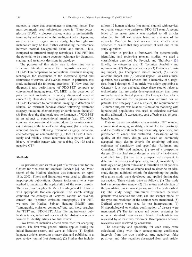
Fig. 1. ROC curve for PET to detect aortic nodal metastasis in newly
diagnosed cervical cancer, with 95% confidence intervals (Area under
curve = 0.952).
Results
Cervical cancer
Thirty-five abstracts were identified of which 20 articles
were deemed potentially relevant to the study questions.
Fifteen original articles met criteria for full text review and
are summarized below. All studies failed to report whether
radiologists were blinded to the pathology results, but none
were excluded for this reason.
Newly diagnosed cervical cancer
Thirteen studies addressed the diagnostic accuracy of
PET in the radiographic assessment of patients with newly
diagnosed cervical cancer. Four prospective studies
addressed the diagnostic accuracy of PET for diagnosis of
aortic node metastasis using histology after aortic lympha-
denectomy as the gold standard [7–10]. The pooled
sensitivity of PET for the detection of aortic node metastasis
is 0.84 (95% CI 0.68–0.94) and pooled specificity is 0.95
(95% CI 0.89–0.98). Results of the individual studies are
summarized in Table 1 and Fig. 1. In three of these studies,
the accuracy of conventional imaging could not be
calculated because inclusion criteria for study entry was a
negative CT or MRI of the abdomen [8–10]. Reinhardt et al.
did not require a negative abdominal imaging study prior to
surgery. Among 12 patients who underwent aortic node
Table 1
Sensitivity and specificity of FDG-PET for detection of aortic node metastasis in
Author (year) N PET MRI
SN (95% CI) SP (95% CI) SN (95%
Reinhardt 2001 12 1.00 (0.29–1.00) 1.00 (0.66–1.00) 0.67 (0.0
Rose 1999 32 0.75 (0.35–0.97) 0.92 (0.73–0.99)
Lin 2003 50 0.86 (0.57–0.98) 0.94 (0.75–0.99)
Yeh 2002 42 0.83 (0.52–0.98) 0.97 (0.32–1.00) 0 (All ha
abdomina
SN = sensitivity.
SP = specificity.
CI = confidence intervals.
sampling, the sensitivity and specificity of MRI for aortic
node metastasis are 0.67 and 1.00 [7]. There was no
evidence of publication bias in these studies using Begg’s
test and Egger’s test.
Four studies addressed the diagnostic accuracy of PET
for diagnosis of pelvic node metastasis (Table 2, Fig. 2)
[7,8,11,12]. Two of these used histology after lymphade-
nectomy as the gold standard [7,8], while two used either
histology or clinical follow-up for confirmation [11,12]. The
pooled sensitivity of PET for detection of pelvic node
metastasis is 0.79 (95% CI 0.65–0.90), while pooled
specificity is 0.99 (95% CI 0.96–0.99). Two studies each
compared PET to MRI and CT. MRI had pooled sensitivity
of 0.72 (95% CI 0.53–0.87) and pooled specificity 0.96
(95% CI 0.92–0.98), while CT had pooled sensitivity of
0.47 (95% CI 0.21–0.73) and not enough data to calculate a
pooled specificity. All four studies concluded that PET has
utility for predicting pelvic nodal disease. Sugarawa et al.
reported the results of patients with both newly diagnosed
and recurrent cancer and included patients in whom the final
diagnosis was not confirmed [11]. For the purposes of this
review, results were calculated from the published raw data
excluding patients with recurrent disease, those without a
confirmed diagnosis, and those in whom studies were read
newly diagnosed cervical cancer
CT
CI) SP (95% CI) SN (95% CI) SP (95% CI)
9–0.99) 1.00 (0.66–1.00)
0 (All had negative
abdominal CT)
0 (All had negative
abdominal CT)
d negative
l MRI)

Table 2
Sensitivity and specificity of FDG-PET for detection of pelvic node metastasis in newly diagnosed cervical cancer
Author (year) N PET MRI CT
SN (95% CI) SP (95% CI) SN (95% CI) SP (95% CI) SN (95% CI) SP (95% CI)
Reinhardt 2001 35 0.91 (0.59–1.00) 1.00 (0.86–1.00) 0.73 (0.39–0.94) 0.83 (0.63–0.95)
Sugarawa 1999 13 1.00 (0.40–1.00) 1.00 (0.66–1.00) 0.50 (0.07–0.93) 1.00 (0.66–1.00)
Rose 1999 32 1.00 (0.71–1.00) 1.00 (0.54–1.00) 0.45 (0.17–0.77)
Belhocine 2002 22 0.56 (0.31–0.78) 0.98 (0.95–1.00) 0.72 (0.46–0.90) 0.97 (0.94–0.99)
SN = sensitivity.
SP = specificity.
CI = confidence intervals.
L.J. Havrilesky et al. / Gynecologic Oncology 97 (2005) 183–191186
as bequivocalQ. Belhocine et al. reported results on a bperlymph nodeQ as opposed to a bper patientQ basis, which may
contribute to bias (by allowing patients with true positive or
true negative findings to be counted multiple times) [12].
The sensitivity and specificity for detection of pelvic node
metastasis from the Belhocine study as presented here were
calculated using the raw data presented in the paper. There
was no evidence of publication bias or heterogeneity in
these four studies.
Two studies addressed the prognostic significance of
PET in newly diagnosed cervical cancer [13,14]. Both
studies are from the same institution and report on patients
treated consecutively during overlapping time periods; it is
unclear whether they represent overlapping patient subsets.
Grigsby et al. retrospectively studied pre-treatment lymph
node staging using PET and CT in 101 consecutive patients
with newly diagnosed stage I–IV cervical cancer prior to
primary radiotherapy [13]. Radiologists were not blinded
and no criteria were given for PET interpretation. The
primary outcome studied was progression-free survival.
Patients with PET-positive and CT-negative aortic lymph
nodes had a 2-year progression-free survival of 18%,
compared to 64% for PET-negative and CT-negative aortic
nodes and 14% for PET-positive, CT-positive aortic nodes.
There were no patients with negative PET and positive CT
findings. In multivariate analysis, PET-positive aortic lymph
Fig. 2. ROC curve for PET to detect pelvic nodal metastasis in newly
diagnosed cervical cancer, with 95% confidence intervals (Area under
curve = 0.970).
node status was the only significant variable predicting
lower progression-free survival (P = 0.025); lymph node
status by CT assessment was not prognostic. However, there
was a difference in treatment between the two groups: aortic
radiation was given to 7 of 7 patients with positive nodes by
CT, but only to 4 of 14 of those with positive nodes only on
PET, and exposure to aortic external radiation was not
included in the survival models. Controlling for treatment
effects is needed to assess whether differences in treatment
contributed to the observed differences in survival.
Miller et al. retrospectively analyzed survival among 47
patients who underwent PET prior to primary radiotherapy
for stage I–IV cervical cancer [14]. PET results were read in
a blinded fashion by 3 radiologists (inter-observer varia-
bility was low); scoring criteria for PET interpretation were
given. Patients with PET-negative lymph nodes had better
overall (P = 0.04) and progression-free (P = 0.03) survival
using Kaplan-Meier analysis at 2 1/2 years than patients
whose lymph nodes were assessed as positive by PET.
When lymph node status was included in a prognostic
scoring system which included primary tumor size and
shape, and heterogeneity of uptake as well as node status,
discrimination was excellent: disease-free survival was 88%
in good prognosis patients and 25% in bad prognosis
patients. Both studies support the prognostic significance of
pre-treatment lymph node staging using PET.
Recurrent cervical cancer
Six retrospective studies addressed the use of PET for
diagnosis of recurrent cervical cancer [12,15–19]. Con-
firmation of radiologic findings was either by histology or
clinical follow-up of 6 months or more in all cases. Three
studies included only patients in whom recurrent cancer was
suspected clinically, with pooled sensitivity of PET 0.96
(95% CI 0.87–0.99) and pooled specificity 0.81 (95% CI
0.58–0.94) [15,18,19]. Chang et al. calculated sensitivity
and specificity on a bper lesionQ as opposed to a bperpatientQ basis, which may contribute to bias (by allowing
patients with true positive or true negative findings to be
counted multiple times) [19]. Results reported here are
analyzed by individual patient (each patient, not each lesion,
is assigned as btrue positiveQ or bfalse negativeQ based on
confirmation of PET results). Two of these studies had a

L.J. Havrilesky et al. / Gynecologic Oncology 97 (2005) 183–191 187
limited ability to calculate specificity since only 1 patient
out of 20 in each study was without recurrence [18,19].
Begg’s test indicates no evidence of publication bias.
Two studies reported the results of surveillance PET
performed without clinical suspicion for recurrence, with
pooled sensitivity of 0.92 (95% CI 0.77–0.98) and pooled
specificity of 0.75 (95% CI 0.69–0.80) [16,17]. Neither
study reported a comparison to conventional imaging
studies, although a prior negative CT or MRI was part of
the inclusion criteria used by Ryu. In a retrospective study,
Belhocine et al. performed PET scans on 38 patients with a
history of cervical cancer. The authors did not separately
analyze PET scans performed for surveillance and those
performed due to clinical suspicion, but reported that PET
had sensitivity and specificity of 1.0 and 0.77 to detect
recurrence, compared to 0.48 and 0.85 for conventional
imaging [12].
The diagnostic accuracy of surveillance PET following
primary treatment was also addressed by Grigsby et al., who
retrospectively reviewed 76 patients undergoing a post-
treatment surveillance PET within 10.4 months of comple-
tion of primary radiotherapy for stage I–IV cervical cancer
[20]. Criteria for PET interpretation were not given. Two-
year progression-free survival was 40% among patients with
persistent PET abnormalities following treatment, compared
to 86% for patients without abnormalities. In a multivariate
analysis, post-treatment abnormal PET was a significant
predictor of death (P b 0.0001). The authors concluded that
post-treatment surveillance PET is predictive of progres-
sion-free survival in patients with a history of cervical
cancer. No data was provided on whether salvage therapies
differed by PET status.
Two studies addressed the possible impact of PET on
therapeutic choices for recurrent cervical cancer [12,21]. In
a retrospective study, Belhocine et al. performed PET scans
on 38 patients with a history of cervical cancer. PET results
contributed to a change in treatment plan in 13/25 (52%)
patients with confirmed recurrences who had an equivocal
result by conventional imaging, suggesting that the addition
of PET to conventional imaging may impact therapeutic
choices for recurrent cervical cancer. Lai et al. [21]
addressed the impact of PET on treatment planning and
clinical outcome in a prospective study of 40 patients with
biopsy-confirmed recurrent or persistent cervical cancer.
Patients underwent PET as well as CT or MRI prior to
salvage treatment planning. The authors found that the
treatment plan was altered by the PET results in 22/40
(55%) of patients. In two-thirds of cases in which treatment
planning was changed by PET, the treatment objective was
changed from curative to palliative intent. The authors
compared the survival of patients who underwent PET prior
to surgery to the survival of a group of historical controls
with recurrent cervical cancer who underwent surgery
without prior PET. Surgically managed patients who had
PET scans prior to treatment planning had a significantly
better 2-year overall survival following surgery compared to
the historical control group managed surgically without PET
(HR 0.21, 95% CI 0.05–0.83).
Ovarian cancer
The literature search identified 36 abstracts. Review of
the abstracts identified 19 articles for full text review. Of the
19 articles, 10 met the criteria for full text review; these are
discussed below and classified by pre-imaging clinical
scenario. There were 3 prospective studies [22–24]. All of
the articles meeting criteria for review addressed the
detection of recurrent or persistent ovarian cancer; none
addressed detection of metastasis at the time of initial
staging. None of the studies addressed whether radiologists
were blinded to the pathology results.
Five studies addressed the use of surveillance PET to
detect recurrent or persistent ovarian cancer in the absence
of clinical suspicion [22,24–27]. Three of these studies
included at least 12 patients and required both negative
serum CA-125 and negative conventional imaging studies
for classification as bno clinical suspicionQ prior to PET
imaging [22,24,25]. The pooled sensitivity of PET in these 3
studies is 0.54 (95% CI 0.39–0.69) and pooled specificity is
0.73 (95% CI 0.56–0.87). There is no evidence of
publication bias using Begg’s test and Egger’s test. In a
prospective study, Rose et al. required a negative abdominal
and pelvic CT and normal CA-125 prior to entry and
performed a second look laparotomy with biopsies on all
patients following the PET scan [22]. The authors reported
that PET has a relatively low sensitivity (0.18) and
specificity (0.45) and concluded that PET is not sensitive
for detection of small volume disease. In a fourth study,
Karlan performed PET on 6 patients who were clinically
free of disease prior to laparotomy. PET failed to detect
microscopic disease in 5 out of 6 patients [27]. In a fifth
study, Cho performed secondary surgery on 31 patients
following primary treatment for ovarian cancer but did not
report pre-operative CA-125 levels and did not require
negative conventional imaging studies [26]. Among 21
evaluable patients, the sensitivity and specificity of PET
were 0.82 and 0.90 compared to 1.0 and 0.90 for CT. The
authors concluded that PET did not improve the diagnostic
accuracy of CT in this setting.
Three studies addressed the use of PET to detect
recurrent ovarian cancer in the setting of a rising CA-125
and negative or equivocal conventional imaging studies
[25,28,29]. The pooled sensitivity of PET is 0.96 (95% CI
0.88–0.99) and pooled specificity 0.80 (95% CI 0.44–0.97).
Two studies were limited by lack of specified minimum
clinical follow-up to confirm PET results [25,29].
Six studies addressed the use of PET to detect recurrent
ovarian cancer when clinical suspicion exists; those includ-
ing at least 12 patients in this category are summarized in
Table 3 and Fig. 3 [23–25,27,30,31]. Pooled PET sensitivity
is 0.90 (95% CI 0.82–0.95) and specificity 0.86 (95% CI
0.67–0.96). Two studies compared PET to the results of CA-

Table 3
Sensitivity and specificity of FDG-PET, conventional imaging, and CA-125 for detection of recurrent ovarian cancer in the presence of clinical suspicion
Author (year) N PET MRI/CT CA-125
SN (95% CI) SP (95% CI) SN (95% CI) SP (95% CI) SN (95% CI) SP (95% CI)
Torizuka 2002 25 0.80 (0.56–0.94) 0.83 (0.36–1.00) 0.55 (0.31–0.77) 0.83 (0.36–1.00) 0.75 (0.51–0.91) 1.00 (0.48–1.00)
Nakamoto 2001 12 0.80 (0.44–0.97) 0.50 (0.01–0.99)
Zimny 2001 58 0.94 (0.85–0.99) 0.75 (0.19–0.99)
Yen 2001 24 0.91 (0.59–1.00) 0.92 (0.64–1.00) 0.91 (0.59–1.00) 0.46 (0.19–0.75) 0.91 (0.59–1.00) 0.77 (0.46–0.95)
Hubner 1993 14 0.91 (0.59–1.00) 1.00 (0.29–1.00)
SN = sensitivity.
SP = specificity.
CI = confidence intervals.
L.J. Havrilesky et al. / Gynecologic Oncology 97 (2005) 183–191188
125 and conventional imaging using MRI or CT [23,30].
The pooled sensitivity of conventional imaging is 0.68 (95%
CI 0.49–0.83), while pooled specificity is 0.58 (95% CI
0.33–0.80). The pooled sensitivity of CA-125 is 0.81 (95%
CI 0.62–0.92), and pooled specificity is 0.83 (95% CI 0.58–
0.96). There was no evidence of publication bias for the
PET results; there were not enough studies reporting on CA-
125 and conventional imaging to report on publication bias.
All authors concluded that PET may be useful in the
detection of recurrent ovarian cancer. Nakamoto also
reported on the possible therapeutic impact of PET imaging,
noting that management of patients changed in 5/12 (42%)
of patients undergoing PET who were initially suspected of
having recurrent disease [24].
Discussion
Cervical cancer
Detection of pre-treatment metastases
Cervical cancer spreads by direct extension and via
lymphatics, with pelvic node metastasis preceding aortic
node metastasis in almost all cases. Sensitive and specific
radiologic imaging modalities that identify occult lymph
Fig. 3. ROC curve for PET to detect recurrent ovarian cancer in the
presence of clinical suspicion, with 95% confidence intervals (Area under
curve = 0.912).
node metastasis may allow avoidance of morbid surgical
procedures and facilitate the planning of such novel, tailored
treatments as intensity-modulated radiation therapy, which
allows a substantial radiation dose to the lymph nodes with
sparing of normal structures [32]. CT and MRI are fairly
inaccurate for the detection of retroperitoneal nodal meta-
stasis; calculated sensitivities range from 24% to 34% for
CT and from 24% to 62% for MRI in studies which include
more than 20 patients [33–35]. Using a meta-analysis of
studies on cervical cancer [36], the pooled sensitivities of
CT and MRI for detecting lymph node metastasis are 47%
and 54%, respectively. Any imaging modality which
improves the accuracy of pre-treatment staging of cervical
cancer has the potential to positively impact survival from
this disease. In this systematic review of the literature, we
found fair evidence that PET is more sensitive than CT or
MRI for detection of retroperitoneal nodal metastasis in
patients with newly diagnosed cervical cancer. Several
prospective studies using pathology report as a gold
standard [7–10] reported that PET has sensitivity superior
to that of conventional imaging in this setting, with
comparable specificities; however, the studies are all limited
by small sample sizes and resulting large confidence
intervals in the estimates of sensitivity and specificity of
both modalities. In addition, two retrospective studies from
the same institution [13,14] demonstrated that pre-treatment
PET findings are predictive of progression-free survival and
possibly overall survival; however, neither study controlled
for potential differences in treatment based on radiology
findings.
It is possible that the addition of PET to the oncologist’s
imaging armametarium may ultimately improve both out-
comes and costs by altering primary management strategies.
For example, a pre-treatment diagnosis of aortic nodal
metastasis changes the patient’s prognosis and has the
potential to change the primary treatment modality from
radical surgery to chemo-radiation, which results in a
different set of expected morbidities. For patients diagnosed
with aortic nodal metastasis, ongoing improvements in
techniques such as intensity-modulated radiation therapy
may result in reduced morbidity [32]. We conclude that
FDG-PET has the potential to have a substantial impact on
treatment choices for newly diagnosed cervical cancer. This

L.J. Havrilesky et al. / Gynecologic Oncology 97 (2005) 183–191 189
potential should be confirmed by performance of properly
designed randomized clinical trials comparing PET to
conventional imaging modalities with treatment based on
PET results.
Persistent and recurrent disease
Earlier detection of recurrent cervical cancer has the
potential to improve survival, since some patients may be
salvaged using radiotherapy or radical surgery [37–39].
Locally recurrent disease is often difficult to detect by
pelvic examination due to thickening of the soft tissue
structures following radiation and/or surgery. Detection of
recurrent disease in the pelvis using MRI and CT is
problematic in this setting because the cancer often grows
by infiltration of tissues which causes only subtle changes in
architecture. Cervical cancer recurrences at the pelvic
sidewall or in pelvic or aortic nodes are unlikely to be
cured, but new treatment modalities such as radical
resection in combination with intraoperative high-dose-rate
brachytherapy have afforded prolonged local control in
preliminary reports [40,41]. As the available treatments for
cervical cancer recurrence improve, the improvement of
imaging modalities to identify recurrences early becomes
more important.
We found fair evidence that PET is useful for the
diagnosis of recurrent cervical cancer. One retrospective
study reported that PET was superior to CT in the presence
of clinical suspicion of recurrence, but small numbers
resulted in wide confidence intervals [15]. Two retrospective
studies found that in the post-treatment surveillance setting,
PET has acceptable sensitivity and specificity but the
authors did not directly compare PET to conventional
imaging [16,17]. A fourth retrospective study [20] demon-
strated that abnormalities on post-treatment surveillance
PET predict lower progression-free survival, while the
appearance of new abnormalities on surveillance PET
predicts poor overall survival. Taken together these data
suggest that PET is a useful adjunct to conventional imaging
and may have the potential to hasten the diagnosis of
recurrent cervical cancer.
If PET is able to improve the detection of recurrent
cervical cancer, there are two potential means of benefit:
improved survival and reduced morbidity. If PET detects
local recurrence earlier and curative salvage therapy is
possible, survival may be improved. If the detection of
metastatic disease by PET leads to the deferral of radical
salvage surgery in favor of palliative measures, unnecessary
morbidity is avoided. Two recent analyses, one prospective
[12,21], suggest that the addition of PET to conventional
imaging to detect recurrent cervical cancer influences
treatment choice in over 50% of cases. In one study, a
comparison to historical controls suggests that patients who
have PET performed prior to surgical salvage have
improved survival [21]. We conclude that PET is a
potentially useful tool for the management of patients with
a history of cervical cancer; again, confirmatory prospective
trials with a direct comparison between PET and conven-
tional imaging modalities are needed.
Ovarian cancer
Recurrence following treatment
Although recurrent ovarian cancer is almost never
curable, early detection of recurrence theoretically affords
a better chance that salvage treatment will result in
prolonged remission and sustained quality of life. Although
CA-125 elevation is often useful in detecting recurrence, it
is not helpful in localizing the disease. Knowledge of the
location of recurrence could guide tailored salvage treat-
ment. For example, a patient with a localized pelvic
recurrence is a candidate for secondary cytoreductive
surgery, while one with miliary peritoneal carcinomatosis
might be better served by treatment with salvage chemo-
therapy. Conventional imaging modalities often give non-
specific results and are suboptimal for the reliable detection
of peritoneal recurrence of ovarian cancer [42–44]. The
identification of more accurate imaging modalities should
improve management decisions for patients with recurrent
ovarian cancer.
Two studies of patients undergoing second look lapa-
rotomy without clinical evidence of recurrence demonstrate
that PET is not sensitive for the detection of microscopic
residual disease [22,27]. In addition, two studies using
histology or clinical follow-up as a standard report that PET
has variable sensitivity for detection of recurrence when
clinical suspicion for recurrence is low [24,25]. When
clinical suspicion for recurrence exists, two studies [23,30]
report that PET has similar sensitivity and specificity to
conventional imaging modalities. Other retrospective stu-
dies [24,25,31] demonstrate that PET has good sensitivity
and specificity in the detection of recurrent ovarian cancer
when there is a clinical suspicion of recurrence. Confidence
intervals are wide due to low numbers. Three studies [25,
28,29] give evidence that PET is helpful for detecting
recurrence when CA-125 is elevated despite negative con-
ventional imaging.
There is one prospective study demonstrating a change in
treatment plan in 25% of cases when PET was added to
conventional imaging for detection of ovarian cancer
recurrence [17]. Most of the impact was among patients
suspected of recurrence based on conventional imaging or
CA-125 prior to PET scan. Evidence that the change in
therapy had any impact on patient outcomes is lacking.
We conclude that PET does not appear to be useful in the
routine surveillance of patients with a history of ovarian
cancer, nor is it likely to improve the sensitivity of
conventional modalities to detect microscopic intraperito-
neal disease. There is fair evidence to support the use of
PET for the detection of recurrent ovarian cancer when the
CA-125 is elevated and conventional imaging is negative or
equivocal, although whether this results in improved patient
outcomes is unclear. A worthwhile prospective study would

L.J. Havrilesky et al. / Gynecologic Oncology 97 (2005) 183–191190
investigate the addition of PET to conventional imaging for
asymptomatic patients with a rising CA-125, with biopsy
confirmation required for diagnosis. Newer technologies
which combine metabolic and anatomic imaging such as
combination PET/CT may have the potential to improve the
accuracy of individual imaging modalities for recurrent
ovarian cancer [45,46].
References
[1] Subak LL, Hricak H, Powell CB, et al. Cervical carcinoma: computed
tomography and magnetic resonance imaging for preoperative staging.
Obstet Gynecol 1995;86(1):43–50.
[2] Matchar D, Kulasingam S, Havrilesky L, Mann L, Myers E, McCrory
D, et al. Evidence report for positron emission testing for six cancers
(brain, cervical, small cell lung, ovarian, pancreatic and testicular).
Agency for Healthcare Research and Quality; 2004.
[3] Fryback DG, Thornbury JR. The efficacy of diagnostic imaging. Med
Decis Mak 1991;11(2):88–94.
[4] Deeks JJ. Systematic reviews of evaluations of diagnostic screening
tests. In: Egger M, Smith GD, Altman DG, editors. Systematic
reviews in health care. Meta-analysis in context. London, England7
BMJ Publishing Group, 2001. p. 248–82.
[5] Begg CB, Mazumdar M. Operating characteristics of a rank correlation
test for publication bias. Biometrics 1994;50(4):1088–101.
[6] Egger M, Davey Smith G, Schneider M, et al. Bias in meta-analysis
detected by a simple, graphical test. BMJ 1997;315(7109):629–34.
[7] Reinhardt MJ, Ehritt-Braun C, Vogelgesang D, et al. Metastatic lymph
nodes in patients with cervical cancer: detection with MR imaging and
FDG PET. Radiology 2001;218(3):776–82.
[8] Rose PG, Adler LP, Rodriguez M, et al. Positron emission
tomography for evaluating para-aortic nodal metastasis in locally
advanced cervical cancer before surgical staging: a surgicopathologic
study. J Clin Oncol 1999;17(1):41–5.
[9] Lin WC, Hung YC, Yeh LS, et al. Usefulness of (18)F-fluorodeoxy-
glucose positron emission tomography to detect para-aortic lymph
nodal metastasis in advanced cervical cancer with negative computed
tomography findings. Gynecol Oncol 2003;89(1):73–6.
[10] Yeh LS, Hung YC, Shen YY, et al. Detecting para-aortic lymph nodal
metastasis by positron emission tomography of 18F-fluorodeoxyglu-
cose in advanced cervical cancer with negative magnetic resonance
imaging findings. Oncol Rep 2002;9(6):1289–92.
[11] Sugawara Y, Eisbruch A, Kosuda S, et al. Evaluation of FDG PET in
patients with cervical cancer. J Nucl Med 1999;40(7):1125–31.
[12] Belhocine T, Thille A, Fridman V, et al. Contribution of whole-body
18FDG PET imaging in the management of cervical cancer. Gynecol
Oncol 2002;87(1):90–7.
[13] Grigsby PW, Siegel BA, Dehdashti F. Lymph node staging by positron
emission tomography in patients with carcinoma of the cervix. J Clin
Oncol 2001;19(17):3745–9.
[14] Miller TR, Pinkus E, Dehdashti F, et al. Improved prognostic value of
18F-FDG PET using a simple visual analysis of tumor characteristics
in patients with cervical cancer. J Nucl Med 2003;44(2):192–7.
[15] Park DHKK, Park SY, Lee BH, Choi CW, Chin SY. Diagnosis of
recurrent uterine cervical cancer: computed tomography versus
positron emission tomography. Korean J Radiol 2000;1(1):51–5.
[16] Ryu SY, Kim MH, Choi SC, et al. Detection of early recurrence with
18F-FDG PET in patients with cervical cancer. J Nucl Med 2003;
44(3):347–52.
[17] Nakamoto Y, Eisbruch A, Achtyes ED, et al. Prognostic value of
positron emission tomography using F-18-fluorodeoxyglucose in
patients with cervical cancer undergoing radiotherapy. Gynecol Oncol
2002;84(2):289–95.
[18] Sun SS, Chen TC, Yen RF, et al. Value of whole body 18F-fluoro-2-
deoxyglucose positron emission tomography in the evaluation of
recurrent cervical cancer. Anticancer Res 2001;21(4B):2957–61.
[19] Chang WC, Hung YC, Lin CC, et al. Usefulness of FDG-PET to
detect recurrent cervical cancer based on asymptomatically elevated
tumor marker serum levels—a preliminary report. Cancer Invest 2004;
22(2):180–4.
[20] Grigsby PW, Siegel BA, Dehdashti F, et al. Posttherapy surveillance
monitoring of cervical cancer by FDG-PET. Int J Radiat Oncol, Biol,
Phys 2003;55(4):907–13.
[21] Lai CH, Huang KG, See LC, et al. Restaging of recurrent cervical
carcinoma with dual-phase [18F]fluoro-2-deoxy-d-glucose positron
emission tomography. Cancer 2004;100(3):544–52.
[22] Rose PG, Faulhaber P, Miraldi F, et al. Positive emission tomography
for evaluating a complete clinical response in patients with ovarian or
peritoneal carcinoma: correlation with second-look laparotomy.
Gynecol Oncol 2001;82(1):17–21.
[23] Torizuka T, Nobezawa S, Kanno T, et al. Ovarian cancer
recurrence: role of whole-body positron emission tomography using
2-[fluorine-18]-fluoro-2-deoxy-d-glucose. Eur J Nucl Med Mol
Imaging 2002;29(6):797–803.
[24] Nakamoto Y, Saga T, Ishimori T, et al. Clinical value of positron
emission tomography with FDG for recurrent ovarian cancer. AJR Am
J Roentgenol 2001;176(6):1449–54.
[25] Zimny M, Siggelkow W, Schroder W, et al. 2-[Fluorine-18]-fluoro-2-
deoxy-d-glucose positron emission tomography in the diagnosis of
recurrent ovarian cancer. Gynecol Oncol 2001;83(2):310–5.
[26] Cho SM, Ha HK, Byun JY, et al. Usefulness of FDG PET for
assessment of early recurrent epithelial ovarian cancer. AJR Am J
Roentgenol 2002;179(2):391–5.
[27] Karlan BY, Hawkins R, Hoh C, et al. Whole-body positron
emission tomography with 2-[18F]-fluoro-2-deoxy-d-glucose can
detect recurrent ovarian carcinoma. Gynecol Oncol 1993;51(2):
175–81.
[28] Chang WC, Hung YC, Kao CH, et al. Usefulness of whole body
positron emission tomography (PET) with 18F-fluoro-2-deoxyglu-
cose (FDG) to detect recurrent ovarian cancer based on asymptoma-
tically elevated serum levels of tumor marker. Neoplasma 2002;49(5):
329–33.
[29] Jimenez-Bonilla J, Maldonado A, Morales S, et al. Clinical impact of
18F-FDG-PET in the suspicion of recurrent ovarian carcinoma based
on elevated tumor marker serum levels. Clin Positron Imaging 2000;
3(6):231–6.
[30] Yen RF, Sun SS, Shen YY, et al. Whole body positron emission
tomography with 18F-fluoro-2-deoxyglucose for the detection of
recurrent ovarian cancer. Anticancer Res 2001;21(5):3691–4.
[31] Hubner KF, McDonald TW, Niethammer JG, et al. Assessment of
primary and metastatic ovarian cancer by positron emission tomog-
raphy (PET) using 2-[18F]deoxyglucose (2-[18F]FDG). Gynecol
Oncol 1993;51(2):197–204.
[32] Portelance L, Chao KS, Grigsby PW, et al. Intensity-modulated
radiation therapy (IMRT) reduces small bowel, rectum, and
bladder doses in patients with cervical cancer receiving pelvic
and para-aortic irradiation. Int J Radiat, Oncol Biol Phys 2001;
51(1):261–6.
[33] Heller PB, Maletano JH, Bundy BN, et al. Clinical–pathologic study
of stage IIB, III, and IVA carcinoma of the cervix: extended diagnostic
evaluation for paraaortic node metastasis—a Gynecologic Oncology
Group study. Gynecol Oncol 1990;38(3):425–30.
[34] Kim SH, Choi BI, Han JK, et al. Preoperative staging of uterine
cervical carcinoma: comparison of CT and MRI in 99 patients.
J Comput Assist Tomogr 1993;17(4):633–40.
[35] Kim SH, Kim SC, Choi BI, et al. Uterine cervical carcinoma:
evaluation of pelvic lymph node metastasis with MR imaging.
Radiology 1994;190(3):807–11.
[36] Scheidler J, Hricak H, Yu KK, et al. Radiological evaluation of lymph
node metastases in patients with cervical cancer. A meta-analysis.
JAMA 1997;278(13):1096–101.

L.J. Havrilesky et al. / Gynecologic Oncology 97 (2005) 183–191 191
[37] Lawhead Jr RA, Clark DG, Smith DH, et al. Pelvic exenteration for
recurrent or persistent gynecologic malignancies: a 10-year review of
the Memorial Sloan-Kettering Cancer Center experience (1972–1981).
Gynecol Oncol 1989;33(3):279–82.
[38] Morley GW, Hopkins MP, Lindenauer SM, et al. Pelvic exenteration,
University of Michigan: 100 patients at 5 years. Obstet Gynecol
1989;74(6):934–43.
[39] Rutledge FN, Smith JP, Wharton JT, et al. Pelvic exenteration:
analysis of 296 patients. Am J Obstet Gynecol 1977;129(8):881–92.
[40] Gemignani ML, Alektiar KM, Leitao M, et al. Radical surgical
resection and high-dose intraoperative radiation therapy (HDR-IORT)
in patients with recurrent gynecologic cancers. Int J Radiat, Oncol
Biol Phys 2001;50(3):687–94.
[41] Leitao Jr MM, Chi DS. Recurrent cervical cancer. Curr Treat Opt
Oncol 2002;3(2):105–11.
[42] Buy JN, Moss AA, Ghossain MA, et al. Peritoneal implants from
ovarian tumors: CT findings. Radiology 1988;169(3):691–4.
[43] Coakley FV, Choi PH, Gougoutas CA, et al. Peritoneal metastases:
detection with spiral CT in patients with ovarian cancer. Radiology
2002;223(2):495–9.
[44] Prayer L, Kainz C, Kramer J, et al. CT and MR accuracy in the
detection of tumor recurrence in patients treated for ovarian cancer.
J Comput Assist Tomogr 1993;17(4):626–32.
[45] Bristow RE, del Carmen MG, Pannu HK, et al. Clinically occult
recurrent ovarian cancer: patient selection for secondary cytore-
ductive surgery using combined PET/CT. Gynecol Oncol 2003;90(3):
519–28.
[46] Pannu HK, Cohade C, Bristow RE, et al. PET-CT detection of
abdominal recurrence of ovarian cancer: radiologic–surgical corre-
lation. Abdom Imaging 2004;18:18.

![FDG-PET in Large Vessel Vasculitis...FDG-PET in Large Vessel Vasculitis 61 5. [18 F]FDG-PET and [18 F]FDG-PET/CT [18 F]FDG-PET is an operator-independent, non- invasive imaging modality](https://img.dokumen.tips/doc/110x75/5f6c13132f0609183b646bce/fdg-pet-in-large-vessel-vasculitis-fdg-pet-in-large-vessel-vasculitis-61-5.jpg)