Embed Size (px)
Citation preview

FDA/ORA/DFS Laboratory Information Bulletin No. 4475 Page 1 of 39
SCREEN FOR THE PRESENCE OF POLYCYCLIC AROMATIC
HYDROCARBONS IN SELECT SEAFOODS USING LC-FLUORESCENCE
Samuel Gratz1*, Angela Mohrhaus1, Bryan Gamble1, Jill Gracie1, David Jackson1, John
Roetting1, Laura Ciolino1, Heather McCauley1, Gerry Schneider1, David Crockett1, Walter Krol2, Terri Arsenault2, Jason White2, Michele Flottmeyer3, Yoko Johnson3,
Douglas Heitkemper1, Fred Fricke1
1Forensic Chemistry Center (FCC), Food and Drug Administration, 6751 Steger Drive, Cincinnati, OH 45237 2Department of Analytical Chemistry, Connecticut Agricultural Experiment Station
3Laboratory Services Division, Minnesota Department of Agriculture
Abstract A liquid chromatography-fluorescence detection (LC-FLD) method has been developed to screen for fifteen targeted parent polycyclic aromatic hydrocarbons (PAHs) at concentrations below the established FDA levels of concern in oysters, shrimp, crabs, and finfish. The procedure was validated by spike recovery experiments of the fifteen parent PAHs at three levels for each matrix, with recoveries ranging from 78 – 99%. Additional spike recovery experiments were conducted for a series of alkylated homologs of naphthalene, dibenzothiophene, and phenanthrene, with recoveries ranging from 87 – 128%. Method accuracy was also evaluated based on analysis of National Institute of Standards and Technology (NIST) Standard Reference Material (SRM) 1974b. PAHs are extracted using a modification of the quick, easy, cheap, effective, rugged, and safe (QuEChERS) sample preparation procedure, employing acetonitrile (CH3CN) as the solvent. The extracts are filtered using 0.2 micron syringe filters, but require no post-extraction sample cleanup for LC-FLD analysis. The chromatographic method employs a polymeric C18 stationary phase designed for PAH analysis with gradient elution to resolve fifteen targeted PAHs in a 35 minute run time. For the analysis of unknowns, a sample that is determined to be positive for a targeted PAH at or above 50% of the FDA level of concern requires that further testing and/or confirmation be performed. Additionally, an estimate of total PAH concentration including alkylated homologs in the sample is calculated. Samples containing total PAH concentrations greater than 50% of the FDA level of concern for naphthalene require that confirmatory analysis be performed. This version of the LIB is an update to the version dated 7/26/2010 that was originally posted on the FDA internet website on 7/27/2010. The Laboratory Information Bulletin is a tool for the rapid dissemination of laboratory methods (or information) which appear to work. It may not report completed scientific work. The user must assure him/her by appropriate calibration procedures that LIB methods and techniques are reliable and accurate for his/her intended use. Reference to any commercial materials, equipment, or process does not in any way constitute approval, endorsement, or recommendation by the Food and Drug Administration.

FDA/ORA/DFS Laboratory Information Bulletin No. 4475 Page 2 of 39
Introduction
Polycyclic aromatic hydrocarbons (PAHs) are found in crude oil in significant amounts, with some exceeding 2000 μg per kg (1). The EPA has reported that the metabolites of many of these compounds have been shown in laboratory test systems to be carcinogens, co-carcinogens, teratogens, and/or mutagens (2). The largest oil spill in U.S. history occurred after the April, 2010 Deepwater Horizon (DWH) oil production platform explosion, with an estimated 4.9 million barrels released from the well into the Gulf of Mexico (3). The sheer volume of oil from this disaster in conjunction with the potential toxicity of the compounds involved is of particular concern to the commercial and recreational fishing industries. Previous environmental tragedies have prompted the development of methods to screen seafood entering the consumer market for the presence of PAHs. However, many previously accepted methods such as the NOAA Technical Memorandum NMFS-NWFSC-59 (4) require extensive clean up procedures as well as fraction collection using size exclusion chromatography. With the large amounts of domestic and exported seafood at risk, a simplified methodology allowing for high sample throughput is necessary. Most of the targeted PAH compounds are strongly fluorescent, and there is an established history of fluorescence-based analysis for the screening of edible seafood for parent PAH compounds, alkylated homologs, and/or metabolites (5-14). Extensive research has been conducted by Krahn, et al. (5) in the use of liquid chromatography equipped with fluorescence detection for the analysis of petroleum related aromatic compounds. Two methods put forth by Ramalhosa, et al. (6) and Pule, et al. (7) make use of the AOAC QuEChERS (quick, easy, cheap, effective, rugged, and safe) method for sample preparation and employ the use of LC-FLD. The current study has been adapted from the two previous methods and tested for applicability on a variety of seafood matrices including oysters, shrimp, finfish, and crab. A variety of sample preparation procedures were explored and it was determined that the required sensitivity could be achieved using 5 g of sample, 15 mL of acetonitrile, and the magnesium sulfate/sodium acetate step of the modified QuEChERS technique, with no additional sample cleanup. Sensitivity of the method is one of the primary concerns. Benzo[a]pyrene, one of the most widely occurring and potent PAHs, as well as six other PAHs have been classified by the EPA as probable human carcinogens (2). The FDA level of concern for benzo[a]pyrene in finfish has been established at 35 ng/g (15). This method’s detection limit has been evaluated at a concentration of 5 ng/g, sufficiently low for the method to be used for screening purposes. Additionally, NIST standard reference material SRM 1974b (16) was used for further verification of the method. This procedure is applicable to screen a variety of seafood matrices including oysters, shrimp, finfish and crab for the presence of parent PAHs and the common alkylated

FDA/ORA/DFS Laboratory Information Bulletin No. 4475 Page 3 of 39 homologs due to oil contamination. The objective of this work is to simplify existing methodology and to increase sample throughput. Experimental
Equipment and Supplies
• Liquid chromatograph (Agilent 1200 series) with binary pump, degasser, autosampler, thermostatted column compartment and multiwavelength fluorescence detector.
• Agilent Chemstation software for controlling LC and data analysis • Zorbax Eclipse PAH analytical column, rapid resolution HT, 4.6 x 50 mm 1.8 μm
(Agilent p/n 959941-918) • Zorbax Eclipse analytical guard column, 4.6 x 12.5 mm 5 μm (Agilent p/n 820950-
939) • Guard column hardware kit, high pressure (Agilent p/n 820888-901) • Centrifuge capable of 3000 x g for 50 mL centrifuge tubes • Balance, sensitivity of 0.1 mg • Adjustable pipettes (10 - 100 μL, 100 - 1000 μL, and 1 - 10 mL) and tips • Centrifuge tube racks for 50 mL (30 mm) tubes • Robot Coupe processor with stainless steel bowl (Robot Coupe p/n R301UB) • Buffered QuEChERS extraction tubes with foil packet containing 6 g of magnesium
sulfate and 1.5 g of sodium acetate (AOAC Method 2007.01, Agilent p/n 5982-5755) • Ceramic homogenizers for 50 mL tubes (100/pk), (Agilent p/n 5982-9313) • Syringes (without needles, nonsterile, BD Luer-Lok Tip, 5 mL capacity), (Fisher p/n
14-823-16D) • PTFE syringe filters (0.20 μm pore size, 25 mm dia.), (Fisher p/n SLFG 025 NK)
Note: to minimize interferences, it is critical that PTFE not be substituted with nylon or other materials.
• 4 mL amber glass vials with PTFE lined caps, (Fisher p/n B7800-2A) All equipment and supplies listed may be substituted with equivalent.
Reagents and Standards
• Acetonitrile (CH3CN), HPLC grade (Fisher p/n A998, or equivalent) • Isopropyl alcohol (IPA), HPLC grade (Fisher p/n A451, or equivalent) • Water, 18.2 MΩ water from a Millipore Milli-Q Gradient A-10 water source (or
equivalent) referred to as DIW (de-ionized water) • NIST Standard Reference Material 1974b, Organics in Mussel Tissue (Mytilus edulis)

FDA/ORA/DFS Laboratory Information Bulletin No. 4475 Page 4 of 39
• Parent PAH Stock Standard mix: 16 parent PAH compounds1 (naphthalene, acenaphthylene, acenaphthene, fluorene, phenanthrene, anthracene, fluoranthene, pyrene, benzo[a]anthracene, chrysene, benzo[b]fluoranthene, benzo[k]fluoranthene, benzo[a]pyrene, dibenzo[a,h]anthracene, benzo[g,h,i]perylene, indeno[1,2,3-cd]pyrene) each at 2000 μg/mL in methylene chloride. Sources include CLP SemiVolatiles PAH standard from Absolute Standards, Inc.2
• Alkylated PAH Homologs Stock Standard Mix: A 20-component PAH compounds stock standard mix (Chiron NPD Cocktail) with components ranging from 100 – 500 μg/mL in isooctane, was obtained from Chiron. Alkylated naphthalenes and alkylated phenanthrenes account for 13 of the 20 components, with concentrations in the range 250 – 500 μg/mL. A listing of the components follows: 1-methylnapthalene; 2-methylnaphthalene; 1,3-dimethylnaphthalene; 1,4-dimethylnaphthalene; 1,5-dimethylnaphthalene; 1,6-dimethylnaphthalene; 1,7-dimethylnaphthalene; 2,6-dimethylnaphthalene; 2,7-dimethylnaphthalene; biphenyl; phenanthrene; 1-methylphenanthrene; 2-methylphenanthrene; 3-methylphenanthrene; 9-methylphenanthrene; dibenzothiophene; 1-methyldibenzothiophene; 2-methyldibenzothiophene; 3-methyldibenzothiophene; and 4-methyldibenzothiophene.
• Parent PAH Individual Solution Standards (200 μg/mL in methanol or methylene chloride) of naphthalene, acenaphthylene, acenaphthene, fluorene, phenanthrene, anthracene, fluoranthene, pyrene, benzo[a]anthracene, chrysene, benzo[b]fluoranthene, benzo[k]fluoranthene, benzo[a]pyrene, dibenzo[a,h]anthracene, benzo[g,h,i]perylene, and indeno[1,2,3-cd]pyrene) were obtained from Supelco Analytical (Bellefonte, PA). Solid state standards of biphenyl and dibenzothiophene were obtained from Aldrich (Milwaukee, WI), and benzo[e]pyrene was obtained from Supelco. Individual standards were used to establish retention times.
• PAH Alkylated Homologs Individual Standards of C1-C4 naphthalenes, C1-C3 fluorenes, and C1-C4 phenanthrenes were obtained as follows: 1-methylnaphthalene, 2-methylnaphthalene, 1,3-dimethylnaphthalene, and 2,7-dimethylnaphthalene were obtained from Acros Organics (Morris Plains, NJ). 1,6-Dimethylnaphthalene, 2,6-dimethylnapthalene, 1-methylfluorene, and 1-methylphenanthrene were obtained from Ultra Scientific (North Kingstown, RI). 1,4-Dimethylnaphthalene, 1,5-dimethylnaphthalene, and 2,3,5-trimethylnaphthalene were obtained from MP Biomedicals (Santa Ana, CA). Stock solutions (500 – 1000 μg/mL) of 1,2,5,6-tetramethylnaphthalene, 1,7-dimethylfluorene, 9-n-propylfluorene, 2-methylphenanthrene, 1,3-dimethylphenanthrene, 1,2,6-trimethylphenanthrene, and 1,2,6,9-tetramethylphenathrene were obtained from Chiron AS (Emeryville, CA).
1Note: Although the mix contains 16 PAH compounds, acenaphthylene does not display any appreciable fluorescence under the method conditions. 2Note: In earlier work, the 2000 μg/mL parent PAH stock standard mix was made using QTM PAH mix from Supelco. The QTM PAH mix does not contain benzo[k]fluoranthene, which must be supplemented (see Appendix for alternate instructions related to the use of the QTM PAH mix).

FDA/ORA/DFS Laboratory Information Bulletin No. 4475 Page 5 of 39 Parent PAH Stock Standards Preparation The CLP SemiVolatiles PAH Stock Standard (or equivalent) is a solution that contains a mixture of sixteen PAHs in methylene chloride, each at a concentration of 2000 μg/mL (ppm). Additional stock solutions were prepared via serial dilution for fortification studies or other uses as follows: 250 μg/mL (ppm) stock standard: 10 mL of this stock spiking solution was prepared by combining 1.25 mL of the CLP SemiVolatiles PAH Stock Standard, followed by dilution to 10 mL with CH3CN. 5.0 μg/mL (ppm) stock standard: 25 mL of a 5 μg/mL spiking solution was prepared by adding 500 microliters of the 250 μg/mL stock standard to a 25 mL volumetric flask, and diluting to volume with CH3CN. 0.5 μg/mL (ppm) stock standard: 25 mL of a 0.5 μg/mL spiking solution was prepared by adding 2.5 mL of the 5 μg/mL stock standard to a 25 mL volumetric flask and diluting to volume with CH3CN. Calibration Standards Calibration standards were prepared at concentrations of 2.5, 25, and 50 ng/mL (ppb) to demonstrate linearity (three points). Dilutions (1:10 and 1:20) of the 0.5 µg/mL (ppm) stock standard with CH3CN were used to prepare the 50 and 25 ng/mL calibration standards; and a further 1:10 dilution of the 25 ng/mL calibration standard was used to prepare the 2.5 ng/mL calibration standard. Three point calibration curves were obtained in triplicate. Check Standards/Continuing Calibration Verification (CCV) Standards For validation studies of laboratory fortified matrices (spike/recovery), the check standard is an external standard made to the same final concentration as the extracts from the spiked matrix samples. Check standards were used for calculation of all sample spike/recoveries based on the peak area ratios of the spiked matrix sample extracts to the appropriate check standard. Parent PAH check standards were prepared by serial dilution of the nominally 250 µg/mL (ppm) stock standard spiking solution. Refer to Table 1 for preparation of check standards at three nominal concentrations. All dilutions are prepared in acetonitrile.

FDA/ORA/DFS Laboratory Information Bulletin No. 4475 Page 6 of 39 For preparation of the alkylated PAH homologs check standard, a 30,000-fold dilution of the alkylated PAH homologs stock mix was made by initial dilution in IPA (25 μL stock mix + 975 μL IPA), followed by serial dilution in CH3CN (25 μL 1st dilution + 975 μL CH3CN, then 53 μL 2nd dilution + 947 μL CH3CN). For batch sample analysis, a continuing calibration verification (CCV) containing parent PAH compounds is analyzed at the beginning and end of each batch of 20 or fewer samples. Typically, this standard is at a concentration near the middle of the calibration range such as the 16.7 ng/mL standard (see also Table 1). Table 1. Dilution scheme for preparation of parent PAH Check/CCV standards (equivalent dilution schemes may be substituted). Spiking Level
Spiked Matrix Sample Solution Final
Concentration(ng/mL)
Check Standard Preparation
First Dilution: Prepare a 1000-fold dilution of the 250 μg/mL stock standard spiking solution by taking a 100 μl aliquot into a 100 ml volumetric flask, and diluting to volume.
High and Mid
33.3
Second Dilution: Prepare a 7.5-fold dilution of the first dilution by taking 133 μl first dilution plus 867 μl CH3CN. Use the second dilution as the check standard for the high and mid level spiked samples.
Low (oysters,
crab, shrimp)
16.7 Third Dilution (oysters, crab, shrimp): Prepare a 2-fold dilution of the second dilution by taking 500 μl second dilution plus 500 μl CH3CN. Use the third dilution as the check standard for the low level spiked samples of oysters, crab, and shrimp.
Low (finfish)
8.31 Third Dilution (finfish): Prepare a 4-fold dilution of the second dilution by taking 250 μl second dilution plus 750 μl CH3CN. Use the third dilution as the check standard for the low level spiked samples of finfish.
Sample Composite Preparation Seafood samples should be prepared by first obtaining the edible portion as described in Table 2, below. Samples are then composited and homogenized by blending in Robot Coupe food processor or equivalent for 2-3 minutes. Seafood samples were stored frozen, but partially thawed prior to preparation for analysis. The matrices used in this validation study included uncooked shrimp purchased at a local grocery store; oysters harvested from Louisiana; and finfish (Spanish mackerel) and blue crab harvested from Alabama. Compositing of multiple individuals from the same site may be appropriate. The minimum sample size for this analysis is 5 grams.

FDA/ORA/DFS Laboratory Information Bulletin No. 4475 Page 7 of 39 Table 2. Directions for obtaining edible tissue portion of selected matrices.
Finfish Remove heads, tails, scales, fins, viscera and bones, save edible portion. If the skin is considered edible, collect it as well.
Crab
Remove the front claw (propus) and the next section of the claw (merus), break the pincher off by pulling down on it. Insert an oyster tool into the opening and break the exoskeleton so that the meat inside can be removed. With the crab head up pull off the top shell (carapace) and discard. Remove viscera and gills. Collect the meat that is around the outer edge of the bottom section of the crab. These are in cartilage sections; an oyster tool can be used to break through the cartilage to obtain the small portion of meat. Approximately 20 grams of meat was typically obtained from a 6 inch blue crab.
Shrimp Remove the head, shell, legs, and tail. Save the remaining edible portion.
Oyster
Find an opening between the top and bottom shell of the oyster to wedge the oyster tool into. When the correct location is found, a small amount of liquid inside the oyster will seep out around the edge where the 2 halves of the oyster come together. Pry the 2 shells apart, then scrape and collect all of the insides including the liquor.
Extraction Procedures Finfish, Shrimp and Crab For analysis of finfish, shrimp and crab, 5 grams of homogenized sample composite and a ceramic homogenizer are transferred to a QuEChERS extraction tube. Five grams of DIW water are then added to the extraction tube followed by vortex mixing or shaking for 1 minute. A 15 mL volume of CH3CN is added to the extraction tube followed by a second one minute vortex or shaking step. Next, the contents of the QuEChERS extraction foil packet (6 g of magnesium sulfate and 1.5 g of sodium acetate) are added to the mixture. The mixture is shaken vigorously for 1 minute; and the extract is centrifuged at 3000 x g for 10 minutes, allowing for removal of the CH3CN (upper) layer. Approximately 6-8 mL of the 15 mL total volume of CH3CN readily separates into the upper layer. A portion (approximately 4 mL) of the supernatant extract is filtered through a 0.2 μm PTFE syringe filter into an amber glass vial and analyzed without further dilution using LC-FLD. Oysters For analysis of oysters, the extraction differs only in that no water is added to the sample, thereby eliminating one mixing step described above for the other matrices. The addition of water to homogenized oyster samples was determined to be unnecessary due to the amount of water present in the native tissue.

FDA/ORA/DFS Laboratory Information Bulletin No. 4475 Page 8 of 39 Fortification / Spike Recovery Samples The analysis of one fortified sample matrix with each batch of 20 or fewer samples is required. Five grams of homogenized composite is fortified with 50 μL of the 5.0 μg/mL stock solution. This provides a fortification level of 0.05 µg/g of sample. SRM 1974b Organics in Mussel Tissue The analysis of SRM 1974b in triplicate is required as an initial demonstration of accuracy. For SRM 1974b, the extraction procedure is identical to that described for oysters. Due to the low levels of PAHs in the SRM, a ten fold concentration step of the filtered extract is necessary. This is accomplished by evaporating 1 mL of extract to dryness under a stream of dry air without heating followed by reconstitution with 100 μL of acetonitrile. Method and Solvent Blanks A method blank must be analyzed with each batch of 20 or fewer samples to monitor for contamination from laboratory sources. Method blanks and fortified method blanks are prepared by substituting 5g of DIW water in place of sample composite and performing the extraction procedure as for oysters described above. Additionally, a solvent blank (CH3CN) should be analyzed between one or more samples or sample types to demonstrate lack of carry over from run to run. Liquid Chromatography with FLD Analysis Samples, standards and blanks were analyzed using an Agilent 1200 Series liquid chromatograph equipped with a binary pump, degasser, autosampler, thermostatted column compartment and a fluorescence detector, all operated under the control of Chemstation software. Separation of PAHs was accomplished at a flow rate of 0.8 mL/min on a Zorbax Eclipse PAH Rapid Resolution HT (4.6 x 50 mm, 1.8 μm) column with a Zorbax Eclipse Analytical Guard Column (4.6 x 12.5 mm, 5 μm). The mobile phase consisted of water and acetonitrile run as a gradient with conditions described in Table 3. The column thermostat was set to 18 oC and all injections were 10 μL. Thorough mobile phase degassing is required to minimize oxygen quenching of PAH fluorescence, with benzo[a]pyrene demonstrating the most susceptibility. Daily purging of the solvent reservoir intake channels, and continuous degassing of mobile phase solvents is required. The use of a helium sparge, especially for startup purging of the solvent reservoir intake channels, has been found to help significantly with quenching issues even when in-line vacuum degassing is used.

FDA/ORA/DFS Laboratory Information Bulletin No. 4475 Page 9 of 39 Table 3. HPLC Mobile Phase Gradient Program
time volume % acetonitrile volume % water Comment 0 60 40
1.5 60 40 7.0 90 10 13.0 100 0 30.00 100 0
Analysis (from 0 to 30 min.)
30.01 60 40 35.00 60 40
Re-equilibration to initial conditions (from 30 – 35 min.)
Fluorescence detection was used with the following parameters: excitation wavelength 260 nm; multiwavelength emission detection (352, 420 and 460 nm); PMT gain setting of 13 for general screening and PMT gain setting 15 for analysis of SRM 1974b only. The three emission wavelengths are necessary for detection of all 15 parent PAH compounds (see Table 4 below). Alkylated homologs of the parent PAHs are generally detected at the same emission wavelength as the parent compound. The PMT gain setting of 13 was chosen to maximize sensitivity for the 0 – 50 ng/mL (ppb) concentration range, and is suitable for this work. Lower PMT gain settings (11 or 12) can be substituted to increase the linear range upwards into the 300 - 500 ng/mL (ppb) range, as needed. Initial work was conducted using programmable wavelength switching, in which the emission wavelength was changed during the chromatographic run. Programmable wavelength switching may provide a minor increase in sensitivity for individual compounds compared to multiwavelength emission detection. Our laboratory observed an average of 16% decrease in signal slope across parent PAH compounds for multiwavelength detection compared to wavelength switching. However, wavelength switching should be limited to the targeted screening of the parent PAHs only, whereas multiwavelength detection may be applied to more general PAH screening, including both the parent PAHs and their alkylated homologs. The conditions for programmable wavelength switching are given in the Appendix for use with fluorescence detectors that do not have multiwavelength capability see Appendix I). (
Table 4. Multiwavelength Emission Detection
Emission λ (nm)
Detected PAH Compounds
352 naphthalene, acenaphthene, fluorene, phenanthrene 420 anthracene, fluoranthene, pyrene, benzo[a]anthracene, chrysene,
benzo[b]fluoranthene, benzo[k]fluoranthene, benzo[a]pyrene, dibenzo[a,h]anthracene, benzo[ghi]perylene
460 indeno[1,2,3-cd]pyrene

FDA/ORA/DFS Laboratory Information Bulletin No. 4475 Page 10 of 39 Data Analysis Blank Run Subtraction for Acetonitrile Gradient Given the gradient conditions, the chromatographic baseline is not flat, and may cause difficulty in obtaining accurate peak integrations, especially for the routine screening of uncontaminated or low level contaminated samples*. The gradient baseline is much more pronounced for detection at 352 nm, but may also be problematic with detection at 420 or 460 nm. For accurate integration, the chromatogram for the solvent blank (CH3CN) should be subtracted out prior to integration using the data system software (this feature is available on Agilent systems). When conducting subtraction of the solvent blank run, ensure that the detection emission wavelengths are matched between the sample and solvent blank chromatograms (i.e., subtract 352 nm solvent blank run from the 352 nm sample run, etc.). Blank run subtraction is recommended for all standards, samples, and method blanks. Choose a mid to late solvent blank run from the sequence for subtraction, i.e., do not use the first solvent blank for subtraction (typically the first injection in the sequence), as the system may not be fully equilibrated. *The issue with the gradient baseline was much less pronounced for the validation studies with the fortified seafood matrices, given the higher signals obtained. Solvent blank run subtraction was not applied to these chromatograms. Method Blank Evaluation Method blanks are used to monitor for background PAH levels associated with all solvents and materials (extraction tubes, reagent packs, etc.) used for sample preparation and extraction. Method blanks frequently show the presence of some naphthalene and phenanthrene, which should be subtracted from the corresponding sample peak areas when calculating the individual amounts. Method blank chromatograms may also be used to assess any unusually high background PAH levels associated with specific batches of solvents or materials. Peak Identification Individual chromatographic peaks were identified based on comparison of their retention times to those of known reference standards. Allowable variability of the LC/FLD retention times should be within 1% of the corresponding standard for peak identification in samples. Q uantitation of Individual PAHs
Concentrations of individual PAHs in the sample extracts are determined by ratioing sample peak areas to the peak areas of reference standards at known concentrations prepared in

FDA/ORA/DFS Laboratory Information Bulletin No. 4475 Page 11 of 39 acetonitrile (external calibration). Calculations may be based on generated external calibration curves or CCV standards. Concentrations of individual PAHs in the samples (w/w) are calculated as follows:
15 mL CH3CN PAH in sample (ng/g) = PAH in extract (ng/mL) X ----------------- 5 g sample
Estimation of Total PAH Concentration A conservative estimate of the total amount of PAHs in samples including alkyl homologues, which are often the major PAH components in weathered crude oil, is determined using the total peak area determined over the RT range of 2.5 – 20 minutes and the sensitivity (slope of the calibration curve) for the least sensitive parent compound which is typically naphthalene. The total area can be obtained as the summation of individually integrated peaks, or via integration of a single “peak” with the baseline drawn from 2.5 – 20 minutes.
Example (using slope data in Table 6): A sample is analyzed and found to contain 10 peaks in the RT range of 2.5 – 20 minutes. Peaks may or may not match RT for
nown parent compounds. k Estimated Total PAHs in extract (ng/mL) =
(total area of peaks integrated from 2.5-20 min after solvent blank run subtraction)
------------------------------------------------------------------------------------------------------- slope for naphthalene (0.43)
Estimated Total PAHs in sample (ng/g) = 15 mL CH3CN Estimated Total PAHs in extract X -------------------- 5 g sample
Criteria for Confirmatory Analysis The LC-FLD method described in this document is considered to be a screening method for PAH contamination in seafood. Any positive or indeterminate findings must be confirmed

FDA/ORA/DFS Laboratory Information Bulletin No. 4475 Page 12 of 39 using the NOAA method (4). Sample results from the LC-FLD method shall be evaluated for 1) individual parent PAH concentrations and 2) estimated total PAH concentration. Individual parent PAH concentrations. The parent PAH compounds for which levels of concern have been established are listed in Table 5. For the LC-FLD screening method, the levels requiring confirmatory analysis have been set at 50% of the FDA established levels of concern. If any one or more parent PAH concentrations exceeds these levels, the sample must undergo confirmatory analysis. Estimated total PAH concentration. The estimated total PAH concentration in the sample is intended as a very conservative estimate that is only used to determine if a confirmatory analysis should be performed, with a confirmatory analysis needed if the total PAH estimate exceeds 50% of the FDA established level of concern for naphthalene, as shown in Table 5. Thus for shrimp and crab the estimated total PAH level requiring confirmatory analysis is 61.5 mg/kg, while in oysters and finfish, the estimated total PAH levels requiring confirmatory analysis are 66.5 and 16.3 mg/kg, respectively. Total fractional amount (Carcinogenic PAH). The total fractional amount is the sum of all percentages for carcinogenic analytes and should be less than 1 for the sample not to be violative. The carcinogenic analytes include benzo[a]anthracene, chrysene, benzo[b]fluoranthene, benzo[k]fluoranthene, benzo[a]pyrene, dibenzo[a,h]anthracene, and indeno[1,2,3-cd]pyrene. Total Fractional Amount = ∑ level determined (mg/kg) / level of concern (mg/kg) QC Elements
• A minimum of three calibration standard levels must be analyzed to demonstrate linearity with r2 ≥ 0.99 for all analytes.
• A single low level spike is required as an initial demonstration of recovery for each
matrix type. Spike recoveries should be demonstrated at 50 ng/g for oysters, crab and shrimp and 25 ng/g for finfish.

FDA/ORA/DFS Laboratory Information Bulletin No. 4475 Page 13 of 39
• The analysis of SRM 1974b in triplicate is required as an initial demonstration of accuracy and precision. Analysis of SRM 1974b should fall within the acceptable range (see Table 11) for 8 or more of the PAHs screened.
• The Method Detection (MDL) for a given analyte should be determined using a
minimum of five replicates of matrix recoveries fortified with approximately 5 ng/g for each of the PAHs identified in Table 5.
• The Limit of Quantitation (LOQ) for a given analyte should be determined according
to 40 CFR Part 1365 using a minimum of 5 replicates of matrix recoveries fortified with approximately 5 ng/g for each of the PAHs identified in Table 5.
• Continuing calibration verification (CCV) standards should be analyzed at the
beginning and end of each batch of 20 or fewer samples. If CCV results do not meet the following criteria, then the entire batch and calibration standards must be reanalyzed. The concentrations of the CCV standards run at the beginning and end of each batch should agree within ≤10% of their known concentrations as determined using the external standard calibration curve (prepared from a minimum of 3 standards).
• A minimum of one fortified sample matrix with each batch of 20 or fewer samples is
required. Recoveries of the 0.05 µg/g PAH spike through the method must be in the range 60% - 130%. The retention times in the spiked samples should be within 1% of the RT of the corresponding standard.
• A minimum of one method blank made with 5 g DIW water in place of sample matrix
must be analyzed with each batch of 20 or fewer samples. The PAH concentrations found in the method blank should be subtracted from the concentrations found in the samples. Some PAHs, such as naphthalene, are ubiquitous and may be difficult to eliminate. With the exception of benzo[a]pyrene, higher background levels may be acceptable. Concentrations in the method blank should not exceed 3 times the certified concentration for the PAH in SRM 1974b.
• A minimum of one sample replicate must be analyzed with each batch of 20 or fewer
samples. For triplicate replicates, the precision is considered acceptable if the percent relative standard deviation (RSD) is < 15 percent for all analytes detected above the LOQ. For duplicate replicates, this translates to a relative percent difference of < 30 percent for all analytes detected above the LOQ.

FDA/ORA/DFS Laboratory Information Bulletin No. 4475 Page 14 of 39 Table 5. FDA established levels of concern for PAHs in shrimp, crab, finfish and oysters and LC-FLD screen levels requiring confirmatory analysis.
FDA level of concern (mg/kg)* LC-FLD screen levels requiring confirmation analysis (mg/kg) Compound Shrimp
and Crab Oysters Finfish Shrimp
and Crab Oysters Finfish
naphthalene 123 133 32.7 61.5 66.5 16.3
acenaphthene NA NA NA NA NA NA
fluorene 246 267 65.3 123 133 32.6
phenanthrene* 923 1000 245
anthracene* 18461 20001 4901 923 1000 245
fluoranthene 246 267 65.3 123 133 32.6
pyrene 185 200 49.0 92.5 100 24.5
benzo[a]anthracene 1.32 1.43 0.35 0.66 0.71 0.17
chrysene 132 143 35.0 66 71.5 17
benzo[b]fluoranthene 1.32 1.43 0.35 0.66 0.71 0.17
benzo[k]fluoranthene 13.2 14.3 3.5 6.6 7.1 1.7
benzo[a]pyrene 0.132 0.143 0.035 0.066 0.071 0.017
dibenzo[a,h]anthracene 0.132 0.143 0.035 0.066 0.071 0.017
benzo[ghi]perylene* NA NA NA NA NA NA
indeno[1,2,3-cd]pyrene 1.32 1.43 0.35 0.66 0.71 0.17
NA = not applicable 1 Represents the sum of level of concern for phenanthrene and anthracene. *For the FDA levels of concern, determination of the alkylated homologues should be included, e.g. C-1, C-2, C-3, C-4 naphthalenes; C-1, C-2, C-3 fluorenes; C-1, C-2, C-3, C-4 anthracene/phenanthrene.

FDA/ORA/DFS Laboratory Information Bulletin No. 4475 Page 15 of 39 Results and Discussion Sample Cleanup Dispersive solid phase extraction (SPE) cleanup has been used in combination with QuEChERS extraction for PAH analysis (7). In this work, the use of primary secondary amine (PSA) and PSA in combination with C18 SPE was evaluated in spike/recovery studies. It was determined that the additional cleanup offered little to no advantage for HPLC with fluorescence detection, and was therefore eliminated from the procedure. As a result, this method requires no post-extraction sample cleanup. Chromatography with FLD The conditions for the chromatographic separation of PAHs were taken directly from previous work (7) and required no further optimization other than adjustments made to the PMT gain setting to increase sensitivity. A representative chromatogram obtained for a standard mixture of the 15 PAHs at concentrations of 33 ng/mL each is presented in Figure 1. Good separation was achieved considering the structural similarities of many of these compounds. The retention window for these compounds is 3.1 to 17.5 minutes (see figure).
0
10
20
30
40
0 5 10 15 20time (min.)
fluor
esce
nce
(LU
)
1 2 13
9
5
14
84
156
12
11
10
73
Figure 1. LC-FLD chromatogram of 15 PAH standard mix, 33 ng/mL each. Peak identifications as follows: 1 naphthalene, 2 acenaphthene, 3 fluorene, 4 phenanthrene, 5 anthracene, 6 fluoranthene, 7 pyrene, 8 benzo[a]anthracene, 9 chrysene, 10 benzo[b]fluoranthene, 11 benzo[k]fluoranthene, 12 benzo[a]pyrene, 13 dibenzo[a,h]anthracene, 14 benzo[g,h,i]perylene, 15 indeno[1,2,3-cd]pyrene.
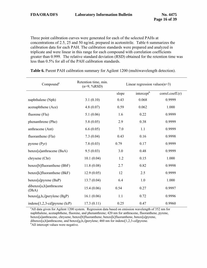
FDA/ORA/DFS Laboratory Information Bulletin No. 4475 Page 16 of 39 Three point calibration curves were generated for each of the selected PAHs at concentrations of 2.5, 25 and 50 ng/mL prepared in acetonitrile. Table 6 summarizes the calibration data for each PAH. The calibration standards were prepared and analyzed in triplicate and were linear in this range for each compound with correlation coefficients greater than 0.999. The relative standard deviation (RSD) obtained for the retention time was less than 0.5% for all of the PAH calibration standards. Table 6. Parent PAH calibration summary for Agilent 1200 (multiwavelength detection).
Compounda Retention time, min. (n=9, %RSD)
Linear regression values(n=3)
slope interceptb correl.coeff.(r)
naphthalene (Nph) 3.1 (0.10) 0.43 0.068 0.9999
acenaphthene (Ace) 4.8 (0.07) 0.59 0.062 1.000
fluorene (Flu) 5.1 (0.06) 1.6 0.22 0.9999
phenanthrene (Phe) 5.8 (0.05) 2.9 0.38 0.9999
anthracene (Ant) 6.6 (0.05) 7.0 1.1 0.9999
fluoranthene (Fla) 7.3 (0.04) 0.43 0.16 0.9998
pyrene (Pyr) 7.8 (0.03) 0.79 0.17 0.9999
benzo[a]anthracene (BaA) 9.5 (0.03) 3.0 0.48 0.9999
chrysene (Chr) 10.1 (0.04) 1.2 0.15 1.000
benzo[b]fluoranthene (BbF) 11.8 (0.08) 2.7 0.82 0.9998
benzo[k]fluoranthene (BkF) 12.9 (0.05) 12 2.5 0.9999
benzo[a]pyrene (BaP) 13.7 (0.04) 6.4 1.0 1.000 dibenzo[a,h]anthracene (DhA) 15.4 (0.06) 0.54 0.27 0.9997
benzo[g,h,i]perylene (BgP) 16.1 (0.06) 1.1 0.72 0.9996
indeno[1,2,3-cd]pyrene (IcP) 17.3 (0.11) 0.25 0.47 0.9960 aAll data given for Agilent 1200 system. Regression data based on emission wavelength of 352 nm for naphthalene, acenaphthene, fluorene, and phenanthrene; 420 nm for anthracene, fluoranthene, pyrene, benzo[a]anthracene, chrysene, benzo[b]fluoranthene, benzo[k]fluoranthene, benzo[a]pyrene, dibenzo[a,h]anthracene, and benzo[g,h,i]perylene; 460 nm for indeno[1,2,3-cd]pyrene. bAll intercept values were negative.

FDA/ORA/DFS Laboratory Information Bulletin No. 4475 Page 17 of 39 V alidation of Method for Determination of PAH Content in Select Seafoods
P arent PAH Validation
The method was applied to the analysis of edible portions of oysters, finfish, shrimp and crabs. A validation protocol was devised and conducted independently in each of the authors’ three laboratories. Validation of the method was accomplished, in part, by evaluating spike recoveries for each matrix, fortified in triplicate at three concentration levels of fifteen (15) parent PAHs. Method detection limits (MDL) and limits of quantitation (LOQ) for the 15
AHs were determined for each matrix using an additional low level sample fortification. P For recovery studies, 5 g portions of homogenized composite of each matrix type were fortified with fifteen selected PAHs at three different concentrations (low, mid and high). The high fortification level in each matrix was 10 μg/g and was accomplished by the addition of 200 μL of a 250 μg/mL spiking solution. The mid level spike was prepared at 1.0 μg/g by the addition of 20 μL of a 250 μg/mL spiking solution. The low level spike for shrimp, crabs and oysters was prepared at 0.05 μg/g and was accomplished by the addition of 50 μL of a 5.0 μg/mL spiking solution. The low spike level for finfish was 0.025 μg/g and was accomplished by the addition of 25 μL of a 5.0 μg/mL spiking solution. Spiked composites were allowed to stand for 30 minutes prior to extraction. For mid and high spike levels, an additional dilution of the extract was required to keep from saturating the detector. For mid level spikes, a 1:10 dilution with CH3CN was performed just prior to analysis. For high level spikes, a 1:100 dilution with CH3CN was performed just prior to analysis. Additionally, five replicates were fortified at 5 ng/g for each matrix PAH. MDL and method
OQ values were determined using the Student's t-test at a 99% confidence interval.. L MDL = , where s is the standard deviation and, for n=5, n-1=4, = 4.6 st ×=99α 99=αt M DL = 4.6 s
LOQ = 10 s Representative chromatograms from oyster tissue, unfortified and fortified with the 15 parent PAHs at the 1.0 μg/g level, are given in Figure 2. Representative chromatograms from finfish edible tissue (Spanish mackerel), unfortified and fortified with the 15 PAHs at the 0.025 μg/g level, are presented in Figure 3. Excellent retention time stability was observed for each of the parent PAH compounds in the chromatograms from the spiked matrix extracts with retention time precisions (%RSD) of 0.5% or less for oysters, 0.65% or less for finfish, 0.15% or less for shrimp, and 0.35% or less for crab. Offsetting was used in the figures for display purposes. PAH recovery results for the three fortification levels in the four seafood types are given in Table 7, with each value based on triplicate experiments in the three validating laboratories (n=9). The naphthalene results were corrected for the background signal from method blanks, and are considered estimates. The majority of recoveries fall in the range 85 - 99% with good precision (< 10% RSD). The lowest recovery was 78% (excluding naphthalene results).

FDA/ORA/DFS Laboratory Information Bulletin No. 4475 Page 18 of 39
0
10
20
30
40
0 5 10 15 20time (min.)
fluor
esce
nce
(LU
)
5
1
11
15
4
3
6
7
8
910
12
13142
A
B
Figure 2. LC-FLD chromatograms obtained from oyster sample. A: Lower trace showing unfortified sample. B: Upper trace showing oyster sample fortified with the 15 parent PAH standard mixture at a level of 1.0 µg/g each (note that an additional 10-fold dilution of the fortified extract was made prior to analysis to prevent saturation of the detector). Peak labels as in Figure 1.
0
5
10
15
20
0 5 10 15 20time (min.)
fluor
esce
nce
(LU
)
1 23
4
5
67
8
910
11
12
131415
A
B
Figure 3. LC-FLD chromatograms obtained from finfish sample. A: Lower trace showing unfortified sample. B: Upper trace showing finfish sample fortified with the 15 parent PAH standard mixture at a level of 0.025 µg/g each. Peak labels as in Figure 1.

FDA/ORA/DFS Laboratory Information Bulletin No. 4475 Page 19 of 39 Table 7. Parent PAH recovery for three fortification levels in edible seafood matrices analyzed in triplicate by three validating laboratories (n=9).
PAH Compound Percent Spike/Recovery (%RSD)
Oysters Shrimp Crab Finfish
fortification level, µg/g fortification level, µg/g fortification level, µg/g fortification level, µg/g
10 1.0 0.05 10 1.0 0.05 10 1.0 0.05 10 1.0 0.025
naphthalene 90 (6.0) 81 (7.2) 94 (9.7) 83 (5.9) 78 (10) 99 (13) 86 (5.5) 73 (18) 92 (22) 84 (3.0) 78 (9.5) 122 (25)
acenaphthene 94 (6.6) 89 (4.0) 87 (13) 89 (3.9) 83 (5.7) 88 (13) 93 (7.5) 84 (9.5) 82 (10) 88 (2.5) 84 (5.0) 85 (4.8)
fluorene 95 (6.0) 90 (4.6) 91 (6.8) 90 (3.6) 85 (6.9) 88 (8.6) 94 (7.7) 85 (6.4) 85 (8.9) 90 (3.3) 86 (5.5) 89 (6.3)
phenanthrene 94 (5.0) 90 (5.7) 99 (4.5) 92 (2.2) 87 (3.9) 86 (5.7) 95 (6.5) 88 (4.8) 87 (7.1) 91 (4.6) 87 (4.2) 98 (8.7)
anthracene 93 (5.1) 85 (5.4) 93 (3.8) 92 (3.1) 87 (3.5) 87 (7.4) 93 (4.2) 88 (5.0) 86 (10) 90 (6.9) 86 (3.8) 91 (6.2)
fluoranthene 94 (4.5) 88 (5.8) 97 (20) 94 (1.8) 88 (3.7) 85 (6.6) 95 (4.8) 89 (4.7) 86 (6.2) 91 (5.2) 86 (2.5) 89 (3.9)
pyrene 92 (6.2) 89 (6.5) 96 (11) 93 (2.9) 86 (4.3) 86 (7.1) 94 (5.0) 87 (5.5) 87 (3.5) 89 (5.9) 85 (5.0) 89 (6.4)
benzo[a]anthracene 93 (5.9) 88 (3.1) 94 (2.7) 94 (2.6) 88 (2.8) 88 (4.8) 94 (3.0) 89 (4.2) 89 (3.7) 90 (5.7) 86 (3.9) 88 (4.7)
chrysene 92 (5.8) 88 (3.3) 94 (3.4) 96 (2.8) 89 (3.6) 87 (4.5) 91 (6.7) 89 (4.5) 88 (3.1) 90 (5.1) 86 (4.2) 87 (5.7)
benzo[b]fluoranthene 92 (7.1) 86 (4.3) 90 (2.8) 94 (3.0) 87 (3.4) 88 (4.4) 96 (4.7) 90 (3.6) 89 (3.3) 90 (6.7) 86 (4.1) 88 (4.8)
benzo[k]fluoranthene 90 (6.6) 85 (3.3) 91 (1.9) 94 (2.7) 88 (3.0) 89 (4.6) 93 (4.0) 89 (4.5) 90 (2.9) 89 (5.3) 85 (4.1) 89 (3.0)
benzo[a]pyrene 93 (9.6) 80 (9.7) 90 (3.9) 93 (3.3) 89 (3.9) 89 (4.8) 95 (4.1) 91 (3.7) 91 (3.6) 86 (8.0) 85 (9.4) 93 (16)
dibenzo[a,h]anthracene 88 (8.2) 83 (4.0) 88 (4.4) 92 (3.6) 87 (4.9) 89 (3.9) 90 6.7) 89 (6.5) 90 (1.2) 89 (7.1) 84 (4.2) 87 (7.2)
benzo[g,h,i]perylene 85 (8.2) 79 (3.7) 87 (5.2) 88 (3.6) 84 (4.7) 85 (4.2) 87 (8.8) 86 (6.7) 90 (1.9) 85 (8.4) 81 (8.2) 86 (11)
indeno[1,2,3-cd]pyrene 88 (10) 81 (5.8) 91 (7.5) 91 (5.3) 84 (6.9) 87 (13) 92 (7.4) 90 (11) 85 (7.0) 86 (8.8) 81 (10) 78 (18)

FDA/ORA/DFS Laboratory Information Bulletin No. 4475 Page 20 of 39 The method detection limits (MDL) and limits of quantitation (LOQ) for the 15 parent PAHs in the four seafood matrices are given in Table 8, and show sub to low ppb (ng/g) MDLs for most of the PAH compounds. The MDLs are directly influenced by the fluorescence response for each of the PAH compounds under the method conditions (compare slopes from Table 6), with the highest MDLs seen for naphthalene, dibenzo[a,h]anthracene, benzo[g,h,i]perylene, and indeno[1,2,3]pyrene. The fluorescence excitation and emission wavelengths in the current method are not optimized for individual PAH compounds, and represent a compromise to allow detection of compound groups. The higher MDL for naphthalene is also related to the lower precision obtained for this compound, which may be attributed to naphthalene background levels (ubiquitous contaminant). All of the MDLs are one to four orders of magnitudes below current guidelines for LOCs in seafood (see Table 5), making the current method well suited for its intended use. While some of the calculated LOQs are below 1.0 ppb (ng/g), the low ppb range is considered a practical LOQ for determination of the PAH compounds using the current method. Table 8. Method detection limits (MDL) and limits of quantitation (LOQ) determined for parent PAHs in edible seafood matrices by three validating laboratoriesa (ng/g).
Oysters Shrimp Crabb Finfish
PAH Compound MDL LOQ MDL LOQ MDL LOQ MDL LOQ
naphthalene 8.1 18 3.9 8.4 6.9 15 8.1 18
acenaphthene 0.80 1.7 0.76 1.7 1.6 3.4 0.80 1.7
fluorene 0.56 1.2 0.22 0.48 0.70 1.5 0.56 1.2
phenanthrene 0.31 0.67 0.59 1.3 0.79 1.7 0.31 0.67
anthracene 0.12 0.27 0.09 0.19 0.73 1.6 0.12 0.27
fluoranthene 2.2 4.8 1.6 3.5 0.86 1.9 2.2 4.8
pyrene 1.7 3.7 0.65 1.4 1.0 2.2 1.7 3.7
benzo[a]anthracene 0.65 1.4 0.46 0.99 0.56 1.2 0.65 1.4
chrysene 0.70 1.5 0.42 0.92 0.60 1.3 0.70 1.5
benzo[b]fluoranthene 0.28 0.61 0.53 1.2 0.54 1.2 0.28 0.61
benzo[k]fluoranthene 0.25 0.53 0.09 0.20 0.38 0.81 0.25 0.53
benzo[a]pyrene 0.27 0.59 0.10 0.22 0.50 1.1 0.27 0.59
dibenzo[a,h]anthracene 2.5 5.5 1.7 3.8 1.7 3.6 2.5 5.5
benzo[g,h,i]perylene 1.6 3.5 1.2 2.6 1.1 2.4 1.6 3.5
indeno[1,2,3-cd]pyrene 3.1 6.7 2.9 6.4 7.6 17 3.1 6.7
aDetermination based on five replicates from each of the three laboratories (n=15). bCrab data was obtained under wavelength switching conditions.

FDA/ORA/DFS Laboratory Information Bulletin No. 4475 Page 21 of 39 Alkylated PAH Homologs Validation Alkylated homologs of the parent PAHs are known to represent a large proportion of the PAH content in crude oils, including alkylated homologs of naphthalene, fluorene, dibenzothiophene, phenanthrene, and chrysene. Analytical standards for many alkylated homologs are commercially available either as individual compounds or as mixes (see Experimental Section). However, given the structural variations which are possible for the alkylated homologs based on the number of alkyl substituents and their ring positions, there are many other alkylated homologs for which analytical standards are not commercially available. Experiments were conducted with the available alkylated homologs or homolog mixes to determine the ability to screen for alkylated PAH homologs using the current method. Retention data for several alkylated homologs of naphthalene, fluorene, and phenanthrene, and other crude oil-related PAHs were obtained based on injection of the individual standards, and are given in Table 9 (see next page) for two different Agilent 1200 HPLC systems. The data show a large amount of retention window overlap among the series of naphthalenes (range 3 – 8 min.), fluorenes (range 5 – 9 min.), and phenanthrenes (range 6 – 11 min.). Response factors relative to naphthalene were calculated for these PAH compounds by ratioing the peak area/concentration (ppb) for each compound to that of naphthalene, and are also given in the table (last column). The relative response factors show an equivalent to higher fluorescence response under the method conditions for most of the PAH compounds vs. naphthalene. Higher responses were obtained as a whole for the series of alkylated fluorenes and phenanthrenes vs. the alkylated naphthalenes. Recovery experiments were conducted in triplicate for the alkylated PAH homologs in oysters, crab, shrimp, and finfish based on spiking the matrices with the alkylated PAH homologs standard mix. The exact composition of the standard mix is given in Table 10. A single fortification level was tested, with resultant spiking levels in the range 25 - 50 ng/g for the individual naphthalene- and phenanthrene-based PAHs. HPLC-FLD chromatograms (352 nm) are given in Figure 4 for an unfortified finfish sample (A), the alkylated PAH homologs standard mix (B), and a fortified finfish sample (C). Recovery results are given in Table 11, and ranged from 87-128% across the four seafood matrices with good precision. As a whole, the alkylated PAH homologs are not chromatographically resolved, and peak or peak cluster assignments are noted in both Figure 4 and Table 11, both below. Results for the validation studies for the parent and alkylated PAH homologs provide the rational basis for making estimates of total PAH content based on a summation of the total peak areas over the retention range (2.5 – 20 min.) Although the alkylated PAH homologs are not chromatographically resolved, conservative estimates (overestimates) of total parent and alkylated PAH content can be made using the parent PAH compound with the lowest response, i.e., naphthalene (see slopes, Table 6 and relative response factors, Table 9).
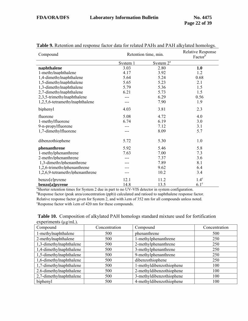
FDA/ORA/DFS Laboratory Information Bulletin No. 4475 Page 22 of 39 Table 9. Retention and response factor data for related PAHs and PAH alkylated homologs.
Compound Retention time, min. Relative Response Factorb
System 1 System 2a
naphthalene 3.03 2.80 1.01-methylnaphthalene 4.17 3.92 1.21,4-dimethylnaphthalene 5.64 5.24 0.681,5-dimethylnaphthalene 5.65 5.23 2.11,3-dimethylnaphthalene 5.79 5.36 1.52,7-dimethylnaphthalene 6.21 5.73 1.52,3,5-trimethylnaphthalene --- 6.29 0.561,2,5,6-tetramethylnaphthalene --- 7.90 1.9biphenyl 4.03 3.81 2.3fluorene 5.08 4.72 4.01-methylfluorene 6.74 6.19 3.09-n-propylfluorene --- 7.12 3.11,7-dimethylfluorene --- 8.09 5.7 dibenzothiophene 5.72 5.30 1.0
phenanthrene 5.92 5.46 5.81-methylphenanthrene 7.63 7.00 7.32-methylphenanthrene --- 7.37 3.6 1,3-dimethylphenanthrene --- 7.89 8.11,2,6-trimethylphenanthrene --- 9.62 6.41,2,6,9-tetramethylphenanthrene --- 10.2 3.4benzo[e]pyrene 12.1 11.2 1.4c
benzo[a]pyrene 14.8 13.5 6.1c
aShorter retention times for System 2 due in part to no UV-VIS detector in system configuration. bResponse factor (peak area/concentration (ppb)) calculated and ratioed to naphthalene response factor. Relative response factor given for System 2, and with λem of 352 nm for all compounds unless noted. cResponse factor with λem of 420 nm for these compounds. Table 10. Composition of alkylated PAH homologs standard mixture used for fortification experiments (μg/mL). Compound Concentration Compound Concentration 1-methylnaphthalene 500 phenanthrene 500 2-methylnaphthalene 500 1-methylphenanthrene 250 1,3-dimethylnaphthalene 500 2-methylphenanthrene 250 1,4-dimethylnaphthalene 500 3-methylphenanthrene 250 1,5-dimethylnaphthalene 500 9-methylphenanthrene 250 1,6-dimethylnaphthalene 500 dibenzothiophene 250 1,7-dimethylnaphthalene 500 1-methyldibenzothiophene 100 2.6-dimethylnaphthalene 500 2-methyldibenzothiophene 100 2,7-dimethylnaphthalene 500 3-methyldibenzothiophene 100 biphenyl 500 4-methyldibenzothiophene 100

FDA/ORA/DFS Laboratory Information Bulletin No. 4475 Page 23 of 39
0
50
100
150
200
0 5 10 15time (min.)
fluor
esce
nce
(mV
)4c
5
3c
21
A
C
B
Figure 4 HPLC-FLD chromatograms (352 nm) obtained for an extract of finfish (lower trace A), the PAH alkylated homologs standard mix (middle trace B), and an extract of finfish fortified with the standard mix (upper trace C). Spiking levels for the individual alkylated naphthalene and phenanthrene based PAHs were in the range 25 – 50 ng/g finfish. Peaks (1, 2, 5) or peak clusters (3c, 4c) used for spike/recovery calculations are noted in the figure. See Table 11 for peak/peak cluster assignments. The large peak in the unfortified extract (A) corresponds to naphthalene. Table 11. Recovery for alkylated PAH homologs in seafood with fortification levels of 25 - 50 ng/g.
Peak/peak cluster no.a
Ret. times, min. Assigned Compound(s) Percent Recovery (n=3, %RSD)
oysters shrimp crab finfish
1 4.1 biphenyl, 1-methylnaphthalene 128 (1.4) 107 (3.5) 120 (4.7) 119 (1.2)
2 4.5 2-methylnaphthalene 122 (3.6) 103 (0.36) 107 (8.2) 116 (3.9)
3c 5.6, 5.9, 6.4
dimethylnaphthalenes, dibenzothiophene,
h th119 (0.31) 109 (1.0) 108 (4.9) 115 (2.1)
4c 7.2, 7.4, 7.6
methylphenanthrenes, methyldibenzothiophenes 107 (1.7) 114 (0.51) 115 (2.2) 117 (2.0)
5 8.0 methylphenanthrenes 87 (1.1) 127 (1.5) 119 (0.8) 122 (0.10) aAs shown in Figure 4.

FDA/ORA/DFS Laboratory Information Bulletin No. 4475 Page 24 of 39 Analysis of NIST SRM 1974b Organics in Mussel Tissue An additional component of the validation protocol involved the analysis of NIST Standard Reference Material 1974b (16), a frozen mussel tissue homogenate containing certified levels for 14 of the 15 parent PAH compounds evaluated in this procedure. SRM 1974b was analyzed in triplicate by each of the three validating laboratories. As previously noted, a 10-fold pre-concentration and PMT gain setting of 15 were used for analysis of the SRM, owing to the extremely low PAH contents. A representative chromatogram generated from the analysis of SRM 1974b is given in Figure 5. Results are summarized in Table 12 for each of the three laboratories, along with the NIST certified values, and allowable control limits established by NOAA for assessment of method accuracy (4). Results are given in units of μg/kg (equivalent to ng/g) to match the NIST certificate. Determined amounts for 11 of the 14 PAHs fell within the NOAA control limits. Although the other three compounds (naphthalene, dibenzo[a,h]anthracene, and indeno[1,2,3-cd]pyrene) did not fall within the control limits, they are considered exempt from consideration (4) because the certified values fall below the method LOQs for these compounds.
0
15
30
45
60
75
0 5 10 15 20time (min.)
fluor
esce
nce
(LU
)
1
3,4
5
6
7
89
10
11
12
1314
Figure 5. LC-FLD chromatogram obtained from NIST SRM 1974b, mussel tissue homogenate, analyzed for 14 target PAHs. Peak labels as in Figure 1.

FDA/ORA/DFS Laboratory Information Bulletin No. 4475 Page 25 of 39
Table 12. Parent PAH certified and determined amounts for NIST SRM 1974b mussel tissue. Compound Certified
Valuea Control Limits Rangeb (µg/kg) Determined Amount (µg/kg)
Lab 1 (n=3)
Lab 2 (n=3)
Lab 3 (n=3)
Overall Ave. (n=9)
naphthalenec 2.43 1.6 - 3.3 8.86 5.74 4.26 6.28
fluorene 0.494 0.3 - 0.7 0.42 0.41 0.41 0.41
phenanthrene 2.58 1.7 - 3.5 2.39 2.57 1.54 2.17
anthracene 0.527 0.3 - 0.8 0.17 0.36 0.30 0.28
fluoranthene 17.1 11.5 - 23.1 15.3 16.4 14.8 15.5
pyrene 18.04 12.2 - 24.2 19.0 15.1 16.9 17.0
benzo[a]anthracene 4.74 2.9 - 6.9 4.07 3.86 4.15 4.02
chrysene 6.3 3.7 - 9.5 9.40 6.87 8.04 8.10
benzo[b]fluoranthene 6.46 4.1 - 9.2 5.22 4.41 4.26 4.63
benzo[k]fluoranthene 3.16 2.1 - 4.3 2.38 2.40 2.20 2.33
benzo[a]pyrene 2.80 1.7 - 4.1 2.03 2.18 2.43 2.22
dibenzo[a,h]anthracenec 0.327 0.2 - 0.5 2.28 1.83 2.69 2.27
benzo[g,h,i] perylene 3.12 2.0 - 4.5 3.01 2.89 2.78 2.89
indeno[1,2,3-cd] pyrenec 2.14 1.4 - 2.9 0.64 0.53 3.20 1.46 aWet mass basis as given in NIST certificate (16). bNOAA (4) defines the lower control limit as 0.7 x (certified value – uncertainty value), and the upper control limit as 1.3 x (certified value + uncertainty value). cThe certified value is below the method LOQ for these compounds, making them exempt in the NOAA criteria (4)

FDA/ORA/DFS Laboratory Information Bulletin No. 4475 Page 26 of 39 Supplementary Information and Validation Data Supplementary information and validation data are given in Appendix I, as listed below. Appendix I Listing of Supplementary Data and Validation
• Alternative Preparation of Parent PAH Stock Standard Mix
• Table I-1. Wavelength switching fluorescence detection signal timetable
• Table I-2. FDA Forensic Chemistry Center Parent PAH calibration summary for Agilent 1200 (wavelength switching conditions).
• Table I-3. Connecticut Agricultural Experiment Station (CAES) parent PAH
calibration summary for Hewlett Packard 1100 system1 (multiwavelength conditions). • Table I-4. Minnesota Department of Agriculture (MDA) parent PAH calibration
summary for Agilent 1200 system1 (multiwavelength conditions).
• Subtraction of Acetonitrile Blank Run
• Total PAH Estimation Protocol with Evaluation

FDA/ORA/DFS Laboratory Information Bulletin No. 4475 Page 27 of 39 References
(1) Moret, S., Dudine, A., and Conte, L.S., (2000) J. Am. Oil Chem. Soc. 77, 1289-1292.
(2) USEPA, Guidance for Assessing Chemical Contaminant Data for Use in Fish Advisories (2000) EPA 823-B-00-007, Third Edition, United States Environmental Protection Agency (3) Lubchenco, J., McNutt, M., Lehr, B., Sogge, M., Miller, M., Hammond, S., Conner, W. (2010) http://www.restorethegulf.gov/release/2010/09/09/bp-deepwater-horizon-oil-budget-what-happened-oil . (4) Sloan, C.A., Brown, D.W., Pearce, R.W., Boyer, R.H., Bolton, J.L., Burrows, D.G., Herman, D.P., and Krahn, M.M. (2004). Extraction, Cleanup, and Gas Chromatography/Mass Spectrometry Analysis of Sediments and Tissues for Organic Contaminants. U.S. Dept. Commer., NOAA Technical Memorandum NMFS-NWFSC-59, 47. (5) Krahn, M.M., Ylitalo, G.M., Chan, S.-L., and Varanasi, U. (1993) Environ. Sci. Technol. 27, 699-708. (6) Ramalhosa, M.J., Paiga, P., Morais, S., Delerue-Matos, C., & Oliveira, M.B.P.P. (2009) J. Sep. Sci. 32, 3259-3538
(7) Pule, B.O., Mmualefe, L.C., and Torto, N. (2010) Analysis of Polycylic Aromatic Hydrocarbons in Fish with Agilent SampliQ QuEChERS AOAC Kit and HPLC-FLD, Agilent Technologies Application Note. 5990-5441EN. (8) Krahn, M.M., Myers, M.S., Burrows, D.G., & Malins, D.C. (1984) Xenobiotica 14, 633-646
(9) Lawrence, J.F., & Weber, D.F. (1984) J. Agric. Food Chem. 32, 789-794.
(10) Krone, C.A., Stein, J.E., & Varanasi, U. (1992) Chemosphere 24, 497-510.
(11) Perfetti, G.A., Nyman, P.J., Fisher, S., Joe, F.L. Jr., & Diachenko, G.W. (1992) J. AOAC Int. 75, 872-877.
(12) Krahn, M.M., Ylitalo, G.M., Buzitis, J., Chan, S-L., & Varanasi, U. (1993) J. Chromatogr. 642, 15-32.
(13) Hufnagle, L.C. Jr., Camarata, S.E., Ernest, D., Krone, C.A. & Krahn, M.M. (1999) Arch. Environ. Contam. Toxicol. 37, 220-226.

FDA/ORA/DFS Laboratory Information Bulletin No. 4475 Page 28 of 39
(14) Kelly, C.A., Law, R.J., & Emerson, H.S. (2000) Sci. Ser., Aquat. Environ. Prot.: Analyt. Meth. Number 12, CEFAS, Lowestoft, 18pp.
(15) Protocol for Interpretation and Use of Sensory Testing and Analytical Chemistry Results for Re-opening Oil-impacted Areas Closed to Seafood Harvesting, June 18, 2010, FDA/CFSAN, College Park, MD.
(16) NIST Certificate of Analysis, Standard Reference Material 1974b, Organics in Mussel Tissue, 2003, Gaithersburg, MD.

FDA/ORA/DFS Laboratory Information Bulletin No. 4475 Page 29 of 39 Appendix I. Supplementary Information and Validation Data Alternative Preparation of Parent PAH Stock Standard Mix Alternative instructions are given below for the preparation of the parent PAH stock standard mix (2000 μg/mL) using the QTM PAH mix supplemented with benzo[k]fluoranthene. The QTM PAH mix supplemented with benzo[k]fluoranthene was used in much of the earlier work, and is acceptable although more work is required. Note that the QTM PAH mix also contains 2-bromonaphthalene. 2-bromonaphthalene does not cause interference in the current method because it exhibits no appreciable fluorescence under the method conditions.
• QTM PAH Mix (contains 16 PAH @ 2000 micrograms/mL each in methylene chloride) (Supelco p/n 47930-U, or equivalent)
• Benzo[k]fluoranthene, (Supelco p/n 48492, or equivalent) If using QTM PAH mix in conjunction with a separate benzo[k]fluoranthene stock solution: A stock standard solution of benzo[k]fluoranthene was prepared at a concentration of 2000 μg/mL in methylene chloride. The QTM PAH Mix is a solution that contains a mixture of sixteen PAHs in methylene chloride, each at a concentration of 2000 μg/mL. 250 μg/mL stock standard: 10 mL of a stock spiking solution was prepared by combining 1.25 mL each of the QTM PAH mix and the 2000 μg/mL benzo[k]fluoranthene stock, followed by dilution to 10 mL with CH3CN 5.0 μg/mL stock standard: 25 mL of a 5 μg/mL spiking solution was prepared by adding 500 microliters of the 250 μg/mL stock standard to a 25 mL volumetric flask and diluting to volume with CH3CN 0.5 μg/mL stock standard: 25 mL of a 0.5 μg/mL spiking solution was prepared by adding 2.5 mL of the 5 μg/mL stock standard to a 25 mL volumetric flask and diluting to volume with CH3CN

FDA/ORA/DFS Laboratory Information Bulletin No. 4475 Page 30 of 39
Table I-1. Wavelength switching fluorescence detection signal timetable
Time (min.)
Excitation nm
Emission nm
PMT-Gain
Baseline PAHs detected
0.00 260 352 13 Zero naphthalene, acenaphthene, fluorene, phenanthrene
6.35 260 420 13 Zero anthracene, fluoranthene, pyrene, benzo[a]anthracene,
chrysene, benzo[b]fluoranthene, benzo[k]fluoranthene,
benzo[a]pyrene, dibenzo[a,h]anthracene,
benzo[ghi]perylene
17.15 260 460 13 Zero indeno[1,2,3-cd]pyrene
In the initial work, the parent PAH calibration was conducted under wavelength switching conditions (see Table I-1 above). The wavelength switching times given in Table I-1 can be adjusted as needed to accommodate individual systems. The calibration summary is given in Table I-2, below. Instrument detection limits (IDL) and limits of quantitation (LOQ) were also determined based on replicate analyses of a 1.7 ng/mL standard mixture (n=7), and are also listed in the table. IDL and LOQ values were determined as outlined below using the Student's t-test at a 99% confidence interval. IDL = , where s is the standard deviation and, for n=7, n-1=6, = 3.7 st ×=99α 99=αt IDL = 3.7 s LOQ = 10 s The parent PAH calibration protocol was also conducted by both the Connecticut Agricultural Experiment Station (CAES) and the Minnesota Department of Agriculture (MDA) with data summaries given in Tables I-3 and I-4, respectively.

FDA/ORA/DFS Laboratory Information Bulletin No. 4475 Page 31 of 39 Table I-2. FDA Forensic Chemistry Center Parent PAH calibration summary for Agilent 1200 system1 (wavelength switching conditions).
Calibration
Range Retention Time Regression IDL LOQ (n = 3) (n = 9) equation1 (n = 7) (n = 7)
Compound ng/mL Min %RSD (n = 3) R2 (ng/mL) (ng/mL)
naphthalene 0.0 - 50.0 3.1 0.49 y = 0.512x + 0.034 0.99996 0.17 0.46
acenaphthene 0.0 - 50.0 4.9 0.27 y = 0.689x + 0.089 0.99986 0.12 0.33
fluorene 0.0 - 50.0 5.1 0.25 y = 1.869x + 0.033 0.99999 0.05 0.15
phenanthrene 0.0 - 50.0 5.9 0.18 y = 3.433x + 0.174 0.99998 0.04 0.11
anthracene 0.0 - 50.0 6.6 0.13 y = 8.454x + 0.088 1.00000 0.03 0.07
fluoranthene 0.0 - 50.0 7.3 0.10 y = 0.517x + 0.025 0.99999 0.32 0.85
pyrene 0.0 - 50.0 7.9 0.08 y = 0.936x + 0.023 0.99998 0.09 0.24
benzo[a]anthracene 0.0 - 50.0 9.5 0.05 y = 3.589x - 0.025 1.00000 0.04 0.11
chrysene 0.0 - 50.0 10.1 0.06 y = 1.409x + 0.137 0.99993 0.12 0.33
benzo[b]fluoranthene 0.0 - 50.0 11.8 0.06 y = 3.186x - 0.132 1.00000 0.05 0.14
benzo[k]fluoranthene 0.0 - 50.0 12.9 0.06 y = 14.209x - 0.272 1.00000 0.01 0.03
benzo[a]pyrene 0.0 - 50.0 13.7 0.06 y = 7.646x + 0.257 0.99999 0.06 0.16
dibenzo[a,h]anthracene 0.0 - 50.0 15.4 0.07 y = 0.671x - 0.072 0.99997 0.16 0.42
benzo[g,h,i]perylene 0.0 - 50.0 16.2 0.07 y = 1.374x - 0.169 0.99997 0.12 0.32
indeno[1,2,3-cd]pyrene 0.0 - 50.0 17.4 0.07 y = 0.268x - 0.204 0.99923 2.5 6.8
1 System components were Agilent 1200 series with G1379B degasser, G1312A binary pump, G1329A auto sampler, G1316A column controller and G1321A fluorescence detector. 2 y = area; x = concentration (ng/mL)

FDA/ORA/DFS Laboratory Information Bulletin No. 4475 Page 32 of 39 Table I-3. Connecticut Agricultural Experiment Station (CAES) parent PAH calibration summary for Hewlett Packard 1100 system1 (multiwavelength conditions).
Calibration
Range Retention Time Regression IDL LOQ (n = 3) (n = 9) Equation2 (n = 7) (n = 7)
Compound ng/mL Min %RSD (n = 3) R2 (ng/mL) (ng/mL)
naphthalene 0.0 - 50.0 2.9 0.75 y = 0.946x - 0.020 0.99998 0.34 1.12
acenaphthene 0.0 - 50.0 4.7 0.65 y = 1.247x - 0.363 0.99986 0.22 0.74
fluorene 0.0 - 50.0 4.9 0.62 y = 3.033x -0.516 0.99990 0.09 0.31
phenanthrene 0.0 - 50.0 5.7 0.54 y = 6.033x + 0.729 0.99998 0.64 2.13
anthracene 0.0 - 50.0 6.5 0.47 y = 11.818x - 0.322 0.99998 0.13 0.44
fluoranthene 0.0 - 50.0 7.3 0.41 y = 0.835x - 0.103 0.99990 0.09 0.31
pyrene 0.0 - 50.0 7.8 0.38 y = 2.040x -0.114 0.99998 0.31 1.03
benzo[a]anthracene 0.0 - 50.0 9.7 0.26 y = 6.390x - 0.196 0.99999 0.07 0.23
chrysene 0.0 - 50.0 10.3 0.23 y = 2.654x - 0.137 0.99998 0.06 0.21
benzo[b]fluoranthene 0.0 - 50.0 12.1 0.17 y = 5.776x - 0.531 0.99998 0.04 0.15
benzo[k]fluoranthene 0.0 - 50.0 13.3 0.13 y = 19.900x – 1.190 1.00000 0.05 0.18
benzo[a]pyrene 0.0 - 50.0 14.1 0.11 y = 14.040x – 1.195 0.99999 0.46 1.52
dibenzo[a,h]anthracene 0.0 - 50.0 15.9 0.06 y = 1.384x - 0.285 0.99990 0.27 0.89
benzo[g,h,i]perylene 0.0 - 50.0 16.7 0.07 y = 3.204x - 0.342 0.99997 0.24 0.79
indeno[1,2,3-cd]pyrene 0.0 - 50.0 18.1 0.10 y = 0.515x - 0.105 0.99984 0.35 1.16
1System components were Hewlett Packard 1100 series with G1322A degasser, G1311A quaternary pump, G1313A auto sampler, G1316A column controller and an Agilent Series 1200 G1321A fluorescence detector. 2 y = area; x = concentration (ng/mL)

FDA/ORA/DFS Laboratory Information Bulletin No. 4475 Page 33 of 39
Table I-4. Minnesota Department of Agriculture (MDA) parent PAH calibration summary for Agilent 1200 system1 (multiwavelength conditions).
Calibration
Range Retention Time Regression IDL LOQ (n = 3) (n = 10) equation1 (n = 7) (n = 7)
Compound ng/mL Min %RSD (n = 3) R2 (ng/mL) (ng/mL)
naphthalene 0.0 - 50.0 2.9 0.10 y = 0.979x + 2.145 0.99484 0.31 1.02
acenaphthene 0.0 - 50.0 4.7 0.08 y = 1.336x + 0.007 1.00000 0.07 0.25
fluorene 0.0 - 50.0 4.9 0.08 y = 3.750x + 0.172 1.00000 0.06 0.21
phenanthrene 0.0 - 50.0 5.7 0.07 y = 7.919x - 0.591 0.99999 0.05 0.18
anthracene 0.0 - 50.0 6.5 0.07 y = 12.805x + 0.729 1.00000 0.10 0.32
fluoranthene 0.0 - 50.0 7.2 0.06 y = 1.036x - 0.111 0.99999 0.32 1.07
pyrene 0.0 - 50.0 7.7 0.06 y = 1.1560x - 0.330 0.99998 0.11 0.36
benzo[a]anthracene 0.0 - 50.0 9.4 0.06 y = 7.270x - 0.940 0.99998 0.02 0.06
chrysene 0.0 - 50.0 10.0 0.07 y = 2.889x - 0.330 0.99991 0.08 0.27
benzo[b]fluoranthene 0.0 - 50.0 11.7 0.09 y = 7.005x - 1.141 0.99998 0.01 0.05
benzo[k]fluoranthene 0.0 - 50.0 12.8 0.10 y = 30.193x - 4.119 0.99999 0.01 0.04
benzo[a]pyrene 0.0 - 50.0 13.6 0.09 y = 12.143x + 3.767 0.99994 0.03 0.10
dibenzo[a,h]anthracene 0.0 - 50.0 15.4 0.11 y = 1.398x - 0.784 0.99992 0.17 0.58
benzo[g,h,i]perylene 0.0 - 50.0 16.1 0.12 y = 2.800x - 1.353 0.99993 0.02 0.08
indeno[1,2,3-cd]pyrene 0.0 - 50.0 17.4 0.13 y = 0.329x + 0.340 0.99998 0.12 0.40
1System components were Agilent 1200 series with G1379B degasser, G1312A binary pump, G1367B auto sampler, G1316B column controller and G1321B fluorescence detector. 2 y = area; x = concentration (ng/mL)

FDA/ORA/DFS Laboratory Information Bulletin No. 4475 Page 34 of 39 Subtraction of Acetonitrile Blank Run Given that the current HPLC method is based on gradient separation, the chromatographic baseline is not flat throughout the run. Examples of chromatograms for an acetonitrile blank injection are given in Figure I-1 below for emission detection at either 352 or 420 nm. The baselines tend to follow the gradient, with the magnitude of the background fluorescence much higher for detection at 352 nm.
0
0.1
0.2
0.3
0.4
0.5
0.6
0 5 10 15 20
time (min.)
fluor
esce
nce
(LU
)
352 nm
420 nm
Figure I-1. Chromatograms for acetonitrile blank injection with detection at 352 nm (upper trace) or 420 nm (lower trace.).
The Agilent software allows subtraction of a blank run chromatogram. After loading the file of interest (standard, sample, method blank, etc.), choose “File…Subtract Blank Run”, and choose a blank acetonitrile injection, making sure to match the emission wavelengths between the two chromatograms, i.e., 352 or 420 nm. The exact profile of the blank run chromatogram may vary widely between sources or lots of acetonitrile, and/or from day to day based on lamp power, etc. Hence, the acetonitrile blank chosen for subtraction should come from the same sequence as the standards and samples. While a blank may be used as the first injection in the sequence, it should not be used as the blank for subtraction as the system may not have been fully equilibrated (choose a different blank injection for subtraction). While subtraction of the acetonitrile blank run is particularly important for estimation of total PAH content (see next section), it will generally make peak area integration easier and more

FDA/ORA/DFS Laboratory Information Bulletin No. 4475 Page 35 of 39 accurate for all data processing. An example is given below for the 1.7 ppb parent PAH standard mix (Figure I-2) with 352 nm detection. Integration of the four standard peaks of interest (peak labels 1 – 4) is simpler and more accurate after subtraction of the blank run.
0
0.3
0.6
0.9
1.2
0 5 10 15 20time (min.)
fluor
esce
nce
(LU
) before subtraction
after subtraction
12
3
4
Figure I-2. Chromatogram with 352 nm detection for the 1.7 ppb parent PAH standard mix before subtraction (upper trace) or after subtraction (lower trace) of the acetonitrile blank chromatogram. Total PAH Estimation Protocol with Evaluation Estimation of total PAH content is based on taking the total fluorescence peak area over the retention window for the parent and alkylated PAH compounds studied (2.5 to 20 min.), and calculating a conservative estimate of the total PAH content. The estimate is intended to be an overestimate to allow an additional margin of safety relative to FDA levels of concern (LOC). The current screening method is highly sensitive with detection limits and limits of quantitation in the sub to low ppb range. Experience with calculating the total PAH content for actual seafood samples from the Gulf of Mexico waters has also shown that the total fluorescence signals are very low.

FDA/ORA/DFS Laboratory Information Bulletin No. 4475 Page 36 of 39 In order to obtain accurate peak and total peak integrations for the estimation of total PAH content, subtraction of an acetonitrile blank run chromatogram should be conducted prior to integration (see previous section). A couple of examples of the use of blank run subtraction are given below and discussed in turn. Figure I-3 shows the 352 nm chromatogram for a sample of crab spiked with the parent PAH standard mix at the 1.0 μg/g level before and after subtraction of the blank run. The dotted lines represent total peak area integration over the range 2.5 – 20 min. In the figure, it is obvious that the total peak area is grossly overestimated prior to subtraction of the blank run due to the contour of the gradient baseline.
0
0.5
1
1.5
2 6 10 14 18time (min.)
fluor
esce
nce
(LU
)
before subtraction
after subtraction
Figure I-3. Chromatogram with 352 nm detection for a spiked crab sample before subtraction (upper trace) or after subtraction (lower trace) of the acetonitrile blank chromatogram. The dotted lines represent total peak area integration over the range 2.5 – 20 min.

FDA/ORA/DFS Laboratory Information Bulletin No. 4475 Page 37 of 39 While the magnitude of the background fluorescence is greater at 352 nm, the background is also present with 420 nm detection, and can also lead to gross overestimates. Figure I-4 (below) shows Figure I-3 shows the 420 nm chromatogram before and after subtraction of the blank run for an unfortified sample of a shrimp sample obtained from the Gulf. Again, the dotted lines represent total peak area integration over the range 2.5 – 20 min. The total peak area is grossly overestimated prior to subtraction, and is seen to be almost nil after subtraction.
0
0.5
1
1.5
2 6 10 14 18time (min.)
fluor
esce
nce
(LU
)
before subtraction
after subtraction
Figure I-4. Chromatogram with 420 nm detection for an unfortified shrimp sample before subtraction (upper trace) or after subtraction (lower trace) of the acetonitrile blank chromatogram. The dotted lines represent total peak area integration over the range 2.5 – 20 min.

FDA/ORA/DFS Laboratory Information Bulletin No. 4475 Page 38 of 39 The Total PAH Estimation Protocol was applied to a series of Gulf coast oyster sample extracts fortified with various PAH solutions and known total PAH amounts (obtained by summing the amount of the individual components in the spiking solutions). The estimation protocol was also evaluated for oyster extracts fortified with source oil1 from the Deepwater Horizon well riser (DWH source oil). The experiments included several different fortification levels for each of the PAH spiking solutions with triplicate testing of the mid spiking level. Fortification levels were chosen to approximate 25%, 60%, and 100% of the FDA Level of Concern (LOC) for naphthalene in oysters which is 133 mg/kg (exception is the DWH source oil mid and high level spikes which represent 80% and 130% of the naphthalene LOC, respectively). All samples were run using the current HPLC-FLD method, and the total peak area (2.5 – 20 min.) was integrated after subtraction of an acetonitrile blank run. The total PAH estimates were then compared to the known total PAH levels to evaluate the estimation protocol. For the DWH source oil, the actual total fortification level was calculated based on an assumed total PAH content of 1.24 % w/w for the oil2. Results are given in Table I-1 (next page) along with the total PAH estimate for the unfortified oysters, conducted in triplicate. Comparison of the total PAH estimates with the known values showed consistent overestimation of total PAH content (i.e., estimates increased proportionately with fortification levels). The actual degree of overestimation varied among the PAH spiking solutions as follows: parent PAHs (9 - 10 fold), parent/alkylated mixes (3 – 5 fold), and DWH source oil (25 – 30 fold). Consistent total PAH estimates were obtained among the triplicate samplings for the unfortified oysters. 1The DWH source oil was collected in May, 2010, from aboard the Enterprise Discoverer drill ship, and was obtained directly from the MC252 well via the riser insertion tube. The source oil was provided to FDA’s Gulf Coast Seafood Laboratory in June, 2010, and a subsample was provided to the FDA Forensic Chemistry Center. 2This figure for the DWH source oil was obtained from “Hydrocarbon Results for MC252 Riser Oil”, ENTRIX, Inc. Memorandum Rob Barrick to Ralph Markarian, John Dimitry, May 3, 2010, Seattle, WA, which was accessed as: publicfiles.dep.state.fl.us/.../Oil.../Riser%20Oil%20Analysis%203May10.pdf

FDA/ORA/DFS Laboratory Information Bulletin No. 4475 Page 39 of 39
Table I-1. Evaluation of total PAH estimation using Gulf oysters. PAH spiking solution mix1 Actual Total Fortification Level Total PAH Estimate (mg/kg) (mg/kg)
16 component parent PAH 32 315 80 712 80 701 80 724 130 1207
14 component parent/alkylated 32 141 78 291 78 288 78 321 130 470
20 component parent/alkylated 32 131 80 278 80 298 80 285 130 417
DWH source oil 35 959 106 2934 106 3024 106 2970 177 4424
none, unfortified 0 4.7 0 5.0 0 4.9 1A listing of PAH components for each of the standard mix spiking solutions follows. 16 component parent PAH mix: naphthalene, acenaphthylene, acenaphthene, fluorene, phenanthrene, anthracene, fluoranthene, pyrene, benzo[a]anthracene, chrysene, benzo[b]fluoranthene, benzo[k]fluoranthene, benzo[a]pyrene, dibenzo[a,h]anthracene, benzo[g,h,i]perylene, indeno[1,2,3-cd]pyrene. 14 component parent/alkylated mix: naphthalene; 2-methylnaphthalene; 2,3-dimethylnaphthalene; 2,3,6-trimethylnaphthalene; dibenzothiophene; 4-methyldibenzothiophene; 2,4,7-trimethyldibenzothiophene; 2,8-dimethyldibenzothiophene; fluoranthene; phenanthrene; 2-methylphenanthrene; 1,6-dimethylphenanthrene; 1,2,8-trimethylphenanthrene; pyrene. 20 component parent/alkylated mix: 1-methylnaphthalene; 2-methylnaphthalene; 1,3-dimethylnaphthalene; 1,4-dimethylnaphthalene; 1,5-dimethylnaphthalene; 1,6-dimethylnaphthalene; 1,7-dimethylnaphthalene; 2,6-dimethylnaphthalene; 2,7-dimethylnaphthalene; biphenyl; phenanthrene; 1-methylphenanthrene; 2-methylphenanthrene; 3-methylphenanthrene; 9-methylphenanthrene; dibenzothiophene; 1-methyldibenzothiophene; 2-methyldibenzothiophene; 3-methyldibenzothiophene; and 4-methyldibenzothiophene.