Embed Size (px)
Citation preview

FAST VIABILITY ASSESSMENT OF CLOSTRIDIUM SPORES—SURVIVAL IN EXTREME ENVIRONMENTS
Thesis by
Wanwan Yang
In Partial Fulfillment of the Requirements
for the Degree of
Doctor of Philosophy
California Institute of Technology
Pasadena, California
2010
(Defended August 7, 2009)

ii
© 2010
Wanwan Yang
All Rights Reserved

iii
"Everything should be made as simple as possible, but not simpler."
Albert Einstein

iv
Acknowledgments
First and foremost I offer my sincerest gratitude to my thesis adviser, Dr. Adrian Ponce,
for his guidance and support throughout my research and study at JPL. It is his
enthusiasm in research and encouragement that have motivated me and ultimately made
this work possible. I am also heartily thankful to my thesis committee members: Prof.
Harry Gray, Prof. Victoria Orphan, and Prof. Alex Sessions. Given their busy schedules,
it has been kind of them to play a role in my course of study at Caltech.
I would like to thank all members of the Ponce Group who have been supporting me
continuously in the past years. Douglas Yung and Morgan Cable have helped me with the
Micro EVA and Spectro EVA instruments, and their friendship also supported me
through tough times. Shannon Beaty deserves a special thanks as a fun and supportive
friend. I would also like to thank other group members for the valuable discussion and
feedback on my thesis work: Dr. James Kirby, Dr. Stephanie Connon, Dr. Donald
Obenhuber, Kevin Hartman, Emma Crow-Willard, Dana Levine, Elizabeth Lester, and
Hannah Shafaat. I owe my gratitude to Dr. Wayne Schubert at JPL who has been
generously lending me instruments whenever I needed them. I also thank Dr. Michael
Russel for helpful scientific discussions.
I especially would like to thank Derrick Bass, Chang Luo, Tudor Stoenescu, and
Marco Seidel for their continuous love and support over the past several years. Without
their friendship, my grad school life would have been a lot more difficult. I also thank Icy
Ma, John Yong, Heywood Tam, Lap-Man Lee, King-Fai Li, and the rest of HKSA
members for being wonderful friends. I would like to thank Prof. Michael Hoffmann,
Prof. Paul Wennberg, and Prof. Janet Hering for helping me out during the tough time

v
when I switched research groups. I appreciate the support and friendship from other staff
members and students in ESE, including Dr. A. J. Colussi, Dr. Nathan Dalleska, Cecilia
Gamboa, Linda Scott, Jie Cheng, and Jina Choi.
Many thanks go to my dance coaches, Valdas and Lilia Padriezas, and dance partner
Florian Gador, for bringing me to the world of ballroom dance. Candy Tong, Rosalyn
Sayaman, Oleg Kogan, Jessie Rosenberg, and Robert and Megan Nissen from the Caltech
ballroom dance club have made my journey at Caltech a lot more enjoyable.
Last but not least, my deepest gratitude goes to everybody in my family: my parents,
sisters, and brother. Their unconditional and endless love has supported me in all my
endeavors in life.

vi
Abstract
Bacterial endospores are formed in genera such as Bacillus and Clostridium in response
to adverse environmental changes. Endospores have remarkable resistance to various
extreme conditions and can remain dormant for extended periods of time. Clostridium
spores are of particular interest due to their significant importance in several industries,
such as food processing, wastewater treatment, pharmaceuticals, and health care. They
are also the ideal candidates to study Panspermia and potential extraterrestrial life.
However, to date, most endospore research has been conducted on Bacillus, and study of
the anaerobic spore former, Clostridium, is not adequate.
In this study, we have developed a general protocol to produce and purify Clostridium
spores. Spectroscopy and microscopy based Endospore Viability Assay (Spectro EVA and
Micro EVA) were developed and validated to assess the viability of Clostridium spores.
Germinability was used as an indicator for spore viability. The basic principle of the two
EVAs is to measure the release of a unique biomarker, dipicolinic acid (DPA), via
germination as a proxy for endospore viability. In particular, a luminescence time-gated
microscopy technique (Micro EVA) has been developed to enumerate germinable
Clostridium endospores within an hour. Micro EVA is based on energy transfer from
DPA to terbium ions doped in a solid matrix upon UV excitation. The distinctive
emission and millisecond lifetime enables time-resolved imaging to achieve single
endospore sensitivity. Comparing to traditional CFU cultivation, EVA probes the early
stage of germination, resulting in a much faster detection rate (within 60 minutes) than
CFU measurement (more than 3 days incubation). Micro EVA has also been successfully

vii
applied to quantify Clostridium spores in an extreme cold biosphere, Greenland ice core,
and a hyper-arid biosphere, Atacama Desert, two Mars analogs on earth.
The development of EVA provides a faster way to assess viability of Clostridium spores,
which has significant importance in various industries. It also enables the determination
of the limit and longevity of life, and provides insight on the search of extinct or extant
life on Mars and other celestial bodies.

viii
Table of Contents CHAPTER 1: INTRODUCTION .................................................................... 1
1.1 OVERVIEW OF SPORE FORMERS ............................................................................. 1
1.2 ANAEROBIC SPORE FORMERS (CLOSTRIDIUM) ................................................. 5
1.3 OVERVIEW OF SPORE DETECTION METHODS ................................................. 10
1.4 OUTLINE OF THESIS .................................................................................................... 14
1.5. REFERENCES................................................................................................................. 17
CHAPTER 2: PRODUCTION AND CHARACTERIZATION OF PURE CLOSTRIDIUM SPORE SUSPENSIONS .................................................... 23
2.1 ABSTRACT ...................................................................................................................... 23
2.2 INTRODUCTION ............................................................................................................ 24
2.3 MATERIALA AND METHODS ................................................................................... 26
Materials .................................................................................................................................. 26
Phase contrast microscopic enumeration to determine spore concentrations .......................... 27
Endospore production .............................................................................................................. 27
Endospore purification ............................................................................................................ 28
Spore culturability ................................................................................................................... 29
D-value measurement of spores .............................................................................................. 30
Measurement of hydrophobicity.............................................................................................. 30
Release of DPA from spores of C. sporogenes and C. hungatei and quantification of DPA per spore using Tb3+-DPA luminescence ...................................................................................... 31
2.4 RESULTS .......................................................................................................................... 31
Production of Clostridium spores ............................................................................................ 31
Characterization of spores of C. sporogenes and C. hungatei. ................................................ 33
2.5 DISCUSSION ................................................................................................................... 34
2.6 REFERENCES .................................................................................................................. 40
CHAPTER 3: RAPID ENDOSPORE VIABILITY ASSAY FOR CLOSTRIDIUM SPORES ............................................................................. 47
3.1 ABSTRACT ...................................................................................................................... 47
3.2 INTRODUCTION ............................................................................................................ 48

ix
3.3 MATERIALS AND METHODS ................................................................................... 50
Materials .................................................................................................................................. 50
Endospore production and purification ................................................................................... 50
Endospore quantification by Tb3+-DPA luminescence ............................................................ 50
Spectro-EVA determination of germinable and total spore concentrations ............................ 51
Phase contrast microscopic enumeration of total and germinable spores ............................... 52
Determination of CFU per milliliter of spore suspension ....................................................... 53
Germination dynamics of temperature dependence ................................................................ 53
Comparison of the effect of D-alanine on Clostridium and Bacillus spore germination ........ 54
3.4 RESULTS .......................................................................................................................... 55
Correlation of Tb3+-DPA luminescence intensity with spore concentration ........................... 55
The release of DPA from spores upon germination ................................................................ 55
Validation of spectroscopy based endospore viability assay (Spectro-EVA) ......................... 56
Temperature dependence of germination rates ........................................................................ 57
Effect of D-alanine on spore germination of C. sporogenes and B. atrophaeus ..................... 57
3.5 DISCUSSION .................................................................................................................... 58
3.6 REFERENCES .......................................................................................... 63 CHAPTER 4: DEVELOPMENT OF A MICROSCOPIC BASED ENDOSPORE VIABILITY ASSAY TO DETECT SINGLE CLOSTRIDIUM SPORE GEMINATION ..................................................... 74
4.1 ABSTRACT ...................................................................................................................... 74
4.2 INTRODUCTION ............................................................................................................ 75
4.3 MATERIALS AND METHODS ................................................................................... 77
Materials .................................................................................................................................. 77
Endospore production and purification ................................................................................... 78
Sample preparation for Micro-EVA experiments ................................................................... 79
The Micro-EVA instrument .................................................................................................... 80
Endospore germination and germinable endospore assignment .............................................. 80
Phase contrast microscopic enumeration to determine spore concentrations .......................... 81
Spore culturability ................................................................................................................... 82
Spectroscopy ........................................................................................................................... 82

x
4.4 RESULTS .......................................................................................................................... 83
Germination time course for single Clostridium spore ........................................................... 83
Validation of Micro-EVA against CFU culturing ................................................................... 83
Discrimination of Clostridium from Bacillus spores ............................................................... 84
4.5 DISCUSSION ................................................................................................................... 85
4.6 REFERENCES .................................................................................................................. 89
CHAPTER 5: APPLICATION OF MICRO-EVA TO DETECT CLOSTRIDIUM SPORES FROM GREENLAND ICE CORE AND ATACAMA DESERT ................................................................................... 98
5.1 ABSTRACT ...................................................................................................................... 98
5.2 INTRODUCTION ............................................................................................................ 99
5.3 MATERIALS AND METHODS ................................................................................ 102
Materials ............................................................................................................................... 102
GISP2 ice core handling ....................................................................................................... 103
Ice core analysis using Spectro-EVA ................................................................................... 105
Ice core analysis using Micro-EVA ..................................................................................... 106
CFU Cultivation ................................................................................................................... 107
Identification of anaerobic spore formers ............................................................................. 108
Atacama Desert soil sampling .............................................................................................. 109
Measure of soil water activity, pH, EH, eC and temperature in field................................... 109
Cell extraction from soils and Micro-EVA measurement .................................................... 110
5.4 RESULTS ....................................................................................................................... 110
Recovery of anaerobic spore formers from Greenland Ice Core and phylogenetic analysis 110
Application of Spectro-EVA on ice core samples ................................................................ 110
Application of Micro-EVA on ice core samples .................................................................. 110
Physical and chemical properties of Atacama soil ............................................................... 110
Application of Micro-EVA on Atacama soil samples .......................................................... 110
5.5 DISCUSSION ................................................................................................................ 110
5.6 REFERENCES ............................................................................................................... 110
CHAPTER 6: SUMMARY ......................................................................... 132
xi
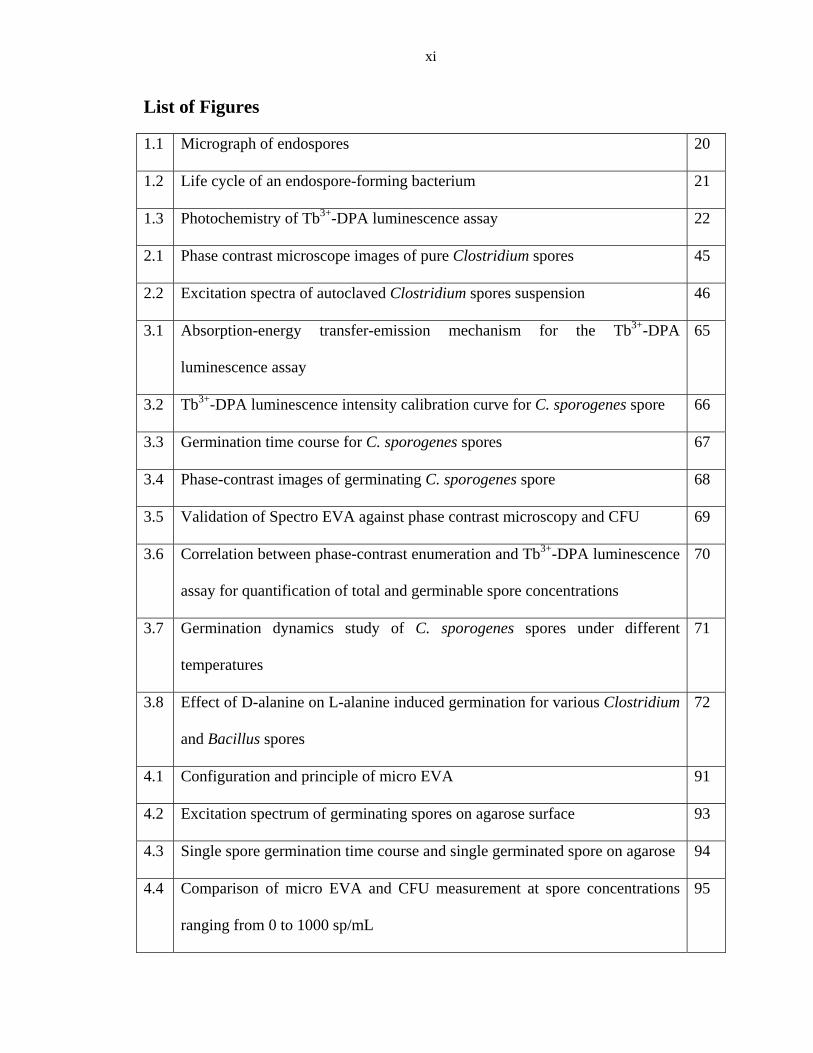
List of Figures
1.1 Micrograph of endospores 20
1.2 Life cycle of an endospore-forming bacterium 21
1.3 Photochemistry of Tb3+-DPA luminescence assay 22
2.1 Phase contrast microscope images of pure Clostridium spores 45
2.2 Excitation spectra of autoclaved Clostridium spores suspension 46
3.1 Absorption-energy transfer-emission mechanism for the Tb3+-DPA
luminescence assay
65
3.2 Tb3+-DPA luminescence intensity calibration curve for C. sporogenes spore 66
3.3 Germination time course for C. sporogenes spores 67
3.4 Phase-contrast images of germinating C. sporogenes spore 68
3.5 Validation of Spectro EVA against phase contrast microscopy and CFU 69
3.6 Correlation between phase-contrast enumeration and Tb3+-DPA luminescence
assay for quantification of total and germinable spore concentrations
70
3.7 Germination dynamics study of C. sporogenes spores under different
temperatures
71
3.8 Effect of D-alanine on L-alanine induced germination for various Clostridium
and Bacillus spores
72
4.1 Configuration and principle of micro EVA 91
4.2 Excitation spectrum of germinating spores on agarose surface 93
4.3 Single spore germination time course and single germinated spore on agarose 94
4.4 Comparison of micro EVA and CFU measurement at spore concentrations
ranging from 0 to 1000 sp/mL
95

xii
4.5 Micro EVA image of Clostridium and Bacillus spores inoculated on Tb3+/D-
alanine-doped agarose
96
5.1 Ice core decontamination and handling procedure 126
5.2 Phase contrast images of sporulating cultures of spore formers from
Greenland ice core
127
5.3 Phylogenetic tree of Greenland ice core isolates 128
5.4 Spectro EVA results on one ice core sample (depth: 1566 m, age: 10,000 yrs) 129
5.5 Micro EVA image of germinated spores from Greenland ice core and
corresponding germination time course
130
5.6 Micro EVA image of germinated spores from Atacama Desert and
corresponding germination time course
131

xiii
List of Tables
2.1 Effect of different combinations of three sporulation conditions on
sporulation of difference Clostridium species
43
2.2 Properties of Clostridium spores 44
5.1 Summary of anaerobic CFU counts of Greenland ice core in 1/10 R2A
and MM2 medium after 6 months incubation at 22 ºC
122
5.2 Summary of micro EVA results for Greenland ice core samples 123
5.3 Physical properties of all sampling sites in Atacama Desert 124
5.4 Summary of micro EVA results for Atacama soil samples 125

1
CHAPTER 1: INTRODUCTION
1.1 OVERVIEW OF SPORE FORMERS
Bacterial spores (i.e., endospores), first discovered by various research groups
independently in 1876 are almost exclusively found in gram positive bacteria [1-3]. The
formation of endospore is one of the defining traits of genera such as Bacillus,
Clostridium, Thermoactinomyces, Sporolactobacillus, and Sporosarcina.
Desulfotomaculum and Sporomusa are two types of spore formers that have gram
negative staining but have a gram positive type cell wall and share 16s rRNA sequences
with Clostridium species. Currently, the only well-known gram negative spore former is
Sporohalobacter [3].
Endospores are formed by intracellular division within the cytoplasm of a mother cell.
Spore-forming bacteria initiate sporulation in response to adverse environmental changes,
such as nutrient limitation. After being through a sequential sporulation stages, the
mature spores are released from the mother cells [3, 4]. The resultant endospore form
exhibits incredible longevity, tenacity and persistence facing extreme environmental
stresses (i.e. extremes of temperature and pH, high/low pressure, desiccation, UV
radiation and attack by a wide variety of oxidants) that would kill growing cells of nearly
all other bacterial species [5]. Several published reports have claimed the isolation of
viable spore-forming bacteria from the gut of a bee trapped in Dominican amber about
25~40 million years ago [6], and even more spectacular, the recovery of spores from 250-

2
million-year-old halite crystals [7]. A six-year space study also showed that spores of
Bacillus subtilis survived after exposure to space environments such as high vacuum and
radiation [8].
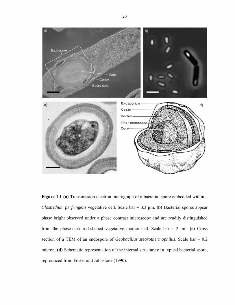
The unique structure and chemical composition of endospore play major roles in its
extreme resistance to various stresses. Figure 1.1 shows the internal structure of a typical
bacterial spore. Starting from outside and proceeding inward, the spore includes
exosporium, coat, cortex, inner membrane and central core. The exosporium is composed
of proteins with unknown functions. The coat is a complex structure with multiple layers,
which is important in spore resistance to some chemicals, such as exogenous lytic
enzymes that can degrade spore cortex. However, the coat has little or no role in spore
resistance to heat, radiation and some other chemicals. The cortex is made up of
peptidoglycan, which is essential for spore formation and the reduction of water content
in spore core. The inner spore membrane serves as a strong permeability barrier that
plays an important role in spore resistance to many chemicals, especially to those that
cross the membrane to damage DNA in the core. The spore core is an analogue of the
protoplast of the growing cell, which contains most spore enzymes, DNA, ribosomes and
tRNAs. One unique feature of endospore is the low water content. Unlike the protoplast
of a growing cell, in which water comprises 75~80% of its wet weight, endospore only
contains 27~55% water as its wet weight. The amount of free water in spore core is
extremely low, which restricts the macromolecular movement. The low water content
affects the refractive index of spore, which leads to the phase bright appearance of spores
under phase contrast microscope. Low water content is also the major factor that accounts

3
for the enzymatic dormancy of endospore and its resistance to wet heat. Another unique
molecule in spore core is pyridine-2,6-dicarboxylic acid (dipicolinic acid, DPA), which
plays significant role in the UV photochemistry of spore DPA and its resistance to UV
radiations [9-11].
The life cycle of a spore-forming bacterium comprises of three processes: vegetative
growth, sporulation and germination (Figure 1.2) [4]. Vegetative growth occurs when
nutrients are available and is characterized by cells growing logarithmically by
symmetric fission. Cells adjust growth when nutrient levels fall. If nutrients become too
scarce (i.e., the onset of starvation), cells cease growing exponentially and enter
stationary phase. During the transition to stationary phase, cells initiate various responses
to adapt to the adverse conditions and increase competitiveness against other species,
such as chemotaxis and motility, synthesis of antibiotics and toxin, expression of
transport systems, induction of catabolic pathways, and activation of the genetic
competence cascade. The function of these responses is to help the cell to reach, to
liberate (by killing neighboring cells), to take up and to metabolize potential secondary
sources of nutrients. The cells will commit to sporulation when all these efforts fail. Once
a cell commits to sporulate, it must complete the process and become a dormant spore [3].
Albeit its resistance to environmental insult and its metabolic dormancy, endospore is
able to recover its metabolism as an active growing cell in response to favorable
environmental conditions, such as presence of water, nutrient and germinants. As an
endospore proceeds through germination towards cell division, there are various stages,
including (1) spore activation; (2) stage I germination, during which water partially

4
rehydrates the spore core and DPA is released; (3) stage II germination, during which
cortex hydrolysis occurs and metabolism resumes; and (4) outgrowth, during which cell
division occurs [4, 12-14]. Each of the stage can be observed by different environmental
technique described below.
Endospores are of significant importance to scientists due to their ubiquity in the natural
environment and their resistance to various stresses. Bacillus and Clostridium contain the
causative species for anthrax, tetanus, botulism, and gas gangrene [15]. Since spores can
survive various sterilization processes, spore formers are causative agents for various
food-borne diseases. Bacillus cereus is a common aerobic food-borne pathogen [16], and
Clostridium botulinum and Clostridium perfringens are anaerobic food-borne pathogens
commonly associated with food poisoning from canned foods [15]. Similarly, the
survival of pathogenic spore-forming bacteria under various disinfectant treatments is one
of the main causes of hospital acquired infections [17, 18]. The most virulent species is B.
anthracis, which has been released as bioweapon [19, 20]. Despite the ominous toxicity,
spore formers have some industry importance as well. The proteases secreted by B.
subtilis are a common additive to laundry detergents; the α-amylases and glucose
isomerase secreted by B. amyloliquefaciens, B. licheniformis, and B. stearothermophilus
are very useful in converting starch to corn syrup and dextrose; B. thuringiensis are
natural insecticides; the xylanase of B. stearothermophilus is widely used in the paper
pulp industry; and B. subtilis natto is the agent that produces a fermented soy product
commonly eaten for breakfast in Japan [3]. C. acetobutylicum has been used to produce
significant amount of acetone/butanol/ethanol by large scale industrial fermentation. C.

5
thermocellum has been used in cellulose degradation. Endospore formers are also the
producers for important antibiotics, such as peptides bacitracin, fengycin, polymyxin,
gramicidin and tyrocidine [3].
Bacillus and Clostridium spores are also of great interests in sterilization control. Spores
of these two genera have been used to monitor the sterility processes in various industries.
Residual endospores on spacecraft surfaces after disinfection post potential jeopardy to
the samples collected from other planets and may also give rise to growth of earth
microorganism on these planets. Understanding properties of endospores aids in NASA’s
objective in planetary protection and preventing forward contamination. Owing to its
high resistance to various environmental insults, bacterial spores can be recovered from
almost all extreme environments on Earth. Study of the viable endospores entombed in
polar ices, suspended in frozen lakes and embedded in permafrost also provides insights
in longevity of life.
1.2 ANAEROBIC SPORE FORMERS (CLOSTRIDIUM)
By far the greatest amount of information is available for aerobic spores-formers,
Bacillus species. There has been little detailed work conducted to study the sporulation
and germination of Clostridium. Unlike Bacillus, Clostridia are anaerobic, which will not
grow or sporulate under aerobic conditions. The vegetative cells are likely killed by
exposure to O2, but their spores can survive long periods of exposure to oxygen [21]. The
sporulation processes is similar in these two genera, however, the initiation of this event
is different. It has been well known that sporulation in Bacillus is triggered by nutrient

6
limitation; however, what triggers the sporulation in Clostridium is unknown. In this
thesis, I will focus my study on Clostridium species.
The genus Clostridium was first proposed by A. Prazmowski in 1880, and since then
more than 100 bacterial species have been assigned to it. To be classified as a
Clostridium species, a microorganism has to meet four criteria: (1) it must be able to form
endospores; (2) it must obligatorily rely on an anaerobic energy metabolism, while the
anaerobic requirement for some pathogenic Clostridium species is less strict; (3) it must
be unable to carry out a dissimilatory sulfate reduction; and (4) it must have a Gram
positive type cell wall. Clostridial cells are straight or curved rods, 0.3~1.6 × 1~14 µm,
with the exception of C. coccoides, which is coccal to rod shape. Cells are usually motile
with peritrichous flagellation [22].
The genus is very heterogeneous, with the GC content of the DNA ranging from 21 to 54
mol%. Clostridium includes psychrophilic, mesophilic and thermophilic species. The
major role of this genus in nature is to degrade organic compounds to acids, alcohols,
carbon dioxide, and hydrogen. A butyric acid smell is frequently associated with
Clostridium species. Some species are moderately aerotolerant (C. aerotolerans, C.
carnis, C. durum, C. histolyticum, and C. tertium), while others, such as C.
aminovalericum are extremely fastidious [22]. Although some species tolerate oxygen
and are even able to grow under air, sporulation only occurs under anaerobic conditions,
which is one of the major features that distinguish Clostridium from Bacillus.

7
Due to the ubiquitous distribution of Clostridium species in natural environment, the
substrate spectrum of the whole genus is extremely broad and covers a wide range of
naturally occurring compounds. On the basis of their preferred substrates, the genus can
be divided into four different nutritional groups: (1) Saccharolytic clostridia, which are
usually nonpathogenic, and able to grown on carbohydrates such as xylose, mannitol,
glucose, fructose, lactose, and raffinose. This group includes starch-utilizing species (e.g.,
C. butyricum), cellulose-utilizing species (e.g., C. cellobioparum), pectin-utilizing
species (e.g., C. felsineum), and chitin-utilizing species (i.e., C. sporogenes); (2)
Proteolytic clostridia, which are able to excrete proteases and digest proteins. An unique
feature of this group is the ability to ferment amino acids and form corresponding
branched-chain fatty acids. Several species of this group are highly pathogenic, such as C.
botulinum and C. tetani; (3) Proteolytic and saccharolytic clostridia, in which most
species are pathogenic (e.g., C. perfringens and C. sordellii), with the exception of C.
oceanicum; (4) Specialists, which are neither proteolytic nor saccharolytic.
Microorganisms in this group are specialized on utilizing one or a few substrates. For
example, C. acidiurici and C. purinolyticum grow on purines such as uric acid and
adenine but not on sugars or amino acids. C. kluyveri ferments only on ethanol, acetate
and bicarbonate to butyrate, caproate and molecular hydrogen. C. propionicum ferments
only on threonine and three-carbon compounds such as alanine, lactate, acrylate, serine
and cysteine. C. cochlearium degrades only glutamate, glutamine, and histidine [22].
The study of Clostridium becomes more and more popular due to its importance in
various areas, such as the food industry, wastewater treatment, medical industry and

8
astrobiology. Spore of Clostridium species may survive various pasteurized processes,
which causes spoilage in food stuffs later on. C. perfringens causes human gas gangrene
and two very different foodborne diseases: a relatively mild type A diarrhea, and a very
serious but rare type C human necrotic enteritis. It also causes many animal diseases such
as enterotoxaemia in mammals and necrotic enteritis in birds. The production of one or
more toxins by C. perfringens is the cause of these diseases. Botulism, a rare but
extremely dangerous disease with high mortality, is caused by a neurotoxin produced by
C. botulinum and related species (C. baratii and C. butyricum). C. tyrobutyricum causes
spoilage of cheese by gas formation and off-flavor development. The butyric acid
clostridia, C. butyricum and C. pasteurianum may spoil canned fruits occasionally with a
pH as low as 3.7. Psychrotrophic clostridia (e.g., C. gasigenes and C. estertheticum)
survive and grow at low temperatures, which cause spoilage of chilled vacuum-packed
meat by forming gas and developing off-odors. These psychrotrophic clostridia can also
multiply and spoil fresh meat, milk and potatoes maintained at low temperatures [23].
The study of Clostridium spore inactivation is crucial to develop the standard of hygiene
in various control procedures in food industry.
Sulfite-reducing clostridia are exclusively fecal in origin [24], and their spores are very
resistant and can survive in water much longer than coliforms or streptococci. Spores of
sulfite-reducing clostridia (SSRC) are also resistant to disinfection and therefore not
ready to reduce by various treatments. So it may not be an ideal candidate to assess water
treatment efficiency; however, they are of use in assessing the efficiency of filtration and
the susceptibility of water sources to intermittent pollutions [24]. Spores can survive very

9
long period of time, and in such cases spores may be detected long after a pollution
incident giving rise to false alarm. This is one of the reasons that they are not used as
fecal indicators in the USA. However, SSRC have been considered as an indicator for
the presence of pathogenic microorganisms in drinking water by European Community
legislators [25].
Clostridium spores have important application in the medical and health industry. C.
difficile and C. tetani are etiological agents that cause hospital acquired infections [17,
18]. Despite the toxicity of some Clostridium species, non-pathogenic clostridia have
been used to target tumors with gene therapy in cancer research. The majority of solid
tumors contain regions of low oxygen or dead tissue, where the traditional radiotherapy
and chemotherapy are ineffective. The anaerobic environment encourages growth of
Clostridium species, and their spores survive oxygen rich environment. If clostridial
spores are injected into an animal with cancer they spread throughout the body, only
spores that reach an oxygen starved area of a tumor will germinate, multiply and become
active. Engineered clostridia have been shown to successfully target tumors and deliver
therapeutic gene, and safe and effective anti-tumor results are reached [26].
Clostridia also produce interesting fermentation products and secrete useful enzymes and
proteins. C. acetobutylicum was used for approximately 30 years on a large scale for
acetone/butanol production. Acetogenic clostridia (e.g., C. thermoaceticum, C.
thermoautotrophicum, and C. formicoaceticum) have been studied as potential producers
for calcium-magnesium acetate as deicer. C. thermohydrosulfuricum, C. thermocellum

10
and C. saccharolyticum have also been investigated to produce ethanol. Some
Clostridium species are good sources of stable enzymes. Hyun and Zeikus reported the
simultaneous and enhanced production of thermostable amylases and ethanol from starch
by cocultures of C. thermosulfurogenes and C. thermohydrosulfuricum. C.
thermosaccharolyticum has also been reported to produce pullulanase, an important
enzyme in sugar syrups production. C. thermocellum is the most abundant cellulolytic
species, which has potential to produce large amount of cellulases, an enzyme responsible
for the conversion of cellulosic biomass to useful chemical products (e.g., ethanol) [27].
Due to its anaerobic property and high resistant to various extreme conditions,
Clostridium spores also have potential applications in astrobiology. Clostridium spores
will be the perfect model microorganism to study the potential growth of earth
microorganisms on Mars, considering a highly anaerobic Mars atmosphere (with 95.3%
CO2). They are also the ideal microorganism to test the Panspermia hypothesis that life is
transported from one planet to another and endospores are one of the microorganisms that
most likely survive an interplanetary journey [28, 29]. In planetary protection,
Clostridium spores can serve as an indicator to monitor the sterility of spacecraft before
launching, which has significant application in preventing forward contamination from
Earth to other planets.
1.3 OVERVIEW OF SPORE DETECTION METHODS
Currently, the standard method for quantifying endospores is to enumerate colony
forming units (CFU) after heat-shock treatment and several days of incubation. The

11
advantage of CFU count is that it gives actual cell number and diversity of the sample
under analysis. However, this method is very time consuming, and moreover, tedious
anaerobic techniques are required for Clostridium sores. Furthermore, when analyzing
environmental samples, the culturability may be as low as 0.1% or 0.01%, known as the
viable but not culturable (VBNC) phenomenon. So, using cultivation-based methods
tends to underestimate the total spore counts.
Recently, direct polymerase chain reaction (PCR) has been applied to detect spores of B.
anthracis [30] and C. tyrobutyricum [31], which require disruption of spores by glass or
zirconia beads or microwaving to release DNA from spores. Quantitative PCR methods
are shown to be extremely sensitive, with detection limits of fewer than 10 cells per mL,
and analysis times of approximately 3 h. However, the PCR technique requires that the
bacteria and spores be disrupted to make the endogenous DNA available for
amplification. Bacterial spores are particularly difficult to process, as their nucleic acid is
encased in a very resistant shell. Therefore, direct PCR is a very expensive and labor
intensive detection method.
Bacterial spores can also be detected with specificity using fluorescent-conjugated
polyclonal antibodies directed towards the spore coat, based on the interaction between
antibodies and bacterial spore cell surface antigens [32-34]. Cardosi et al. described a
sensitive two-site, enzyme-linked immunoassay for C. perfringens phospholipase C
(atoxin). The approach incorporated an electrochemical detection step based on thin-layer
hydrodynamic voltammetry coupled with fast liquid chromatography with

12
electrochemical analysis (LCEC) [35]. The advantage of this technique is its specificity
to the particular microorganisms. However, its limitations are due to the requirement of
intensive labor and expensive reagents.
We propose a new method for spore detection, which targets the unique biomarker of
bacterial spores, DPA. DPA can be release by induced germination (simply adding
germinant to spore suspensions) or autoclaving spores. DPA is released in the early stage
of germination, which makes a rapid detection method possible. The new method also
requires minimal labor work, and most germinants are inexpensive amino acids.
Terbium3+-DPA (Tb3+-DPA) luminescence assay forms the basis of spore detection
method developed in my thesis. Tb3+, a lanthanide ion, is a unique fluorescence metal
with decay time as milliseconds [36-38]. When Tb3+ absorbs a photon or is otherwise
supplied with a sufficient quantum of energy, it reaches an electronically excited state.
The excited state is not stable, and energy loses via radiative transition, with the emission
of a photon as the electron transfer back into its lower energy orbital. This is known as
fluorescence. The emission of Tb results from transitions involving 4f orbitals, which are
forbidden transitions with very low absorption coefficients. That is why the emissive
rates are low for Tb, resulting in long lifetimes. Also, due to its weak absorption, Tb is
usually not directly excited but rather excited through chelated organic ligands by energy
transfer [37].

13
When absorbing photons, optimally in the UV range, DPA is promoted from its ground
state to a vibrationally excited singlet state. This excited singlet can lose its excess energy
by both radiative and nonradiative decay. DPA has a matched electronic transition levels
with Tb3+, which makes an efficient intersystem crossing possible. In addition, the energy
difference is well above the 5D4 state of Tb3+, so the back transfer into DPA triplet state is
not possible. When chelating with Tb3+, energy is transferred from the lowest-lying DPA
triplet excited state to the emissive 5D4 state of Tb3+. In this case, DPA acts as a light
harvesting antenna to receive UV excitation and then transfers the energy to Tb3+, and
Tb3+ will emit photons at its characteristic wavelengths as it returns back to its lower
energy stages. The original Tb3+ excitation spectrum (λex = 270 nm) will be changed to a
characteristic dual-peak spectrum of DPA (λex = 273, 279 nm) upon complexation with
DPA. Because the energy gap between the Tb3+ ground state and emissive states is very
large (20,500 cm-1), the luminescence enhancement is very significant (>20,000 times)
[37], according the energy gap law. Figure 1.3 shows the Jablonski diagram of the
absorption-energy transfer-emission (AETE) mechanism from DPA to Tb3+.
Lanthanide ion fluorescence is chosen to detect DPA due to two of their useful analytical
properties: the fluorescence lifetime of lanthanide ions are as long as several milliseconds
and the ion fluorescence are substantially enhanced by energy transfer from a chelated
DPA. Tb is the best lanthanide for DPA detection due to its brighter fluorescence, longer
fluorescence lifetime and higher chelating enhancement ratio [36-38]. Background and
autofluorescence signals usually decay rapidly with lifetimes on the timescale of 1~100
nanoseconds [37]. Terbium dipicolinate has a lifetime in the range of milliseconds.

14
Therefore, when excited by UV, the emitted Tb3+-DPA intensity can be observed without
interference from autofluorescence and scattering light, resulting in a substantial increase
in detection sensitivity.
1.4 OUTLINE OF THESIS
Clostridium species are ubiquitous, with significant industrial and scientific importance.
However, due to the tedious anaerobic growth requirements and the lack of genetic tools
for Clostridium, studies on this genus are inadequate. In my thesis, my goals are to
develop and validate spore detection techniques for Clostridium spores, to distinguish
them from their aerobic counterpart, and to apply these techniques to various
environmental samples. My thesis work contributes to filling in the knowledge gap for
anaerobic spore-forming microorganisms, providing useful tools for scientists to study
Clostridium species in the food industry, wastewater treatment and pharmaceuticals, and
assessing the distribution of Clostridium spores in various extreme ecosystems on Earth
and the longevity of this toughest life form.
Chapter 2 describes a novel method to successfully sporulate various Clostridium
species. The production of pure Clostridium endospore suspensions is indispensable for
investigating their physiology, chemistry, and industrial applications. The biggest
challenges for the production of pure Clostridium spore suspensions are strain
degeneration and the unsynchronized growth of this genus. I have overcome these
challenges and provided a general protocol for optimizing the production of pure, high

15
concentration Clostridium endospores. This protocol is used through all my thesis work
to produce various pure Clostridium spore suspensions.
Chapter 3 describes the development and validation of a spectroscopy based method,
Endospore Viability Assay (Spectro-EVA) to detect Clostridium spores in liquid samples.
Spectro-EVA is based on the detection of a bacterial spore biomarker, DPA with Tb3+-
DPA luminescence. DPA is released either by germination and autoclaving, and the
resulting DPA concentrations are correlated to the concentrations of germinable spores
and total spores in a given sample. The ratio of germinable to total spores indicates the
proportion of germinable spores in a given spore suspension. Specto-EVA has been
applied to study the germination dynamics of C. sporogenes spores as a function of
temperature in the range of 30 °C to 60 °C, and to study the difference between Bacillus
and Clostridium spores responding to various germinants.
Chapter 4 details the development of a microscopy based, Tb3+-DPA luminescence
method to detect and quantify Clostridium spores on solid matrix, known as the
microscopic endospore viability assay (Micro-EVA). Micro-EVA has been fully
validated with cultivation and phase contrast microscopy on enumerating pure endospore
suspension. Unlike Spectro-EVA described in Chapter 3, Micro-EVA is developed to
quantify Clostridium spores from solid samples from various environments, and diluted
liquid sample with minimal viable microorganisms. This detection method has also been
applied to distinguish aerobic and anaerobic spores.

16
Chapter 5 describes the application of these two spore detection methods (Spectro-EVA
and Micro-EVA) to detect and enumerate viable Clostridium spores from various
extreme environment samples. This includes samples from Greenland ice core and
Atacama Desert. The ice core samples include a transect of different depths extending
from 600 to 110,000 years old. The Atacama samples include soils from subsurface of the
most arid site, Yungay, and three depths at a mine pit. The goal is to assess the
distribution and abundance of viable anaerobic Clostridium spores in various
environments and to shed light on the longevity of this genus.
Chapter 6 concludes the thesis with various potential applications of these detection
methods. With the importance of Clostridium in food and medical industry, EVAs will be
able to find its applications in monitoring the food safety and evaluate the cleanness of
various medical devices. Since EVAs target the early stage of spore germination, by
coupling with other methods, such as monitoring optical density change, ATP methods,
phase contrast microscopy and enumeration of CFUs, EVAs will provide insight on the
different stages of Clostridium spores germination under various conditions.

17
1.5. REFERENCES
1. Cohn, F., Untersuchungen uber Bacterien. IV. Beitrage zur Biologie der Bacillen. Beitr. Biol. pflanz., 1876. 2: p. 249-276.
2. Tyndall, J., Further researches on the department and vital persitence of putrefactive and
infective organisms from a physical point of view. Phil. Trans. Roual Soc., 1877. 167(149-206).
3. Brun, Y.V. and L.J. Shimkets, eds. Prokaryotic Development. Endospore-forming
bacteria: an overview., ed. A.L. Sonenshein. 2000, American Society for Microbiology: Washington, D.C. 133-150.
4. Cutting, S., ed. Molecular Biology Methods for Bacillus. Sporulation, germination and
outgrowth, ed. W.L. Nicholson and P. Setlow. 1990, John Wiley and Sons: Sussex, England. 391-450.
5. Nicholson, W.L., et al., Resistance of Bacillus endospores to extreme terrestrial and
extraterrestrial environments. Microbiol Mol Biol Rev 2000. 64(3): p. 548-572. 6. Cano, R.J. and M.K. Borucki, Revival and identification of bacterial spores in 25-
million-year-old to 40-million-year-old Dominican amber. Science, 1995. 268: p. 1060-1064.
7. Vreeland, R.H., W.D. Rosenzweig, and D.W. Powers, Isolation of a 250 million-year-old
halotolerant bacterium from a primary salt crystal. Nature, 2000. 407: p. 897-900. 8. Horneck, G. and H. Bucker, et al, Long-Term Survival of Bacterial-Spores in-Space. Life
Sciences and Space Research Xxv(2), 1994. 14: p. 41-45. 9. Setlow, P., Spores of Bacillus subtilis: their resistance to and killing by radiation, heat
and chemicals. J Appl Microbiol, 2006. 101: p. 514-525. 10. Church, B.D. and H. Halvorson, Dependence of the heat resistance of bacterial
endospores on their dipicolinic acid content. Nature, 1959. 183: p. 124-125. 11. Byrne, A.F., T.H. Burton, and R.B. Koch, Relation of dipicolinic acid content of
anaerobic bacterial endospores to their heat resistance. J Bacteriol, 1960. 80: p. 139-140. 12. Setlow, P., Spore germination. Curr Opin Microbiol, 2003. 6: p. 550-556. 13. Foster, S.J. and K. Johnstone, Pulling the trigger: the mechanism of bacterial spore
germination. Mol Microbiol, 1990. 4: p. 137-141. 14. Moir, A., B.M. Corfe, and J. Behravan, Spore germination. Cell Mol Life Sci, 2002. 59: p.
403-409. 15. Murray, P., et al., eds. Manual of clinical microbiology, 8th ed., vol. 1. 2003, ASM Press:
Washington, D.C.

18
16. Lukasova, J., J. Vyhnalkowa, and Z. Pacova, Bacillus species in raw milk and in the farm environment. . Milchwissenschaft, 2001. 56: p. 609-611.
17. McFarland, L.V., Epidemiology of infectious and iatrogenic nosocomial diarrhea in a
cohort of general medicine patients. Am. J. Infect. Control, 1995. 23: p. 295-305. 18. McFarland, L.V., et al., Nosocomial acquisition of Clostridium-difficile infection. N. Engl.
J. Med., 1989. 320: p. 204-210. 19. Sanderson, W.T., et al., Bacillus anthracis contamination and inhalational anthrax in a
mail processing and distribution center. J. Appl. Microbiol., 2004. 96: p. 1048-1056. 20. Weis, C.P., et al., Secondary aerosolization of viable Bacillus anthracis spores in a
contaminated US Senate Office. JAMA, 2002. 288: p. 2853-2858. 21. Smith, L.D., ed. Practical Handbook of Microbiology. The Clostridia. 127-135. 22. Balows, A., et al., eds. The Prokaryotes. The genus Clostridium-nonmedical, ed. H.
Hippe, J.R. Andreesen, and G. Gottschalk. Vol. 2. 1992, Springer-Verlag: New York. 1800-1866.
23. Peck, M.W., et al., eds. Foodborne clostridia and sporulation. 2004, Institute of Food
Research: Norwich, UK. 24. Gray, N.F., ed. Biology of Wastewater Treatment. Public health. 2004, Imperial College
Press. 885-1056. 25. Committee on Indicators for Waterborne Pathogens, N.R.C., ed. Indicators for
Waterborne Pathogens 2004, The National Academy Press: Washington, D.C. 26. Mellaert, L.V., S. Barbe, and J. Anne, Clostridium spores as anti-tumour agents. Trends
in Microbiol., 2008. 14: p. 190-196. 27. Minton, N.P. and D.J. Clarke, eds. Clostridia. Clostridial enzymes, ed. B.C. Saha, R.
Lamed, and J.G. Zeikus. 1989, Plenum Press: New York. 28. Hoch, J.A. and R. Losick, Genome sequencing-panspermia, spores and the Bacillus
subtilis genome. Nature, 1997. 390: p. 237-238. 29. Parsons, P., Exobiology-dusting off panspermia. Nature, 1996. 383: p. 221-222. 30. Johns, M., et al., Improved methods for the detection of Bacillus anthracis spores by the
polymerase chain reaction. Lett. in Appl. Microbiol. , 1994. 18: p. 236-238. 31. Herman, L.M.F., J.H.G.E. De Block, and G.M.A.V.J. Waes, A direct PCR detection
method for Clostridium tyrobutyricum spores in up to 100 milliliters of raw milk. Appl. Environ. Microbiol., 1995. 61: p. 4141-4146.
32. Phillips, A.P. and K.L. Martin, Immunofluorescence analysis of Bacillus spores and
vegetative cells by flow cytometry. Cytometry, 1983. 4: p. 123-131.

19
33. Phillips, A.P. and K.L. Martin, Dual-par arnet er scatter-flow immunofluorescence analysis of Bacillus spores. Cytometry, 1985. 6: p. 124-129.
34. Phillips, A.P. and K.L. Martin, Investigation of spore surface antigens in the genus
Bacillus by the use of polyclonal antibodies in immunofluorescence tests. J Appl Microbiol, 1988. 64: p. 47-55.
35. Cardosi, M., et al., An electrochemical immunoassay for Clostridium perfringenes
phosholipase C. Electroanalysis, 2005. 3: p. 169-176. 36. Hindle, A.A. and E.A.H. Hall, Dipicolinic acid (DPA) assay revisited and appraised for
spore detection. Analyst, 1999. 124: p. 1599-1604. 37. Lakowicz, J.R., ed. Principles of Fluorescence Spectroscopy. 1983, Plenum: New York. 38. Jones, G. and V.I. Vullev, Medium effects on the stability of terbium complexes with
pyridine-2,6-dicarboxylate. Journal of Physical Chemistry A, 2002. 106: p. 8213-8222.
20
Figure 1.1 (a) Transmission electron micrograph of a bacterial spore embedded within a
Clostridium perfringens vegetative cell. Scale bar = 0.3 μm. (b) Bacterial spores appear
phase bright observed under a phase contrast microscope and are readily distinguished
from the phase-dark rod-shaped vegetative mother cell. Scale bar = 2 μm. (c) Cross
section of a TEM of an endospore of Geobacillus stearothermophilus. Scale bar = 0.2
micron. (d) Schematic representation of the internal structure of a typical bacterial spore,
reproduced from Foster and Johnstone (1990).

21
Figure 1.2 Three-stage life cycle of an endospore-forming bacteria: vegetative growth,
sporulation and germination.

22
Figure 1.3 Jablonski diagram of the absorption-energy transfer-emission (AETE)
mechanism from DPA to Tb3+. UV radiation is absorbed by the conjugated π-electron
system of the DPA leading to a singlet excited state, which flows to the ligand triplet
excited state and then the emittive level (5D4) of the Tb ion through intersystem crossing.
Luminescence is observed through radiative decay from the excited state to the seven
energy levels of the Tb3+ heptet ground state (7FJ).

23
CHAPTER 2: PRODUCTION AND CHARACTERIZATION OF
PURE CLOSTRIDIUM SPORE SUSPENSIONS*
2.1 ABSTRACT
As the study of anaerobic spore-forming clostridia becomes increasingly important due to
their ubiquity in the natural environment and their importance in various industries, the
production of pure Clostridium endospore suspensions is indispensable for investigating
their physiology, chemistry, and industrial applications. In this chapter, we have
developed two sporulation methods that yielded high concentrations of notably pure
Clostridium sporogenes and C. hungatei spore suspensions (10 mL of 109 spores/mL
with >99% purity each). Each method was derived by evaluating combinations of three
sporulation conditions, including freeze drying of inocula, heat-shock treatment of
cultures, and subsequent incubation at suboptimal temperatures that yielded the highest
percentage of sporulation. Pure spore suspensions were characterized in terms of
dipicolinic acid content, culturability, decimal reduction time (D)-value for heat
inactivation (100 ˚C) and hydrophobicity. Our results show that while some Clostridium
species produce a high percentage of spores with heat-shock treatment and suboptimal
temperature incubation, other species require the additional step of freeze drying the
inocula to achieve a high percentage of sporulation. The protocol we derived here
optimizes the production of pure, high concentration Clostridium endospore suspensions,
which are required for investigating species of medical and environmental importance.
* Adapted from Journal of Applied Microbiology 106 (2009) 27-33 W.-W. Yang1, E. N. Crow-Willard2 and A. Ponce1,2 1California Institute of Technology, Pasadena, CA 91125 2 Jet Propulsion Laboratory, Pasadena, CA 91101

24
Defining the conditions for optimal spore production also provides insight into the
underlying mechanisms of Clostridium sporulation.
2.2 INTRODUCTION
Endospore-forming bacteria (e.g., Bacillus and Clostridium) initiate sporulation [1, 2] to
survive periods of environmental extremes that are unfavorable for growth and readily
kill vegetative cells [3]. The study of anaerobic spore-forming clostridia has become
increasingly important due to their ubiquity in the natural environment [4-6] and their
importance in medical and industrial applications, which has been thoroughly illustrated
in Chapter 1 and can be briefly summarized as follow: Clostridium botulinum and C.
perfringens are common food-poisoning agents that produce toxins which cause diseases
such as botulism and human necrotic enteritis [7, 8]. In addition, C. perfringens, C.
difficile and C. tetani are causative agents of gas gangrene, pseudomembranous colitis
and tetanus [7, 9]. Some psychrotrophic clostridia are also responsible for the spoilage of
chilled vacuum-packed meat [8]. Clostridium species have been used in industry for
beneficial purposes. For example, C. acetobutylicum has been used to produce significant
amounts of acetone/butanol/ethanol by large-scale industrial fermentation [10, 11].
Finally, C. perfringens has been used as an indicator of present fecal contamination as
well as a conservative tracer for recent past fecal contamination events [12, 13], because
it is present in large numbers in human and animal wastes.
25
The production of pure Clostridium endospore suspensions is indispensable for
investigating their physiology, chemistry, and industrial applications. In contrast to
aerobic Bacillus species, research of Clostridium species has been limited by the
difficulty in producing pure endospore suspensions and tedious anaerobic growth
requirements. The biggest challenge for the production of pure Clostridium spore
suspensions is its typically unsynchronized growth habits. Most Clostridium cultures
contain all possible cell forms: young vegetative cells, cells in various stages of
sporulation, free spores, and germinated spores [14-16]. Past efforts to harvest clean
spores from such a mixture have been unsuccessful [15, 17]. Another challenge to spore
production is due to a process known as strain degeneration [18, 19], where loss of spore
production occurs after repeated subculturing or during growth in continuous culture.
Degeneration appears to be facilitated by excessive acidification of cultures during
exponential growth, and by a global regulatory gene responsible for strain degeneration
[19].
The first successful attempts to overcome these obstacles and increase synchronized
sporulation were applied to C. sporogenes and C. roseum by increasing inoculum size (up
to 10% v/v), heat-shock treatment of cultures, and multiple successive transfers of log
phase culture before scaling up for spore production, resulting in up to 95% sporulation
[14-16, 20]. However, these techniques did not apply universally to all Clostridium
species. For example, Kihm et al. found that a zinc containing medium stimulated 70%
sporulation of C. botulinum 113 B, although this stimulating effect was not pronounced
with other C. botulinum strains [21]. Long et al. showed a high degree of variation

26
between strains of C. acetobutylicum, with respect to their ability to sporulate on various
media, and was only able to obtain a maximum of 70% sporulation for one of the strains
tested while the other strains had yields as low as 10% [22]. A similar conclusion was
reached by other investigators in attempts to optimize the process of C. perfringens
sporulation [23, 24], and their results also indicated strain-dependent effects on various
sporulation media.
We have adapted and modified the most successful techniques previously developed and
have demonstrated that synchronized growth and subsequent high endospore yields can
be readily purified to a 99% pure endospore suspension for C. spororgenes and C.
hungatei. Specifically, we examined the effects of freeze drying, suboptimal growth
temperature and heat-shock treatment on the sporulation of C. sporogenes and C.
hungatei and determined optimal sporulation conditions for these two species. Finally,
we characterized these pure spore suspensions in terms of the dipicolinic acid (DPA)
content, decimal reduction time (D)-value for heat inactivation (100 ˚C), and
hydrophobicity of spores.
2.3 MATERIALA AND METHODS
Materials
Deionized water (18.2 MΩ/cm) was obtained from an ultrafilter system (Water Pro PS,
LabConco, Kansas City, MO). Terbium (III) chloride hexahydrate-(99.999%),
dipicolinic acid (99%) (2,6-pyridinedicarboxylic acid, DPA) were obtained from Aldrich

27
(Milwaukee, WI). Reinforced clostridial medium (RCM), trypticase and yeast extract
were purchased from BD Diagnostics (Franklin Lakes, NJ). Ammonium sulfate was
purchased from Mallinckrodt (Carlsbad, CA). Clostridium sporogenes (ATCC No.7955)
and Clostridium hungatei (ATCC No.700212) [25] were purchased from American Type
Culture Collection (ATCC) (Manassas, VA) as freeze-dried pellets.
Phase contrast microscopic enumeration to determine spore concentrations
Aliquots (5 µL) of spore suspension were placed in a Petroff-Hausser counting chamber
(Model 3900, Hausser Scientific, Horsham, PA,), and spores were observed at 400×
magnification using a phase contrast microscope (Nikon Eclipse 80i, AG Heinze Co,
Lake Forest, CA) mounted with a digital camera (Nikon Digital Sight DS-5M). The
smallest squares in the counting chamber are 0.05 mm × 0.05 mm, of which 80 were
analyzed to obtain the average spore count per square. To obtain statistically significant
counts, the spore concentration was held above 107 spores/mL, which resulted in at least
10 cells per sixteen squares.
Endospore production
C. sporogenes and C. hungatei were revived from frozen, dry pellets in a small volume
(5~6 mL) of growth media at the optimal growth temperatures of 37 °C and 30 °C,
respectively. The growth media for C. sporogenes and C. hungatei was reinforced
clostridial medium (RCM) and ATCC 2135 broth (GS-2CB medium), respectively.
Incubation commenced under strict anaerobic conditions with 100% N2 headspace. For
28
sporulation of C. sporogenes, a 10% inoculum from a growth culture was transferred to
75 mL of sporulation medium 3% trypticase, 1% peptone and 1% (NH4)2SO4 [26].
After heat-shock treatment at 80 ºC for 15 min, it was incubated at the suboptimal
temperature of 30 ºC (7 °C below optimal) while shaking at 180 rpm. C. hungatei was
sporulated in the same manner as sporogenes, however, its sporulation medium was the
same as its growth medium and it was grown at the suboptimal temperature of 23 °C. To
facilitate sporulation, a frozen 15% glycerol stock of C. hungatei was freeze-dried while
still in the cryovial in a benchtop freeze dry system (Freezone 4.5, Labconco, Kansas
City, MO). This dry pellet was then revived in 10 mL growth medium. A similar
procedure was applied to sporulate an environmental isolate, Clostridum G5A-1, a strain
isolated in our laboratory from Greenland Ice Sheet Project 2 (GISP-2) at 1566 m, an ice
core dated to be 10,000 years old. The sporulation temperature for this new isolate was
23 °C, since the optimal growth temperature of this strain has not been determined yet.
The cultures were monitored by phase contrast microscopy for spore production on a
daily basis using the method described above.
Endospore purification
After incubation in sporulation media at suboptimal temperature for 5 to 6 days when
most spores were released from the mother cells and total sporulation had reached above
90%, spores were harvested and cleaned following a revised procedure adapted from
methods for Bacillus and Clostridium spores [1, 14, 16]. Our method is summarized as
follows. Endospores were harvested and purified by centrifugation. The spore
suspension (80 mL) was centrifuged at 12,850 g for 10 min at 4ºC. The endospores were

29
washed once by resuspending the pellet in 20 mL (1/4 volume) of deionized water
followed by repeat centrifugation. The spore pellet was then resuspended in 1/4 volume
of 1× phosphate buffered saline (PBS) (137 mM NaCl, 2.7 mM KCl, 10 mM
Na2HPO4/KH2PO4, pH 7.4) containing 500 µg/mL lysozyme (Sigma, St. Louis, MO),
sonicated for 5 minutes to release spores from mother cells and incubated for 2 hours at
37ºC to digest the vegetative cells. To remove vegetative cell debris, spores were washed
10 to 14 times with 1/4 volume of deionized water followed by centrifugation at 2,050 g
for 20 minutes. The purity of the spore suspension was verified with phase contrast
microscopy to have less than 1% vegetative cell material.
Spore culturability
To determine the endospore culturability, heat-shock treatment at 80°C for 15 minutes
was applied to a purified spore suspension with a known concentration that was first
determined by microscopy as described above. A series of dilutions were made to reach
an expected concentration range of 1000 spores/mL and 100 spores/mL. One hundred µL
aliquots from each dilution were plated in triplicate on solidified growth medium (15 g
agar per liter) and incubated in GasPakTM EZ Anaerobe pouch system (Becton Dickinson,
Sparks, MD) at optimal growth temperature for 3 days. Colonies were counted and the
average number was designated as colony forming units (CFU) per volume of original
sample. The culturability was determined as the percentage of spores capable of forming
colonies.

30
D-value measurement of spores
The D-value (i.e., decimal reduction time) was determined as the time required to kill
90% of spores at 100ºC. To determine the D-value of C. sporogenes a spore suspension
was made to reach an expected concentration range of 100-1000 spores/mL where plating
and incubation were done as detailed above. 2 mL of a dilute spore suspension was
heated to 100 ºC, in a digital dry bath (Labnet International Inc., Edison, NJ). 100 µL
samples were removed every 5 minutes and plated on solidified growth medium. Samples
were also plated before heating to measure the original spore culturability. Samples were
taken directly from the tube at 100°C to avoid temperature decrease during sampling. A
similar procedure was followed for C. hungatei, however, samples were removed for
plating every 1 minute.
Measurement of hydrophobicity
The hydrophobicity measurement is based on the partition of spores between an aqueous
phase and a hydrocarbon phase. A spore suspension with an optical density (OD)
between 0.4 and 0.5 as measured on a UV-Visible spectrophotometer (Varian Inc., Palo
Alto, CA), was mixed with a 1/10 volume of hexadecane by vortexing for 30 seconds.
The mixture was settled for 30 minutes to allow a hexadecane-aqueous partition to form.
The upper phase was removed and the OD of the lower phase was recorded. The OD
value of the hexadecane treated sample was divided by the original OD to get the
hydrophobicity value of the spore suspension.

31
Release of DPA from spores of C. sporogenes and C. hungatei and quantification of
DPA per spore using Tb3+-DPA luminescence
A spore suspension in water of known concentration was autoclaved at 134 °C for 45 min
at in a Tuttnauer 3870EA autoclave (Tuttnauer USA Co., Hauppauge, NY) to completely
release DPA. After autoclaving, 10 µM TbCl3 was added to spore suspension and DPA
was detected using Tb3+-DPA luminescence. Tb3+-DPA luminescence excitation spectra
(λex=250-360 nm, λem=544 nm) and emission spectra (λex=278 nm, λem=450-650 nm)
were recorded with a fluorimeter model FL-1089 (Jobin Yvon, Edison, NJ) consisting of
a 500-W Xe-lamp for excitation, two double-monochromators set at 4-nm bandpass, and
a Pelletier-cooled photomultiplier tube model R928 (Products for Research, Inc., Danvers,
MA). A 500 nm cut-off filter (Omega Filters, Brattleboro, VT) was placed at the entrance
of the emission monochromator. Emission intensities recorded on different days were
normalized to a Tb3+-DPA standard solution. DPA released from spores was quantified
by comparing the normalized intensity with a standard curve obtained by measuring the
intensity of a known concentration range of pure DPA.
2.4 RESULTS
Production of Clostridium spores
For all three Clostridium species, sporulation commenced after 2 days of incubation, at
which time premature spores appeared at the terminal ends of the vegetative cells. The
cultures were incubated for a total of 5 to 6 days, which allowed spores to mature and the
majority to be released from the mother cells. Our optimized sporulation procedure

32
allowed us to obtain pure spore suspensions of these three Clostridium species. Each
batch of 75 mL of sporulated culture had a concentration of ~108 spores per mL (sp/mL).
Upon subsequent purification and washing, 1 mL of a highly purified and concentrated
spore suspension (1010 sp/mL) was obtained. Figure 2.1 shows the phase contrast
microscopy image of purified spores of C. sporogenes, C. hungatei and the Greenland Ice
Core isolate, C. 5GA-1. Spores can be seen as round phase bright bodies approximately
2.4 ± 0.3 µm in diameter for C. sporogenes. Spores of C. hungatei are elliptical, the size
of which are 2.4 ± 0.3 µm wide and 3.6 ± 0.3 µm long. The average ratio of length to
width is 1.55 ± 0.2. The spores of the Greenland isolate, C. G5A-1 are round shape, with
approximately 2.4 ± 0.3 µm in diameter. The final purity of spores was greater than 99%
with less than 1% vegetative cells observed as phase dark bodies.
Heat-shock treatment before incubation is extremely important for enhanced sporulation.
Cultures of C. sporogenes without heat-shock resulted in only 45-50% sporulation, while
those with heat-shock reached sporulation yields of 70-95% (Table 2.1). This effect of
heat-shock treatment was even more striking with C. hungatei, but only when combined
with a freeze-dried inoculum where heat-shock resulted in 75-90% sporulation, while
non-heat-shocked cultures contained only 10% spores (Table 2.1). Freeze drying
treatment is vital for C. hungatei sporulation. Our results showed that sporulation yields
reached as high as 90% when a freeze-dried inoculum was used, while <1-5% sporulation
was obtained without freeze drying (Table 2.1). The freeze drying treatment is also
indispensable for endospore production from Clostridium G5A-1. Without freeze drying,
the percentage of sporulation was less than 5% even after application of an appropriate

33
heat-shock treatment of 60 ºC for 15 min. However, when freeze drying was applied
along with heat-shock treatment the percentage of sporulation reached 90% (Table 2.1).
Incubation temperature affected the sporulation to a lesser extent than heat-shock and
freeze drying treatments. The optimal growth temperatures for C. sporogenes and C.
hungatei were 37 ºC and 30 ºC, while the suboptimal sporulation temperatures were 30ºC
and 23 ºC, respectively. When sporulated at an optimal growth temperature, C.
sporogenes reached 70% sporulation, while C. hungatei reached 75%. When the
incubation temperature was decreased by 7 ºC to a suboptimal temperature, both species
reached greater than 90% sporulation (Table 2.1). We also observed that when heat-shock
treatment was combined with growth at suboptimal temperature (3 °C below the optimal
growth temperature of 7 °C) and was applied to the psychrophilic C. frigoris, ~80%
sporulation was achieved (data not shown). A high degree of sporulation makes the
downstream harvest proceed with little difficulty. Heavy inoculum (~10%) is also
necessary for high percentage sporulation since in most cases, an inoculum size less than
2% resulted in no growth in sporulation medium.
Characterization of spores of C. sporogenes and C. hungatei.
For a given concentration of C. sporogenes spores (measured by phase contrast
microscopy), ~45% formed colonies on agar plates on the growth medium RCM, and
only ~30% on tryptic soy agar (TSA). For a given suspension of C. hungatei spores,
~36% formed colonies on ATCC 2135 agar. The hydrophobicity of C. sporogenes and C.
hungatei were 0.625 and 0.602 respectively. C. sporogenes is one of the most heat

34
resistant spore formers, with a D value of 15 min at 100 ºC; C. hungatei spores were
much less heat-resistant with a D value of 5 min at 100 °C. Table 2.2 summarizes the
properties of these two spore formers. DPA, a unique biomarker only found in
endospores [27], was detected by Tb3+-DPA luminescence where Figure 2.2 shows the
spectral overlap of two different excitation spectra: an autoclaved spore suspension and a
DPA reference control for both C. sporogenes and C. hungatei. This indicates that the
spores of both C. sporogenes and C. hungatei contain DPA which can be released upon
spore lysis by autoclaving. The lysis of spores was also confirmed by microscopy where
spores turned from phase bright to phase dark under phase contract microscope after
autoclaving. The average DPA content, determined by comparison with a DPA standard
curve, was 3.7×10⁸ and 3.6×10⁸ molecules per spore for C. sporogenes and C. hungatei,
respectively (Table 2.2). And the DPA content for Clostridium G5A-1 was 4.5 ×10⁸
molecules per spore.
2.5 DISCUSSION
The diversity of the Clostridium genus provides a significant challenge to deriving a
universal sporulation medium for these anaerobic endospore-forming microorganisms.
This is exacerbated by characteristic unsynchronized sporulation and strain degeneration,
which results in a low degree of sporulation. Moreover, the resistance of the tough cell
wall to lysing agents makes the spore cleaning process very difficult. To overcome these
problems, we have adopted a technique to synchronize the sporulation of two mesophilic
Clostridium species resulting in a high concentration of clean Clostridium spores. The
main factor for clean spore production is a high degree of sporulation, which can be

35
obtained by combinations of heat-shock, suboptimal sporulation temperature and freeze-
dried inoculum.
We focused these initial efforts on optimizing the sporulation conditions of C.
sporogenes and C. hungatei, two distinctive Clostridium species based on their G-C
content (26% for C. sporogenes and 40% for C. hungatei), and detailed the sporulation
outcomes for various combinations of heat-shock treatment, freeze-dried inoculum and
suboptimal temperature incubation. When treatments that were optimal for sporulation of
C. sporogenes and C. hungatei were applied to C. frigoris and Clostridium sp. G5A-1 we
also observed high sporulation of 80% and 90% respectively, without further
optimization. While the successful application of these conditions for these varied
Clostridium species may indicate general utility, further species will need to be analyzed
to verify this.
Various combinations of the three treatment options produced, in some cases,
dramatically different sporulation outcomes. When the heat-shock treatment technique
was applied alone the sporulation increased from 45% to 70% for C. sporogenes, but
there was no increase for C. hungatei. Incubation at suboptimal temperature alone does
not appear to significantly increase sporulation for either species; however, the
combination of heat-shock treatment and growth at suboptimal temperature increased the
percentage of C. sporogenes sporulation dramatically from 45% to 95% while only
having a limited effect on C. hungatei with an increase from <1% to 5%. However, the

36
addition of a freeze-dried inoculum for C. hungatei increased sporulation to 90% when
applied in combination with heat-shock and suboptimal temperature. The same effect was
also observed with a Greenland Ice Core isolate, Clostridium G5A-1. When a freeze-
dried inoculum of isolate G5A-1 was followed by heat-shock treatment, sporulation
reached 90%, while heat-shock alone resulted in sporulation of <5%. We also found that
the heat-shock temperature needed to activate synchronized sporulation varies with
different species. For mesophilic species, 80 ºC was applied, while such a high
temperature kills both vegetative cells and spores of the psychrophilic C. frigoris. In this
case, a much lower temperature (40 ºC heat-shock treatment) was used to activate the
culture. Our study indicates that combinations of these three treatments are necessary for
high percentage sporulation and point to varied factors that control sporulation of
Clostridium.
Strain degeneration (i.e., loss of the ability to sporulate) was observed in both C.
sporogenes and C. hungatei. After 5 or 6 subcultures without heat-shock treatment, C.
sporogenes no longer formed spores, while C. hungatei failed to sporulate after only 2 to
3 subcultures. We found that heat-shock treatment and suboptimal temperature
incubation not only increased sporulation but also reduced the degeneration of
sporulation. This is in agreement with Kashket and Cao, who concluded that heat-shock
directly selects for non-degenerated cells while lowering the incubation temperature
decreases the growth rate and thus the rate of acidification of the medium, allowing the
sporulation pathway to be induced fast enough to outcompete degeneration. However,
some degeneration resistant mutants have been isolated, which imply that the decrease in

37
degeneration may not simply be due to decreased growth rate and acidification, but a
global gene regulatory mechanism may also play a role in strain degeneration [19]. We
were able to recover the sporulation ability of degenerated C. hungatei and Clostridium
sp. G5A-1 after the degenerated culture was freeze-dried and subsequently used as
inoculum without heat-shock treatment to subsequently obtain a sporulation of 60-70%
(data not shown). To date, there are no studies showing a correlation between freeze
drying and sporulation.
A direct correlation of freeze drying to sporulation has not been previously documented.
Freeze drying treatment of the inoculation, which contains a mixture of vegetative cells
and spores, may affect sporulation by the up or down regulation of genes involved in the
sporulation pathway in Clostridium. In Bacillus subtilis, the autostimulatory loop of
spo0A results in a bistable gene expression which is responsible for the heterogeneity in
the sporulating culture of this strain [28, 29]. If that is the case for Clostridium
sporulation, freeze drying treatment may change the bistable distributions by up
regulating the expression of spo0A, which results in high degree of sporulation.
Desiccation has been shown to change the expression profiles of bacterial genes. For
example, desiccation results in the up-regulation of genes responsible for drought
survival for both Bradyrhizobium japonicum and Rhodococcus jostii [30, 31]. Freeze
drying, heat-shock treatment and suboptimal growth conditions are all stressful situations
for microorganism. The up-regulation of genes responsible for the survival of less than
ideal condition is a natural response of microorganisms. Since sporulation is a
mechanism of surviving conditions that are not suitable for growth it does not seem

38
surprising that the treatments used in this study would induce a subsequent sporulation
response.
Quorum sensing, the ability of cells to sense their population density and communicate
with each other and control cellular functions has been demonstrated in aerobic spore-
forming Bacillus to regulate the sporulation pathway [32]. Whether or not quorum
sensing plays a role in the sporulation of Clostridium species remains to be answered.
Culture filtrates of a Clostridium strain, putrefactive anaerobe (PA) 3679 have been
found to enhance sporulation of this species when adding to fresh media [33], and a
sporulation factor has been found in the supernatant fluid of C. perfringens NCTC 8239
to stimulate sporulation of the same strain [34]. These results suggest Clostridium species
may communicate with each other (“quorum sensing”) during sporulation. L. Zhao et al.
have shown that a molecule produced during C. botulinum growth has a similar chemical
composition and structure with a quorum sensing signal molecule, AI-2 [35]. G. P. Carter
et al. have identified a homologue of luxS (a gene that controls the synthesis of quorum
sensing signal AI-2 in Vibrio harveyi) in the genome of C. difficile [36]. To date, quorum
sensing has not yet been found to have any direct regulatory effect on the cellular
function of Clostridia. It is also proposed that LuxS in C. botulinum may be simply a
metabolic protein, while the signal molecule AI-2 is merely a metabolic product [36].
However, the inoculum size effect on sporulation, and the heat-shock synchronized
growth shown in our results indicate quorum sensing may play a role in the sporulation of
Clostridium. A large inoculum size could guarantee a high enough cell density for
quorum sensing, while heat-shock treatment may activate some yet-to-be-found protein(s)

39
to synchronize the sporulating behavior of the cells in the culture. Our results contribute
to an understanding of the varied and unknown factors that control sporulation of
Clostridia.
In summary, for the case of C. sporogenes, inoculating with a large (10% v/v), post-log
growth population, followed by heat-shock treatment and incubation under suboptimal
temperature (30 ºC vs. 37 ºCoptimal) was sufficient to obtain a high percentage of
sporulation. For the case of C. hungatei, the additional step of freeze drying the inocula
was required to obtain high percentages of sporulation. To date, there are no previous
studies showing a correlation between freeze drying and sporulation. Our results
contribute to an understanding of the varied factors that control sporulation of
Clostridium.

40
2.6 REFERENCES
1. Cutting, S., ed. Molecular Biology Methods for Bacillus. Sporulation, germination and outgrowth, ed. W.L. Nicholson and P. Setlow. 1990, John Wiley and Sons: Sussex, England. 391-450.
2. Brun, Y.V. and L.J. Shimkets, eds. Prokaryotic Development. Endospore-forming bacteria: an overview., ed. A.L. Sonenshein. 2000, American Society for Microbiology: Washington, D.C. 133-150.
3. Nicholson, W.L., et al., Resistance of Bacillus endospores to extreme terrestrial and extraterrestrial environments. Microbiol Mol Biol Rev, 2000. 64(3): p. 548-572.
4. Hungate, R.E., The anaerobic mesophilic cellulolytic bacteria. Bacteriol Rev, 1950. 14: p. 1-48.
5. Petit, L., M. Gibert, and M.R. Popoff, Clostridium perfringens:toxinotype and genotype. Trends in Microbiol, 1999. 7: p. 104-110.
6. Myllykoski, J., et al., The detection and prevalence of Clostridium botulinum in pig intestinal samples. Int J Food Microbiol, 2006. 110: p. 172-177.
7. Hatheway, C.L., Toxigenic clostridia. Clin Microbiol Rev, 1990. 3(1): p. 66-98.
8. Peck, M.W., et al., eds. Pathology and Ecology of the genus Clostridium in Humans, Animals, and Foodstuffs: Identification, Epidemiology and Prophylaxis. Genus Clostridium: Foodborne clostridia and sporulation. 2004. 1-50.
9. Russell, A.D., Bacterial spores and chemical sporicidal agents. Clin Microbiol Rev, 1990. 3(2): p. 99-119.
10. Durre, P. and C. Hollergschwandner, Initiation of endospore formation in Clostridium acetobutylicum. Anaerobe, 2004. 10(2): p. 69-74.
11. Jones, D.T. and D.R. Woods, Acetone-butanol fermentation revisited. Microbiol Rev, 1986. 50(4): p. 484-524.
12. Committee on Indicators for Waterborne Pathogens, N.R.C., Indicators for Waterborne Pathogens. 2004, Washington, D.C., The National Academy Press.
13. Chauret, C.P., et al., Chlorine dioxide inactivation of Cryptosporidium parvum Oocysts and bacterial spore indicators. Appl Environ Microbiol, 2001. 67: p. 2993-3001.
14. Brown, W.L., Z.J. Ordal, and H.O. Halvorson, Production and cleaning of spores of putrefactive anaerobe-3679. Appl Microbiol, 1957. 5(3): p. 156-159.
15. Halvorson, H.O., ed. Spores. An approach to synchronous growth for spore production in Clostridium roseum. R.E. Collier. 1957, American Institute of Biological Sciences: Washington, D. C. 10-17.
16. Powers, E.M., Method for obtaining free bacterial spores of Bacillus subtilis var. niger. Appl Microbiol, 1967. 16: p. 180-181.

41
17. Zoha, S.M.S. and H.L. Sadoff, Production of spores by a putrefactive anaerobe. J Bacteriol, 1958. 76: p. 203-206.
18. Kutzenok, A. and M. Aschner, Degenerative processes in a strain of Clostridium butylicum. J Bacteriol, 1952. 64: p. 829-836.
19. Kashket, E.R. and Z.Y. Cao, Clostridial strain degeneration. Fems Microbiol Rev, 1995. 17(3): p. 307-315.
20. Uehara, M., R.S. Fujioka, and H.A. Frank, Method for obtaining cleaned putrefactive anaerobe 3679 spores. J Bacteriol, 1965. 89(3): p. 929-930.
21. Kihm, D.J., et al., Zinc stimulates sporulation in Clostridium botulinum 113b. Curr Microbiol, 1988. 17(4): p. 193-198.
22. Long, S., D.T. Jones, and D.R. Woods, Sporulation of Clostridium acetobutylicum P262 in a defined medium. Appl Environ Microbiol, 1983. 45(4): p. 1389-1393.
23. De Jong, A.E.I., R.R. Beumer, and F.M. Rombouts, Optimizing sporulation of Clostridium perfringens. J Food Prot, 2002. 65(9): p. 1457-1462.
24. Ellner, P.D., A medium promoting rapid quantitative sporulation in Clostridium perfringens. J Bacteriol, 1956. 71(4): p. 495-496.
25. Monserrate, E., S.B. Leschine, and E. Canale-Parola, Clostridium hungatei sp nov., a mesophilic, N2-fixing cellulolytic bacterium isolated from soil. Int J Syst Evol Microbiol, 2001. 51: p. 123-132.
26. Perkins, W.E., Production of clostridial spores. J Appl Bacteriol, 1965. 28(1): p. 1-16.
27. Gould, G.W. and A. Hurst, eds. The Bacterial Spore. 1969, Academic Press: New York.
28. Veening, J.W., L.W. Hamoen, and O.P. Kuipers, Phosphatases modulate the bistable sporulation gene expression pattern in Bacillus subtilis. Mol Microbiol, 2005. 56(6): p. 1481-1494.
29. Losick, R. and C. Desplan, Stochasticity and cell fate. Science, 2008. 320: p. 65-68.
30. Cytryn, E.J., et al., Transcriptional and physiological responses of Bradyrhizobium japonicum to desiccation-induced stress. J Bacteriol, 2007. 189(19): p. 6751-6762.
31. LeBlanc, J.C., E.R. Goncalves, and W.W. Mohn, Global response to desiccation stress in the soil actinomycete Rhodococcus jostii RHA1. Appl and Environ Microbiol, 2008. 74(9): p. 2627-2636.
32. Lazazzera, B.A., Quorum sensing and starvation: signals for entry into stationary phase. Curr Opin Microbiol, 2000. 3(2): p. 177-182.
33. Lund, A.J., F.W. Janssen, and L.E. Anderson, Effect of culture filtrates on sporogenesis in a species of Clostridium. J Bacteriol, 1957. 74(5): p. 577-583.
34. Tseng, W.-J. and R.G. Labbe, Characteristics of a sporulation stimulating factor from Clostridium perfringens type A. Lett Appl Microbiol 2001. 30: p. 254-257.

42
35. Zhao, L., T.J. Montville, and D.W. Schaffner, Evidence for quorum sensing in Clostridium botulinum 56A. Lett Appl Microbiol, 2006. 42(1): p. 54-58.
36. Carter, G.P., et al., Quorum sensing in Clostridium difficile: analysis of a luxS-type signalling system. J Med Microbiol, 2005. 54(2): p. 119-127.

43
Table 2.1. Effect of different combinations of three sporulation conditions on
sporulation of difference Clostridium species.
Sporulation Conditions Species
Freeze Dry Heat-shock Suboptimal T C. sporogenes C. hungatei C. G5A-1
– – – 45% <1% <1%
– – + 50% <1% nd
– + – 70% <1% <5%
– + + 95% 5% nd
+ – + nd 10% nd
+ + – nd 75% 90%
+ + + nd 90% nd
nd: not determined

44
Table 2.2. Properties of Clostridium spores.
C. sporogenes C. hungatei
Optimal growth temperature 37ºC 30ºC
Optimal Sporulation temperature 30ºC 23ºC
Spore size (µm) 2.4 diameter 3.6 × 2.4
DPA content (molecules/spore) 3.7 × 10⁸ 3.6 × 10⁸
Culturability 45% (RCM)a 30% (TSA)b 36%c
D100ºC Value 15 min 5 min
Hydrophobicity 0.625 0.602
a. C. sporogenes spores wereincubated on RCM (reinforced clostridial medium)
agar.
b. C. sporogenes spores were incubated on TSA (tryptic soy agar).
c. C. hungatei spores were inculbated on ATCC 2135 agar.

45
Figure 2.1 Phase contrast microscope image of (A) C. sporogenes, (B) C. hungatei and
(C) Clostridium G5A-1 spores. Scale bar: 10 μm.

46
Figure 2.2 Excitation spectra (λobs=543 nm) autoclaved spores (106 spoes/mL) from (a)
C. sporogenes, (b) C. hungatei and (c) Clostridium G5A-1, and (d) DPA reference (1 μM)
with [Tb3+] = 10 μM. Spectra are offset for clarity.

47
CHAPTER 3: RAPID ENDOSPORE VIABILITY ASSAY FOR
CLOSTRIDIUM SPORES†
3.1 ABSTRACT
A rapid spectroscopy based Endospore Viability Assay (Spectro-EVA), previously
developed for Bacillus spores, was modified for enumeration of germinable Clostridium
sporogenes spore. The Spectro-EVA is based on the detection of dipicolinic acid (DPA),
which is released during stage I germination and quantified by terbium ion (Tb3+)-DPA
luminescence. Germination of C. sporogenes spores in aqueous suspension was induced
by L-alanine and NaHCO3 addition, and germinable endospores concentrations were
determined by reference to a standard curve. Determination of the fractions of
germinable C. sporogenes spores by Spectro-EVA and phase-contrast microscopy
yielded comparable results of 54.0% ± 2.9% and 59.3% ± 2.6%, respectively, while only
32.3 % ± 5.3% of spores produced colonies on reinforced clostridial medium (RCM).
Rates of germination were measured as a function of temperature (30 °C~60 °C) using
Spectro-EVA, yielding a linear relationship between the square root of the rate constant
and inverse temperature. Spectro-EVA was also applied to study the effect of D-alanine,
a germination inhibitor for most Bacillus spores, on the germination of Clostridium
spores. Our results showed that while D-alanine inhibited the germination of various
Bacillus spores, it germinated Clostridium spores at a similar germination rate and extent
† Adapted from International Journal of Food Microbiology 133 (2009) 213-216 W.-W. Yang1 and A. Ponce1,2 1California Institute of Technology, Pasadena, CA 91125 2Jet Propulsion Laboratory, Pasadena, CA 91101

48
as L-alanine. These findings indicate that Spectro-EVA could potentially be used to
distinguish aerobic and anaerobic spores.
3.2 INTRODUCTION
Bacterial spores (endospores) are dormant cellular structures in the life cycle of Bacillus
[1, 2] and Clostridium [2, 3] and are arguably the hardiest life forms on Earth [4-8].
Given the importance of bacterial spores in various industries as illustrated in chapter 1, a
rapid and accurate method to detect and quantify viable spores in any given samples is
desirable.
Currently, the standard method to quantify endospore viability is based on culture
dependent methods such as determination of numbers of colony forming units (CFU)
from plate counts, which requires several days of incubation and tedious anaerobic
techniques for Clostridium species. To facilitate quantification, Shafaat and Ponce (2006)
developed a spectroscopy based Endospore Viability Assay (Spectro-EVA) for Bacillus
spores based on the detection of a unique biomarker, dipicolinic acid (DPA). Despite
exhibiting no detectable metabolic activity, spores are able to monitor the nutritional
status of their environment and germinate when conditions are favorable [9-12]. When
specific germinants are added to a spore suspension, spores proceed towards germination
and outgrowth via the following steps: (1) spore activation, (2) stage 1 germination,
during which water partially rehydrates the spore core and DPA is released, (3) stage 2
germination, during which cortex hydrolysis and metabolic activity resumes, and (4)

49
outgrowth [9, 11, 13]. DPA, released from the spore core during stage 1 germination, can
be detected by adding TbCl3 to the spore suspension and measuring the resulting Tb3+-
DPA luminescence intensities (Figure 3.1) [14-17]. Spore can also be lysed and DPA is
released from spore core by physical forces, such as microwaving and autoclaving.
The DPA concentration can be correlated to the germinable spore population, given an
average DPA content per spore, and full DPA release during germination [10, 13, 18].
This is the underlying principle of Spectro-EVA, in which the germinable spore
population is determined by measuring the proportion of DPA released after germination
versus DPA released from the total spore population after lysis by autoclaving.
We have adapted Spectro-EVA, previously used for Bacillus spores, to quantify total
numbers of Clostridium spore and the fractions of these spores that could germinate.
Spectro-EVA has been validated against traditional methods of enumeration by phase
contrast microscopy and plate counts and applied to study the germination dynamics of C.
sporogenes spores as a function of temperature. Spectro-EVA has also been applied to
study the effect of D-alanine, a germination inhibitor of Bacillus spores, on the
germination of Clostridium spores.

50
3.3 MATERIALS AND METHODS
Materials
Deionized water (18.2 MΩ/cm) was obtained from an ultrafilter system (Water Pro PS,
LabConco, Kansas City, MO, USA). Terbium (III) chloride hexahydrate-(99.999%),
dipicolinic acid (2,6-pyridinedicarboxylic acid, DPA, 99%), L-alanine and L-lactate were
purchased from Aldrich (Milwaukee, WI, USA). C. sporogenes, ATCC 7955 (American
Type Culture Collection, Manassas, VA, USA) was obtained as a freeze-dried pellet. B.
atrophaeus (ATCC 9372) endospores were purchased from Raven Biological
laboratories. Endospore suspension was stored at 4 ºC in the dark before use.
Endospore production and purification
C. sporogenes was revived in approximately 5 mL of reinforced clostridial medium
(RCM; Becton Dickinson, Sparks, MD, USA) at 37 ºC for 3 days. The culture was
transferred to fresh medium after development of visible turbidity. The production and
purification of C. sporogenes spores was performed using a protocol developed
specifically for Clostridium species [19]. The spore suspension contained less than 1%
vegetative cell material as determined by phase contrast microscopy.
Endospore quantification by Tb3+-DPA luminescence
Tb3+-DPA luminescence excitation spectra (λex=250-360 nm, λem=544 nm) and emission
spectra (λex= 278nm, λem= 450-650 nm) were recorded with a fluorimeter (Model FL-
1089, Jobin Yvon, Edison, NJ, USA) consisting of a 500-W Xe-lamp for excitation, two

51
double-monochromators set at 4-nm bandpass, and a Pelletier-cooled photomultiplier
tube (Model R928, Products for Research Inc, Danvers, MA, USA). A 500 nm cut-off
filter (Omega Filters, Brattleboro, VT, USA) was placed at the entrance of the emission
monochromator. Emission intensities recorded on different days were normalized by
reference to the emission intensity of a Tb3+-DPA standard solution.
To correlate emitted intensity of luminescence with spore concentration, 13 triplicate
suspensions of spores at numbers ranging from 106 to 109 spores/mL were autoclaved at
136 °C for 60 minutes to completely release DPA from spores. The luminescence
intensity obtained with each spore suspension was then recorded and a calibration curve
was obtained (Figure 3.2). The possibility of interference of germinants on the
luminescence intensity was eliminated by adding to suspensions the same concentrations
of L-alanine (100 mM) and NaHCO3 (50 mM) as were used in validation experiments
described below.
Spectro-EVA determination of germinable and total spore concentrations
To determine the number of germinable spores in samples, germination was induced in
triplicate samples of suspensions containing known total numbers of spores by adding 0.3
mL of 1 M L-alanine and 0.15 mL of 1 M NaHCO3 to 2.55 mL of each spore suspension.
The final concentrations of germinants were 100 mM for L-alanine and 50 mM for
NaHCO3. The supplemented suspensions were then incubated at 37 °C for 48 h to allow
the germination process to go to completion. Following germination, 30 µL of 1 mM
TbCl3 was added to each suspension to obtain a final concentration of 10 µM TbCl3.

52
Tb3+-DPA luminescence emission and excitation spectra of the supsensions were
recorded. The luminescence intensity was converted to the number of germinable
spores/mL by reference to the calibration curve (Figure 3.2).
To determine the total number of spores, each supsension use for determination of
germinable spore numbers was sealed in a microwavable tube and autoclaved at 136 °C
for 60 minutes, to release DPA from spores that had not germinated. After cooling to
room temperature, the supsensions were transferred to plastic cuvettes, and Tb3+-DPA
luminescence emission and excitation spectra were determined. The total numbers of
spores/mL were obtained by reference to the calibration curve. The proportion of spores
that germinated was then calculated as the ratio of germinated spores to total spores.
Phase contrast microscopic enumeration of total and germinable spores
Five µL aliquots of spore suspensions were placed in a Petroff-Hausser counting chamber
(Model 3900, Hausser Scientific, Horsham, PA, USA), and spores were observed at
400× magnification using a phase contrast microscope (Nikon Eclipse 80i, AG Heinze
Co, Lake Forest, CA, USA) fitted with a digital camera (Nikon Digital Sight DS-5M).
The smallest squares in the counting chamber are 0.05 mm × 0.05 mm, of which 80 were
analyzed to obtain the average spore count per square. To obtain statistically significant
counts, the spore concentration was kept above 107 spores/mL, which resulted in at least
10 cells per sixteen squares. Spore suspensions with numbers in the range 5×10⁷- 5×10⁸
spores/mL were prepared by appropriate dilution of the stock suspension. Non-

53
germinated, intact spores are phase bright, while germinated spores and vegetative cells
are phase dark under phase contrast illumination. Phase bright spores were counted
before and after germination. The total numbers of spores were determined from counts
of phase bright spores before germination, while the numbers of germinable spores were
determined from the differences in the numbers of phase bright spores before and after
germination.
Determination of CFU per milliliter of spore suspension
A heat-shock treatment of 80 °C for 15 minutes was applied to a spore suspension for
which the total number of spores had been determined by phase contrast microscopy.
Dilutions were prepared to obtain spore suspensions containing between 102 and 103
spores/mL. One hundred µL aliquots from each dilution were plated in triplicate on RCM
supplemented with agar at 15 g/L. The plates were incubated in a GasPakTM EZ
Anaerobe pouch system (Becton Dickinson) at 37 °C for 3 days. Colonies were counted
and the mean number of CFU/mL in each original sample was calculated.
Germination dynamics of temperature dependence
The temperature dependence of spore germination over time was determined by
measuring the Tb3+-DPA luminescence intensity increase as spores germinated (Figure
3.3). Spore suspensions containing ~107 spores/mL, supplemented with 100 mM L-
alanine, 10 mM NaHCO3 and 10 µM TbCl3, were incubated at 30 °C, 37 °C, 45 °C and
60 °C. The germinants, L-alanine and NaHCO3, were added to the spore suspensions at
time zero. Suspensions were buffered by 500 mM sodium acetate buffer at pH 6.5. Tb3+-

54
DPA luminescence emission spectra intensity of each supsension was recorded at various
intervals during incubation until the luminescence intensity reached a plateau. The
plateau is reached when all germinable spores have germinated and completely released
DPA under the germination conditions provided. Spore germination time courses were
modeled using Gt = A – e-rt [28], where Gt is the luminescence intensity at time t, A is the
plateau luminescence intensity, and r is the germination rate constant. The best fit to the
experimental data yielded the plateau intensity parameter (A) for each incubation
temperature. The time course data in terms of extent of germination was obtained by
normalizing the luminescence intensity time course data with respect to A.
Comparison of the effect of D-alanine on Clostridium and Bacillus spore
germination
Competition experiments were conducted to spores of three Clostridium species (C.
sporogenes, C. hungatei and a Greenland Ice Core isolate, C. G5A-1) and three Bacillus
species (B. atrophaeus, B. subtilis isolated from Atacama Desert, and B. simplex isolated
from Kilimanjaro) with different concentration combinations of L-alanine and D-alanine.
Spore suspensions containing ~5×10⁶ spores/mL, supplemented with 100 mM germinants
(L-alanine and D-alanine at various ratios, while the total germinant concentrations were
constant) and 10 µM TbCl3, were incubated at 37 °C. The ratios of D-alanine to L-alanine
used were as follow: 9:1, 3:1, 1:1, 1:3, and 1:9. Spore suspensions supplemented with
only D-alanine (100 mM) or L-alanine (100 mM) were also used as controls. The
germinants were added to the spore suspensions at time zero. Suspensions were buffered
by 500 mM sodium acetate buffer at pH 6.5. Tb3+-DPA luminescence emission spectra

55
intensity of each suspension was recorded at various intervals during incubation until the
luminescence intensity reached a plateau. Following germination, each suspension was
sealed in a microwavable tube and autoclaved at 134 °C for 60 minutes. The Tb3+-DPA
luminescence intensity at each interval was normalized to the intensity measured after
autoclaving with the same suspension.
3.4 RESULTS
Correlation of Tb3+-DPA luminescence intensity with spore concentration
The plot of Tb3+-DPA luminescence intensity versus microscopic enumeration of spores
exhibited a linear correlation (R2 = 0.999) for spore numbers in the range 106 to 109
spores/mL (Figure 3.2). On the assumption that there is an average DPA content per
spore, the average DPA content per C. sporogenes spore was calculated to be 3.7 × 10⁸
molecules per spore, which is comparable to the value of 1.7~2.2 ×10⁸ molecules per
spore for Bacillus spores [14, 17].
The release of DPA from spores upon germination
We have tested the effects of a variety of germinants and germinants combinations on C.
sporogenes germination. These germinants include L-alanine, L-lacte and NaHCO3. For
optimal germination of C. sporogenes spores, a combination of 100 mM L-alanine and 50
mM NaHCO3 was required. Clostridium spores also germinated upon the addition of L-
lactate, but at a much slower rate and to a lesser extent (data not shown). Even under
56
optimized germination conditions, germination of the tested strain of C. sporogenes is
approximately 10 times slower than that of Bacillus atrophaeus [17].
On addition of L-alanine and NaHCO3 to a C. sporogenes spore suspension, DPA was
released from the core of the germinated spores into the bulk solution (Figure 3.3). Tb3+-
DPA luminescence intensity increased over time and reached a plateau at approximately
60% after 30 h. The phase contrast image after germination also shows approximately
60% spore germination. Figure 3.4 shows the transition in one sample from phase bright
to phase dark before and after germination, respectively.
Validation of spectroscopy based endospore viability assay (Spectro-EVA)
To validate Spectro-EVA, we applied Spectro-EVA, phase contrast enumeration and
CFU counts on the same spore suspension to measure germinability/viability, the
percentage of spores that germinated after two days of incubation. The percentage of
spores that germinated after two days of incubation was 54.0% ± 2.9% determined by
phase contrast microscope enumeration and 59.3% ± 2.6% by Spectro-EVA. For the
same spore suspension, only 32.3% ± 5.3% formed colonies on RCM agar plates
incubated anaerobically at 37 °C after three days (Figure 3.5).
To further validate Spectro-EVA, a set of side-by-side comparison experiments between
Spectro-EVA and phase contrast enumeration was performed over the concentration
range of 106-107 spores/mL. There was a one-to-one correlation between phase-contrast

57
and Spectro-EVA determination of the total and germinable spores/mL, indicated by a
good fit to a line with a slope of unity (Figure 3.6). This validated the Spectro-EVA
methodology with respect to the traditional phase contrast microscopy methodology to
enumerate total and germinable spores over a range of 106-107 spores/mL in spore
concentration. These data show that the Spectro-EVA we developed for Clostridium
spores can be used to quantify the concentration of total and germinable spores in a given
sample.
Temperature dependence of germination rates
The germination dynamics were accurately described by the equation for Gt for each
temperature shown (Figure 3.7A, R2 > 0.99 for each time course). From the best fit, we
obtained the germination rate (r), which increased as temperature (T) increased from 30
ºC to 60 ºC. The temperature dependence data yielded a linear relationship between √r
and 1/T (Figure 3.7B). This shows that we can estimate the germination rate at any
temperature between 30 ºC and 60 ºC. This model predicts that germination would cease
at 12 ºC, however, at 4 ºC we still observed minimal germination and release of DPA
(data not shown). This may be due to different germination dynamics at lower
temperatures.
Effect of D-alanine on spore germination of C. sporogenes and B. atrophaeus
Figure 3.8 showed the effect of D-alanine on the germination of various Bacillus and
Clostridium spores. For a given Clostridium spore suspension, when the concentration

58
ratio of D-alanine to L-alanine increased while the total germinant concentration
remained constant (100 mM), the germination rates and extent remained constant (Figure
3.8 D, E, F). However, when the same experiment conducted with Bacillus spores, we
observed a different pattern. B. subtilis only germinated in 100 mM L-alanine, while
replacing 10% of L-alanine with D-alanine completely inhibited spore germination
(Figure 3.8 A). As for B. atrophaeus, D-alanine started to inhibit spore germination when
the concentration ratio of D-alanine to L-alanine exceeded one, indicated by the slower
germination rate and lesser germination extent. When D-alanine served as the sole
germinant, we still observed the release of DPA from B. atrophaeus spores; however, it
may be due to the spontaneous release of DPA, not through spore germination, since
DPA was released from B. atrophaeus spores without the presence of any germinant at a
faster rate and a higher extent (Figure 3.8 B). Our results also showed that D-alanine
does not have obvious inhibitory effect on the germination of B. simplex spores (Figure
3.8 C). This indicated that D-alanine could be used interchangeable as a germinant with
L-alanine for Clostridium spores, but not for Bacillus spores.
3.5 DISCUSSION
We have developed a spectroscopy based Endospore Viability Assay (Spectro-EVA) to
detect anaerobic spores, and to measure the germinable spores and total spores in aqueous
suspensions. Spectro-EVA has also been validated with respect to traditional phase
contrast microscopy method and CFU measurement, which shows comparable results
with these three methods (Figure 3.5). Spectro-EVA measurements probe DPA release
during stage 1 germination, while phase contrast microcopy measurements probe water

59
uptake by spores which leads to the loss of refractivity under phase contrast microscope.
CFU measurements probe the cell division, which is the last stage of the germination
process, and requires proper growth conditions (e.g., enough nutrient, and anaerobic
environment for Clostridium spores). The advantages of Spectro-EVA over phase
contrast microscopy and CFU measurements are that it only requires simple analytical
method to quantify DPA release by Tb3+-DPA luminescence, and it probes for viability at
an earlier stage so as to avoid the specific growth requirements and longer time for full
outgrowth.
Endospore germination was assumed to indicate spore viability. In any spore population,
a fraction of the total spores is germinable, and a fraction of germinable spores will give
vegetative cells that will grow to form colonies. Within the germinable population, a
subset may be germinable-but-not-viable, and within the viable spore population, subsets
may be viable-but-not-germinable (i.e., superdormant), viable-but-not-culturable (VBNC),
and viable-and-germinable-but-not-culturable. CFU measurement will most likely
underestimate the number of viable spores due to the existence of VBNC spores [20]. In
contrast, EVA will most likely overestimate the number of viable spores due to the
existence of germinable-but-not-viable spores, although the possibility of superdormant
endospore populations would reduce that overestimation.
From the germination kinetics study, we observed a linear correlation between r and
1/T. This leads to the definition of a theoretical lowest temperature that supports

60
germination and outgrowth of Clostridium spores, at which germination rate r equals to
zero. The theoretical lowest temperature could be used as the upper limit of the food
processing temperature to avoid the potential survival and growth of C. botulinum spores.
However, germination was observed at a temperature below the theoretical lowest point,
which suggests the germination dynamics may be different from the model at lower
temperatures. In this case, the relation of r and 1/T may not be linear any more at
lower temperatures. If this is true, in the food processing, the upper limit of processing
temperature should be set to be a couple degree lower than the theoretical temperature.
D-alanine has been reported as a germination inhibitor for Bacillus spores by competing
against with L-alanine for the binding site on the spore coat [21-23]. However, these
studies have not been conducted with Clostridium spores. We have shown that D-alanine
can be interchangeable with L-alanine as a germinant for Clostridium spores, while it
inhibits the germination of 2 out of 3 Bacillus spores tested. D-alanine can be potentially
served to distinguish Clostridium spores from Bacillus ones. Our results also indicate that
the difference between Bacillus and Clostridium spores may lie in the different properties
of the binding site on the spore coat of these two genera.
The possible survival and outgrowth of various Clostridium spores during processing and
storage of food is of concern. Because Clostridium spores are more resilient than other
pathogens, they are employed as biological indicators for evaluating the efficacy of
sterilization regimens [25, 26]. Ultimately, we envision that a further optimized Spectro-
EVA protocol for Clostridium spores will be applied to evaluate the efficiency of

61
different food processing procedures. Moreover, C. sporogenes is also a great surrogate
for C. botulinum because it is a common spoilage organism found in sealed products that
are maintained under anaerobic conditions. As new processing technologies, such as
ultra-high pressure processing (UPP), ionizing radiation, high voltage electric discharges,
microwave process, are being considered, we envision that Spectro-EVA will be
employed to monitor the inactivation of a surrogate C. sporogenes spores to the process.
Spectro-EVA also has the potential to monitor the in situ inactivation dynamics, which
would be helpful in validating a given processing technology. Given these factors, we
believe that Spectro-EVA is poised to provide a faster and more accurate way to design
inactivation regimens and monitor the efficiencies of various new and emerging food
preservation technologies as they continue to expand [27].
Other potential applications of Spectro-EVA for Clostridium endospores include
evaluating the efficacy of sterilization regimens for wastewater and medical equipment.
In the waste water treatment industry, spores of sulfite-reducing clostridia (SSRC) have
been considered as an indicator for the presence of pathogenic microorganisms. We
envision that Spectro-EVA will be applied to measure the viable SSRC before and after
treatment to evaluate the efficiency of different treatments. In the medical field, there is
also strong evidence suggesting that contact transmission from contaminated articles or
the hands of staff is responsible for the spread of diseases. This is due to the persistence
of spores of C. difficile on inanimate articles, such as lamps, door handles or bed rails.
Measurements that guarantee rooms are thoroughly cleaned between patients are
indispensable in preventing the transmission of this disease. We envision that Spectro-

62
EVA will be applied to evaluate the effectiveness of any process that is used to sterilize
surfaces in the hospital.
In summary, we have developed a Spectro-EVA to detect and quantify Clostridium
spores. Spectro-EVA has been validated against phase contrast microscopy and CFU
measurements, which indicates that Spectro-EVA can be used as a rapid and simpler
alternative to CFU measurement for quantifying Clostridium spores. Spectro-EVA has
been applied study the germination kinetics C. sporogenes spores, and a theoretical
lowest germination temperature was derived. EVA has potential applications in food
industry, waste water treatment and astrobiology due to its significant importance in these
areas.

63
3.6 REFERENCES
1. Doi, R.H., Sporulation and Germination. Bacillus, ed. C.R. Harwood. 1989, New York Plenum press.
2. Sonenshein, A.L., Endospore-forming bacteria: an overview. Prokaryotic Development, ed. Y.V.a.S. Brun, L.J. 2000, Washington, DC: American Society for Microbiology.
3. Andreesen, J.R., H. Bahl, and G. Gottschalk, Introduction to the physiology and biochemistry of the genus Clostridium. Clostridia, ed. N.P. Minton and D.J. Clarke. 1989, New York: Plenum Press.
4. Nicholson, W.L., et al., Resistance of Bacillus endospores to extreme terrestrial and extraterrestrial environments. MMBR, 2000. 64(3): p. 548-572.
5. Dose, K., et al., Survival of microorganisms under the extreme conditions of the Atacama Desert. Origins of Life and Evolution of the Biosphere, 2001. 31(3): p. 287-303.
6. Cano, R.J. and M.K. Borucki, Revival and identification of bacterial spores in 25-million-year-old to 40-million-year-old dominican amber. Science, 1995. 268(5213): p. 1060-1064.
7. Vreeland, R.H., W.D. Rosenzweig, and D.W. Powers, Isolation of a 250 million-year-old halotolerant bacterium from a primary salt crystal. Nature, 2000. 407(6806): p. 897-900.
8. Horneck, G., H. Bucker, and G. Reitz, Long term survival of bacterial spores in space, in Life Sciences and Space Research Xxv(2). 1994. p. 41-45.
9. Foster, S.J. and K. Johnstone, Pulling the trigger - the mechanism of bacterial spore germination. Mol Microbiol, 1990. 4(1): p. 137-141.
10. Gould, G.W. and A. Hurst, eds. The Bacterial Spore. 1969, Academic Press.
11. Nicholson, W.L. and P. Setlow, Sporulation, Germination and Outgrowth, in Molecular Biology Methods for Bacillus, S. Cutting, Editor. 1990, John Wiley and Sons: Sussex, England. p. 391-450.
12. Setlow, P., Spore germination. Curr. Opin. Microbiol., 2003. 6: p. 550-556.
13. Setlow, B., A.E. Cowan, and P. Setlow, Germination of spores of Bacillus subtilis with dedecylamine. J. Appl. Microbiol., 2003. 95: p. 637-648.
14. Hindle, A.A. and E.A.H. Hall, Dipicolinic acid (DPA) assay revisited and appraised for spore detection. Analyst, 1999. 124(11): p. 1599-1604.
15. Pellegrino, P.M., et al., Bacterial endospore detection using terbium dipicolinate photoluminescence in the presence of chemical and biological materials. Analytical Chemistry, 1998. 70(9): p. 1755-1760.
16. Rosen, D.L., C. Sharpless, and L.B. McGown, Bacterial spore detection and determination by use of terbium dipicolinate photoluminescence. Analytical Chemistry, 1997. 69(6): p. 1082-1085.

64
17. Shafaat, H.S. and A. Ponce, Application of a rapid endospore viability assay for monitoring UV inactivation and characterizing arctic ice cores. Appl. Environ. Microbiol., 2006. 72: p. 6808-6814.
18. Woese, C. and H.J. Morowitz, Kinetics of the release of dipicolinic acid from spores of Bacillus subtilis. J. Bacteriol., 1958. 76: p. 81-83.
19. Yang, W.-W., E. Crow-Willard, and A. Ponce, Production and characterization of pure spore suspensions of Clostridium sporogenes and C. hungatei. Appl. Microbiol., 2008. 106: p. 27-33.
20. Colwell, R. and D. Grimes, eds. Nonculturable Microorganisms in the Environment. 2000, ASM Press: Washington, D.C.
21. Fey, G., G.W. Gould, and A.D. Hitchins, Identification of d-alanine as the auto-inhibitor of germination of Bacillus globigii spores. J. Gen. Microbiol., 1964. 35: p. 229-236.
22. Preston, R.A. and H.A. Douthit, Germination of Bacillus cereus spores: critical control by DL-alanine racemase. J. Gen. Microbiol., 1984. 130: p. 3123-3133.
23. Yasuda-Yasaki, Y., S. Namiki-Kanie, and Y. Hachisuka, Inhibition of Bacillus subtilis spore germination by various hydrophobic compounds: demonstration of hydrophobic character of the L-alanine receptor site. J. Bacteriol., 1978. 136: p. 484-490.
24. Yung, P.T., M.J. Kempf, and A. Ponce, A rapid single spore enumeration assay. IEEE Aerospace Conference, 2006.
25. Administration, U.S.F.a.D., Kinetics of microbial inactivation for alternative food processing technologies. 2000, http://www.cfsan.fda.gov/~comm/ift-toc.html.
26. foods, N.a.c.o.m.c.f., Requisite scientific parameters for establishing the equivalence of alternative methods of pasteurization. Journal of Food Protection, 2005. 69: p. 1190-1216.
27. Peck, M.W., et al., eds. Genus Clostridium Genus Clostridium Foodborne clostridia and sporulation. 2004.
28. Chea, F.P., Chen, Y., Montville, T.J., Schaffner, D.W., Modeling the germination kinetics of Clostridium botulinum 56A spores as affected by temperature, pH, and sodium chloride. J. Food Protection, 2000. 63: p. 1071-1079.

65
Figure 3.1 (A) Absorption-energy transfer-emission mechanism for the Tb3+-DPA
luminescence assay. (B) Cuvettes containing solutions of 1 mM Tb3+, 1 mM Tb3+ plus 1
mM DPA, and 1 mM DPA under UV (254 nm) light.

66
Figure 3.2 Tb3+-DPA luminescence intensity standard curve. Error bars represent
standard deviation.

67
Figure 3.3 Germination timecourse of C. sporogenes spores, which shows Tb3+-DPA
luminescence intensity as a function of time.

68
Figure 3.4 Phase contrast images of spores, which appear as (A) phase bright bodies
when intact, and (B) after germination is complete.
(A)
(B)

69
Figure 3.5 Percentage of germinable spores in the total population that can be determined
by Spectro-EVA, phase contrast microscopy, and CFU enumeration.
0
10
20
30
40
50
60
70
Via
ble
endo
spor
es (%
)
Spectro-EVA Phase contrast CFU

70
Figure 3.6 (A) Correlation between phase-contrast enumeration and Tb3+-DPA
luminescence assay for quantification of total spore concentration. (B) Correlation
between phase contrast enumeration and Tb3+-DPA luminescence assay for quantification
of germinable spores. The lines shown indicate a 1:1 correspondence.
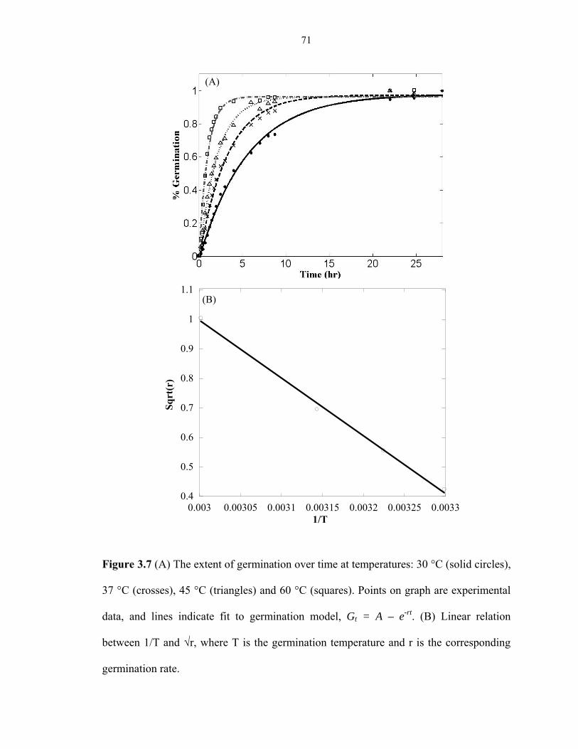
71
0.4
0.5
0.6
0.7
0.8
0.9
1
1.1
0.003 0.00305 0.0031 0.00315 0.0032 0.00325 0.0033
Sqrt
(r)
1/T
(B)
Figure 3.7 (A) The extent of germination over time at temperatures: 30 °C (solid circles),
37 °C (crosses), 45 °C (triangles) and 60 °C (squares). Points on graph are experimental
data, and lines indicate fit to germination model, Gt = A – e-rt. (B) Linear relation
between 1/T and √r, where T is the germination temperature and r is the corresponding
germination rate.
(A)

72
Figure 3.8 Competition experiments on spore germination of (A) B. subtilis from
Atacama Desert, (B) B. atrophaeus, (C) B. simplex from Kilimanjaro, (D) C. sporogenes,
(E) C. hungatei, and (F) C. G5A-1 from Greenland ice cores. The ratios of D-/L-alanine
0
0.2
0.4
0.6
0.8
1
0 50 100 150 200
Frac
tion
of
Ger
min
atio
n
Time (hr)
(A)
(B)
0
0.2
0.4
0.6
0.8
1
0 10 20 30 40 50 60 70
10:09:17.5:2.55:52.5:7.51:90:10Negative control
Frac
tion
of
Ger
min
atio
n
Time (hr)
0
0.2
0.4
0.6
0.8
1
0 50 100 150 200
Frac
tion
of
Ger
min
atio
n
Time (hr)
0
0.2
0.4
0.6
0.8
1
0 50 100 150
Frac
tion
of
Ger
min
atio
n
Time (hr)
0
0.1
0.2
0.3
0.4
0.5
0.6
0.7
0.8
0 10 20 30 40 50 60 70
Frac
tion
of
Ger
min
atio
n
Time (hr)0
0.2
0.4
0.6
0.8
1
0 20 40 60 80 100
Frac
tion
of
Ger
min
atio
n
Time (hr)
(C)
(D)
(E)
(F)

73
used are 10:0, 9:1, 7.5:2.5, 5:5, 2.5:7.5, 1:9 and 0:10. At any given sample, the total
germinant concentration (D- plus L-alanine) is 100 mM.

74
CHAPTER 4: DEVELOPMENT OF A MICROSCOPIC BASED
ENDOSPORE VIABILITY ASSAY TO DETECT SINGLE
CLOSTRIDIUM SPORE GEMINATION
4.1 ABSTRACT
A microscopy based rapid endospore viability assay (Micro-EVA) has been developed to
enumerate germinable Clostridium endospores in 15-30 minutes. Germinating
Clostridium endospores release ~108 molecules of dipicolinic acid (DPA) when
immobilized onto terbium ion (Tb3+)/D-alanine-doped agarose, which enables
enumeration under UV excitation of resultant green luminescent spots with time-gated
Tb3+-DPA luminescence microscopy. The intensity timecourses of the luminescent spots
were characteristic of the stage I germination dynamics. Micro-EVA was validated
against traditional CFU cultivation over three orders of magnitude in endospore
concentration, from 0 to 1000 spores/mL. Determination of the fractions of germinable C.
sporogenes spores by Micro-EVA yielded 56.4% ± 1.5%, while only 43.0% ± 1.0%
spores were culturable after 10 days incubation at room temperature. The ratio of
germinable/culturable was 1.31, which was consistent with the fact that a subset of the
total endospore population is germinable-but-not-culturable. With Micro-EVA, D-alanine
only germinated three Clostridium spores, but not the five different Bacillus spores tested.
Our results indicate that D-alanine is a potential solution to distinguish Clostridium
spores and Bacillus spores.

75
4.2 INTRODUCTION
Bacterial endospores are dormant microbial structures that are highly resistant to
chemical, physical, and radiation sterilization processes [1, 2]. They are formed when
spore formers (e.g., Bacillus and Clostridium) face unfavorable growth conditions, such
as starvation. Sporulation is considered one of the survival strategies of microorganisms.
Once they are formed, spores can stay dormant for extended periods of time [3, 4]. The
isolation of viable spore-forming bacteria from the gut of a bee trapped in Dominican
amber about 25~40 million years ago, and the recovery of spores from 250-million-year-
old halite crystals have been previously reported [5, 6].
The significant importance of studying endospores has been illustrated in chapter 1,
which can be summarized as follow. Bacillus cereus is a common aerobic food-borne
pathogen, and Clostridium botulinum and C. perfringens are anaerobic food-borne
pathogens associated with food poisoning from canned foods [7]. C. perfringens, C.
difficile and C. tetani are etiological agents in hospital acquired infection [8, 9]. A
number of Bacillus species are deliberately released because they produce toxins for
agricultural pest control, including B. thuringiensis, which accounts for approximately
90% of the bioinsecticide market [10]. The most ominous use of spore formers is the
release of B. anthracis as a bioweapon [11-13]. Spores of sulfite-reducing clostridia
(SSRC) have been considered as an indicator for the presence of pathogenic
microorganisms in drinking water by European Community legislation [14]. Due to their
extreme resistance to various sterilization processes that readily kill vegetative cells,

76
endospores are routinely employed as biological indicators to validate and monitor the
effectiveness of sterilization methods employed in various industries, such as medical
device, pharmaceuticals, health care, food preparation, and wastewater remediation [15].
Currently, the standard method for quantifying endospores is measuring colony forming
units (CFU) after heat-shock treatment. This method requires several days of incubation,
tedious anaerobic culturing technique for anaerobic spore formers, and it is only able to
culture only less than 1% of environmental spores [16]. Other detection methods include
DNA assay and immunoassay, which require extensive sample preparation and are labor
intensive. Chapter 3 describes a spectroscopy based endospore viability assay (Spectro-
EVA) to detect and quantify Clostridium spores via induced germination. Germination is
triggered by various germinants, such as L-alanine/NaHCO3, L-lactate or D-alanine,
which causes the release of approximately 108 molecules of a unique biomarker,
dipicolinic acid (DPA) from the spore core [17]. Spectro-EVA is based on the detection
of DPA, which is detected by adding TbCl3 to the spore suspension and measuring the
resulting Tb3+-DPA luminescence intensities [18, 19].
Despite the short detection time, Spectro-EVA has its limit. For example, it has to be
conducted in liquid suspension, and it is sensitive to other chemical interferences. The
limit of detection for Spectro-EVA is about 1000 spores/mL. To study the endospores
from various environmental samples, a much lower detection limit is desired.
77
Here we report details of a rapid microscopy based endospore viability assay (Micro-
EVA) in which C. sporogenes endospores were immobilized on terbium ion (Tb3+)/D-
alanine-doped agarose (Figure 4.1). D-alanine serves to trigger germination, during
which DPA is released from endospores. The Tb3+ subsequently binds DPA, resulting in
green luminescent spots under UV excitation in a microscope field of view, which were
enumerated as germinable endospores using time-gated Tb3+-DPA luminescence
microscopy (i.e., Micro-EVA). We validate Micro-EVA against culturing as a method
for rapid endospore viability assessment. We also applied Micro-EVA to distinguish
Clostridium spores from their aerobic counterparts.
4.3 MATERIALS AND METHODS
Materials
Deionized water (18.2 MΩ/cm) was obtained from an ultrafilter system (Water Pro PS,
LabConco, Kansas City, MO, USA). Terbium (III) chloride hexahydrate-(99.999%),
dipicolinic acid (2,6-pyridinedicarboxylic acid, DPA, 99%), L-alanine and D-alanine
were purchased from Sigma-Aldrich (Milwaukee, WI, USA). Agarose for culturing
experiments was obtained from MacConnell Research (San Diego, CA). Agarose for
Micro-EVA experiments was obtained from Invitrogen (Carlbad, CA). C. sporogenes,
ATCC 7955 and Clostridium hungatei (ATCC No.700212) from American Type Culture
Collection, (Manassas, VA, USA) was obtained as freeze-dried pellets. B. atrophaeus
(ATCC 9372) and B. cereus endospores were purchased from Raven Biological
Laboratories.

78
Endospore production and purification
C. sporogenes and C. hungatei were revived from frozen, dry pellets in a small volume
(5~6 mL) of growth media at the optimal growth temperatures of 37 °C and 30 °C,
respectively. The growth media for C. sporogenes and C. hungatei was reinforced
clostridial medium (RCM) and ATCC 2135 broth (GS-2CB medium), respectively.
Incubation commenced under strict anaerobic conditions with 100% N2 headspace. The
culture was transferred to fresh medium after development of visible turbidity. The
production and purification of various Clostridium spores was performed using a protocol
developed specifically for Clostridium species [20]. The spore suspension contained less
than 1% vegetative cell material as determined by phase contrast microscopy.
Bacillus spores were obtained by applying the protocol described as follow: Bacillus
vegetative cells were grown on TSA and inoculated onto a sporulation medium after
reaching exponential growth phase. The sporulation medium contained 1.6% nutrient
broth, 1.6% agar, 0.2% KCl, 0.05% MgSO4, 1 mM Ca(NO3)2, 100 µM MnCl2·4H2O, 1
µM FeSO4 and 0.1% glucose (pH 7.0) [4]. After incubation at 37 °C for 1 week, cells
were suspended into sterile deionized water. With phase contrast microscopy, 95% of the
cells formed endospores free of sporangia. Endospores were harvested and separated
from vegetative cells and debris by centrifugation at 6,300 g, washing 10 times and
sonication (25 kHz) for 5 min. The endospore suspension was incubated in lysozyme (0.2
mg/mL) and trypsin (0.1 mg/mL) at 30 °C with constant stirring overnight to lyse and
degrade vegetative cells. Endospores were purified by 8 cycles of centrifugation (6,300 g)

79
and washed with sterile deionized water until >99.9% of the cells were fully refractile
with no noticeable cellular debris.
Both Clostridium and Bacillus spore suspensions were stored at 4 °C in the dark before
use. Total spore concentrations were determined using a Petroff-Hausser hemocytometer
and CFU concentrations were determined using spread plating in triplicate measurements.
Sample preparation for Micro-EVA experiments
Endospore suspension with known concentration was filtered onto a 1.5 mm2 spot on a
0.2 µm polycarbonate membrane filter (Whatman, Florham Park, NJ) using a 96-well
micro-sample filtration manifold (Schleicher & Schuell, Keene, NH). To ensure that the
endospore surface density was optimal for a given initial endospore concentration, each
microscopic field of view contains less than 300 endospores. Endospores concentrated on
the filter were transferred to a ~0.5 mm thick, 9 mm diameter slab of 1.5% agarose
substrate containing 5 mM TbCl3 and 100 mM L-alanine (or D-alanine) mounted in a
silicone isolator (Molecular Probes, Eugene, OR) on a quartz microscope slide. After
endospore transfer, the agarose surface was covered with a piece of 0.2 mm thick
polydimethylsiloxane (PDMS).
Polydimethylsiloxane (PDMS) was prepared by mixing the polymer base and curing
agent (Sylgard, Dow Corning) in 10 to 1 ratio. After degassing, the mixture was cast over

80
a 0.2-mm thick stainless steel mold and cured in an oven for 2 hours at 65 °C. Agarose,
silicone isolator and PDMS were autoclaved at 121 °C for 15 minutes before use. A piece
of PDMS was peeled off and attached on top of an endospore-laden agarose surface for
sealing.
The Micro-EVA instrument
The instrument consists of a time-gated camera (Photonics Research Systems, Salford,
UK) mounted on a Nikon SMZ800 stereoscopic microscope (large working distance for
xenon lamp), a xenon flashlamp (Perkin-Elmer, Waltham, MA) mounted at 45 degrees
with respect to the sample, and a temperature controlled microscope slide holder
(Thermal). The slide holder enabled endospores to germinate at 37 °C. The CCD camera
has a resolution of 752 × 582 pixels at 14 bits with a chip size of 2/3 inch. The camera
has 50% sensitivity between 430 and 730 nm, with peak sensitivity at 550 nm. It was
Peltier-cooled to 40 °C below ambient temperature and was synchronized to the xenon
lamp via TTL pulses (300 Hz with tail time up to 50 µs). A highpass filter (03FCG067,
Melles Griot) centered at 500 nm was placed along the light path on the emission side
before reaching the microscope objective. We collected time stacks of time-gated images
by real-time streaming with a delay of 100 µs and exposure time of 5 s in each frame.
Endospore germination and germinable endospore assignment
Endospore germination on agarose surface followed the reported micro-germination
dynamics [21]. DPA released from single endospores manifested as individual bright

81
spots in 30 minutes under time-gated microscopy due to local formation of Tb3+-DPA.
Assignment of germinable endospores was made based on intensity, size and germination
timecourse. Adaptive thresholding was applied to segment pixels that were 20% brighter
than the background with a characteristic rising intensity. Each bright spot must exhibit a
continuous rising intensity over the course of germination in order to register an
endospore count. This criterion eliminated false positives by not counting sporadic bright
spots and long-lived luminescent interference. The 8-connected adjacent pixels were
analyzed to screen for endospore clumps. The number of endospores present was
calculated by dividing the squared sum of neighboring pixel brightness by the mean
brightness of an individual endospore determined empirically. This was done in a
recursive way until all of the pixels were counted and marked.
Phase contrast microscopic enumeration to determine spore concentrations
Aliquots (5 µL) of spore suspension were placed in a Petroff-Hausser counting chamber
(Model 3900, Hausser Scientific, Horsham, PA), and spores were observed at 400×
magnification using a phase contrast microscope (Nikon Eclipse 80i, AG Heinze Co,
Lake Forest, CA) mounted with a digital camera (Nikon Digital Sight DS-5M). The
smallest squares in the counting chamber are 0.05 mm × 0.05 mm, of which 80 were
analyzed to obtain the average spore count per square. To obtain statistically significant
counts, the spore concentration was held above 107 spores/mL, which resulted in at least
10 cells per sixteen squares.

82
Spore culturability
To determine the endospore culturability, heat-shock treatment at 80 °C for 15 minutes
was applied to a purified spore suspension with a known concentration that was first
determined by microscopy as described above. A series of dilutions were made to reach
an expected concentration range of 0 to 1000 spores/mL. For higher concentration (1000,
333 spores/mL), one hundred µL aliquots from each dilution were plated in triplicate on
solidified Reinforced Clostridial Medium (15 g agar per liter). For lower concentration
(below 100 spores/mL), 1 mL aliquots from each dilution were plated in triplicate on the
same solidified medium. Plates were incubated in a vinyl anaerobic chamber (Type C,
Coy Laboratory Products Inc., Grass Lake, Michigan) at room temperature for 10 days.
Colonies were counted and the average number was designated as colony forming units
(CFU) per volume of original sample. The culturability was determined as the percentage
of spores capable of forming colonies.
Spectroscopy
Tb3+-DPA luminescence excitation spectra (λex=250-360 nm, λem=544 nm) and emission
spectra (λex=278 nm, λem=450-650 nm) were recorded with a fluorimeter (Model FL-
1089, Jobin Yvon, Edison, NJ, USA) consisting of a 500-W Xe-lamp for excitation, two
double-monochromators set at 4 nm bandpass, and a Pelletier-cooled photomultiplier
tube (Model R928, Products for Research Inc, Danvers, MA, USA). A 500 nm cut-off
filter (Omega Filters, Brattleboro, VT, USA) was placed at the entrance of the emission
monochromator to prevent second-order diffraction. Endospores immobilized on agarose
were measured in front face configuration.

83
4.4 RESULTS
Germination time course for single Clostridium spore
Spores of C. sporogenes were used as the model microorganism for this study.
Germination of spores on Tb3+/germinant (L-alanine or D-alanie) doped agarose was
confirmed by the detection of DPA release via excitation spectroscopy using a Fluorolog
fluorimeter in a front face configuration (Figure 4.2). Figure 4.3 A showed the Micro-
EVA time-lapse images of a germinating C. sporogenes spore. DPA release was observed
with micor EVA via DPA complexation with the Tb3+-doped into the agarose matrix,
which can be visualized as a bright spot around the spore. The timecourse showed the
increase of intensity during germination as DPA molecules were released, and the
intensity reached a plateau after 15 minutes, indicating the completion of germination.
Figure 4.3 B showed single germinated spores under Micro-EVA in one field of view. In
combination, these data established that Micro-EVA can be used to observe single spore
germination on agarose.
Validation of Micro-EVA against CFU culturing
To validate Micro-EVA, we performed parallel germination and culturing experiments
over three orders of magnitude in endospore concentration, from 0 to 1000 spores/mL.
Figure 4.4 shows the germinable endospore concentrations measured with Micro-EVA,
and culturable endospore concentrations measured with plate counting plotted against
total endospore concentrations as determined with phase contrast microscopy. Sterile
samples did not yield false positive counts, which enabled us to achieve the ultimate

84
sensitivity of one germinable endospore per Micro-EVA field of view. Of the total
endospore concentrations, Micro-EVA revealed that 56.4% ± 1.5% of the population
were germinable within 60 minutes and CFU culturing determined that 43.0% ± 1.0%
were culturable within 10 days incubation. The ratio of germinable/culturable was 1.31,
which is consistent with the fact that a subset of the total endospore population is
germinable-but-not-culturable.
Discrimination of Clostridium from Bacillus spores
D-alanine was used as the germinant to distinguish Clostridium spores from Bacillus
spores. D-alanine has been reported as a germination inhibitor for various Bacillus spores,
however, its effect on Clostridium spore germination has not been extensively studied.
We tested the effect of D-alanine on germination with various Clostridium and Bacillus
spores with Spectro-EVA, and found that in liquid suspension, D-alanine germinated all
Clostridium spores tested, while it inhibited the germination of all but one Bacillus spores,
B. simplex isolated from Kilimanjaro (Chapter 3). This indicated that D-alanine may be a
potential solution to distinguish Clostridium spores and Bacillus spores.
We conducted Micro-EVA experiment with D-alanine as the only germinant on five
different Bacillus spores and three Clostridium spores. The Bacillus spores used in this
study are: B. subtilis isolated from Atacama Desert, B. simplex isolated from Kilimanjaro,
B. atrophaeus and B. cereus from ATCC, B. longisporus isolated from JPL soil samples.
The Clostridium spores used in this study are C. sporogenes and C. hungatei from ATCC,

85
and Clostridium G5A-1 isolate from Greenland ice core. With all the Bacillus spores
tested, none of them germinated with D-alanine as the sole germinant on agarose with
Micro-EVA, including the Kilimanjaro strain that germinated by D-alanine in liquid
suspension with Spectro-EVA. All three Clostridium spores germinated by D-alanine,
indicated as bright spots under UV light on agarose after 1 hour germination at 37 °C
(Figure 4.5).
4.5 DISCUSSION
We have developed a microscopy based Endospore Viability Assay (Micro-EVA) to
detect anaerobic spores, and to measure the germinable spores in a solid phase. Micro-
EVA has also been validated with respect to traditional CFU cultivation. Micro-EVA
yielded 56.4% ± 1.5% germinable spores with 60 minutes, while CFU revealed 43.0% ±
1.0% culturable spores for the same suspension after 10 days incubation at room
temperature. The difference can be explained as only a subset of germinable spores is
culturable. Micro-EVA measurements probe DPA release during stage 1 germination,
while CFU measurements probe the cell division, which is the last stage of the
germination and outgrowth process, and requires proper growth conditions (e.g., enough
nutrient, and anaerobic environment for Clostridium spores). Micro-EVA proved to be a
faster method to assess spore viability, as germinability has been proposed as the
indicator of spore viability [17, 22].

86
In Micro-EVA experiments, individual germinable spores are counted in a microscope
field of view after germination commences. As a spore germinates, ~108 DPA molecules
are released into the immediate area surrounding the spore. DPA then combines with
Tb3+ in the agarose matrix to form the Tb3+-DPA luminescence halos under UV
excitation. The germinating endospores manifest as bright spots that grow in intensity
over a period of 3-5 minutes, and are enumerated in a microscope field of view. The
characteristic germination timecourse allows unambiguous assignments of germinable
spores.
In chapter 3, we reported a related method where germinating spores were enumerated in
bulk suspension by luminescence spectroscopy (i.e., Spectro-EVA), where Tb3+-DPA
luminescence intensities were tabulated against a C. sporogenes spore calibration curve
[17]. The Micro-EVA approach is superior to Spectro-EVA because Micro-EVA is
capable of enumerating single spores while the limit of detection of Spectro-EVA is 1000
spores/mL. This advantage is gained because in Micro-EVA experiments the mM DPA
halos surrounding single germinated endospores are readily imaged with high contrast,
whereas germination of single endospores in bulk suspension (~1 mL) gives rise to mere
fM DPA concentrations, which are far below LOD for Spectro-EVA. With Micro-EVA,
we take advantage of the long luminescent lifetime (τ = 0.5 to ~2 ms) of Tb3+-DPA [23],
enabling the use of time gating to effectively remove background fluorescence (i.e.,
interferent fluorophores with nanosecond lifetimes). Time gating eliminates potential
false positive causing features and renders the image background dark. Elimination of
this background enables a striking increase in image contrast and detection sensitivity

87
even for the most challenging environmental extracts, such as soil samples. This will be
illustrated in chapter 5.
The difference between Bacillus and Clostridium spores has received increasing attention
recently due to the advance of molecular technology. It has been reported that the
difference of these two genera lies in the initiation of sporulation [24]. However, the
study to distinguish spores from these two genera has not been reported. Here, we
investigate this issue by studying the response of Clostridium and Bacillus spores to
different germinants. Our preliminary results show that D-alanine, D-serine and lactic
acid inhibit Bacillus spores germination, while all chemicals facilitate the germination of
Clostridium spores (data not shown). We picked D-alanine for further study. Our results
show that on solid phase with Micro-EVA, D-alanine does not germinate all Bacillus
spores tested, while it germinates all Clostridium spores. We have not investigated in
why spores of B. simplex from Kilimanjoro germinate in liquid solution (Spectro-EVA)
with D-alanine as the sole germinant but not on solid phase (Micro-EVA). Our results
support that D-alanine can indeed distinguish Clostridium spores from Bacillus spores on
solid phase with Micro-EVA. In fact, D-alanine has been reported as a germination
inhibitor for various Bacillus spores by competing with L-alanine for the active site of
dehydrogenase [25-27]. However, D-alanine does not have any inhibitory effect on the
Clostridium spore germination, despite the similarity between these two genera. Our
results provide insight to understand the germination mechanism for Clostridium spores.

88
Micro-EVA is not only much more rapid than culture dependent methods (30 minutes vs.
3-10 days), but the simple chemistry, instrumentation, and image analyses are all
amenable for automation. Automated viability assessment of endospores will have the
potential to find application in many industries where microbial inactivation needs to be
monitored and assured. Potential applications of Micro-EVA for Clostridium endospores
include evaluating the efficacy of sterilization regimens for food, wastewater, and
medical equipment. To test a given sterilization regimen, we envision that surrogate
microorganisms (e.g., C. sporogenes spores) would be applied to the appropriate matrix
(i.e., food, wastewater, medical equipment), and after the sterilization process is
complete, the matrix is extracted, and the extract analyzed with the Micro-EVA. By
comparing the percentage of germinable spores before and after a specific sterilization
treatment, we are able to evaluate if the treatment is efficient or not. This is particularly
important for the new sterilization technologies, since Micro-EVA can potentially
monitor the in situ inactivation dynamics, which is indispensable for identifying the
inactivation regime and validating the new technology. Ultimately, with implementation
of automated sample handling and analysis, we envision that Micro-EVA will enable
online monitoring of sterilization processes for various sterilization processing facilities.
89
4.6 REFERENCES
1. Nicholson, W.L., et al., Resistance of Bacillus endospores to extreme terrestrial and extraterrestrial environments. MMBR, 2000. 64: p. 548-572.
2. Setlow, P., ed. Bacterial Stress Response. ed. G. Storz and R. Hengge-Aronis. 2000, ASM Press: Washington, D.C. 217-230.
3. Sonenshein, A.L., ed. Endospore-forming bacteria: an overview. Prokaryotic Development, ed. Y.V. Brun and L.J. Shimkets. 2000, American Society for Microbiology: Washington, D.C. 133-150.
4. Nicholson, W.L. and P. Setlow, eds. Sporulation, germination and outgrowth. Molecular Biology Methods for Bacillus, ed. Y.V. Brun and L.J. Shimkets. 1990, John Wiley and Sons: Sussex, England. 391-450.
5. Cano, R.J. and M.K. Borucki, Revival and identification of bacterial spores in 25-million-year-old to 40-million-year-old Dominican amber. Science, 1995. 268: p. 1060-1064.
6. Vreeland, R.H., W.D. Rosenzweig, and D.W. Powers, Isolation of a 250 million-year-old halotolerant bacterium from a primary salt crystal. Nature, 2000. 407: p. 897-900.
7. Murray, P., et al., eds. Manual of Clinical Microbiology. 8th ed. Vol. 1. 2003, ASM Press: Washington, D.C.
8. McFarland, L.V., Epidemiology of infectious and iatrogenic nosocomial diarrhea in a cohort of general medicine patients. Am. J. Infect. Control, 1995. 23: p. 295-305.
9. McFarland, L.V., et al., Nosocomial acquisition of Clostridium difficile infection. N. Engl. J. Med., 1989. 320: p. 204-210.
10. Chattopadhyay, A., N.,, B. Bhatnagar, and R. Bhatnagar, Bacterial insecticidal toxins. Crit. Rev. Microbiol., 2004. 30: p. 33-54.
11. Jernigan, D.B., et al., Investigation of bioterrorism related anthrax, United States, 2001: epidemiologic findings. Emerg. Infect. Dis., 2002. 8: p. 1019-1028.
12. Sanderson, W.T., et al., Bacillus anthracis contamination and inhalational anthrax in a mail processing and distribution center. J. Appl. Microbiol., 2004. 96: p. 1048-1056.
13. Weis, C.P., et al., Secondary aerosolization of viable Bacillus anthracis spores in a contaminated US Senate Office. JAMA, 2002. 288: p. 2853-2858
14. Committee on Indicators for Waterborne Pathogens, N.R.C., ed. Indicators for Waterborne Pathogens 2004, The National Academy Press: Washington, D.C.

90
15. Rutala, W.A. and D.J. Weber, Disinfection of endoscopes: review of new chemical sterilants used for high-level disinfection. Infection control and hospital epidemiology, 1999. 20: p. 69-76.
16. Ponce, A., S.A. Connon, and P.T. Yung, eds. Detection and viability assessment of endospore-forming pathogens. Principles of Bacterial Detection: Biosensors, Recognition Receptors and Microsystems, ed. M. Zourob, S. Elwary, and A. Turner. 2008, Springer: New York. 481-523.
17. Yang, W.-W. and A. Ponce, Rapid endospore viability assay of Clostridium sporogenes spores Int. J. Food Microbiol., 2009. 133: p. 213-216.
18. Hindle, A.A. and E.A.H. Hall, Dipicolinic acid (DPA) assay revisited and appraised for spore detection Analyst, 1999. 124: p. 1599-1604.
19. Jones, G. and V.I. Vullev, Medium effects on the stability of terbium(III) complexes with pyridine-2,6-dicarboxylate. J. Phys. Chem., 2002. 106: p. 8213-8222.
20. Yang, W.-W., E.N. Crow-Willard, and A. Ponce, Production and characterization of pure Clostridium spore suspensions. J. Appl. Microbiol., 2009. 106: p. 27-33.
21. Hashimoto, T. and W.R. Frieben, Microgermination of Bacillus cereus spores. J. Bacteriol., 1969. 100: p. 1385-1392.
22. Shafaat, H.S. and A. Ponce, Applications of a rapid endospore viability assay for monitoring UV inactivation and characterizing arctic ice cores. Appl. Environ. Microbiol., 2006. 72: p. 6808-6814.
23. Jones, G. and V.I. Vullev, Medium effects on the photophysical properties of terbium (III) complexes with pyridine-2,6-dicarboxylate (supporting information). Journal of Physical Chemistry A, 2002. 106: p. 8213-8222.
24. Dürre, P. and C. Hollergschwandner, Initiation of endospore formation in Clostridium acetobutylicum. Anaerobe, 2004. 10: p. 69-74.
25. Fey, G., G.W. Gould, and A.D. Hitchins, Identification of d-alanine as the auto-inhibitor of germination of Bacillus globigii spores. J. Gen. Microbiol., 1964. 35: p. 229-236.
26. Preston, R.A. and H.A. Douthit, Germination of Bacillus cereus spores: critical control by DL-alanine racemase. J. Gen. Microbiol., 1984. 130: p. 3123-3133.
27. Yasuda-Yasaki, Y., S. Namiki-Kanie, and Y. Hachisuka, Inhibition of Bacillus subtilis spore germination by various hydrophobic compounds: demonstration of hydrophobic character of the L-alanine receptor site. J. Bacteriol., 1978. 136: p. 484-490.

91
Figure 4.1 (a) Configuration of the Micro-EVA instrument used in this investigation,
consisting of a stereomicroscope mounted with a time-gated camera and a xenon flash
lamp for UV excitation. (b) Sample well on quartz microscope slide containing Tb3+/D-
alanine doped agarose. (c) Schematic representation of the sample slide, consisting of a
quartz slide on which Tb3+/D-alanine doped agarose is confined by red rubber gasket well.
Endospores (brown circles) are inoculated onto agarose substrate and subsequently
covered with a thin layer of PDMS. (d) Inoculated endospores germinate due to D-
alanine, causing the release of ~108 molecules of DPA and subsequent formation of
highly luminescent Tb3+-DPA complexes that appear as discrete bright spots in the
microscope field of view. (e) Absorption-energy transfer-emission photophysics of the
Tb3+-DPA luminescence assay. DPA acts as a light harvester that transfers excitation

92
energy to luminescent terbium ion. (f) Energy (Jablonski) diagram of the Tb3+-DPA
photophysics.

93
0
2 106
4 106
6 106
8 106
1 107
260 280 300 320 340 360
Lum
ines
cenc
e In
tens
ity
Wavelength (nm)
Figure 4.2 Excitation spectrum (λem = 545 nm) of the agarose substrate of Micro-EVA
when 108 C. sporogenes endospores were inoculated, taken with a Fluorolog-3
fluorimeter at a front-face configuration. The dual excitation peaks (λex = 272, 280 nm)
indicated the presence of Tb3+-DPA released from germinating spores. Red plot
represents germinating endospores on agarose and the green plot shows the agarose
substrate.
280 nm 272 nm

94
Figure 4.3 (a) Germination timecourses of single C. sporogenes spores at 37 °C
monitored by Tb3+-DPA luminescence using Micro-EVA. (b) Two single germinated
spores under Micro-EVA in one field of view. Purple color is the background intensity;
germinated spores are luminescent as green spots under UV excitation.
(a)
(b)
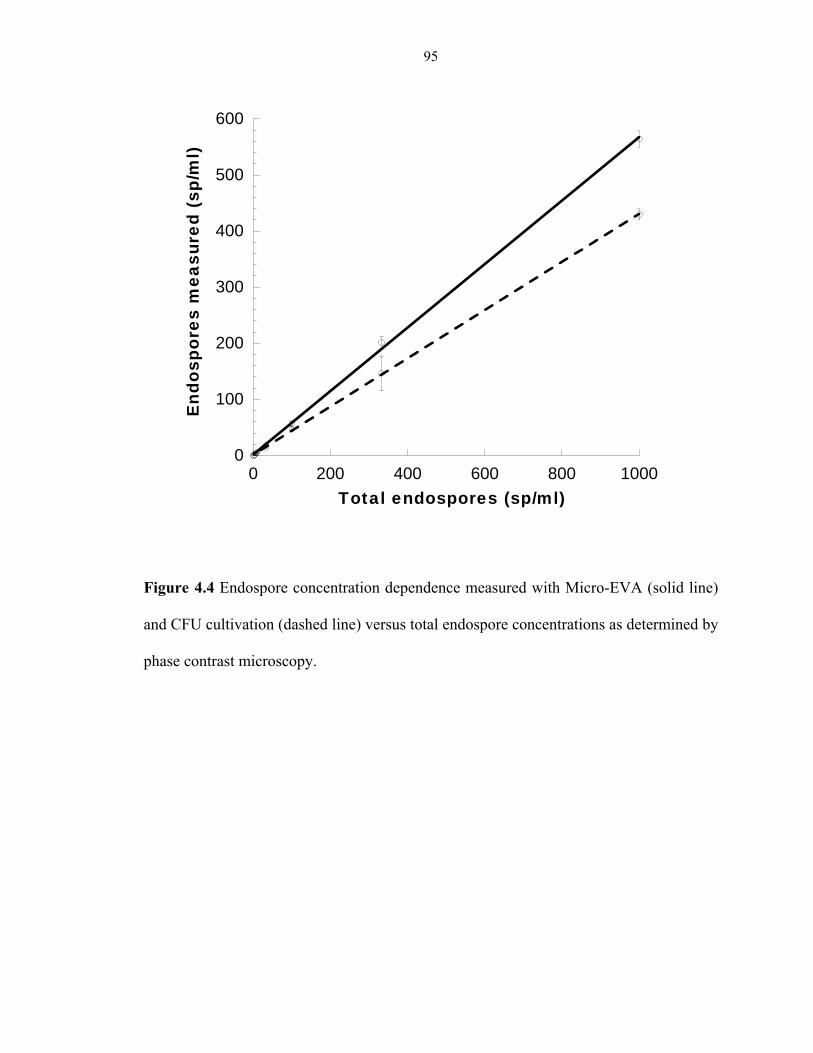
95
Figure 4.4 Endospore concentration dependence measured with Micro-EVA (solid line)
and CFU cultivation (dashed line) versus total endospore concentrations as determined by
phase contrast microscopy.
0
100
200
300
400
500
600
0 200 400 600 800 1000
End
ospo
res
mea
sure
d (s
p/m
l)
Total endospores (sp/ml)

96
(a) (b)
(c) (d)
(e) (f)
(g) (h)

97
Figure 4.5 Endospores inoculated on Tb3+/D-alanine-doped agarose. (a) B. subtilis from
Atacama Desert, (b) B. simplex from Kilimanjaro, (c) B. atrophaeus (ATCC 9372), (d) B.
cereus, (e) B. longisporus from JPL soil, (f) C. hungatei (ATCC 700212), (g) Clostridium
G5A-1 from Greenland ice core, (h) C. sporogenes (ATCC 7955). The bright spots on
images of Bacillus spores are Tb microspheres, which serve as internal standard to
facilitate locating the focal plane. Scale bar: 100 µm.

98
CHAPTER 5: APPLICATION OF MICRO-EVA TO DETECT
CLOSTRIDIUM SPORES FROM GREENLAND ICE CORE AND
ATACAMA DESERT
5.1 ABSTRACT
Understanding the boundary conditions for survival of microorganisms in extreme
environments will aid the NASA effort to search for life on Mars and prevent forward
contamination from spacecraft before launching. We investigated the limit and longevity
of life in two Mars analogs on Earth, Greenland ice core and Atacama Desert. While the
spectroscopy based endospore viability assay (Spectro-EVA) failed to quantify
germinable endospores in most environmental samples, the microscopy based endospore
viability assay (Micro-EVA) has been successfully employed to detect and quantify
viable Clostridium spores from these extreme environments. Micro-EVA is based on the
detection of a unique biomarker of endospores, DPA, via terbium ion (Tb3+-DPA)
luminescence. DPA is released during D-alanine induced germination, an indicator for
spore viability. Greenland ice core samples yielded 1 to 2 germinable Clostridium spores
per milliliter of melt ice water after 2-hour germination, and our culture based study also
showed that 10 different Clostridium species are revived from the ice core samples with
age up to 60,000 years old. The Atacama Desert samples yielded 66 to 157 germinable
Clostridium spores/g soil after 90-minute germination. Our preliminary findings support
the hypothesis that the higher water activity of the environmental sample, the more
germinable spores are detected from it. In conclusion, Micro-EVA has been successfully

99
applied to assess germinability of Clostridium spores in extreme environments. Studying
these extreme habitats on Earth gleans invaluable information on the search for extinct or
extant life on the dry and icy biosphere, Mars.
5.2 INTRODUCTION
The first astrobiological experiment conducted by the Viking missions more than three
decades ago concluded that life had little chance of developing on Mars due to the harsh
conditions detected on the surface of the planet: intense UV radiation, generation of
oxidant compounds, absence of water and low temperatures [1]. However, in recent years,
the discovery of extremophiles, organisms that can thrive under conditions formerly
considered unsuitable for life, such as extreme temperatures, pressures, pH, opens the
window on possibilities that life exists in these extreme conditions [2, 3]. There are
various places on Earth considered as Mars analogs due to their extreme physical and
chemical conditions [4], such as the Antarctic ice sheets and underground lakes, Atacama
Desert [2]. Research on life in these extreme environments sheds light on several
important exobiological questions in the past: What are the environmental limits for life?
How long can microorganisms survive? Is there life beyond Earth? Can life travel from
one planet to another?
Approximately 75% of Earth’s biosphere is cold and covered by oceans at temperatures
below 5°C [5]. More than 70% of Earth’s fresh water occurs as ice and a large portion of
the soil ecosystem exists as permafrost. These permanently frigid habitats are unique
repositories of microbes frozen at various points in the geologic past. A diversity of

100
viable microorganisms, including fungi, bacteria and archaea have been isolated from
glacial ice up to 400,000 years old and permafrost up to 3 million years old, originating
from many geographical locales [6-11]. The extent to which organisms can survive
extended periods in metabolic inactive form in ice cores is one of the key questions in the
study of life in extreme environments. Ice cores, as an environment with low and more or
less constant temperature, is the region where remnants of life or even dormant forms of
life would be preserved for the longest time. The ice sheets of Greenland are considered
suitable sites for researchers to study longevity of microbial cells at low temperatures
[12]. GISP2 (Greenland Ice Sheet Project 2) is an international collaborative project that
lasted from 1993 to 1998 to drill and collect ice cores from different depths along the
3053.44 m ice, which corresponding to samples aged ~110,000 years old. Only a few
studies have been carried out on bacterial communities within ice cores, however, the
population of bacterial spores in these communities has never been directly assessed [13-
16]. We will address this question in this chapter.
As one of the oldest and driest desert on Earth, Atacama Desert in Northern Chile is
considered one of the Mars analogs on Earth. It is a cold desert with a hyper arid habitat
[2]. The average annual air temperature is -5 °C to 45 °C, depending on elevation and
distance from the sea. The annual precipitation is less than 1 mm. Some of the weather
stations in the Atacama Desert have never received rainfall records. The cold water
brought from Antarctica along the Chilean coast creates a phenomenon called the
temperature inversion in which cold air is below a ceiling of warm air, preventing water
evaporation and formation of clouds. Also, the desert is in the rain shadow of the Andes

101
on the west and Chilean Coast Range on the north, within the dry subtropical climate belt
[17, 18]. Owing to the combined effects of these physical factors, the Atacama Desert has
been devoid of water for over 150 million years [19-21], and it has been reported to
contain near sterile soils with only trace organic carbon and nitrogen content, and these
conditions have existed for at least several millions years [22]. The upper soil layers are
dry and weathered. The soil environment precludes any downward infiltration. For this
reason, the possibility of penetration of model microorganisms into the soil subsurface is
precluded, making it a very good model to assess the dry limit of life, effect of water on
microbial abundance and preservation of organic material along a depth transect of soil. It
is also one of the best repositories for life on Earth because of minimal biological
degradation of dead cell debris. Our research group went on an expedition to the Atacama
Desert in 2007, and collected samples from the most arid zone, the Yungay area, and a
depth transect in a soil pit 80~220 cm underneath the surface soil. Samples from two sites
in the Yungay area were collected: Site A and Site E. Site A is the location of a past
effort to extract DNA and cover culturable microorganisms from the soils of this region
[22], which did not yield detectable concentrations of DNA and low culturable cell
counts. Site E is 5 km northeast of Site A, and is the location of prior investigations that
focused on soil chemistry, identification of cultured isolates and DNA extraction [23, 24].
Formation of the remarkably resistant and dormant endospores in genera such as Bacillus
and Clostridium is observed as a response to adverse conditions, such as nutrient
depletion and low temperature [25]. Due to their resistance to various extreme stresses,
endospores are one of the life forms that are most likely to survive in the extreme

102
environments mentioned above. In fact, a six-year space study also showed that spores of
Bacillus subtilis survived after exposure to space environments such as high vacuum and
radiation [26]. If endospores were present in an early warm and wet Martian environment,
they may currently be surviving in the subsurface ice/permafrost. Assessment of viable
endospores from Greenland Ice Core and Atacama Desert provides insight on the limit
and longevity of life, and the potential of extraterrestrial life. We have described two
endospore viability assays, Spectro-EVA and Micro-EVA, which are based on the
detection of the release of a unique biomarker, dipicolinic acid (DPA) upon induced
germination. In this chapter, we applied of these assays to assess of viable Clostridium
spores from these two Mars analogs. As comparison, culture-based and DNA extraction
studies have been also included.
5.3 MATERIALS AND METHODS
Materials
Deionized water (18.2 MΩ/cm) was obtained from an ultrafilter system (Water Pro PS,
LabConco, Kansas City, MO). Terbium (III) chloride hexahydrate-(99.999%), dipicolinic
acid (99%) (2,6-pyridinedicarboxylic acid, DPA), and D-alanine were obtained from
Sigma-Aldrich (Milwaukee, WI), and were used without further purification. Sodium
pyrophosphate was purchased from Mallinckrodt (Paris, KY). Agarose for culturing
experiments was obtained from MacConnell Research (San Diego, CA). Agarose for
Micro-EVA experiments was obtained from Invitrogen (Carlbad, CA). All utensils were
either prepackaged one-use only or glassware was kilned at 500 °C for 4 hours for

103
sterilization and removal of biomolecules such as DNA, RNA and DPA. All media and
utensils were pre-chilled to 4 °C before use for the Greenland ice core experiments.
GISP2 ice core handling
The ice core was received as a gift from the Greenland Ice Sheet Project (GISP 2) and
corresponded to 14 depths from 158 to 2804 m. The GISP2 ice cores, arranged in
ascending depth order (MCA.02 # 158, depth 157.45-157.70 m, age 600 years; MCA.02
# 480, depth 480.15-480.40 m, age 2000 years; MCA.02 # 835, depth 834.10-834.35 m,
age 4000 years; MCA.02 # 1566, depth 1565.75-1566.00 m, age 10,000 years; MCA.02
#1923, depth 1922-1922.25 m, age 20,000 years; MCA.02 #2096, depth 2095.75-2096.00
m, age 30,000 years; MCA.02 #2253, depth 2252.10-2252.35 m, age 40,000 years;
MCA.02 #2429, depth 2428.00-2428.25 m, age 50,000 years; MCA.02 #2517, depth
2516.75-2517.00 m, age 60,000 years; MCA.02 #2593, depth 2592.31-2592.56 m, age
70,000 years; MCA.02 #2657, depth 2656.20-2656.45 m, age 80,000 years; MCA.02
#2703, depth 2702.11-2702.36 m, age 90,000 years; MCA.02 #2746, depth 2745.65-
2745.90 m, age 100,000 years; MCA.02 #2804, depth 2803.65-2803.90 m, age 110,000
years), were obtained from the National Ice Core Laboratory (NICL) and stored at -80 °C.
In preparation for decontamination, the ice core temperature was ramped to 0 °C via
storage first at -25 °C for eight hours, followed by two hours at 0 °C. Temperature
equilibration of the ice core was necessary to prevent the decontamination solutions from
freezing onto the outside of the ice core.

104
Decontamination was carried out under aseptic conditions, as illustrated in figure 5.1.
The core sections were placed on an aluminum block (pre-chilled at -80 °C freezer and
disinfected by bleach). A heat-sterilized saw was used to cut the core into half. One half
underwent the decontamination process and the remaining portion was stored at -80 °C
for future processing. A beaker containing 1600 mL of cold (4 °C) 6.25% sodium
hypochlorite (NaOCl) solution and 3 beakers containing 1600 mL of cold (4 °C) sterile
distilled water (18.2 MΩ, < 1 ppb TOC, DNase-free and RNase-free) were prepared. The
ice core was submerged in the NaOCl solution for 10 s and then rinsed with sterile water
3 times, each with 10 s, and finally placed in another beaker for melting aseptically at
room temperature inside a biohazard cabinet hood (Sterilgard III Advance, The Baker
Company, Sanford, ME). The decontamination procedure has been validated using a
synthetic ice core sections (a cylinder 5 cm in diameter and 15 cm in length), consisting
of frozen suspensions of 104 B. atrophaeus endospores in the interior, and 105 B.
megaterium endospores smeared on the exterior surfaces.
Before melting, 1/10 of the meltwater was reserved for culturing. The remaining
meltwater was vacuum filtered through a 45 mm 0.1 µm membrane filter (Nucleopre
Track-etch membrane, Sterlitech Corporation, Kent, WA) backed with a 0.45 µm Supor
backing filter. The meltwater was analyzed with 24 hours by culturing. The filter(s)
was/were resuspended in 4.4 mL of the original cold filtrate (or cold sterile water for the
first three pieces ice core, depth 158 m, 480 m and 835 m). The suspension and filter(s)
were vortexed for 5 minutes, with 1-minute chilling increment in between to prevent cells
from overheating. The membrane filter(s) was/were removed from the suspension and

105
stored in -20 °C freezer for clone library analysis. The concentrated suspension was used
for subsequent endospore viability assays.
Ice core analysis using Spectro-EVA
For the older ice cores (age 10,000 years old and above), the ice core suspension was
prepared in 300 µL containing 100 mM L-alanine, 10 mM NaHCO3 and 1 µM TbCl3 at
pH 6.5 to induce germination of Clostridium spores, and for remaining ice cores, ice core
suspension was prepared in 250 µL containing 100 mM D-alanine to induce Clostridium
spores germination. The ice core suspensions were incubated at 37 °C for 48 hours to let
germination complete. After germination, ice core suspension was transferred to an
eppendorf tube and autoclaved at 134 °C for 45 minutes to release DPA from
ungerminated spores. Before and after germination, and after autoclaving, Tb3+-DPA
luminescence excitation (λex = 250 – 360 nm, λem = 544 nm) and emission (λex = 278 nm,
λem = 450 - 650 nm) spectra were recorded at room temperature with a fluorimeter model
FL-1089 (Jobin Yvon, Edison, NJ) consisting of a 500-W Xe-lamp for excitation, two
double-monochromators set at 4-nm bandpass, and a Pelletier-cooled photomultiplier
tube model R928 (Products for Research, Inc., Danvers, MA). Emission intensities were
calibrated with respect to an external Tb3+-DPA luminescence standard solution.
Luminescence intensities were quantified by integrating the area under the 544-nm
emission peak with a baseline subtraction. Quartz micro-cuvettes (Starna Cell) were used
for minimal dilution factor. The aerobic version of Spectro-EVA has also been employed
on all ice cores with a change in the germinant used. 100 mM L-alanine was used as a
sole germinant for Bacillus spores.

106
Ice core analysis using Micro-EVA
Micro-EVA experiments were performed as previously described in chapter 4. Briefly, a
time-gated microscopy technique was used to count individual endospores in a
microscope field of view. The contrast in the image is generated by the formation of the
long-lived and brightly luminescent Tb3+-DPA complex as DPA is released during the
germination process. DPA release is observed as a spreading halo originated from each
endospore during germination, where DPA molecules bind with Tb3+ that has been added
into the surrounding matrix. The germinating endospores manifest as bright spots in the
field of view that grow in intensity over a period of 3-5 minutes.
A 100 µL aliquot of the concentrated sample was filtered onto a 1.5 mm2 spot on a 0.2
µm polycarbonate membrane filter (Whatman, Florham Park, NJ) using a 96-well micro-
sample filtration manifold (Schleicher & Schuell, Keene, NH). The sample was
transferred aseptically to 1.5% agarose doped with 5 mM TbCl3 and 100 mM D-alanine,
contained in a 9 mm silicone well (Molecular Probes, Eugene, OR) on a quartz slide. The
sample was transferred by streaking the membrane filter across the agarose surface. The
D-alanine within the agarose triggers Clostridium spores present in the sample to
germinate and release DPA. A piece of 4.5 µm thick polydimethylsiloxane (PDMS) film
was placed on top of the agarose to prevent aerial contamination, minimize water
evaporation and DPA diffusion across the agarose substrate. Images were taken with a
time-gated microscope, which consists of a pulse xenon flashlamp, a stereoscopic
microscope (1 mm × 0.8 mm field of view at 63× magnification), mounted with a time-
gated camera (Photonics Imaging System, UK). The timing of gating is governed by the

107
xenon flashlamp, which serves as the master oscillator at 300 Hz. After a delay of 100 µs
after the lamp flash, the CCD camera begins integration of single photons. Over a
germination course of 120 minutes, formation of luminescence spots could be observed
under time-gated microscopy, which was indicative of the localized formation of Tb3+-
DPA due to DPA release during endospore germination. Background fluorescence and
autofluorescence were minimized because of time gating, rendering a low intensity
background. Characteristic germination timecourse allows unambiguous assignment of
germinating endospores.
CFU Cultivation
Cultivation experiments were only conducted with older ice cores (age 10,000 and above).
1/10 R2A was used for the cultivation of aerobic bacteria. Two solid media, MM2 and
1/10 R2A, were used for the growth of anaerobic bacteria. Both aerobic and anaerobic
cultures were incubated at two different temperatures: 7 °C and 22 °C. MM2 consists of
basal mineral salts (CaCl2·2H2O, 0.2 g/L; KH2PO4, 0.5 g/L; K2HPO4, 0.5 g/L, Na2SO4, 1
g/L; NH4Cl, 0.5 g/L; MgSO4·7H2O, 0.2 g/L; NaCl, 0.8 g/L), trace elements (FeCl2·4H2O,
0.003 g/L; CuCl2·2H2O, 2×10⁻⁴ g/L; MgCl2·6H2O, 0.001 g/L; CoCl2·6H2O, 0.0017 g/L;
ZnCl2, 0.001 g/L; H3BO3·2H2O, 10-4 g/L; NiSO4·7H2O, 3×10⁻⁴ g/L; Na2SeO3, 2×10⁻⁴ g/L),
redox indicator (resazurine, 10-4 g/L), reducing agent (cysteine HCl, 0.5 g/L; Na2S·9H2O,
0.3 g/L), carbonate buffer (NaHCO3, 4 g/L) and various nutrients (trypticase, 1 g/L;
yeast extract, 1 g/L; glucose, 5 g/L). For anaerobic samples that are incubated at 7 °C,
plates were stored inside a BBL GasPak 100TM Anaerobe vented system (Becton
Dickinson, Sparks, MD). For anaerobic samples that are incubated at 22 °C, plates were

108
incubated in a vinyl anaerobic chamber (Type C, Coy Laboratory Products Inc., Grass
Lake, Michigan). The liquid media were overgassed with N2, inoculated from the melted
ice (~1:200 dilution), and incubated at 7 °C and 22 °C respectively. All experiments are
conducted in triplicate. Cell counts reported are based on solid agar cultures after ~6
months of incubation at room temperature.
Identification of anaerobic spore formers
Each isolated was grown in MM2 liquid medium until the OD reached ~0.5. Cells were
harvested and DNA was extract using a protocol for isolation of genomic DNA from
gram positive bacteria in QIAGEN’s DNeasy Tissue Kit (QIAGEN, Inc., Valencia, CA,
USA). DNA amplifications were performed in a reaction containing ~50 ng of pure
genomic DNA, 2.5 U of Taq DNA polymerase (Applied Biosystems), 5 µL of 10× PCR
buffer, 2.5 mM MgCl2, 200 µM each deoxynucleotide triphosphate, 1 µM each 8-forward
and 1492-reverse primers, and sterile water to a total volume of 50 µL. Reaction mixtures
were irradiated with ultraviolet light for 5 minutes at 254 nm (1000 J/m2) before the
addition of genomic DNA and Taq DNA polymerase to eliminate potential contamination.
All negative controls with water instead of DNA template were tested. The PCR product
was purified with QIAquick PCR Purification Kit (QIAGEN, Inc., CA). DNA sequencing
was performed by the routine automated methods at Genewiz, Inc. (South Plainfield, NJ).
Sequencing primers, 8-forward, 1492-reverse and one internal primers, 338-forward,
were used to sequence the PCR products. Sequence reads were assembled and edited by
using Invitrogen Vector NTI ContigExpress. Web-based similarity searches were

109
performed at GenBank and the Ribosomal Database Project (RDP) [27]. Sequences were
phylogenetically analyzed in an ARB database (http://www.arb-home.de/, [28])
Sequences were aligned using the Fast Aligner in ARB and the tree was constructed
using the Neighbor Joining distance method with a Jukes and Cantor correction.
Atacama Desert soil sampling
All utensils were either packaged sterile or pre-treated at 240 °C overnight. The sampling
sites were approached downwind, and gloves and masks worn to reduce the chance of
contamination. Care was taken to minimize contamination of the experimental sites by
reducing foot traffic to designated paths and areas. Soil samples were collected by spoons
and scoops aseptically into sterile Whirl-Paks with the collector wearing masks, apron,
and sterile gloves collected when facing the wind. The rhizosphere domain is defined as
extending approximately 5 cm from the root surface in all directions. Some root nodules
were collected together with soils. Sieving, homogenizing and sample handling took
place in a station in Yungay. Soil samples were sealed in amber jars to protect from
sunlight and moisture, transported in coolers with ice packs/dry ice or placed in dry
shipper dewars for transport back for laboratory analysis.
Measure of soil water activity, pH, EH, eC and temperature in field
Most of the instrumentation and procedure were outlined in Connon et al. [23]. Briefly,
water activity was measured by a field compatible water activity meter (Decagon,
Pullman, WA). Soil hydrogen ion activity (pH), redox potential (EH), and conductivity

110
(eC) were performed in the field on a 1:5 solids:deionized water suspension. An IQ150
Handheld pH/mV/temperature meter (I.Q. Scientific Instruments) or similar was used to
measure pH and redox potential. Soil conductivity was measured in triplicate with an
Orion 4-cell conductivity probe model 125A+ (Thermo Electron, Beverly, MA) or
similar and calibrated using two traceable conductivity calibration solutions, 1413 µS/cm
and 12.9 mS/cm (Cole Palmer, Vernon Hills, IL). Soil samples were analyzed for anion
composition by anion exchange chromatograph (AEC) and total organic carbon (TOC).
AEC was performed following the protocols of the EPA’s Method 300.0 and TOC
followed a modified version of a Lloyd Kahn Method by Datachem Labs (Salt Lake City,
UT). A field compatible Pawkit water activity meter (Decagon, Pullman, WA) was
calibrated using standards of 6.0 M NaCl (aw = 0.76) and 13.41 M LiCl (aw = 0.25).
Following calibration, a thin layer of homogenized soil was placed along the bottom of a
disposable sample cup and placed in the instrument for measurement.
Cell extraction from soils and Micro-EVA measurement
Thirty mL of autoclaved phosphate-buffered saline (PBS) (EMD Chemicals Inc.,
Gibbstown, NJ) with 10 mM sodium pyrophosphate and 0.1% Tween-80 (Sigma-Aldrich,
St. Louis, MO) was added to 15 g of soil in a 50-mL centrifuge tube. The tube was
vortexed at maximum speed for 5 minutes, with intervening 1-minute cooling in ice. The
tube was then allowed to sediment for 20 – 40 minutes until a clear layer of supernatant
was visible. The supernatant was aliquoted for Micro-EVA measurement. 500 µL of soil
extract was filtered onto 13 mm2 spots using a 96-well micro-sample filtration manifold
(Schleicher & Schuell, Keene, NH). All these steps were carried out inside a biohazard

111
cabinet hood (Sterilgard III Advance, The Baker Company, Sanford, ME). Micro-EVA
measurements to quantify viable Clostridium spores were conducted following the
procedures described in the Ice Core session.
5.4 RESULTS
In this section, we will first describe the successful recovery of anaerobic spores from
two depths of Greenland ice core samples, and the 16s rDNA phylogenetic analysis of
these spore formers. Spectro-EVA and Micro-EVA have also been applied to quantify
viable anaerobic spores from ice core samples. Due to the sensitivity of Spectro-EVA,
only Micro-EVA has been applied to the soil samples from Atacama Desert.
Recovery of anaerobic spore formers from Greenland Ice Core and phylogenetic
analysis
Cultivation experiments have only been conducted to older ice cores (age 10,000 years
and above). Both aerobic and anaerobic cultivation were performed. For aerobic culturing,
no colonies were observed in the spread plating culture on either 1/10 R2A medium, B10
medium, Butlin’s medium, nitrate medium, or N-deficient medium supplemented with
native ice core meltwater at different pHs 3, 5, 7, 9, 11 at 7 °C and 22 °C after 6 months
of incubation. Anaerobic spread plating yielded 0 to 11 cfu/mL with both 1/10 R2A and
MM2 medium at 22 °C. No colonies were found in all cultures incubated at 7 °C. Table
5.1 summarizes the anaerobic CFU counts after 6 months incubation at 22 °C.

112
With the liquid culture, turbidity was only observed at the MM2 media inoculated with
ice core from depth 1566 m (age 10,000 years old) and depth 2517 m (age 60,000 years
old), and 1/10 R2B inoculated with ice core from depth 2593 m (age 70,000 years old).
Liquid from turbid MM2 cultures was replated on agar plates, and heat-shock treatment
(80 °C for 15 minutes) was applied to select for spore formers. 10 different spore-forming
strains were isolated from these two depths, 1566 m and 2517 m. Figure 5.2 showed the
sporulating cultures of four selected strains. The vegetative cells are phase dark, rod
shape, and about 4 to 6 µm long. Spores are phase bright, with about 2 µm in diameter.
Phylogenetic analysis showed that all 10 strains belong to the genus of Clostridium, and
are closely related to each other. The closest related Clostridium species are: C.
putrificum (X73442), C. novyi (M59100 and C. sporogenes (X68189), with more than
97% similarity. There are also two unknown species clustering in the group: uncultured
bacterium (EU118956) and Rumen bacterium (EU124832) (Figure 5.3).
Application of Spectro-EVA on ice core samples
Figure 5.4 illustrates an excitation spectrum of a 1566 m deep ice core section. The green
spectra represented the excitation spectra of 1 µM DPA with 5 µM TbCl3 in water (solid
line) and native ice core filtrate with 0.1 µm membrane filter (dotted line). The filtrate
was shown to slightly broaden the characteristic Tb3+-DPA characteristic dual-peak
spectroscopic handle. Endospore spectra were compared with the dotted DPA reference.
The blue, orange and pink plots represented the DPA content by autoclaving, induced

113
aerobic germination and induced anaerobic germination of the ice core sample,
respectively. The control negative sample, shown in black, did not manifest any spectral
similarity to the Tb3+-DPA complex. The excitation spectrum showed that there was no
DPA release from induced anaerobic germination sample, and even in the autoclaving,
induced aerobic germination samples, there may be DPA release, however, the DPA
concentration was too low, the dual-peak spectroscopic handle was ambiguous
comparing with what we have seen from a shallower ice core sample before. The spectro-
EVA analysis failed to give us conclusive results with low spore concentration samples
(e.g., Greenland ice cores), due to its limit of detection.
Application of Micro-EVA on ice core samples
Micro-EVA has only been applied to quantify Clostridium spores from the three
shallower ice cores (depth 158 m, age 600 years; depth 480 m, age 2000 years; depth 834
m, age 4000 years). Figure 5.5 showed representative Micro-EVA images of germinated
Clostridium spores in one ice core sample and corresponding germination time course
plots of two different Clostridium spores. DPA release during germination resulted in
bright luminescent spots due to Tb3+-DPA complex formation with the Tb3+ that was
present in the surrounding agarose medium, and the intense Tb3+-DPA luminescence
emanating from the location of germinating endospores enabled rapid enumeration. From
Micro-EVA experiments, we determined that the number of germinable Clostridium
spores from the three depths (arranged in ascending depth order ) are 1.97 ± 0.32 sp/mL,
1.21 ± 0.41 sp/mL and 1.97 ± 0.77 sp/mL (Table 5.2). From the germination timecourse
(Figure 5.5 b), we observed that the spore from depth 834 m (age 4000 years old) started

114
to germinate within the first 5 minutes, and the luminescence intensity of the spot reached
a plateau in 50 minutes. However, another spore from depth 158 m (age 600 years old)
showed a different germination profile. It started with a long lag phase (~20 minutes),
which was the luminescence intensity stayed unchanged, and then the intensity started to
increase gradually. The rapid release of DPA started after 40 minutes, and the intensity
reached a plateau after 100 minutes. The distinctive germination timecourses could
distinguish different species of Clostridium spores.
Physical and chemical properties of Atacama soil
Hyper-arid and highly oxidizing soil in Atacama Desert, the habitat predominantly
studied to date as a proxy for extraterrestrial life, differs from other extreme
environments on Earth, such as permafrost, deep-sea hydrothermal vent, ice cores, in its
absence of culturable bacteria and zero water activity, which precludes metabolism of all
types of bacteria. The total organic carbon content varies from 0.05% to 0.1% in the soil.
The pH is acidic. The redox potential (Eh) of the investigated samples varied from +260
to +480. The water activity recorded ranged from 0.01 to 0.08 during the day and peaked
at 0.52 in the early predawn hours, which is also below the lowest reported aw for
microbial growth. The total organic carbon content was between 880 and 1700 µg/g soil,
which were around the borderline of the limit of quantitation (1000 µg/g soil). Most
desert soils contain significantly more organic carbon. There were no significant
differences in water activity, soil chemistry and TOC levels between surface and
subsurface samples from sites A and E. Table 5.3 summarizes the physical and chemical
properties of all sampling sites.

115
Application of Micro-EVA on Atacama soil samples
Navarro-Gonzalez et al. has reported in the Science journal in 2003 that site A is a near-
sterile region with no recoverable DNA and extremely low culturable counts [22]. We
have recovered both aerobic and anaerobic microorganisms from all the sampling sites,
including aerobic and anaerobic spore formers (data not shown). Here, we picked the
subsurface of site E, and three depths in the mine pit for Micro-EVA study.
Figure 5.6 showed a representative Micro-EVA image of germinated Clostridium spores
in one soil sample and corresponding germination time course plots of two different
Clostridium spores. DPA release during germination resulted in bright luminescent spots
due to Tb3+-DPA complex formation with the Tb3+ that was present in the surrounding
agarose medium. The intense Tb3+-DPA luminescence emanating from the location of
germinating endospores enabled rapid enumeration. From Micro-EVA experiments, we
determined that the number of germinable Clostridium spores from all the sites tested
ranged from 66 ~ 173 spores/g soil (Table 5.4). Our data suggested that the higher water
activity, the higher germinable Clostridium spore counts. From the germination
timecourse (Figure 5.6 b), we observed that the spore from depth 1 (the bottom of the
mine pit) started to germinate after 5-minute lag time, and the luminescence intensity of
the spot reached a plateau in 60 minutes. The germination dynamics of this Clostridium
spore is biphasic. The spore from depth 6 (the top of the mine pit) had a much longer lag
phase, ~ 30 minutes. After the lag phase, the intensity increased continuously and reached
a plateau around 70 minutes. The distinctive germination time courses could distinguish
different Clostridium spores.

116
5.5 DISCUSSION
We have successfully applied Micro-EVA to quantify viable Clostridium spores from
two Mars analogs on Earth, Greenland ice cores and Atacama Desert. With the low
detection limit (theoretically one germinable spore/field of view), Micro-EVA was able
to identify viable spores from diluted samples, such as Greenland ice core (less than 10
spores/mL, below the limit of detection of Spectro-EVA). That explains why Spectro-
EVA fails to provide distinctive signals, while Micro-EVA is able to give conclusive
results. Due to the long luminescent lifetime (τ = 0.5 to ~2 ms) of Tb3+-DPA complex
[29], the use of time gating effectively removes background fluorescence (i.e., interferent
fluorophores with nanosecond lifetimes), which eliminates potential false positive
causing features and renders the image background dark. Elimination of various
background signals enables a striking increase in image contrast and detection sensitivity
even for the most challenging environmental extracts, such as Atacama soil samples.
The dormancy of endospores, combined with their remarkable resistance to various
environmental extremes, enables them to survive for extended periods of time. Therefore
endospores have been used as a model organism to understand the limit of life in various
extreme environments. In microbiology, the definition of viability is often vague with the
presence of microbes existing in viable-but-not-culturable, viable-but-not-germinable and
germinable-but-not-viable states. A commonly accepted definition of viability is the
potential of growth, however, this make it impossible to measure viability [30]. Here, we
employ germinability of endospores as the indicator of viability, which will set an upper
117
boundary for the actually viability. The germinability of endospores is measured by the
release of a unique biomarker, DPA from the spore core upon induced germination [31].
Greenland ice cores have been considered as unique repositories of microbes frozen at
various points in the geologic past. Due to the extreme low temperature, Greenland ice
also serves as a model to study life in another planet [3, 32]. Research on the viability of
endospores in the ice core samples provides insight on the limit and longevity of life.
Several efforts have been reported to recover spore formers from cold environments. For
example, spore-forming bacteria have been isolated from 750,000 year old Guliya ice [33]
and from a Malan Glacier ice core (0-102 m) [34, 35]. Miteva and colleagues found spore
formers among the isolates acquired from GISP2 3,043 m, a visibly silty layer, which had
a total microbial concentration of 1 to 9 × 10⁷ cells/mL [14-16]. However, they note that
spore formers were not dominant in their culture collection. While spore formers have
been previously isolated from deep ice cores, no studies have been previously undertaken
to determine the abundance of the endospores themselves in these systems. Our Micro-
EVA experiments reported here successfully detect and quantify Clostridium spores from
Greenland ice cores, which show about 1 to 2 spores/mL melt ice water. Currently, no
other technologies have been able to detect viable spores at such low level, and Micro-
EVA is the first technology that made such a success.
As one of the driest and oldest deserts on Earth, Atacama Desert is considered as another
Mars analog on Earth [2, 22]. It serves as a proving ground for future life detection

118
instrumentation, and characterizing the environmental microbiology will provide an
important context for data sets to be obtained by future instruments slated for flight.
Furthermore, understanding which phylogenetic groups and species of microorganisms
are most likely to survive and grow in Mars special regions will aid in establishing
planetary protection requirements to help prevent forward contamination [36]. Bacterial
spores are highly resistant to both dehydration and high temperatures and, for this reason;
endospore-forming bacteria are extremely common in soils. Past effort has shown a near-
sterile region (site A in the Yungay region) with no recoverable DNA and extremely low
culturable cell counts [22]. However, our research group is able to recover both aerobic
and anaerobic spore formers from multiple sites in Atacama Desert (data not shown).
Moreover, using Micro-EVA, we are able to detect and quantify germinable Clostridium
spores from all sample sites tested. Our data suggest that life can exist in much harsher
conditions than scientists thought before, and life may exist on some special regions on
another planet, such as the subsurface with less UV radiation and higher water activity.

119
5.6 REFERENCES
1. Margulis, L., et al., The Viking Mission: implications for life on Mars. J. Mol. Evol., 1979. 14: p. 223-232.
2. Rothschild, L.J. and R.L. Mancinelli, Life in extreme environments. Nature, 2001. 409: p.
1092-1101. 3. Horneck, G., The microbial world and the case for Mars. Planetary and Space Science,
2000. 48: p. 1053-1063. 4. Amils, R., et al., Extreme environments as Mars terrestrial analogs: the Rio Tinto case.
Planetary and Space Science, 2006. 55: p. 370-381. 5. Cavicchioli, R., Cold-adapted archaea. Nature Rev. Microbiol., 2006. 4: p. 331-343. 6. Catranis, C. and W.T. Starmer, Microorganisms entrapped in glacial ice. Antarctic
Journal of the United States 1991. 26: p. 234-236. 7. Christner, B.C., et al., Isolation of bacteria and 16S rDNAs from Lake Vostok accretion
ice. Environ. Microbiol. , 2001. 3: p. 570-577. 8. Christner, B.C., Recovery and identification of viable bacteria immured in glacial ice.
Icarus 2000. 144: p. 479-485. 9. Karl, D.M.e.a., Microorganisms in the accreted ice of Lake Vostok, Antarctica. Science,
1999. 286: p. 2144-2147. 10. Ma, L.J., et al., Detection and characterization of ancient fungi entrapped in glacial ice.
Mycologia, 2000. 92: p. 286-295. 11. Priscu, J.C.e.a., Geomicrobiology of subglacial ice above Lake Vostok, Antarctica. .
Science 1999. 286: p. 2141-2144 12. Llano, G., presented at the Proceeding of the Colloquium on Conservation Problems in
Antarctica, Lawrence, Kansas. unpublished, 1972. 13. Christner, B.C., et al., Bacterial recovery from ancient glacial ice. Environ. Microbiol. ,
2003. 5: p. 433-436. 14. Miteva, V.I. and J.E. Brenchley, Detection and isolation of ultrasmall microorganisms
from a 120,000-year-old Greenland glacier ice core. Appl. Environ. Microbiol. , 2005. 71: p. 7806-7818.
15. Miteva, V.I., P.P. Sheridan, and J.E. Brenchley, Phylogenetic and physiological diversity
of microorganisms isolated from a deep Greenland glacier ice core. Appl. Environ. Microbiol. , 2004. 70: p. 202-213.

120
16. Sheridan, P.P., V.I. Miteva, and J.E. Brenchley, Phylogenetic analysis of anaerobic psychrophilic enrichment cultures obtained from a Greenland glacier ice core. Appl. Environ. Microbiol. , 2003. 69: p. 2153-2160.
17. Rech, J.A., et al., Neogene climate change and uplift in the Atacama Desert, Chile.
Geology 2006. 34: p. 761-764. 18. Houston, J. and A.J. Hartley, The central andean west-slope rainshadow and its potential
contribution to the origin of hyper-aridity in the Atacama desert. IJC, 2003. 23: p. 1453-1464.
19. Hartley, A.J., et al., 150 million years of climatic stability: evidence from the Atacama
Desert, northern Chile. J. Geol. Soc., 2005. 162: p. 421-424. 20. Rech, J.A., J. Quade, and W.S. Hart, Isotopic evidence for the source of Ca and S in soil
gypsum, anhydrite and calcite in the Atacama Desert, Chile. Geochimica et Cosmochimica Acta 2003. 67: p. 575-586.
21. Clarke, J.D.A., Antiquity of aridity in the Chilean Atacama Desert. Geomorphology, 2006.
73: p. 101-114 22. Navarro-González, R., et al., Mars-like soils in the Atacama Desert, Chile, and the dry
limit of microbial life Science, 2003. 302: p. 1018-1021. 23. Connon, S.A. and A. Ponce, Bacterial diversity in hyperarid Atacama Desert soils. J.
Geophys. Res., 2007. 112(G04S17). 24. Lester, E.D., M. Satomi, and A. Ponce, Microflora of extreme arid Atacama Desert soils.
Soil Biol. Biochem., 2007. 39: p. 704-708. 25. Gould, G.W. and A. Hurst, eds. The Bacterial Spore. 1969, Academic Press: New York. 26. Horneck, G., H. Bucker, and G. Reitz, Long-term survival of bacterial spores in space.
Adv. Space Res., 1994. 14(10): p. 41-45. 27. Maidak, B.L.e.a., The RDP-II (Ribosomal Database Project). Nucleic Acids Res., 2001.
29: p. 173-174. 28. Ludwig, W.e.a., ARB: a software environment for sequence data. Nucleic Acids Res.,
2004. 32: p. 1363-1371. 29. Jones, G. and V.I. Vullev, Medium effects on the photophysical properties of terbium(III)
complexes with pyridine-2,6-dicarboxylate (supporting information). Journal of Physical Chemistry A, 2002. 106: p. 8213-8222.
30. Colwell, R. and D. Grimes, eds. Non-culturable microorganisms in the environment.
2000, ASM Press: Washington, D.C. 31. Yang, W.-W. and A. Ponce, Rapid endospore viability assay for Clostridium sporogenes
spores. Int. J. Food Microbiol., 2009. 133: p. 213-216.

121
32. Abyzov, S.S., et al., Antarctic ice sheet as an object for methodological problems of exobiology. Adv. Space Res., 1999. 23: p. 371-376.
33. Christner, B.C., et al., Bacterial recovery from ancient glacial ice. Environ. Microbiol.,
2003. 5(5): p. 433-436. 34. Zhang, X.J., et al., Microorganisms in a high altitude glacier ice in Tibet. Folia
Microbiologica, 2002. 47(3): p. 241-245. 35. Zhang, X.J., et al., Analysis of the characteristics of microorganisms packed in the ice
core of Malan glacier, Tibet, China. Science In China Series D-Earth Sciences, 2001. 44: p. 369-374.
36. Mars., C.o.p.t.f.c.o., ed. Preventing the Forward Contamination of Mars. 2006, The
National Academies Press: Washington, D.C.

122
Table 5.1 Summary of anaerobic CFU counts in 1/10 R2A and MM2 medium after 6
months incubation at 22 ºC.
Depth
(m)
Age (years) 1/10 R2A (CFU/mL ice
core meltwater)
MM2 medium (CFU/mL ice
core meltwater)
1566 10,000 0.6 0.3
1923 20,000 2 0
2096 30,000 0 4.1
2253 40,000 0.3 1.3
2429 50,000 0.6 0
2517 60,000 0 2
2593 70,000 11 0
2657 80,000 0 0.6
2703 90,000 0 0.6
2746 100,000 0 0
2804 110,000 1 0

123
Table 5.2 Summary of Micro-EVA results for Greenland Ice Core Samples.
Depth
(m)
Age
(years)
Time to filter
(min)a
pH No. of viable Clostridium spores per mL
ice core meltwater
158 600 14 5.0 1.97 ± 0.32
480 2000 23 5.0 1.21 ± 0.41
834 4000 35 5.0 1.97 ± 0.77
a. Time to filter: the amount of time that takes to finish filtering 450 mL ice core
meltwater. This parameter indicates the turbidity of ice core sample.

124
Table 5.3 Physical properties of all sampling sites in Atacama Desert.
Designation Site description Temperature,
°C pH eH, mV
eC, µS/cm
Water activity, aw
A
Surface 36.3 5.97 60.5 2373 0.01
Subsurface 23.5 6.92 5.3 2740 0.06
E Surface 2 25.1 5.89 65 2710 0.10
Subsurface 2 19.7 6.73 15.5 2360 0.07
D1 Depth profile 1, bottom
(Depth: 214 cm)
14.8 8.14 -68.2 7.12 0.37
D2 Depth profile 2
(Depth: 189 cm)
34.2 ND ND ND 0.20
D3 Depth profile 3
(Depth: 163 cm)
15.7 8.19 -71.8 5883 0.20
D4 Depth profile 4
(Depth: 139 cm)
23.9 ND ND ND 0.30
D5 Depth profile 5
(Depth: 115 cm)
20.0 8.72 -103.2 2177 0.25
D6 Depth profile 6, top
(Depth: 88 cm)
20.8 8.67 -98.7 9120 0.25

125
Table 5.4 Summary of Micro-EVA results for Atacama soil samples.
Description pH Water activity
(aw)
No. of Viable Clostridium spores
(No./g soil)
Site E subsurface 6.73 0.07 66 ± 23
Depth profile 6, Top
(Depth: 88 cm)
8.67 0.25 98 ± 19
Depth profile 1, Bottom
(Depth: 214 cm)
8.14 0.37 157 ± 18

126
Figure 5.1 Ice core handling procedure. Ice cores were obtained form the National Ice
Core Laboratory and stored in -80 °C freezer before use. (a) A cylinder of ice core,
wrapped in sterile packaging, was placed on top of a chilled aluminum plate as a cold
operating bench. (b), (c) A heat-sterilized saw was used to cut the core into two halves
for duplicate analysis. (d) The cut ice core was decontaminated by a 10-s dipping in full-
strength ultra-chlorox, followed by 3 sequential 10-s rinses in filter-sterilized ultrapure
water. (e) The decontaminated ice core was allowed to melt under room temperature
inside a biohazard safety cabinet. (f) The ice core meltwater was filtered and concentrated
on 0.1-µm polycarbonate membrane filter for spectroscopic and microscopic analyses.
(a) (b) (c)
(d) (e) (f)

127
Figure 5.2 Sporulating cultures of four selected spore formers from Greenland ice core.
Depth: 1566 m. Age: 10,000 years old. (a) C. G5A-1, (b) C. G5A-2, (c) C. G5A-3, (d) C.
G5B-1.
10 μm
(a) (b)
(c) (d)

128
Figure 5.3 Phylogenetic tree of Greenland Ice Core isolates. 5A-1, 5A-2, 5A-3, 5B-2,
and 5B-3 are isolated from depth 1566 m (10,000 years old); 6A-1, 6A-3-1, 6A-3-2, 6B-1,
and 6B-3 are isolated from depth 2517 m (60,000 years old).

129
Figure 5.4 Excitation spectra of GISP2 ice core (depth 1566 m, age 10,000 years old)
concentrate measured in 10 µM TbCl3 following the Spectro-EVA protocol, monitoring
emission intensity at 544 nm. Here are the color assignments: autoclaved sample (plot in
solid blue); sample induced to aerobic germination (plot in solid orange); sample induced
to anaerobic germination (plot in solid pink); 1 μM DPA (plot in solid green); 100 µM
DPA in filter-sterilized ice core meltwater (plot in dotted green) and the negative control
brine water control (plot in black).

130
Figure 5.5 (a) Micro-EVA image showing germinated spores in the ice core sample
(depth 158 m, 600 years old). Bright spots indicate germinated spores. It was taken after
120 minutes of germination. (b) Corresponding timecourse plots of two germinating
spores.
0
0.2
0.4
0.6
0.8
1
1.2
0 20 40 60 80 100 120
Nor
mal
ized
Int
ensi
ty
Germination Time (min)
Spore from 4000 yrs ago
Spore from 600 yrs ago
(a)
(b)

131
Figure 5.6 (a) Micro-EVA image showing germinated spores in Atacama soil sample
(Depth profile 6, top). Bright spots indicate germinated spores, and the big bright spot
represents 25 germinated spores clustering together. It was taken after 90 minutes of
germination. (b) Corresponding time course plots of two germinating spores.
0
0.2
0.4
0.6
0.8
1
1.2
0 10 20 30 40 50 60 70 80
Nor
mal
ized
Int
ensi
ty
Germination Time (min)
(a)
(b)
Depth profile 1, bottom (214 cm)
Depth profile 6, top (88 cm)

132
CHAPTER 6: SUMMARY
Ever since endospores had been discovered independently by Cohn, Koch and Tyndall in
the latter part of the 19th century, their extreme resistance and hardy structure have
fascinated the field of microbiology. They are one of the most differentiated and
evolutionary capable living organisms on Earth. They are omnipresent and omnipotent,
and inhabit the air, soil, deep-sea sediments, permafrost and ice cores. They can remain
dormant in soil and amber for millions of years before waking up and germinating back
to vegetative form. They are even postulated to have traveled billions of light years from
other planets to the Earth via meteorites.
I focus my thesis study on the anaerobic spore former, Clostridium. Clostridium has
important application in various industries. For example, Clostridium botulinum and C.
perfringens are common food-poisoning agents that produce toxins which cause diseases
such as botulism and human necrotic enteritis; C. perfringens, C. difficile and C. tetani
are causative agents of gas gangrene, pseudomembranous colitis and tetanus; Some
psychrotrophic clostridia are also responsible for the spoilage of chilled vacuum-packed
meat; C. acetobutylicum has been used to produce significant amounts of
acetone/butanol/ethanol by large-scale industrial fermentation; C. perfringens has been
used as an indicator of present fecal contamination as well as a conservative tracer for
recent past fecal contamination events, because it is present in large numbers in human
and animal wastes.

133
This thesis can be broken down into four parts: (1) I developed a general protocol to
produce pure Clostridium spores for three different Clostridium species. Producing pure
Clostridium spores is a big challenge for scientists who studied this genus in the past
several decades, which is also one of the reasons why the studies of Clostridium are not
adequate. I solved this problem by optimizing the sporulation conditions for Clostridium,
which include freeze drying the inoculums, heat-shocking the culture before incubation,
and incubating at suboptimal growth temperature (3~8 ºC below optimal growth
temperature). The optimized protocol yielded more than 95% degree of sporulation for
three different species: C. sporogenes (ATCC), C. hungatei (ATCC), and C. G5A-1
(isolated from Greenland ice core). Pure spore suspensions of these species were
produced, and DPA was detected in all three types of spores. (2) I applied and validated a
spectroscopy based endospore viability assay (Spectro-EVA) to detect viable Clostridium
spores in liquid suspension. Spectro-EVA is based on the detection of a unique biomarker
of endospores, dipicolinic acid (DPA), which is released during induced germination via
terbium ion (Tb3+)-DPA luminescence. Spectro-EVA can only be applied to clean liquid
samples, and its detection limit is 1000 spores/mL. However, Spectro-EVA serves as the
basis for the development of a more advanced spore detection assay, Micro-EVA. (3)
Micro-EVA is a microscopy based endospore viability assay, which can detect single
spore germination. I validated Micro-EVA against the traditional CFU measurement to
assess Clostridium spore viability. Micro-EVA takes the advantage of the long
luminescent lifetime (τ = 0.5 to ~2 ms) of Tb3+-DPA, and enables the use of time gating
to effectively remove background fluorescence (i.e., interferent fluorophores with
nanosecond lifetimes). Elimination of this background enables a striking increase in

134
image contrast and detection sensitivity even for the most challenging environmental
extracts, such as soil samples. With Micro-EVA, I was also able to distinguish
Clostridium spores from Bacillus spores, by using a germinant (D-alanine) that only
germinates Clostridium spores. (4) Micro-EVA was applied to detect viable Clostridium
spore from two Mars analogs, Greenland ice core and Atacama Desert. Our results show
that Greenland ice core samples contain very low viable spore counts, ~1 or 2 viable
Clostridium spores/mL ice core meltwater. In Atacama Desert, one of the driest deserts
on Earth that has also been reported to contain near sterile soil, we found that there are
around 66~157 viable Clostridium spores/g soil. Our preliminary conclusion is that in the
extreme environments, the higher water activity, the more Clostridium spores are
germinable (or viable). Studying the survival and longevity of Clostridium spores in these
extreme habitats on Earth gleans invaluable information on the search for extinct or
extant life on the dry and icy biosphere, Mars.
In the future, we will expand the application of Micro-EVA. Due to their ubiquitousness,
Clostridium spores have applications in various industries: food processing, wastewater
treatment, health care and pharmaceuticals. Clostridium spores can serve as indicators to
evaluate the efficiency of a sterilization process. Micro-EVA can also be applied directly
to quantify the number of viable Clostridium spores in real food and water samples, and
to assess the viability of Clostridium spores in medical devices and health care facilities.
Ultimately, we envision that Micro-EVA will be integrated with automated sample
handling and analysis, and will enable online monitoring of various sterilization
processes and facilities in the various industries mentioned above. As the application in

135
astrobiology, Micro-EVA will be applied to assess the sterility of spacecrafts before
launching to prevent forward contamination, and be continuously used to analyze
samples from more extreme environments, such as Kilimanjaro, deep sea sediments, and
ultimately samples return from other planets.