Embed Size (px)
Citation preview
Gut, 1970, 11, 323-329
Familial polyposis coli associated withextracolonic abnormalities
T. G. PARKS, H. J. R. BUSSEY, AND H. E. LOCKHART-MUMMERYFrom St. Mark's Hospital, London
SUMMARY The pedigree of a polyposis coli family, in which several individuals had extra-colonic abnormalities associated with the condition, is presented. Some of the problemsencountered in management are discussed. The literature is reviewed and a simple classificationofthe extracolonic abnormalities is suggested.
The first description of adenomatous polyposis(familial polyposis coli) is probably that ofChargelaigne (1859) but the hereditary natureof the disease was not recognized until 1882when Harrison Cripps reported the presence ofthe disease in a brother and sister. The frequentassociation with intestinal cancer was mentionedfirst by Handford (1890) and during the next30 years both the precancerous nature and thehereditary character of the disease had becomefirmly established. An association of polyposiswith subcutaneous tumours was first noted byDevic and Bussy (1912) but it was not until 1953that Gardner and Richards reported the syndromenow known generally as Gardner's syndrome.They investigated a family in which sevenmembers were suffering from a combinationof lesions consisting of (a) multiple adenomasof the large intestine, (b) multiple osteomas ofthe skull and mandible, and (c) multiple epider-moid cysts and soft tissue tumours of the skin.This family has been followed up and furtherreported by Gardner (1962). Since the originalpaper there has arisen a considerable biblio-graphy of the syndrome which now includesreports of similar cases but with a numberof associated lesions other than those mentionedin the triad and whose connexion with thesyndrome has yet to be established.
It is the purpose of this paper to describe afamily, members of which have not only poly-posis coli but also some usual and some unusual
features of Gardner's syndrome, and to discussthe unexpected difficulties encountered in themanagement of the disease in these patients.
Family History
The pedigree of the family, which first cameunder observation at St. Mark's Hospital in1953, is shown in Figure 1. The family consistsof 46 individuals, of whom 15 are consideredto have polyposis. In three of these (I 2, I 3, I 4)the only information available is that theydied of intestinal cancer at the ages of 49, 51,and 49 years. Another member (II 4) is knownto have had polyposis and intestinal cancerfrom which he died aged 33 years. The twochildren of I 2 emigrated to Canada where bothunderwent colectomy for benign polyposisaround the age of 45 years (II 1 and II 2). Oneof these two patients had a son (III 1) whodeveloped multiple adenomas at the age of 14years but died from a brain tumour two yearslater and is a possible example of Turcot'ssyndrome.The remaining eight patients with polyposis
comprise the group treated at St. Mark's Hospital.Four of them (III 2, III 3, III 6, and III 10)have just begun to develop adenomas and have,so far, received no surgical treatment. The re-maining four patients (II 7, II 8, II 9 and III 9)
on 14 February 2019 by guest. P
rotected by copyright.http://gut.bm
j.com/
Gut: first published as 10.1136/gut.11.4.323 on 1 A
pril 1970. Dow
nloaded from

T. G. Parks, H. J. R. Bussey, and H. E. Lockhart-Mummery
mIR
MALES FEMALES h
UNAFFECTED ORD] O UNEXAMINED
El O) POLYPOSIS
MALES FEMALES
FM e~ INTESTINAL CANCER
m] m) LESIONS OFGARDNER'S SYNDROME
Fig. 2 The colectomy specimen of case 1 showingan annular carcinoma of the splenic flexure and flatpapillary adenoma with a focus ofadencarcinomaof the transverse colon in addition to multipleadenomata.
form the basis of this study and their case historiesare given below.
Case Histories
CASE 1
V.P. (II 9), born on 11 November 1918, was
first seen at St. Mark's on 13 April 1966. Hisprevious history was that at the age of 29 years
he was admitted to hospital elsewhere with a
four-year history of diarrhoea and the passageof bright red blood per rectum. He was found tohave multiple polypi in the rectum and anabdominoperineal excision of the rectum was
performed with the establishment of a left iliaccolostomy. Thereafter, he remained well until1963, when he developed a polyp on the colostomystoma; this was trimmed.
In December 1965 he had an episode ofsubacute intestinal obstruction, and anotherin April 1966, and after conservative managementin another hospital, he was referred to St. Mark'swhere he was first seen and admitted on 13 April1966.On examination there were signs of subacute
intestinal obstruction. In addition, he had an
osteoma (2 cm in diameter) on the left frontalregion and sebaceous cysts on the left wrist andright forearm. Three sebaceous cysts had beenremoved from the scalp 10 years previously.Barium enema via the colostomy revealed an
annular carcinoma of the splenic flexure and an
irregular polyp (2 cm in diameter) in the proximaltransverse colon was also considered suspiciousof malignancy. The rest of the colon was studdedwith innumerable polypi up to 1 cm in diameter.At operation on 22 April the presence of a
constricting carcinoma at the splenic flexure was
confirmed, in addition to multiple polypi through-out the remainder of the colon. No hepaticmetastases were found. Colectomy and ileostomywas performed and the postoperative course was
uneventful.
Pathology (first specimen)The colectomy specimen (Fig. 2) containedabout 550 adenomatous tumours and twodefinitely malignant growths were also present.One of these was a flat papillary adenoma witha focus of adenocarcinoma of an average grade
Fig. 1 The pedigree of the polyposis family
324 on 14 F
ebruary 2019 by guest. Protected by copyright.
http://gut.bmj.com
/G
ut: first published as 10.1136/gut.11.4.323 on 1 April 1970. D
ownloaded from

Familial polyposis coli associated with extracolonic abnormalities
Fig. 3 The pancreatic-duodenectomy specimen ofcase 1 showing a papillary adenocarcinoma involvingthe ampulla of Vater. Numerous sessile tutmours,particularly of the pyloric end of the stomach, hadhistologicalfeatures of a hamartomatoas nature.
of malignancy limited to the muscle coat in thetransverse colon. The second carcinoma was a
deeply ulcerated tumour, 5*5 cm in diameter,at the splenic flexure. This had spread extensivelyin the pericolic fat and two lymphatic metastaseswere present in 28 regional nodes examined(C 1 case).The patient was seen at four-monthly intervals
and in the year after the colectomy gained1i stones in weight. He remained well untilDecember 1967 when he was readmitted withjaundice.He gave a history of the onset of a 'flu-like'
illness three weeks previously, associated withanorexia and nausea but no vomiting. One weekafter the onset of symptoms he developedgeneralized itching and aching across the upperabdomen, but no colicky pain. The ileostomyeffluent became light in colour and urine wasmahogany coloured, and he became obviouslyjaundiced. He felt much iller at this stage, withmarked anorexia, malaise, sweating, and rigors.On admission, on 19 December 1967, temperaturewas 103°F. There was mild tenderness in theepigastrium and resistance to palpation in theright hypochondrium and epigastrium, and thiswas thought to be due to hepatic enlargement,although the liver edge could not be felt.
InvestigationsBile pigment and bile salts were present in theurine but no urobilinogen. Serum bilirubin was6.1 mg/100 ml; alkaline phosphatase 54 KAunits/100 ml; SGPT 170 units/ml; LDH 140IU/litre; fasting blood glucose 95 mg/100 ml;Hb 76%; WCC 7,500/cmm; ESR 23 mm/hour.At this stage, the diagnosis was considered
to be either obstructive jaundice with cholangitis,probably due to recurrent carcinoma, or possiblyan infective hepatitis of a cholangitic type in viewof the 'flu-like' onset, but investigations weremore in keeping with an obstructive phenomenon.
Ampicillin was prescribed but by 22 December1967 he was more ill, had developed neckrigidity and Kernig's sign was positive, andrigors were more frequent. The WCC was1 3,200/cmm. On lumbar puncture cloudycerebrospinal fluid was obtained and, althoughno organisms were cultured, biochemical testswere in keeping with a bacterial menigitis, whichcould have been secondary to cholangitis. Heresponded well to a course of chloramphenicoland sulphadiazine. Jaundice, however, continuedto deepen and reached 18 mg/100 ml one weekafter admission.On 24 December 1967 the patient began to pass
altered blood via the ileostomy and this becamemore marked on 27 December 1967 when thehaemoglobin was estimated at 46 %. Seven unitsof blood were transfused over the next two daysandlaparotomy was undertaken on 29December 1967.At operation the liver was deep green and
enlarged three fingerbreadths below the costalmargin. There was no evidence of secondarydeposits. The gallbladder was large, distended andtense, and the common bile duct measured 2 cmin diameter. Palpation of the second part ofthe duodenum revealed a growth (3 cm diameter)in the region of the ampulla of Vater. As themass was not adherent to the portal vein orsuperior mesenteric artery, pancreatico-duodenec-tomy was performed. Postoperatively the patientmade excellent progress and only a slight purulentdischarge was noted from the drain wound.He was discharged on 22 January 1968. Follow-uphas revealed no evidence of recurrence to date.
Pathology (second specimen)A protuberant growth (3 cm diameter) involvedthe ampulla of Vater and constricted the termin-ation of the main pancreatic duct (Fig. 3).A tongue of growth blocked the common bileduct for a distance of 1 cm. Histologically thetumour was a papillary adenocarcinoma of anaverage grade of malignancy with early invasionof the pancreas but no lymphatic involvement.Numerous sessile tumours measuring up to1 cm diameter were present in the pyloric endof the stomach as well as a few very smallduodenal polyps. These tumours had the histologyof non-neoplastic lesions and were probablyhamartomatous in nature.
325 on 14 F
ebruary 2019 by guest. Protected by copyright.
http://gut.bmj.com
/G
ut: first published as 10.1136/gut.11.4.323 on 1 April 1970. D
ownloaded from

T. G. Parks, H. J. R. Bussey, and H. E. Lockhart-Mummery
CASE 2
C.P. (III 9), born on 20 December 1946, wasfirst seen at St. Mark's Hospital in July 1966,at the age of 19 years. Although symptom-freeshe was found to have several sessile adenomason sigmoidoscopy. She was seen again in January1967 when there was little obvious change, butone of the polyps at 19 cm from the anus wasrather larger than the others. Colectomy andileorectal anastomosis was performed. On theeleventh postoperative day, the patient de-veloped mild subacute intestinal obstruction,which settled within 48 hours on a conservativeregime. Fulguration of polypi in the rectum wascarried out two weeks after colonic resection andthe patient was discharged a few days later.
PathologyExamination of the excised specimen revealednearly 500 small sessile polypi which were morenumerous in the left of the colon and micro-scopically these proved to be adenomas. Therewas no evidence of malignancy.At review in January 1968 the patient was quite
well and no polypi were visible in the rectum.In July 1968 she was noted to have a hard,mobile intraabdominal mass, about 10 cm indiameter, in the umblical region. There waskeloid in the abdominal scar. A provisionaldiagnosis of intraabdominal fibroma was madeand the patient was admitted for laparotomy.At operation there were several fibromata in
the mesentery of the small intestine, the largestbeing 10 cm in diameter. There were no intestinaladhesions. As it was quite impossible to removethese tumours, biopsy of the largest was takenand histology confirmed the diagnosis of fibromawith no signs of malignancy.
CASE 3G.E.P. (II 7), born on 7 November 1912, wasfirst seen at St. Mark's Hospital on 7 August1953 with a five-year history of intermittentloose bowel actions, two or three times daily.He also complained of occasional bleedingwith defaecation and occasional episodes oflower abdominal pain. About the time of onsetof intestinal symptoms the patient noticed twohard tumours in the left frontal region and alsoanother swelling at the left angle of the mandible,and these proved to be osteomata. He alsohad sebaceous cysts of the scalp and right fore-arm. No abnormality was detected in the abdo-men, but multiple polypi were palpated in therectum and sigmoidoscopy revealed polypi ofvariable size. There were large polypid masses at9 cm and 17 cm from the anus and several othersmaller polyps from 6 cm upwards. Biopsies ofthe mass at 9 cm showed histological features ofa villous papilloma and biopsies of the massat 17 cm showed a papillary adenoma. No
evidence of malignancy was demonstrated inany of these sections.On 25 August 1952 colectomy was carried
out, the rectum being divided well below the lowerof the two large masses. A low ileorectal ana-stomosis was effected. Progress was satisfactoryfor three days and then the patient developedintestinal obstruction and, on the fifth post-operative day, laparotomy was necessary tofree adhesions and kinking just above the levelof the ileorectal anastomosis. Thereafter, hemade steady progress, although for a time hehad urgency of defaecation, sometimes leadingto incontinence because of the very small rectalreservoir.
PathologyThe operation specimen showed numerouspolyps scattered throughout the colon whichwere smaller and less numerous on the right sidethan on the left. One tumour in the recto-sigmoidregion was definitely malignant but the carcino-matous tissue was confined to the submucosa (Acase).The remaining polypi in the rectum were
subsequently fulgurated on a number of occasionsand also dilatation of the anastomosis wasperformed because of a minor degree of stenosis.The patient remained very well and was free
of all symptoms when reviewed in May 1969.
CASE 4G.I.P. (II 8), born on 3 August 1915, firstattended St. Mark's Hospital on 20 March 1954because his brother was found to have polyposiscoli. He stated that he always had two or threeloose motions per day and occasional rectalbleeding. In adolescence he had had severallumps on the limbs and trunk, presumablysebaceous cysts.When first seen at St. Mark's Hospital no
abnormality was detected on abdominal examin-ation, but sigmoidoscopy revealed multiplesessile and polypoid lesions of the rectum.Barium enema showed that polyposis involvedthe colon at least as far proximally as the trans-verse colon.The patient was admitted on two occasions
for fulguration of rectal polypi before colectomyand ileorectal anastomosis, in June 1954, afterwhich he made an uneventful recovery.
Pathological examination of the resectedspecimen revealed many polyps throughoutthe colon but particularly in the left side. Inthis region the polyps were larger and hadacquired stalks up to half an inch in length. Micro-scopy confirmed the adenomatous nature ofthese polypi. There was no evidence of malig-nancy.During the subsequent 15 years of follow up,
the patient was readmitted on five occasions forfulguration of rectal adenomas. A sebaceous
326 on 14 F
ebruary 2019 by guest. Protected by copyright.
http://gut.bmj.com
/G
ut: first published as 10.1136/gut.11.4.323 on 1 April 1970. D
ownloaded from

Familial polyposis coli associated with extracolonic abnormalities
cyst was removed from the right shoulder inDecember 1963.
Discussion
The occurrence of extracolonic tumours inassociation with familial polyposis has been welldocumented on many occasions. The sites oforigin, the nature of the tumours, and the pointin time when they make their appearance donot closely follow any particular pattern. Almostany tissue in the body can be involved to a greateror lesser degree, although such tumours seem toarise more commonly in the head, abdomen, andthe extremities. Virtually no associated chestlesion has ever been described. The severityof the extracolonic abnormalities does not appearto be closely related to the number or size of thepolypi in the large bowel or to an early onsetin an individual.
PERIAMPULLARY CARCINOMACarcinoma in the region of the ampulla of Vaterin patients with Gardner's syndrome is probablynot just coincidental. Cabot (1935) reported apatient with polyposis coli who had multiplebony tumours of the cranium and mandibleas well as soft tissue tumours over the entirebody. The patient died at the age of 36 years,and at necropsy he was found to have a carci-noma of the ampulla of Vater, in addition tomultiple polyps in the duodenum, jejunum,ileum, and colon. MacDonald, Davis, Crago,and Berk (1967) reported two cases of peri-ampullary carcinoma associated with Gardner'ssyndrome. In addition, one of these cases hadtwo papillary adenomas in the duodenum, neitherof which was adjacent to the ampulla, and theother had an adrenal adenoma. Recently,Capps, Lewis, and Gazzaniga (1968) reporteda case of triple primary carcinoma affecting thecolon, the ampulla of Vater, and the urinarybladder in a patient with familial polyposis coli,but without other stigmata of Gardner's syn-drome. McFarland, Scheetz, and Knisley (1968)have also reported the association of peri-ampullary carcinoma in a case of polyposis withother stigmata of Gardner's syndrome.The finding in case 1 of a carcinoma of the
ampulla of Vater, causing intestinal bleeding,in addition to the sequence of common bileduct obstruction, cholangitis, bacteraemia, andmeningitis, was of considerable interest. Thepresumptive diagnosis of secondary carcinoma,two years after resection of a carcinoma of thetransverse colon, did not fit the clinical picturecompletely unless there was erosion into theintestinal tract to cause bleeding as well as thesequel of common bile duct obstruction. It wasof paramount importance in this patient that
the cause of the jaundice was fully pursuedrather than assuming that it was due to recurrentgrowth, as the lesion discovered was removable.
Finally, it may be added that two otherpatients who were possible examples of Gardner'ssyndrome underwent colectomy for polyposis atSt. Mark's Hospital. One of these subsequentlydeveloped carcinoma of the duodenum and theother developed carcinoma of the pancreas.
In view of these reports it is suggested thatbefore colectomy and ileorectal anastomosis inpolyposis cases with Gardner's syndrome theupper intestinal tract should be radiographedand that at operation the duodenum should becarefully palpated in a search for polyps or acarcinoma of the periampullary region, especiallyin older patients. During the follow-up period,if symptoms warrant it, investigations of theupper intestinal tract should be performed.
SMALL INTESTINAL ADENOMAS ANDDUODENAL HAMARTOMASAdenomata of the small intestine have beenreported in association with polyposis (McKittrick.et al 1935; Pollackand Swinton, 1955; GumpelandCarballo, 1956). Two patients in the series ofThomas, Watne, Johnson, Roth, and Zimmer-man (1968) had polypoid masses in the terminalileum, but these were found to be hypertrophiedlymphoid tissue on microscopy. Thomford andGreenberger (1968) also noted lymphoid polypsin the ileum in a polyposis patient who hadosteochondromata of the mandible. The findingin case 1 of duodenal and gastric polypi of ahamartomatous variety is emphasized, as thistype of polyp does not appear to have beendescribed before in patients with polyposis colialthough other pathological varieties have beennoted in the gastroduodenal region.
MESODERMAL TUMOURSFibromata may be found superficially (Devicand Bussy, 1912; Gardner and Richards, 1953)or in the mesentery of the small or large intestine(Pugh and Nesselrod, 1945; O'Brien and Wels,1955; Gumpel and Carballo, 1956; Shepherd,1958). Fibromata may also be encounteredin or near the scar after colectomy (O'Brien andWels, 1955; Shepherd, 1958).One of the cases reported here (case 2) de-
veloped extensive mesenteric and retroperitonealfibromata within one year of colectomy andileorectal anastomosis. At laparotomy thesetumours were also so extensive that removalwas out of the question. This tendency to developintraabdominal fibrous tumours postoperativelyraises questions of management in cases ofpolyposis coli. It would seem that such tumourstend to appear after operation and thatsurgical trauma is a precipitating factor in theircausation in patients predisposed to them. In
327 on 14 F
ebruary 2019 by guest. Protected by copyright.
http://gut.bmj.com
/G
ut: first published as 10.1136/gut.11.4.323 on 1 April 1970. D
ownloaded from

T. G. Parks, H. J. R. Bussey, and H. E. Lockhart-Mummery
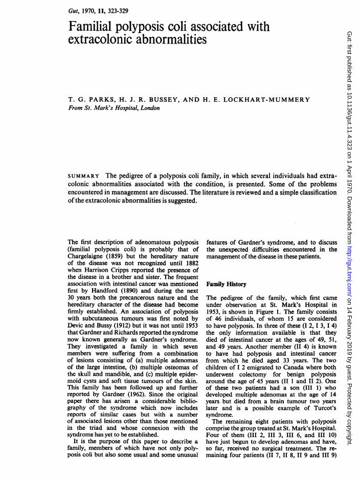
Abnormality ofPrimary Embryonic Layer
Ectodermal Origin Endodermal Origin Mesodermal Origin
Sebaceous cysts Adenomata of the large intestine, small Connective tissueEpidermoid cysts intestine, duodenum, and stomach Fibroma and fibrosarcomaTumours of the central nervous system Desmoid tumours(Turcot's syndrome) Diffuse fibrosis-
Adenomata of adrenal gland, pancreas, mesenteric, mesocolic,and thyroid and retroperitonealCarcinoma of large intestine, ampulla of Excessive intraabdominalVater, thyroid gland, and adrenal gland adhesionsHamartomata of stomach
BoneOsteomaOdontomataSupernumerary teeth
Table Abnormalities grouped according to derangement in primary embryonic layers
view of this definite trend towards abnormalfibrous growth within the abdomen in patientsfrom a Gardner's family, perhaps surgeryshould be delayed as long as feasible. Obviouslyall affected members will require surgery sooneror later as the risk of colon cancer remains. Itmight be wise to delay surgery for some yearsin young people who are known to be membersof a Gardner's family and who, therefore, mightdevelop mesenteric fibromata or possibly otherdangerous lesions, as there does not appear tobe any evidence that carcinoma develops at anearlier age in patients with Gardner's syndromethan in other patients with polyposis coli.
In those cases in which intraabdominalmesodermal growths do occur, the prognosismay be reasonable. Other patients with Gardner'ssyndrome, treated at St. Mark's Hospital, havehad large irremovable fibromata, yet have re-mained otherwise very well for many years.
Intraabdominal tumours may cause pain andobstruction within two or three years ofcolectomyfor polyposis. They are not infrequently mistakenfor malignant secondaries. Simpson, Harrison,and Mayo (1964) studied the association offamilial polyposis of the colon with mesentericfibromatosis in 15 cases from the literature andseven cases seen at the Mayo Clinic. In thesecases, the mesenteric lesion was a benign fibroustumour with infiltrative tendencies, which usuallyappeared after intestinal surgery, but, in spiteof the alarming appearances, the prognosiswas favourable in most cases. Some of thepatients developed severe intraperitoneal adhe-sions and obstruction.Fibrosarcoma has been reported on a few
occasions (Miller and Sweet, 1937; Gumpeland Carballo, 1956; Collins, 1959). Some authors(Weary, Linthicum, Cawley, Coleman, andGraham, 1964; Jones and Cornell, 1966) suggestthat so-called fibrosarcomata may, in actualfact, be desmoid tumours, since up until thetime of their reports there was no evidence ofmetastases in any of the reported cases, andpossibly some of these are examples of mis-
diagnosis. However, Thomas et al (1968) in-cluded in their series a patient who presented withmesenteric fibrosarcoma and later died of theeffects of infiltration of the wall of the smallintestine.Desmoid tumours may appear in or near the
scar after colectomy (O'Brien and Wels, 1955;Hughes and Hueston, 1960). Seventeen of 211polyposis patients reported by Smith (1958)had one or more abnormalities of Gardner'ssyndrome, and six out of 17 had histologicallyconfirmed desmoid tumours associated with theabdominal scars. Two other patients had nodulesin the scars which were never biopsied, but atleast one of these was almost certainly a desmoid.An incidence of 3.5% in Smith's collected seriescontrasts with an incidence of only 0.03% in astudy of 50,346 admissions to the MemorialHospital, reported by Pack and Ehrlich (1944).
In 112 cases of polyposis coli operated on atSt. Mark's Hospital, desmoid tumours werenoted in six patients (Lockhart-Mummery,1967). Five of these had previous surgery. Twopatients with desmoid tumours of the abdominalwall also had multiple fibromata of the smallbowel mesentery and retroperitoneal tissues.In addition to colectomy and ileorectal anastom-osis, one of these patients had desmoid tumours ofthe anterior abdominal wall excised on three oc-casions within a period of 18 months. Two yearslater, in 1961, a further operation was carriedout to remove the superficial part of a large massin the suprapubic region, as complete excisionwas impossible. Since that time there has beenno further enlargement of the lesion and thepatient remains generally well.
In addition to the discrete tumour masses inthe form of fibromata or desmoid tumours,the excessive fibrous reaction may take a morediffuse form, infiltrating the mesentery ormesocolon or retroperitoneal tissues (Simpsonet al, 1964).Thomas et al (1968) observed that thismanifestation of excessive fibrous tissue prolifera-tion is sometimes seen in patients with familial
328 on 14 F
ebruary 2019 by guest. Protected by copyright.
http://gut.bmj.com
/G
ut: first published as 10.1136/gut.11.4.323 on 1 April 1970. D
ownloaded from
329 Familial polyposis coli associated with extracolonic abnormalities
polyposis without any other stigmata of Gardner'ssyndrome.Kaplan (1961) observed a high incidence of
postoperative intestinal obstruction due tofibrous bands in polyposis cases. This was con-firmed in a report from St. Mark's Hospitalof a series by Lockhart-Mummery (1967) inwhich 20% of the polyposis patients who hadabdominal operations subsequently required afurther operation for intestinal obstruction. Thisis exemplified in this communication by case 3in whom laparotomy for adhesions was carriedout on the fifth postoperative day.
ECTODERMAL TUMOURS
Oldfield (1954) described the association ofmultiple sebaceous cysts and polyposis coli inone family group in which three membershad both conditions and three other membershad multiple sebaceous cysts but no colonicdisease. Both conditions appeared to be inheritedon the basis of a dominant gene. Smith (1958)recorded the association of familial polyposisand sebaceous cysts in at least four of his cases.Tumours ofneuroectoderm have been described
in association with polyposis coli by Turcot,Despres, and St. Pierre (1959) in two membersof one family, one of whom had a frontalglioblastoma and the other a medulloblastoma.Unfortunately, little information is available onone of the cases in this present pedigree butenough has been obtained to support stronglya diagnosis of Turcot's syndrome.
GENETICSGardner and Richards (1953) suggested that asingle dominant gene was responsible for all thelesions seen in the syndrome and most subsequentworkers concur with this concept of a pleio-trophic gene. McKusick (1962) held that thereis a single gene which is responsible for Gardner'ssyndrome and that this is different from thegene responsible for simple familial polyposis.On the other hand, Smith (1958) submittedthat the variation in penetrance of the gene couldexplain not only the presence or absence but alsothe degree to which any of the features exists.
In conclusion we have attempted to classify theincreasing variety of tumours that have beenreported in patients with familial polyposisbut it is not yet clear whether some of theseare actually associated with the condition orwhether they are only incidental findings. Allembryonic layers may be involved and it maybe found helpful to group the abnormalitiesaccording to derangement in the primary embry-onic layers (Table).Some of the lesions, as well as other varieties
not listed, have been recorded on only a fewoccasions and only the passage of time, withadditional cases and larger series, will clarifywhether, in actual fact, the association is anintegral part of the syndrome.
We wish to thank Dr B. C. Morson for his help inthe pathological diagnoses and Mr N. Mackie forproviding the photographs.
References
Capps, W. F., Jr., Lewis, M. I., and Gazzaniga, D. A. (1968).Carcinoma of the colon, ampulla of Vater and urinarybladder associated with familial multiple polyposis.Dis. Colon Rect., 11, 298-305.
Chargelaigne, A. (1859). Des Polypes du Rectum. Thesis, Paris.Collins, D. C. (1959). The frequent association of other body tumors
with familial polyposis. Amer. J. Gastroent., 31, 376-381.Devic, A., and Bussy, N. M. (1912). Un cas de polypose ad6nom-
ateuse g6n6ralis6e a tout l'intestin. Arch. Mal. Appar. dig.,6,278-299.
Gardner, E. J. (1962). Follow-up study of a family group ex-hibiting dominant inheritance for a syndrome includingintestinal polyps, osteomas, fibromas and epidermalcysts. Amer. J. hum. Genet., 14, 376-390.
Gardner, E. J., and Richards, R. C. (1953). Multiple cutaneousand subcutaneous lesions occurring simultaneously withhereditary polyposis and osteomatosis. Amer. J. hum.Genet., 5, 139-147.
Gumpel, R. C., and Carballo, J. D. (1956). A new concept offamilial adenomatosis. Ann. intern. Med., 45, 1045-1058.
Handford, H. (1890). Disseminated polypi of the large intestinebecoming malignant. Trans. path. Soc. Lond., 41, 133-137.
Hughes, E. S. R., and Hueston, J. T. (1960). Desmoid tumour infamilial polyposis of the colon: report of a case. Aust.N.Z. J. Surg., 30, 131-139.
Jones, E. L., and Cornell, W. P. (1966). Gardner's syndrome.Review of the literature and report on a family. Arch.Surg., 92, 287-300.
Kaplan, B. J. (1961). Gardner's syndrome: heredo-familialadenomatosis associated with 'soft and hard' fibroustumors and epidermoid cysts. Dis. Colon Rect., 4,252-262.
Lockhart-Mummery, H. E. (1967). Intestinal polyposis: thepresent position. Proc. roy. Soc. Med., 60, 381-388.
MacDonald, J. M., Davis, W. C., Crago, H. R., and Berk, A. D.(1967). Gardner's syndrome and peri-ampullary malig-nancy. Amer. J. Surg., 113, 425-430.
McFarland, P. H., Jr., Scheetz, W. L., and Knisley, R. E. (1968).Gardner's syndrome: report of two families. J. oral Surg.,26,632-638.
McKittrick, L. S., et al. (1935). Case record of the MassachusettsGeneral Hospital, Case No. 21061. New Engl. J. Med., 212,263-267.
McKusick, V. A. (1962). Genetic factors in intestinal poloposis.J. Amer. med. Ass., 182, 271-277.
Miller, R. H., and Sweet, R. H. (1937). Multiple polyposis of thecolon: a familial disease. Ann. Surg., 105, 511-515.
O'Brien, J. P., and Wels, P. (1955). The synchronous occurrenceof benign fibrous neoplasia in hereditary adenosis of thecolon and rectum. N. Y. St. J. Med., 55, 1877-1880.
Oldfield, M. C. (1954). The association of familial polyposis ofthe colon with multiple sebaceous cysts. Brit. J. Surg., 41,534-541.
Pack, G. T., and Ehrlich, H. E. (1944). Neoplasms of the anteriorabdominal wall with special consideration of desmoidtumors. Experience with 391 cases and a collective reviewof the literature. Int. Abstr. Surg., 79, 177-198.
Pollack, J. L., and Swinton, N. W. (1955). Congenital polyposisof the colon with extension to the small intestine andstomach. Lahey Clin. Bull., 9, 174-179.
Pugh, H. L., and Nesselrod, J. P. (1945). Multiple polypoiddisease of the colon and rectum. Ann. Surg., 121, 88-99.
Shepherd, J. A. (1958). Familial polyposis of the colon withassociated connective tissue tumours. J. roy. Coll. Surg.Edinburgh., 4, 31-38.
Simpson, R. D., Harrison, E. G., Jr., and Mayo, C. W. (1964).Mesenteric fibromatosis in familial polyposis; a variantof Gardner's syndrome. Cancer (Philad.), 17, 526-534.
Smith, W. G. (1958). Multiple polyposis, Gardner's syndromeand desmoid tumors. Dis. Colon. Rect., 1, 323-332.
Thomas, K. E., Watne, A. L., Johnson, J. G., Roth, E., andZimmermann, B. (1968). Natural history of Gardner'ssyndrome. Amer. J. Surg., 115, 218-226.
Thomford, N. R., and Greenberger, N. J. (1968). Lymphoidpolyps of the ileum associated with Gardner's syndrome.Arch. Surg., 96, 289-291.
Turcot, J., Despr6s, J. P., and St. Pierre, F. (1959). Malignanttumors of the central nervous system associated withfamilial polyposis ofthe colon. Dis. Colon. Rect., 2, 465-468.
Weary, P. E., Linthicum, A., Cawley, E. P., Coleman, C. C., Jr.,and Graham, G. F. (1964). Gardner's syndrome. A familygroup study and review. Arch. Derm., 90, 20-30.
on 14 February 2019 by guest. P
rotected by copyright.http://gut.bm
j.com/
Gut: first published as 10.1136/gut.11.4.323 on 1 A
pril 1970. Dow
nloaded from