1
False Sense of EPI-to-Structural Alignment with False Sense of EPI-to-Structural Alignment with Common Cross-Modality Registration Methods Common Cross-Modality Registration Methods RW Cox 1 , ZS Saad 1 , DR Glen 1 , MS Beauchamp 2 , R Desai 3 1 NIMH/Bethesda MD USA 2 UT Health Science Center/Houston TX USA 3 Medical College of Wisconsin/Milwaukee WI USA The Problem The Problem Aligning EPI volumes to T 1 -weighted volumes using Mutual Information (MI) or Correlation Ratio (CR) as the cost functional can produce registrations that look good but are actually bad Brain edges from the two volumes might match well, but this can be very misleading: Interior structures (ventricles, fissures, sulci) that are visible in both types of images often are displaced 5 mm or more This is not a software issue: AFNI (3dAllineate), SPM (COREG), and FSL (FLIRT) all often fail to give good anatomical matchings, upon close visual inspection Examples : T 1 -weighted images as the grayscale background, each one registered to the EPI volume with a distinct method EPI volume is edge-detected and only its edges are shown in the color overlay Two implementations of MI (AFNI & SPM/COREG) Two implementations of CR (AFNI & FSL/FLIRT) Our new LPC cost functional (AFNI) EPI interior edges track structural edges only with LPC Local Pearson Correlation Cost Functional (LPC) Weighted correlation coefficient calculated over neighborhood N(x) of any point x, then nonlinearly combined to give final cost: where E(x)=EPI, S(x)=T 1 , s(r)=tanh -1 (r), N(x)=rhombic dodecahdron centered at x, P=FCC lattice of non-overlapping rhombic dodecahdra covering the brain volume, and W(x)=weight proportional to E(x) to accentuate matching of CSF (bright in EPI, dark in T 1 ) as the program minimizes C LPC [E(x),S(T(x,θ)) over affine transformations T() [looking for the most negative correlation] CSF tracks ventricles, fissures, sulci fairly well in EPI, producing a robust match between central and cortical anatomical structures that are visible in both EPI and structural volumes Computing correlations locally helps insure against shading artifacts (especially with multi-coil acquisitions) Conclusions Conclusions Accurate and robust alignment structural and EPI volumes requires a special cost functional And requires visual inspection of results, especially if you are relying on the function-to-structure correspondence projection to cortical surface models; surgical planning Assessment Methodology Three raters (blinded to method and presentation order) each scored each of 27 {EPI,T 1 } volume pairs for alignment on a 4 point scale (from awful to excellent): 1 =awful 2 =errors > 5 mm 3 =errors 2..5 mm 4 =errors 0..2 mm while viewing edge-enhanced images, in all three planes, overlaid in color and/or flickering between viewing E and S Sample datasets from 1.5 and 3.0 Tesla, from diverse sites Raters agreed remarkably well (Spearman correlations0.8)

False Sense of EPI-to-Structural Alignment with Common ...rhdesai/posters/hbm08_cox_align_poster.pdf · False Sense of EPI-to-Structural Alignment with Common Cross-Modality Registration

Embed Size (px)

Citation preview

False Sense of EPI-to-Structural Alignment withFalse Sense of EPI-to-Structural Alignment withCommon Cross-Modality Registration MethodsCommon Cross-Modality Registration Methods

RW Cox1, ZS Saad1, DR Glen1, MS Beauchamp2, R Desai31NIMH/Bethesda MD USA

2UT Health Science Center/Houston TX USA3Medical College of Wisconsin/Milwaukee WI USA

The ProblemThe Problem Aligning EPI volumes to T1-weighted volumes using MutualInformation (MI) or Correlation Ratio (CR) as the cost functionalcan produce registrations that look good but are actually bad

Brain edges from the two volumes might match well, but thiscan be very misleading:

Interior structures (ventricles, fissures, sulci) that are visible inboth types of images often are displaced 5 mm or more

This is not a software issue: AFNI (3dAllineate), SPM (COREG),and FSL (FLIRT) all often fail to give good anatomical matchings,upon close visual inspection
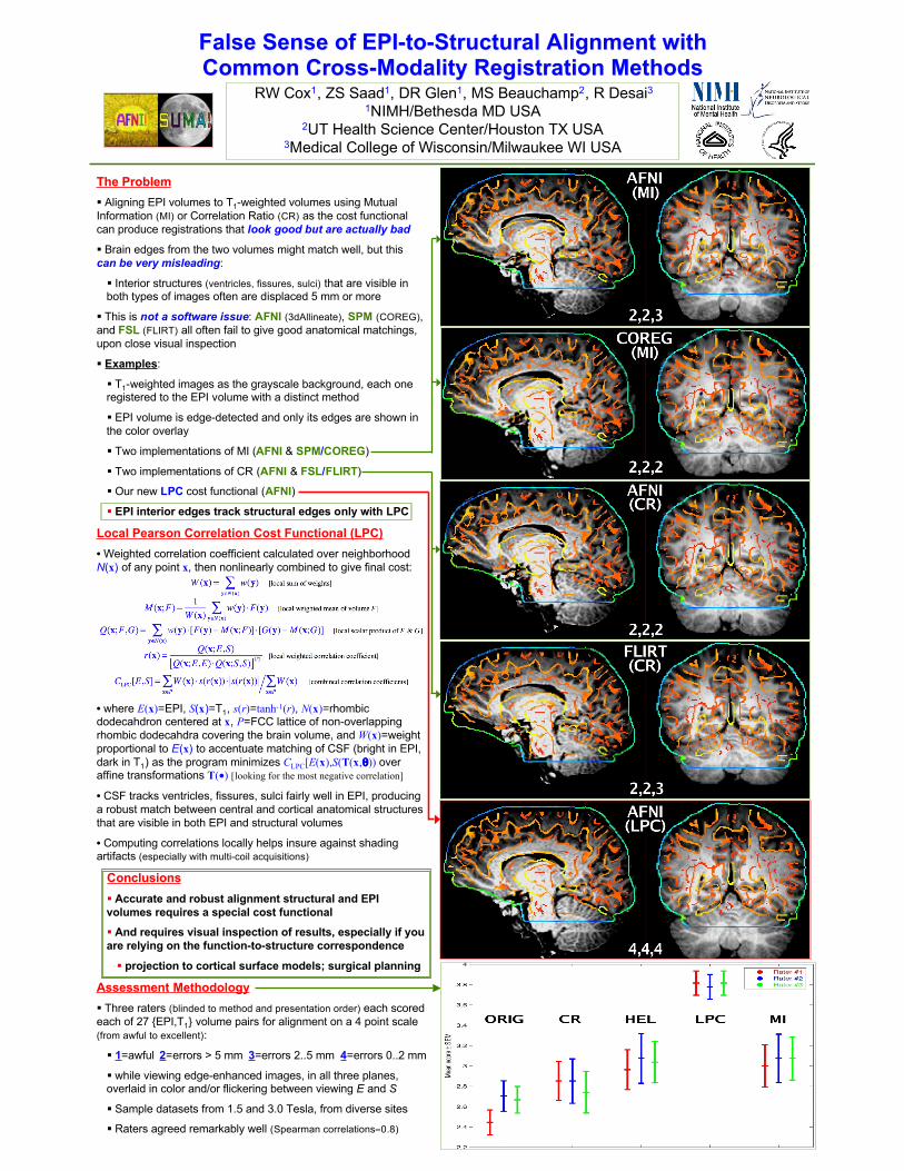
Examples:

T1-weighted images as the grayscale background, each oneregistered to the EPI volume with a distinct method

EPI volume is edge-detected and only its edges are shown inthe color overlay

Two implementations of MI (AFNI & SPM/COREG)

Two implementations of CR (AFNI & FSL/FLIRT)

Our new LPC cost functional (AFNI)

EPI interior edges track structural edges only with LPC

Local Pearson Correlation Cost Functional (LPC)• Weighted correlation coefficient calculated over neighborhoodN(x) of any point x, then nonlinearly combined to give final cost:

• where E(x)=EPI, S(x)=T1, s(r)=tanh-1(r), N(x)=rhombicdodecahdron centered at x, P=FCC lattice of non-overlappingrhombic dodecahdra covering the brain volume, and W(x)=weightproportional to E(x) to accentuate matching of CSF (bright in EPI,dark in T1) as the program minimizes CLPC[E(x),S(T(x,θ)) overaffine transformations T(•) [looking for the most negative correlation]

• CSF tracks ventricles, fissures, sulci fairly well in EPI, producinga robust match between central and cortical anatomical structuresthat are visible in both EPI and structural volumes

• Computing correlations locally helps insure against shadingartifacts (especially with multi-coil acquisitions)

ConclusionsConclusions Accurate and robust alignment structural and EPIvolumes requires a special cost functional

And requires visual inspection of results, especially if youare relying on the function-to-structure correspondence

projection to cortical surface models; surgical planning

Assessment Methodology Three raters (blinded to method and presentation order) each scoredeach of 27 {EPI,T1} volume pairs for alignment on a 4 point scale(from awful to excellent):

1=awful 2=errors > 5 mm 3=errors 2..5 mm 4=errors 0..2 mm

while viewing edge-enhanced images, in all three planes,overlaid in color and/or flickering between viewing E and S

Sample datasets from 1.5 and 3.0 Tesla, from diverse sites

Raters agreed remarkably well (Spearman correlations≈0.8)