Embed Size (px)
Citation preview
Fallacies of Coagulation Testing
Ravi Sarode, MD
John H Childers Professorship in Pathology
Professor of Pathology,
Chief of Pathology and Medical Director
of Clinical Laboratory Services UT Southwestern Medical Center
Director, Transfusion Medicine and Hemostasis
UT Southwestern Medical Center, Dallas, TX
Disclosure
Dr. Sarode has nothing to disclose.

Routine Coagulation Tests
PT and PTT
Fibrinogen and D-Dimers
PT and PTT have diagnostic value in patients with bleeding disorders
They have not been shown to assess bleeding risk in a non-bleeding patient.
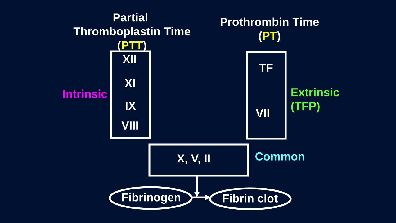
Partial
Thromboplastin Time
(PTT)
Prothrombin Time
(PT)
Fibrin clotFibrinogen
XII
XI
IX
VIII
Intrinsic
TF
VII
Extrinsic
(TFP)
X, V, II Common

Pt Plasma Tissue
Thromboplastin
and Ca++
Prothrombin Time
Clotting time
9 - 12.5”
SourcesBrains: Human
RabbitGoat
Placenta: Human
Recombinant
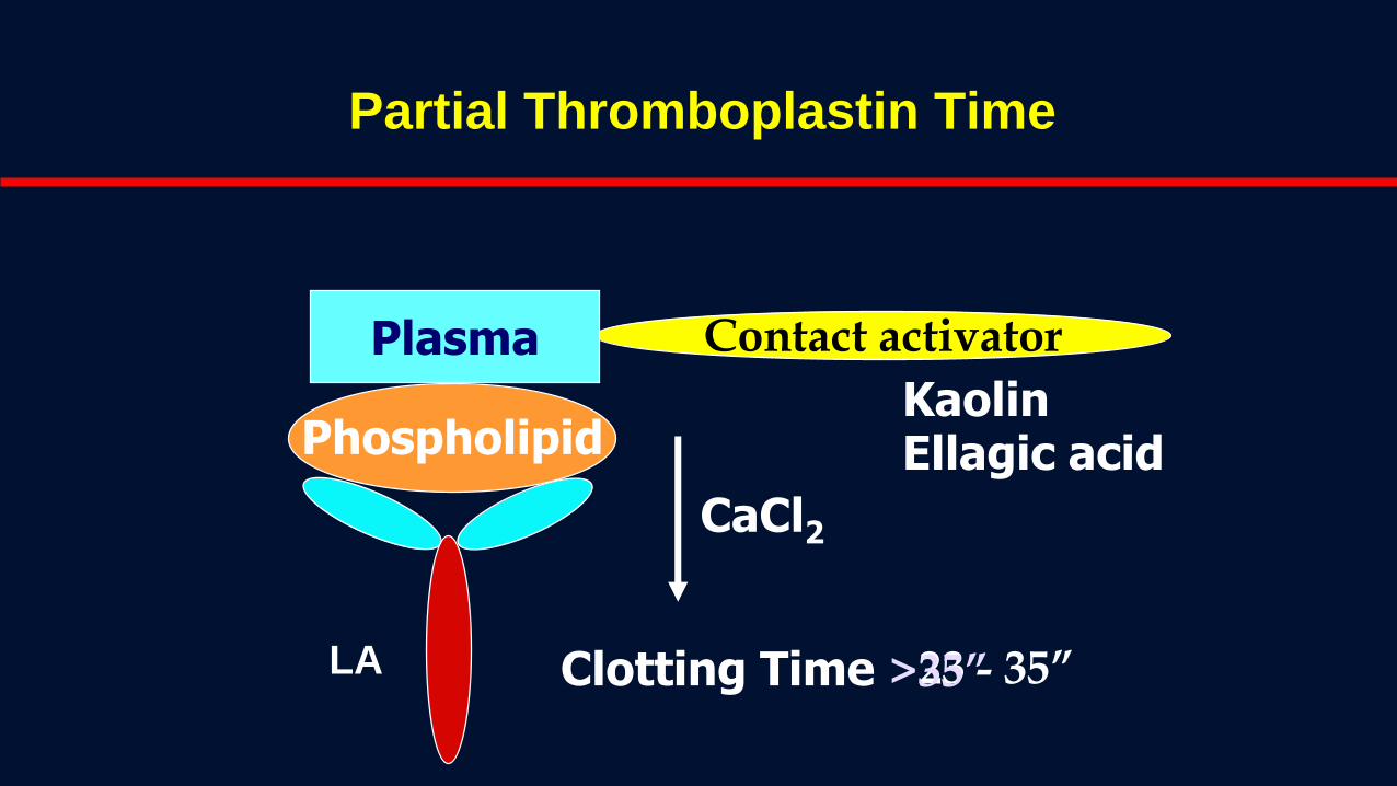
Partial Thromboplastin Time
KaolinEllagic acid
Contact activatorPlasma
Phospholipid
CaCl2
Clotting Time 23 - 35”LA >35”

Case 1
A 4 year old WB seen by a Pediatrician in his office for severe cough and fever for last 4 days. The child had enlarged tonsils. He has had similar bouts several times last year. The ENT surgeon wanted to remove tonsils. This time pediatrician convinced the parents for surgery. As part of pre-op w/u CBC, CMP, UA and PT/PTT were performed. All labs were normal except for PTT of 40” (normal 23-33”).
The pediatrician ordered further w/u for long PTT as per medical school and residency learning.
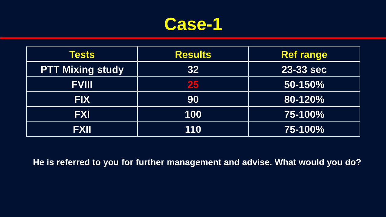
Case-1
Tests Results Ref range
PTT Mixing study 32 23-33 sec
FVIII 25 50-150%
FIX 90 80-120%
FXI 100 75-100%
FXII 110 75-100%
He is referred to you for further management and advise. What would you do?


Case -1
No personal and family history of bleeding
The Doctor’s office is in a suburb
The sample was drawn in the morning by a nurse, kept at RT before shipping out to a reference lab in late afternoon.
The patient referred to our center for further evaluation
Repeat PT and PTT normal – FVIII was 95%

Case -2
45 Year old AA male with a diagnosis of SCC of tongue was scheduled for lymph node dissection. The surgeon had ordered PT/PTT. The PTT was 40”
A hematology consult was placed on Monday.
PTT mixing study was ordered on Tuesday.
The mixing study showed PTT of 33” (ref range 23-33.5) on Wednesday.

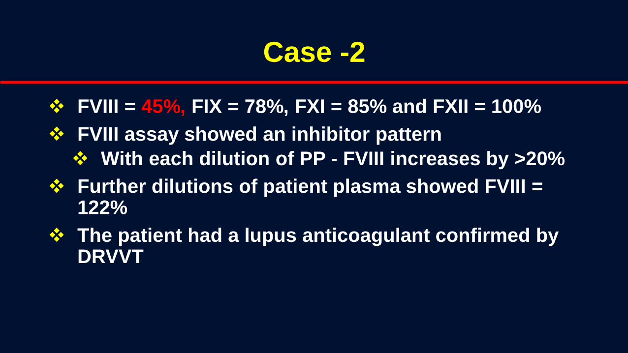
Case -2
FVIII = 45%, FIX = 78%, FXI = 85% and FXII = 100%
FVIII assay showed an inhibitor pattern
With each dilution of PP - FVIII increases by >20%
Further dilutions of patient plasma showed FVIII = 122%
The patient had a lupus anticoagulant confirmed by DRVVT

Case 3
74 AAM (a JW) presented to the ED with hematuria on Wednesday (PTT = 65”, PT 12”)
His past Hx included DVT 2 years back.
PTT at that time was 45” which on mixing study partially corrected to 38”.
LA was confirmed by DRVVT.
He was treated with VKA for 1 year.

Case 3
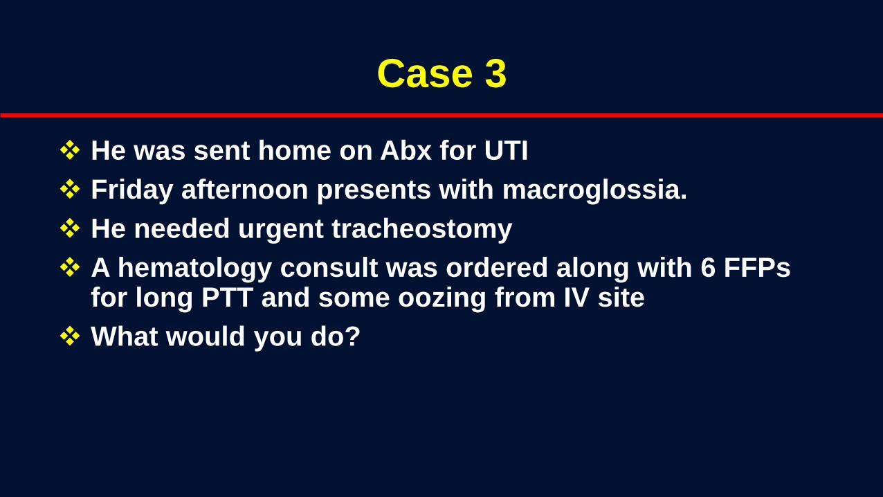
He was sent home on Abx for UTI
Friday afternoon presents with macroglossia.
He needed urgent tracheostomy
A hematology consult was ordered along with 6 FFPs for long PTT and some oozing from IV site
What would you do?

Case -3
His FVIII was <1% and FVIII inhibitor titer of >250 BU
Treatment options inlcuded:
• FEIBA (plasma derived)
• rVIIa
Given rFVIIa 90 ug/kg 2-4 hours being a JW
3 days later space out to 6 hours.
Bled from tracheostomy wound to Hb 2 g/dl – died
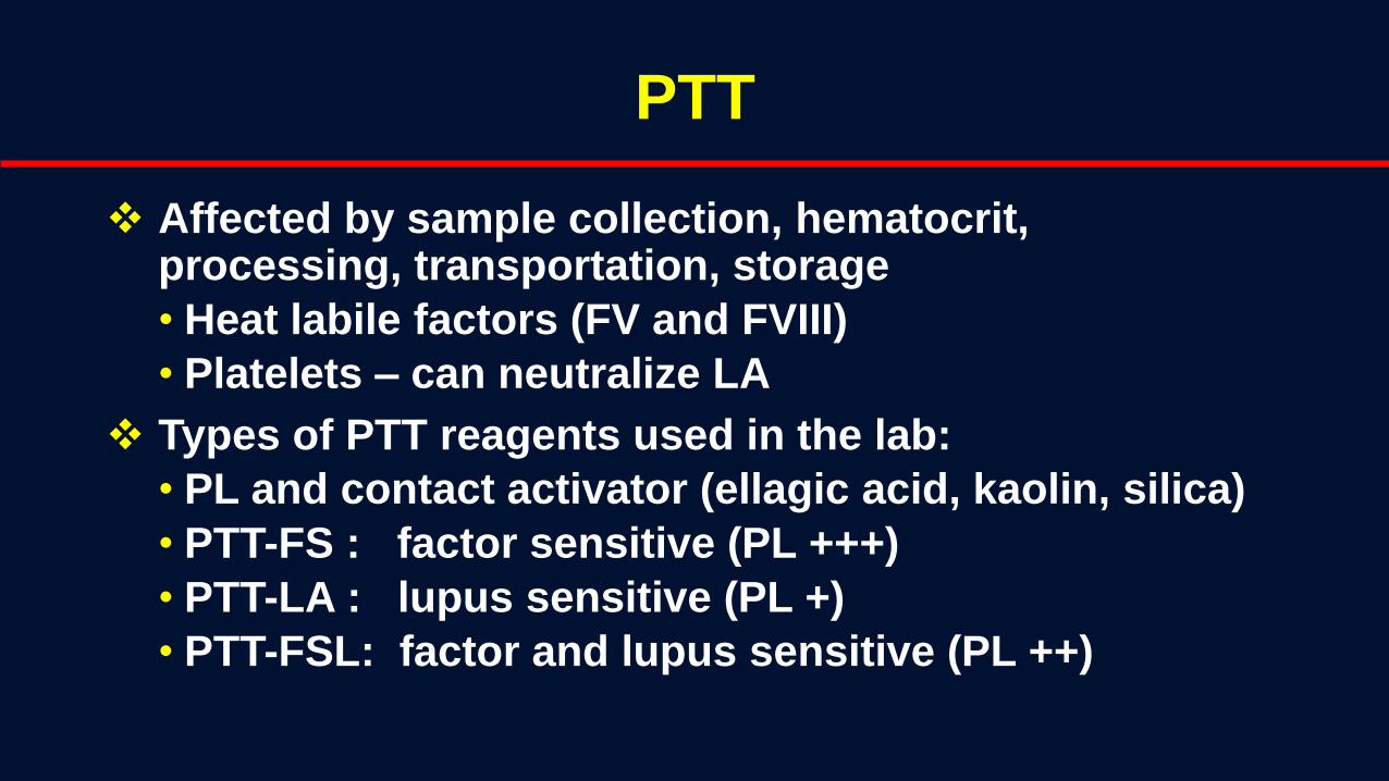
PTT
Affected by sample collection, hematocrit, processing, transportation, storage
• Heat labile factors (FV and FVIII)
• Platelets – can neutralize LA
Types of PTT reagents used in the lab:
• PL and contact activator (ellagic acid, kaolin, silica)
• PTT-FS : factor sensitive (PL +++)
• PTT-LA : lupus sensitive (PL +)
• PTT-FSL: factor and lupus sensitive (PL ++)
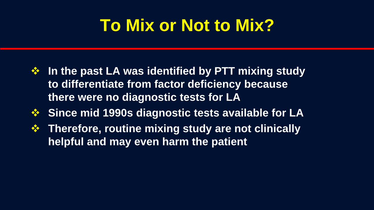
To Mix or Not to Mix?
In the past LA was identified by PTT mixing study
to differentiate from factor deficiency because
there were no diagnostic tests for LA
Since mid 1990s diagnostic tests available for LA
Therefore, routine mixing study are not clinically
helpful and may even harm the patient
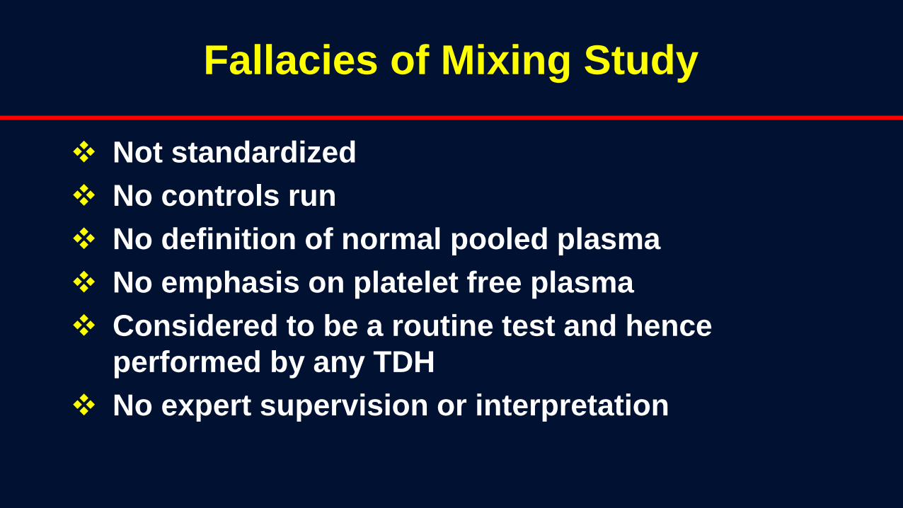
Fallacies of Mixing Study
Not standardized
No controls run
No definition of normal pooled plasma
No emphasis on platelet free plasma
Considered to be a routine test and hence
performed by any TDH
No expert supervision or interpretation

Any Indication of Mixing Study?
Very selected situations
Only under expert supervision
Always 2 step PTT mixing
0 hour and 2 hours at 370C
Suspected exposure to bovine
thrombin

Case - 4
32 year old man with PVD, DMT2, HIV, and HBV presents to ED with increased LLE pain.
Patient’s LLE pain is associated with coolness and foot swelling.
CTA reveals occlusion from distal L-superficial femoral artery to the trifurcation of vessels and of R-peroneal artery in distal calf
Thrombectomy of SF, popliteal and peroneal
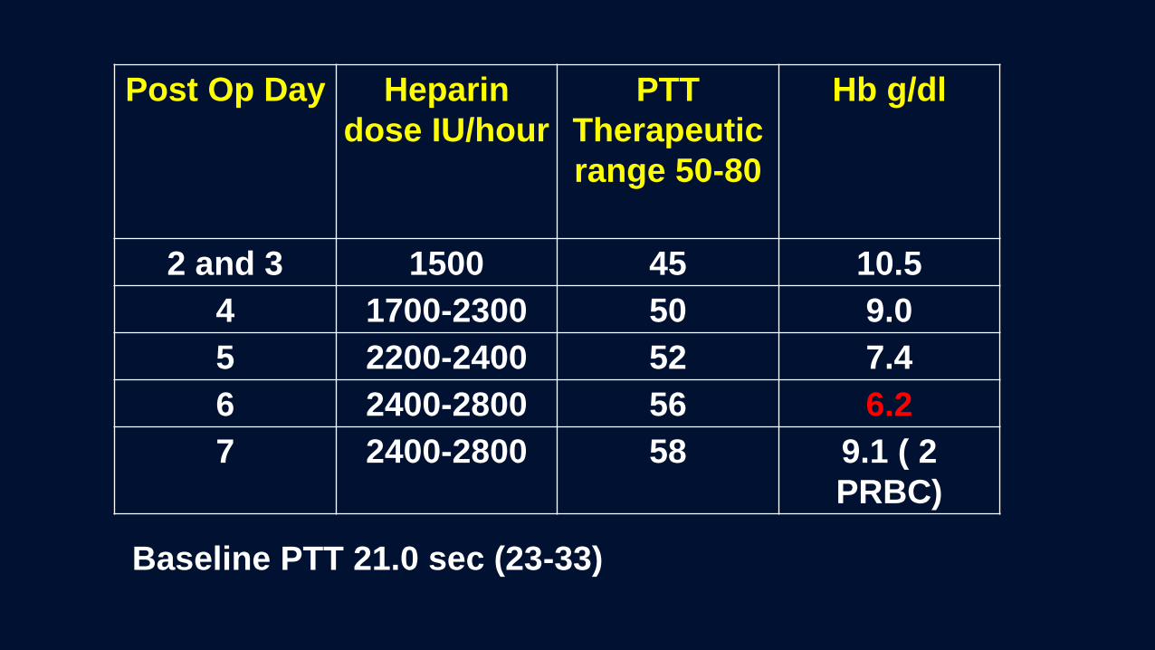
Post Op Day Heparin
dose IU/hour
PTT
Therapeutic
range 50-80
Hb g/dl
2 and 3 1500 45 10.5
4 1700-2300 50 9.0
5 2200-2400 52 7.4
6 2400-2800 56 6.2
7 2400-2800 58 9.1 ( 2
PRBC)
Baseline PTT 21.0 sec (23-33)


Unpredictable Dose Response of UFH
= Heparin
= ATIII
= Other Plasma Proteins
Endothelial cell
Macrophage
= Fibrinogen
= VWF
= Platelets
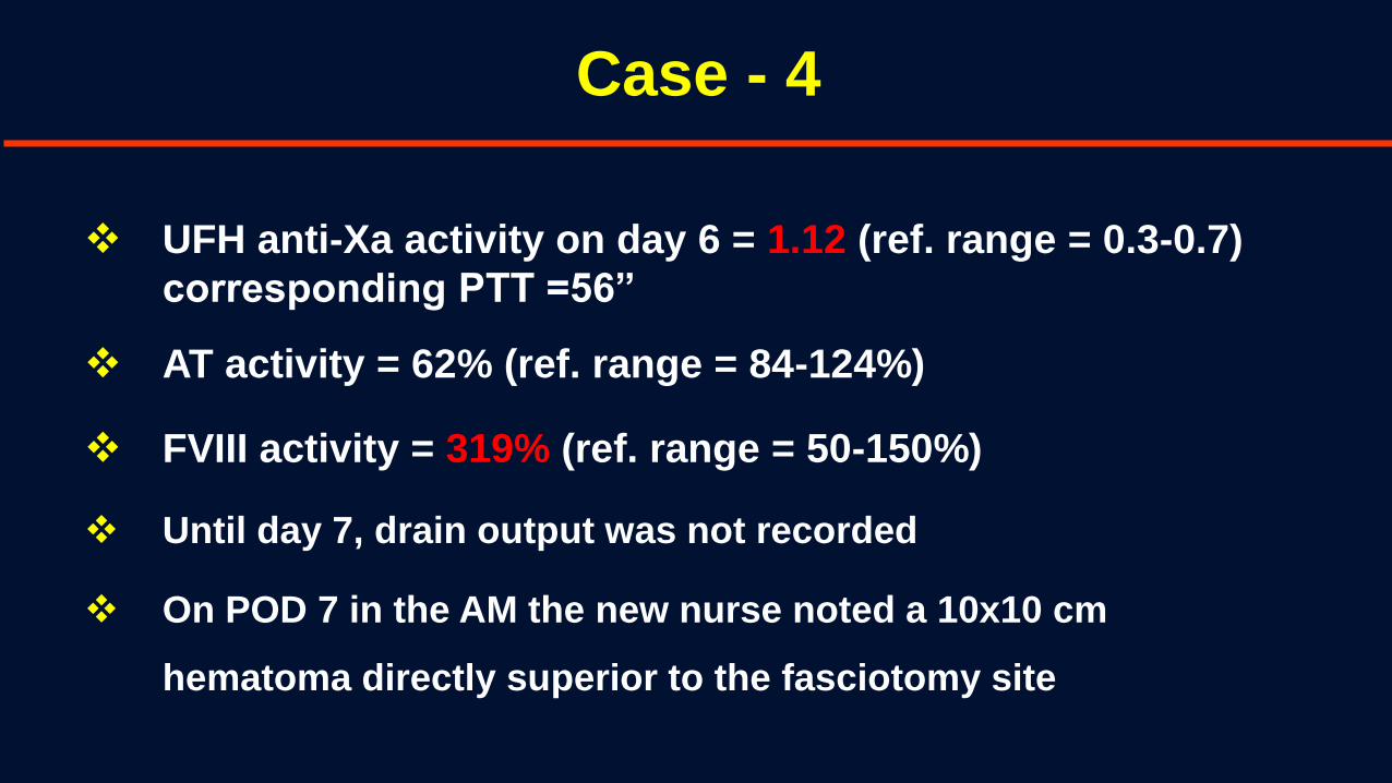
Case - 4
UFH anti-Xa activity on day 6 = 1.12 (ref. range = 0.3-0.7)
corresponding PTT =56”
AT activity = 62% (ref. range = 84-124%)
FVIII activity = 319% (ref. range = 50-150%)
Until day 7, drain output was not recorded
On POD 7 in the AM the new nurse noted a 10x10 cm
hematoma directly superior to the fasciotomy site

True Heparin Resistance
Increased heparin binding proteins
• Acute phase reactants
• Need higher doses of heparin
Increased FVIII
• May not need higher dose
• Monitor with anti-Xa assay
AT deficiency
Perform stat AT levels
Infusion of AT
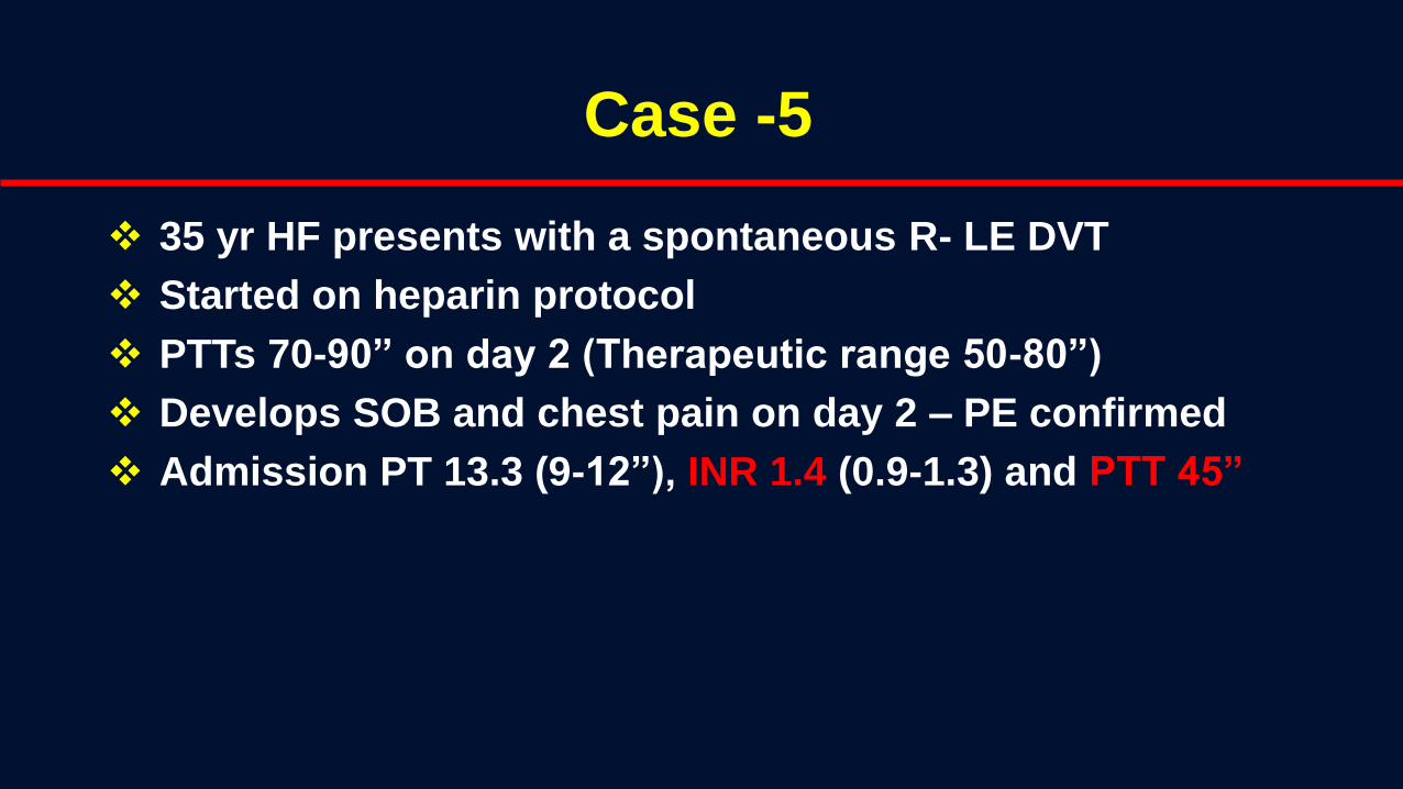
Case -5
35 yr HF presents with a spontaneous R- LE DVT
Started on heparin protocol
PTTs 70-90” on day 2 (Therapeutic range 50-80”)
Develops SOB and chest pain on day 2 – PE confirmed
Admission PT 13.3 (9-12”), INR 1.4 (0.9-1.3) and PTT 45”


Monitoring anticoagulation in LA
Baseline prolonged PTT
• PTT cannot be used for monitoring
• Heparin assay (anti-Xa assay = 0.3-0.7 U/ml)
Baseline prolonged PT/INR
• INR cannot be used to monitor
• Chromogenic FX (15-30% Therapeutic range)

Case – 5
The house staff ordered thrombophilia work – up
LA was confirmed positive and her ACA and Beta 2 GPI IgG were >100 confirming the APS
Her PS activity was 35% (AT and PC were normal)
Does she have a concomitant PS deficiency?
PS: Bound (60%) to C4b and free (40%)

Protein S Activity Assay
XRVV
Xa
Xa + Va
Ca++
PLII IIa
I Ia C.T.
PS - Cofactor
APC
Va i ↓IIa
FVL False ↓ PS
LA False ↑ PS
↑C4b False ↓ PS
↑C.T.
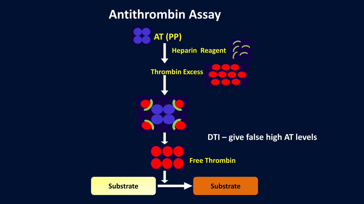
AT (PP)
Heparin Reagent
Thrombin Excess
Substrate
Free Thrombin
Substrate
Antithrombin Assay
DTI – give false high AT levels
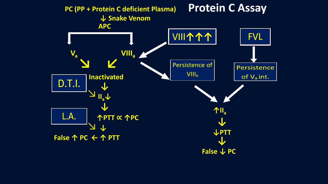
PC (PP + Protein C deficient Plasma) ↓ Snake VenomAPC
Va VIIIa
↘ ↙Inactivated
↓IIa↓
↓↑PTT ∝↑PC
↘
↘ ↓
False ↑ PC ← ↑ PTT
↑IIa
↓↓PTT↓
False ↓ PC
Protein C Assay
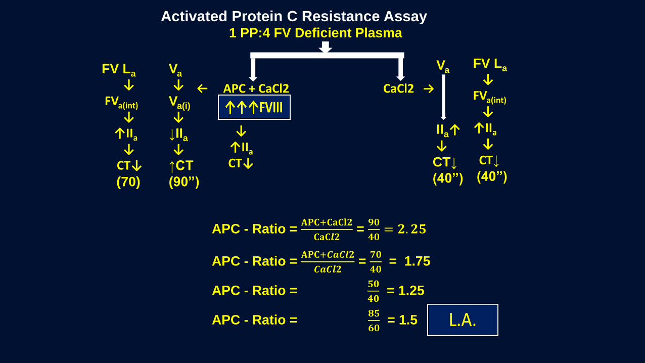
Activated Protein C Resistance Assay1 PP:4 FV Deficient Plasma
APC + CaCl2 CaCl2
Va
↓Va(i)
↓↓IIa↓↑CT
(90”)
Va
IIa↑↓CT↓
(40”)
APC - Ratio = 𝐀𝐏𝐂+𝐂𝐚𝐂𝐥𝟐
𝐂𝐚𝐂𝒍𝟐=
𝟗𝟎
𝟒𝟎= 𝟐. 𝟐𝟓
FV La
↓FVa(int)
↓↑IIa
↓CT↓(70)
APC - Ratio = 𝐀𝐏𝐂+𝑪𝒂𝑪𝒍𝟐
𝑪𝒂𝑪𝒍𝟐=
𝟕𝟎
𝟒𝟎= 1.75
APC - Ratio = 𝟓𝟎
𝟒𝟎= 1.25
↓↑IIa
CT↓
APC - Ratio =𝟖𝟓
𝟔𝟎= 1.5
FV La
↓FVa(int)
↓ ↑IIa
↓CT↓
(40”)
↓↓

Case – 6
TMR consulted to manage bleeding in a 66 yrs old cirrhotic patient
Overnight developed melena
Hb dropped from 7.0 to 5.9
Past Medical History• COPD• Non-alcoholic steatohepatitis (NASH) Cirrhosis MELD on admission: 18 Child-pugh class on admission: Class C INR on admission 1.6 and now 1.8


ROTEM
The Labs
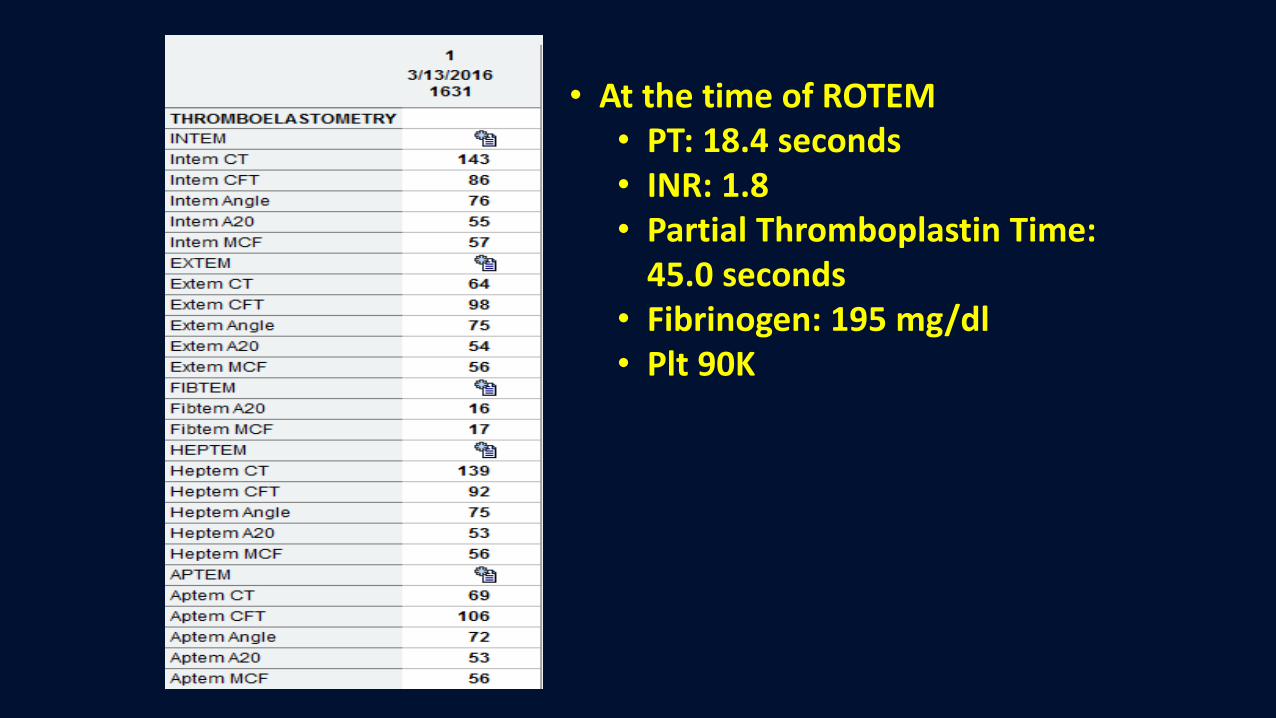
• At the time of ROTEM• PT: 18.4 seconds• INR: 1.8• Partial Thromboplastin Time:
45.0 seconds• Fibrinogen: 195 mg/dl• Plt 90K

Evaluation of Hemostasis in a
Surgically Bleeding Patient
Current tests have poor TAT and correlation with surgical bleeding
In a profusely bleeding patient • Surgical vs coagulopathic bleeding
Trauma – multifactorial – hypothermia, acidosis and coagulopathy (consumption, dilutional and ? trauma induced coagulopathy)
Most non-trauma surgical bleeds in controlled environment (no hypothermia or acidosis) –mostly dilutional coagulopathy
Obstetrical hemorrhage – mostly surgical since body shifts to hypercoagulable state antepartum – could be consumptive or dilutional

Viscoelastic (?) Point of Care Testing
Used in trauma and surgical settings to manage bleeding and coagulopathy
• Thromboelastography (TEG®, Haemonetics, Braintree, MA)
• Rotational thromboelastometry (ROTEM®, TEM International GmbH, Munich, Germany)
Allows for real time in-vitro analysis of clot formation, clot strength, and fibrinolysis on whole blood samples.
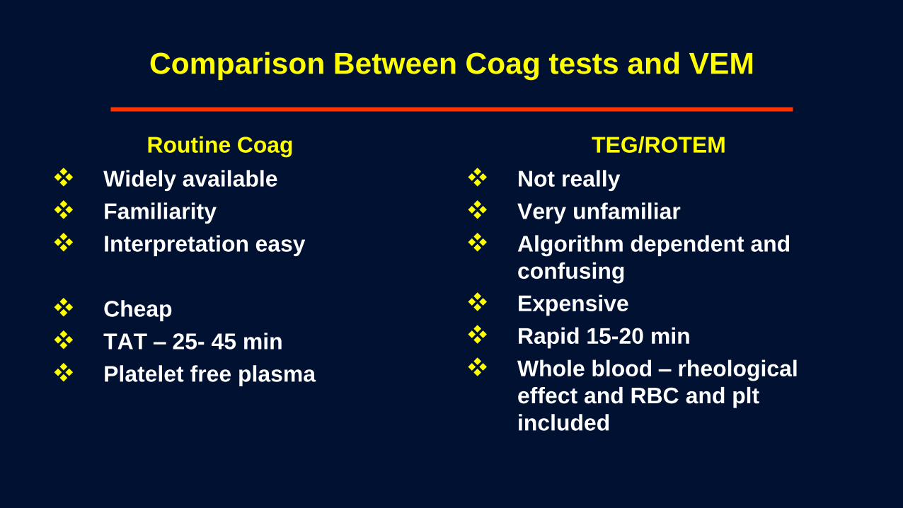
Comparison Between Coag tests and VEM
Routine Coag
Widely available
Familiarity
Interpretation easy
Cheap
TAT – 25- 45 min
Platelet free plasma
TEG/ROTEM
Not really
Very unfamiliar
Algorithm dependent and
confusing
Expensive
Rapid 15-20 min
Whole blood – rheological
effect and RBC and plt
included
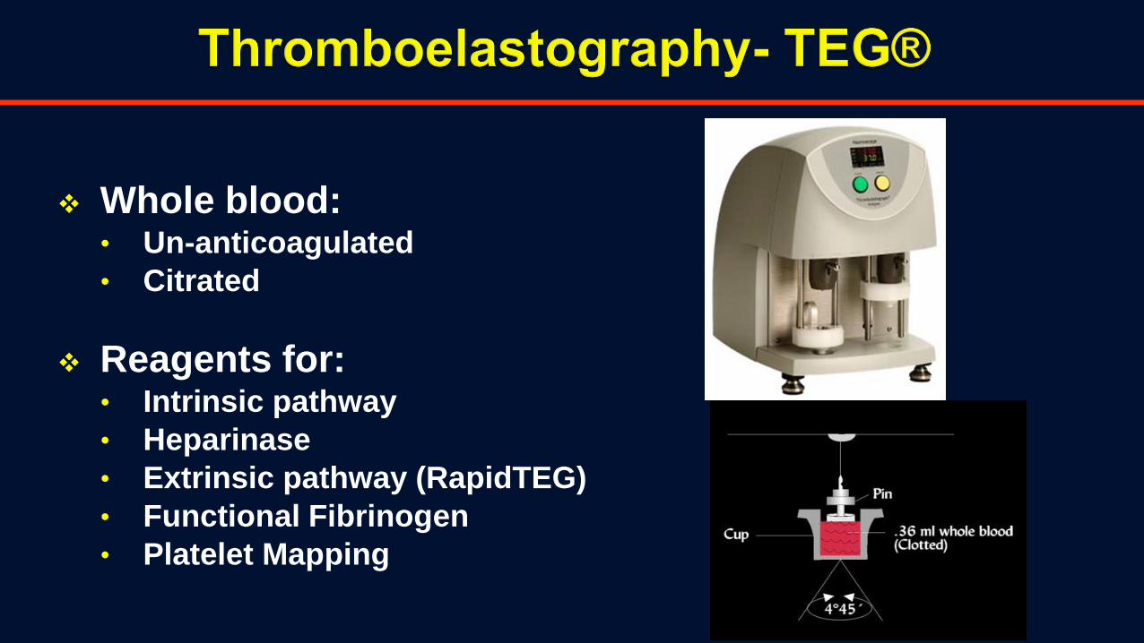
Whole blood:• Un-anticoagulated
• Citrated
Reagents for:• Intrinsic pathway
• Heparinase
• Extrinsic pathway (RapidTEG)
• Functional Fibrinogen
• Platelet Mapping

Whole blood:• Citrated
Reagents for:• INTEM
• HEPTEM
• EXTEM
• FIBTEM
• APTEM
Rotational Thromboelastometry - ROTEM®
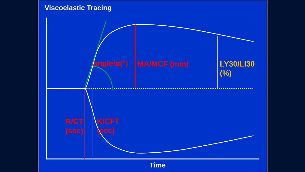
Time
MA/MCF (mm)angle/α(°)
K/CFT
(sec)R/CT
(sec)
Viscoelastic Tracing
LY30/LI30
(%)
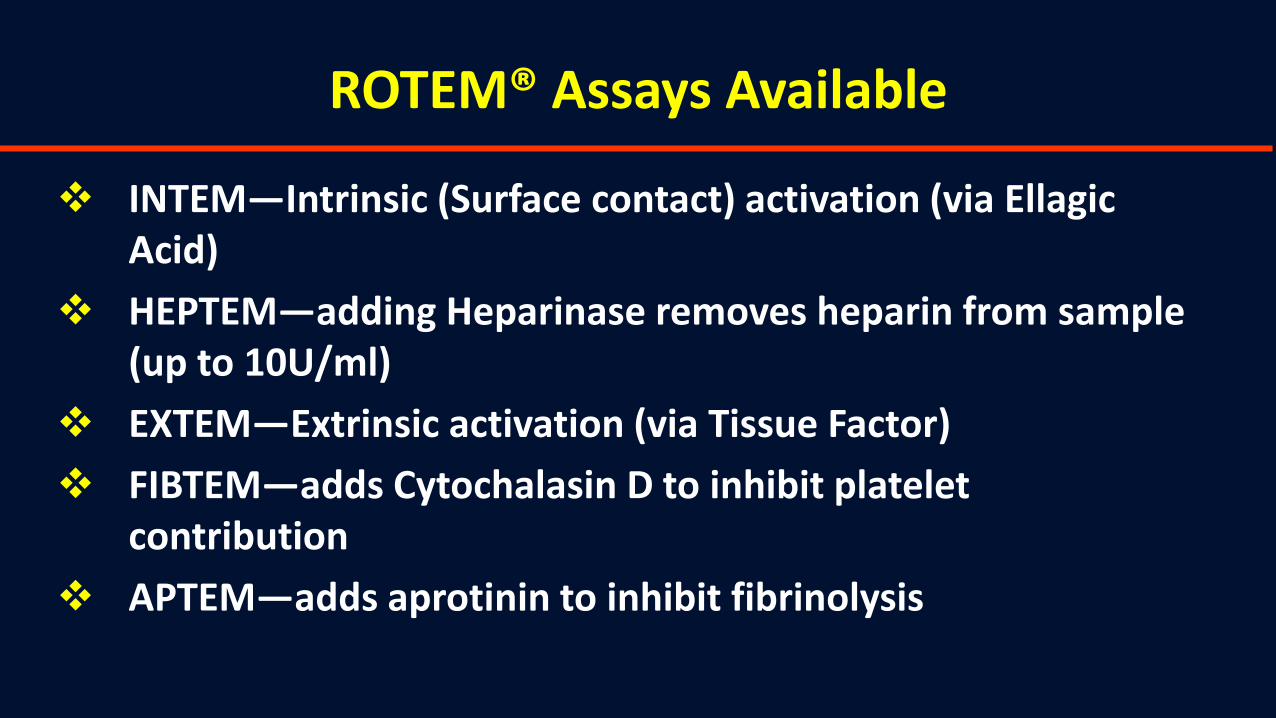
ROTEM® Assays Available
INTEM—Intrinsic (Surface contact) activation (via EllagicAcid)
HEPTEM—adding Heparinase removes heparin from sample (up to 10U/ml)
EXTEM—Extrinsic activation (via Tissue Factor)
FIBTEM—adds Cytochalasin D to inhibit platelet contribution
APTEM—adds aprotinin to inhibit fibrinolysis
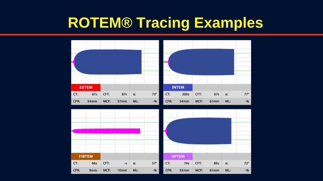
ROTEM® Tracing Examples

ROTEM ®
Normal Platelet Defect Fibrinogen Defect
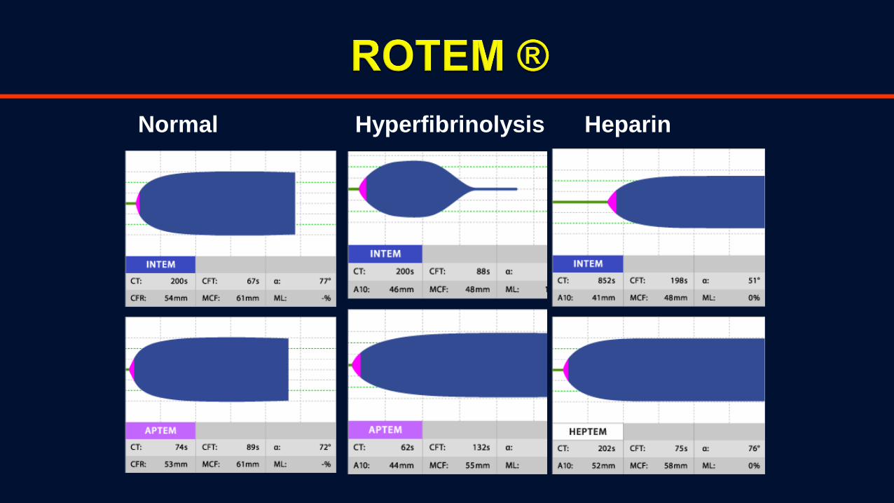
Normal Hyperfibrinolysis Heparin

EXTEMCase -6

How to use in clinical settings?

Rapid Interpretation of Results
Assessment using 3 Assay parameters: CT, A10, and ML:
1. CTIN,EX = Coagulation factors (heparin on INTEM).
severe hypofibrinogenemia or thrombocytopenia.
2. A10IN,EX = Clot firmness (platelets, fibrinogen and
?FXIIIa).
A10FIB = Clot firmness (fibrinogen) if normal =
platelet problem.
3. MLIN,EX, FIB = Clot lysis during the test
(Hyperfibrinolysis)
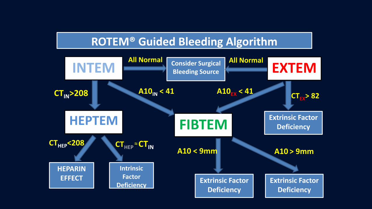
ROTEM® Guided Bleeding Algorithm
INTEM
HEPTEM
HEPARIN EFFECT
Intrinsic Factor
Deficiency
CTIN>208
CTHEP<208 CTHEP≈CTIN
Consider Surgical Bleeding Source
FIBTEM
A10IN < 41
All Normal
EXTEMAll Normal
Extrinsic Factor Deficiency
A10EX < 41CTEX> 82
Extrinsic Factor Deficiency
Extrinsic Factor Deficiency
A10 < 9mm A10 > 9mm
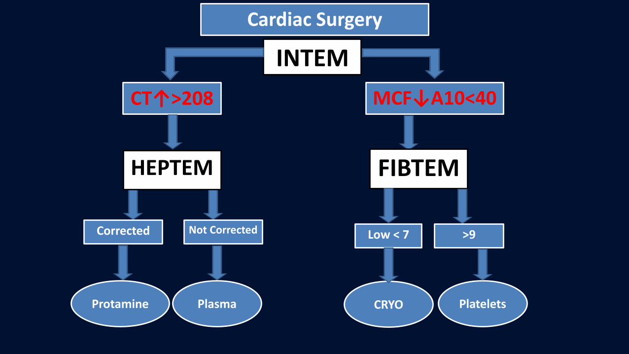
INTEM
CT↑>208 MCF↓A10<40
Corrected Not Corrected
Protamine Plasma
FIBTEM
Low < 7 >9
CRYO Platelets
HEPTEM
Cardiac Surgery
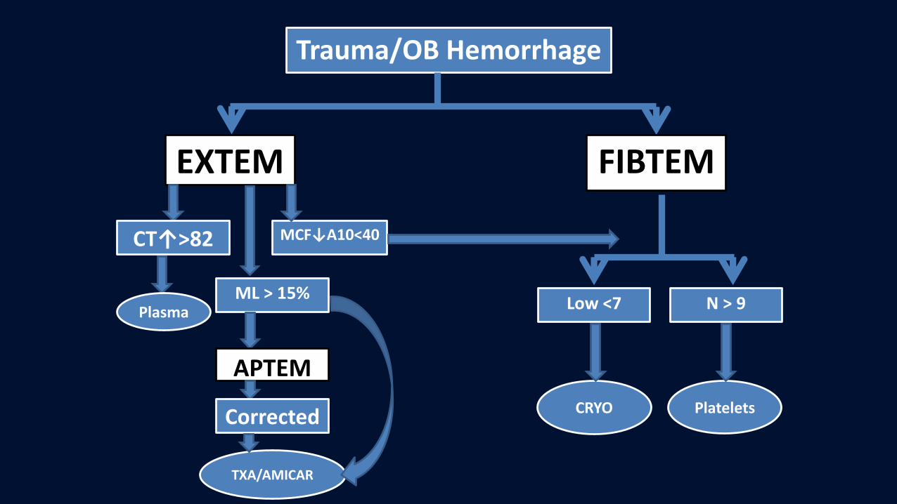
Trauma/OB Hemorrhage
EXTEM
CT↑>82
PlasmaML > 15%
APTEM
Corrected
TXA/AMICAR
FIBTEM
MCF↓A10<40
Low <7 N > 9
CRYO Platelets

FFP is not Amrut!
Amrut =
Indian
mythology
“Nectar of
Goddess =
giving them
immortality!”