Embed Size (px)
Citation preview

Facultad de Farmacia y Nutrición
Targeting JNK for the diagnosis and treatment of Alzheimer’s disease
Silvia Vela Lumbreras
Pamplona, 2017

Facultad de Farmacia y Nutrición
Memoria presentada por Dña. Silvia Vela Lumbreras para aspirar al grado de Doctor
por la Universidad de Navarra.
Fdo. Silvia Vela Lumbreras
El presente trabajo ha sido realizado bajo nuestra dirección en el Departamento de
Farmacología y Toxicología de la Facultad de Farmacia y Nutrición de la Universidad
de Navarra y autorizamos su presentación ante el Tribunal que lo ha de juzgar.
VºBº Directora VºBº Co-Directora
María Javier Ramírez Gil Maite Solas Zubiaurre


Este trabajo ha sido posible gracias a la financiación de dos entidades: el Instituto de
Salud Carlos III (ISCIII) [PFIS (PI13/00858)] y a la Asociación de Amigos de la
Universidad de Navarra (beca predoctoral 2014-2017).


A mi familia


-All truths are easy to understand once they are discovered;
the point is to discover them.
Galileo Galilei


Acknowledgments
Me gustaría expresar mi agradecimiento a todas aquellas personas que han hecho
posible la realización de este trabajo durante los últimos tres años y medio y que han
contribuido de una manera o de otra a facilitar esta tarea, que ha supuesto mucho
esfuerzo pero que a la larga ha sido más que recompensado.
En primer lugar, querría agradecer a la Asociación de Amigos de la Universidad de
Navarra y al Instituto Carlos III las ayudas recibidas que han hecho posible la
realización de este proyecto. A continuación, me gustaría mostrar mis más sinceros
agradecimientos a la Universidad de Navarra y a la Facultad de Farmacia y Nutrición
por haberme formado tanto profesional como personalmente durante los años de mi
carrera universitaria y el doctorado. Pero en especial, quiero expresar mi gratitud al
Departamento de Farmacología y Toxicología por darme la oportunidad de desarrollar
este trabajo de investigación e iniciar mi camino en la carrera investigadora y docente.
Sin embargo, no puedo concluir aquí mis agradecimientos a todo lo que me ha
proporcionado este Departamento, el “depar” ha sido una gran familia para mí
durante este tiempo en el que ha habido momentos buenos y no tan buenos, porque
teniendo la suerte de estar rodeada de gente tan excepcional no existen los momentos
malos.
Especialmente, quiero expresar mi gratitud a mis directoras, la Dra. María Javier
Ramírez y la Dra. Maite Solas, por haber confiado en mí desde el primer momento para
formar parte del este proyecto, por haberme iniciado en el mundo de la investigación
y la docencia. Gracias por todos vuestros consejos, por ser mis guías durante este
proceso y por todo el tiempo que habéis invertido en mí. Creo que las palabras no
pueden expresar mi gratitud por la oportunidad dada, sois un ejemplo a seguir.
Además, también me gustaría agradecer profundamente a la Dra. Rosa Tordera, la
Directora de este Departamento, por su dedicación y accesibilidad, así como todas sus
muestras de apoyo durante este tiempo. A la Dra. Elena Puerta, a la que guardo un
profundo cariño, ya que fuimos su primera promoción de alumnos de Grado en
Farmacia y, al llegar al Departamento de “Farma”, me di cuenta de que el afecto era
mutuo. Quisiera expresar mi gratitud por todos tus consejos y muestras de cariño, eres
una gran persona y mejor profesional. Agradecer al Dr. José López Guzmán y a la Dra.
Guadalupe Beitia (la “parte asistencial y ético-social” del Departamento) su buen

Acknowledgments
hacer, su prudencia, su preocupación por mi trabajo y sus constantes muestras de
afecto; me gustaría deciros que, a pesar de lo que muchos piensen, vuestro trabajo
también es Farmacia y, en mi opinión, una de las partes más importantes de esta
profesión a la que tenemos el orgullo de pertenecer. Gracias por defender la Farmacia
en todo su contenido.
Siguiendo con la gente de este Departamento de Farmacología y Toxicología,
quisiera agradecer especialmente a la Dra. Beatriz Marcos por su interés diario,
disposición en todo momento y su apoyo constante. Bea, ¿Cuántas veces me has dicho
que me parezco a la Goñi?, sin embargo, hay gente que me ve más parecido a ti y para
mí es un orgullo. Gracias por impulsarme a la docencia, quizá sea mi futuro. A la Dra.
Elena Beltrán por guiarme y ayudarme en mi experiencia doctoral, compartir cuartito
contigo fue un verdadero placer. A Mikel Aleixo, por enseñarme tanto de la conducta
murina y permitirme compartir momentos de tu vida tan importantes. Eres un
trabajador nato y lo has demostrado con creces; recuerda que, hasta el día de hoy,
sigo siendo tu alumna más aventajada en Ethovision, pero bien sabes que el mérito no
es sólo mío. También, me gustaría expresar mi gratitud de forma muy especial a
Sandra Lizaso ya que ha sido más que una compañera, una madre y una amiga. Tienes
un corazón enorme y sabes escuchar, valoras lo que tienes porque te ha tocado
pelearlo a pulso. He aprendido mucho de ti tanto en el terreno profesional como en el
personal. Sólo espero que sigamos siendo compañeras de zumba, de risas y cenas
muchos años más. Gracias por todo.
No puedo olvidarme de Pili y Mari Luz que, aunque ya las he conocido trabajando
a tiempo parcial por su prejubilación, siempre han tenido un momento para conversar
y tiempo para escuchar, he aprendido mucho de vuestra experiencia. En este sentido,
también quisiera agradecer a la Dra. Berta Lasheras por sus buenos consejos y por las
continuas muestras de interés que ha mostrado hacia mí durante todo este tiempo
tanto en el plano profesional como en el personal. Fue un placer colaborar en la
organización y desarrollo de tu jubilación.
Cuando comencé la tesis, llegué por sorpresa al Departamento, pero creo que no
pude tener mejor acogida ni una integración más rápida en el grupo. Eso debo
agradecérselo a los doctorandos que allí estaban en ese momento: Manuel Rodríguez
Perdigón, Merche Erburu, Xabier Bengoetxea, Irene Muñoz-Cobo y, muy
Acknowledgments
especialmente Hilda Ferrero, gracias por aconsejarme, enseñarme y ayudarme todo lo
que sabíais o podíais. Hilda merece mención aparte en estos agradecimientos ya que,
además de ser la mejor compañera que una hubiera podido desear, ha sido la mejor
AMIGA durante todos estos años. Hilda, gracias por escucharme, por compartir mis
problemas, por ahuyentar mis preocupaciones, por hacer fácil lo difícil, por tu apoyo
incondicional, por enseñarme todo lo que sé, en definitiva, por estar ahí cada vez que
te he necesitado. Lo que la tesis ha unido no lo va a separar ni los años ni los
kilómetros que separan Borja de Alicante. Gracias por todo, gracias por tanto.
Los nuevos doctorandos que se han ido incorporando desde que comencé en este
Departamento en 2014 han sabido mantener la esencia de los que se han ido
marchando, así pues, me gustaría agradecer a Teresa y Borja los momentos
compartidos: experimentos, prácticas, excursiones, planes, cenas, cañas, etc. Con
vosotros la rutina se ha hecho más divertida. Asimismo, me gustaría agradecer a
Carmen y Patricia los cafés compartidos. No quisiera olvidarme de agradecer también
a todos aquellos alumnos de Trabajos Fin de Master y Fin de Grado o Erasmus que me
han permitido aprender tanto o más de lo que les he intentado enseñar yo a ellos:
Jamie, Yelissa, Urtzi, Dani, Egoitz, Ollie, María Lanz, Puja y Bejal; pero de forma muy
especial, a Fany, por tu sinceridad, por abrirme las puertas de tu casa y de tu corazón,
por tantos viajes y tan buenos ratos, en definitiva, por estar siempre que te he
necesitado. Apenas coincidimos unos meses en el departamento, pero me llevo una
amiga para toda la vida.
Además, querría agradecer a la parte de Toxicología, que aunque está diferenciada
físicamente de Farmacología por la ubicación en los edificios, compartimos no sólo
Departamento, sino también docencia, directiva, facultad, reuniones, juevintxos y
comidas de Navidad y fin de curso. Gracias a Adela, Ariane, Celia, Ana Gloria, Amaya,
Tamara, Laura, Josema, Ismael, Maite, Violeta, Julen y Damián por vuestra
profesionalidad y colaboración. Asimismo me gustaría agradecer a Idoia Beltrán, Itxaso
Ruiz de Lasheras y Elena Gascón por su bondad, disposición y eficacia.
A todos los compañeros de los departamentos vecinos: Aitor, María, Itziar, Ana,
Edu, Mikel Domeño, Hugo, Julen, Rodri, Sergio, Miriam, Yadira y Bea Aragón me
gustaría agradeceros las experiencias compartidas, pero debo hacer mención especial
al grupo de Fisiología, con los que he compartido experimentos y proyectos. A la Dra.

Acknowledgments
María Jesús Moreno gracias por su colaboración y sus muestras de interés hacia mi
trabajo, a la Dra. Silvia Lorente por su apoyo y simpatía, al Dr. Fermín Milagro siempre
dispuesto a resolver cualquier duda, al Dr. Pedro González Muniesa por preocuparse
siempre por mi trabajo y la complicidad de compartir raíces aragonesas y,
especialmente, me gustaría expresar mi gratitud a Laura, Neira y Asún por su
disposición, su apoyo y por los momentos compartidos, haciendo los sacrificios más
amenos junto con Xabier, Rosa, Eva, Elisa, Leire o Miguel. Gracias por hacerme sentir
una más en vuestro departamento. No podría faltar mi agradecimiento a la Dra. Itziar
Velaz asesora y consejera desde mis primeros años en la facultad, gracias por guiarme
y aconsejarme. De igual modo, quisiera agradecer a la Dra. Juana Fernández por su
alegría e interés por mí.
Mis agradecimientos aquí son extensibles a los bedeles en particular a Javi y
Enrique por su sonrisa por las mañanas y por mostrarme siempre su cariño; y a Juan,
Eneko y demás personal del animalario por su ayuda con los animales y su compañía
en los largos días en CIFA.
Quisiera agradecer también a mis amigos del Grado en Farmacia el apoyo y el
interés que han mostrado en todo momento y porque, a pesar de que ya han pasado
los años, siempre he podido contar con ellos: Paty, Vicky, Lucía, Javi, Mikel, Aitor y
Pablo. Gracias por hacer posibles nuestras tan necesarias quedadas a pesar de la
distancia que nos separa, sintiendo que nada ha cambiado desde la última vez que nos
vimos. Agradecer también a dos amigos que estudiaron Periodismo pero casi
convalidaron la carrera de Farmacia a base de compartir momentos con sus amigos
farmacéuticos: Óscar y Edu, gracias por estar siempre ahí.
No me podría olvidar de agradecer a mis compañeras y amigas de la Oficina de
Farmacia de Bayona 37: a Berta Arriazu por ser la mejor jefa que hubiera podido
imaginar, a Maite por ser tan buena compañera, enseñarme tanto y tener tanta
paciencia conmigo y a Alejandra por convertirse en mi confidente y apoyo
incondicional estos años, por tu bondad y cariño, sabes que me has dado mucho más
de lo que merezco. Este agradecimiento se hace extensible por supuesto a Boni,
Fernando, Rut y Joaquín.
A mis amigas, gracias por compartir 25 años a mi lado, sé que siempre lo estaréis.
Lidia, Elsa, Ana y Melani gracias por ser las mejores amigas que se pueda desear.

Acknowledgments
Asimismo, me gustaría dar las gracias a mis amigos de Borja, a mis amigos del instituto
y a mi cuadrilla de Gallur, gracias por alegraros de cada pequeño éxito a pesar de no
entender nada de lo que os contaba.
A Sergio, eres mi mejor casualidad y un ejemplo de superación y optimismo para
mí. Gracias por escucharme, comprenderme y apoyarme en todo momento. Pero
sobre todo gracias por creer en mí y ayudarme a crecer personal y profesionalmente.
Por último pero no menos importante, me gustaría dar las gracias a toda mi
familia, por todo el esfuerzo que han hecho por mí, por todo su apoyo. Especialmente
a mis padres, Javi y Chon, porque siempre he podido contar con vuestro apoyo y con
vuestros buenos consejos. Porque me habéis enseñado a levantarme tras una caída y
me habéis ayudado a ser más positiva cuando todo se veía negro. Porque siempre he
tenido en casa un abrazo que me daba fuerzas para seguir luchando y porque sin
vosotros nada de esto hubiera sido posible.
A mis hermanas, Cristina y Elena, por ayudarme y apoyarme cada vez que lo
necesito. Por estar dispuestas siempre a escuchar y a alegrarme los días. Por ser las
mejores compañeras de este viaje que es la vida.
Finalmente, a mis abuelos, por haberme enseñado a luchar para conseguir mis
sueños y porque, gracias a vosotros, nunca me ha faltado una oportunidad. Por ser
ejemplo de humildad, amor y fortaleza, ojalá algún día pueda devolveros la cuarta
parte lo que me habéis dado sin esperar nada a cambio. Habéis sido las personas que
más me habéis cuidado y mimado.
Muchas gracias a todos, por todo, por tanto.


Table of contents
INTRODUCTION .................................................................................................................. 1
1. C-JUN N-TERMINAL KINASE (JNK) ................................................................................ 3
1.1. JNK distribution and functions in CNS ..........................................................................4
1.2. JNK signaling .................................................................................................................5
1.2.1. JNK activation ........................................................................................................5
1.2.2. JNK substrates .......................................................................................................7
2. JNK AND ALZHEIMER’S DISEASE .................................................................................. 8
2.1. Alzheimer’s disease ......................................................................................................8
2.2. JNK and pathological markers of Alzheimer’s disease ............................................... 11
2.3. Apoptosis regulation mediated by JNK activation ..................................................... 13
2.4. Role of JNK in neuroinflammation ............................................................................. 14
3. NOVEL COMPOUNDS TARGETING JNK INHIBITION ..................................................... 15
3.1. Direct ATP-competitive inhibitors: SP600125............................................................ 16
3.2. Mixed linage kinase inhibitors ................................................................................... 18
3.3. Cell-permeable peptide inhibitors ............................................................................. 19
3.4. Natural inhibitors ....................................................................................................... 22
4. ω-3 DERIVATIVES AND ALZHEIMER’S DISEASE ........................................................... 24
4.1. ω-3 polyunsaturated fatty acid .................................................................................. 24
4.1.1. ω-3 polyunsaturated fatty acid and Alzheimer’s disease .................................. 25
4.2. Specialized pro-resolving lipid mediators .................................................................. 27
4.2.1. Maresin 1 in Alzheimer’s disease ....................................................................... 30
HYPOTHESIS AND AIMS .................................................................................................... 33
EXPERIMENTAL DESIGN AND METHODS ............................................................................ 39
1. HUMAN SAMPLES ............................................................................................... 41
2. ANIMALS ............................................................................................................ 42
2.1. Tg2576 mice ....................................................................................................... 43
2.2. SAMP8 mice ....................................................................................................... 43
3. Aβ INTRACEREBROVENTRICULAR INJECTION IN WT MICE ..................................... 45
4. BEHAVIORAL TEST .............................................................................................. 45
4.1. Open field ........................................................................................................... 46
4.2. Morris water maze (MWM) ............................................................................... 46
4.3. Novel object recognition test (NORT) ................................................................ 47
5. BIOCHEMICAL MEASUREMENTS .......................................................................... 47
5.1. Tissue collection ................................................................................................. 47
5.2. Western blotting ................................................................................................ 48

Table of contents
5.3. Measurement of Aβ levels ................................................................................. 49
5.4. Measurement of pJNK levels in cerebrospinal fluid (CSF) ................................. 49
5.5. Immunofluorescence staining ............................................................................ 50
6. STATISTICAL ANALYSIS ........................................................................................ 51
RESULTS ........................................................................................................................... 53
1. SPECIFIC INCREASE OF pJNK IN AD ....................................................................... 55
1.1. Increased pJNK levels in AD experimental mouse models ................................ 55
1.2. Specific increases in pJNK levels in AD human samples..................................... 56
1.3. pJNK as a potential AD biomarker ..................................................................... 57
2. RELATIONSHIP BETWEEN pJNK AND Aβ LEVELS IN AD .......................................... 58
2.1. Aβ levels in mice models of AD .......................................................................... 58
2.2. Aβ levels in human samples of AD and other dementias .................................. 59
2.3. pJNK and Aβ co-localize in AD human samples and in Tg2576 mouse model ... 60
2.4. Aβ42 intracerebroventricular administration increases pJNK levels in wild type
mice frontal cortex ............................................................................................. 62
3. JNK INHIBITION RESTORES COGNITIVE IMPAIRMENT IN AN AD MOUSE MODEL .... 63
3.1. Effects of ω-3 polyunsaturated fatty acids in cognition .................................... 64
3.2. Involvement of pJNK on cognitive improvement............................................... 66
3.3. Mechanisms and consequences of JNK inhibition by ω-3 derivatives ............... 68
DISCUSSION ..................................................................................................................... 71
CONCLUSIONS .................................................................................................................. 87
REFERENCES ..................................................................................................................... 91


Abbreviations
A
Aβ: Amyloid beta
ABCA1: ATP-Binding cassete transporter 1
AD: Alzheimer’s disease
ADRDA: Alzheimer’s disease and related disorders association.
ALA: Alpha-linoleic acid
AP-1: Activator protein-1
APP: β-amyloid precursor protein
ApoE: Apolipoprotein E
ASK-1: Apoptosis signal-regulating kinase 1
ATF: Activator transcription factor
ATP: Adenosin triphosphate
B
BA10: Broadmann Area 10
BACE-1: Beta-site amyloid precursor protein cleaving enzyme 1
BBB: Blood-brain barrier
Bcl-2: B-cell lymphoma 2
Bcl-xL: B-cell lymphoma-extra large
BDR: Brains for dementia research initiative network
C
CA1: Cornu ammonis region 1
CD14: Cluster of differentiation 14
CERAD: Consortium to establish a registry for Alzheimer’s disease
CHME3: Human microglial cells line
CMS: Chronic mild stress
CNS Central nervous system
COX2: Cyclooxygenase 2
COX: Cytochrome C oxidase
CPP: Cell-penetrating peptide
CPPi: Cell-permeable peptide inhibitor
CSF: Cerebrospinal fluid
Abbreviations
CxF: Frontal cortex
Cyt C: Cytochrome C
D
dH2O: Distilled water
DHA: Docosahexaenoic acid
D-JNKi1: D-isomer c-Jun N-terminal kinase inhibitor 1
DLK: Dual leucine zipper kinase
DMSO: Dimethyl sulfoxide
DPR: Dipeptide repeat protein
DSM-IV: Diagnostic and statistical manual of mental disorders (4th edition)
E
ELISA: Enzyme-linked immunosorbent assay
ER: Endoplasmic reticulum
ERS: Endoplasmic reticulum’s stress
ERK: Extracellular signal-regulated kinase
EPA: Eicosapentaenoic acid
F
FA: Fatty acids
ω-3-FA Omega-3 fatty acids
FTD: Frontotemporal dementia
G
GAP-43: Growth-associated protein 43
GC: Glucocorticoid
GM-CSF: Granulocyte-macrophage colony-stimulating
GR: Glucocorticoid receptor
GROα: Growth related oncogene-alpha
H
HFD: High fat diet
I
ICV: Intracerebroventricular
IL: Interleukin
Abbreviations
Ire-1: Endoplasmic reticulum to nucleus signaling 1
IRS-1: Insulin receptor substrate 1
J
JBD: c-Jun N-terminal kinse binding domain
JIP: c-Jun N-terminal kinase interacting protein
JLP: c-Jun N-terminal kinase leucine zipper protein
JNK: c-Jun N-terminal kinase
JNKi: c-Jun N-terminal kinase inhibitors
K
KO: Knock out
L
LBD: Lewy body dementia
LXA4: Lipoxin A4
LOX: Lipoxygenase
LPS: Lipopolysaccharide
LTP: Long term potentiation
LZK: Leucine zipper-bearing kinase
M
MAPK: Mitogen activated protein kinase
MaR: Maresins
MaR1: Maresin 1 (Macrophage-derived mediator of inflammation
resolution 1)
MCI: Mild cognitive impairment
MetS: Metabolic syndrome
miRNA: Micro ribonucleic acid
MIP-1β: Machrophage inflammatory protein-1beta
MKK: Mitogen activated protein kinase kinase
MLK: Mixed-linage kinase
MLKi: Mixed-linage kinase inhibitors
MMSE: Mini-mental state examination
MPTP: 1-metil-4-phenyl-1,2,3,6-terahydropyridine

Abbreviations
MWM: Morris water maze
N
NF-κB: Nuclear factor-kappa B
NFT: Neurofibrillary tangles
NGF: Nerve growth factor
NINCDS: National Institute of neurologic, communicative disorders and
stroke
NORT: Novel object recognition test
NPD1: Neuroprotectin D1
O
OD: Optical density
P
PBS: Phosphate buffered saline
PBMC: Peripheral blood mononuclear cells
PDX: Protectins
PD1: Protectin D1
PET: Positron emission tomography
PHFs: Paired helical filaments
PI3K: Phosphatidylinositol-4,5-bisphosphate 3-kinase
PMCI: Progressive mild cognitive impairment
PPAR-γ: Peroxisome proliferator-activated receptor-gamma
PS1: Presenilin-1
PUFA: Polyunsaturated fatty acids
Puma: p53 up-regulated modulator of apoptosis
R
ROI: Reactive oxygen intermediates
ROS: Radical oxygen species
RvD: Resolvin D
RvE: Resolvin E
S
SAMP8: Senescence accelerated mouse prone-8

Abbreviations
SAMR1: Senescence accelerated mouse resistant-1
SCI: Subjective cognitive impairment
SNAP-25: Synaptosomal-associated protein 25
SPMs: Specialized pro-resolving mediators
STZ: Streptozotocin
T
TBS: Tris buffered saline
TDP-43: Trans-activator regulatory DNA-binding protein 43
TLR4: Toll like receptor 4
TNF-α: Tumor necrosis factor alpha
TRAF2: TNF receptor-associated factor 2
U
UPR: Unfolded protein response
UV: Ultraviolet
V
VaD: Vascular dementia
VLP-1: Visinin-like protein-1
W
WB: Western blot
WHO: World health organization
WT: Wild type
Y
YLK-40: Chitinase-3 like -1
Z
Zym: Zymosan


INTRODUCTION

Introduction
3
1. C-JUN N-TERMINAL KINASE (JNK)
Since its discovery more than 20 years ago, the c-Jun N-terminal kinase family (JNK)
has remained a subject of intense research interest with continued efforts to evaluate
its biochemistry and regulation, and its contribution to cellular events under
physiological and pathophysiological conditions. The JNK family of protein kinases is one
of the three identified families of mitogen activated protein kinases (MAPK). Three
genes, namely jnk1 (MAPK8), jnk2 (MAPK9), and jnk3 (MAPK10), encode for 10 different
splice variants with molecular weights of 46 and 54kDa (Davis, 2000). The 10 different
variants are grouped depending on the homologous protein regions in the 3 known
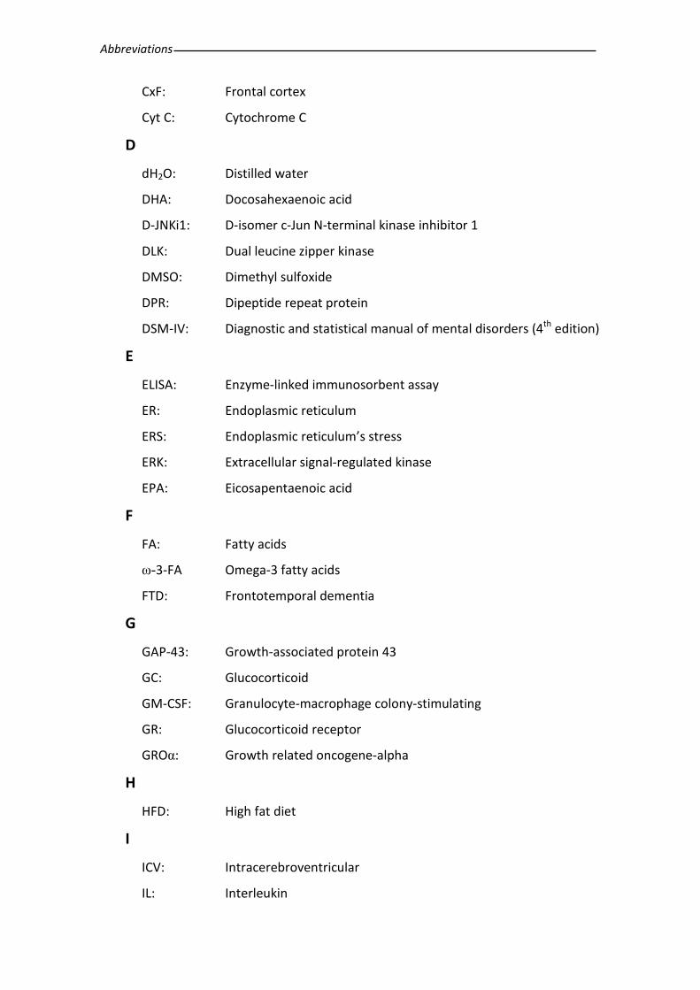
isoforms of JNK: JNK1, JNK2 and JNK3 (Figure 1). Whereas JNK1 and JNK2 have a broad
tissue distribution, JNK3 is mainly localized in neurons and to a lesser extent in the heart
and the testis (Coffey, 2014).
Figure 1. Structural features of JNK isoforms. JNK is a multifunctional enzyme beginning with an amino terminal end (NH2 – terminal, in purple) and, in the other side, a carboxyl terminal end (COOH-terminal) where the long (54KDa, orange) or short (46KDa, green) JNK variants are determined. The schematic illustration depicts the ten JNK isoforms grouped by their homologous region. All isoforms presents 11 kinase subdomains (I-XI, in blue), but the alternative splicing took place in subdomains IX and X (light blue). Modified from Coffey, 2014.
The discovery of JNK pathway scaffolds such as JNK-interacting protein-1 (JIP1) and
related proteins, as well as the identification of JNK inhibitors have contributed to
unmask the roles for the JNKs in both normal physiology and disease. JNK signaling
process has been studied as an active pathological mechanism in many different
diseases, especially in the field of oncology. To mention a few, JNK has been involved in
regulation of the natural killer cells cytokine production and secretion (Lee et al., 2014),

Introduction
4
oncology models and drug-resistant tumor cells (Chuang et al., 2014; Kim et al., 2014;
Okada et al., 2014; Volk et al., 2014) or myeloproliferative disorders (Funakoshi-Tago et
al., 2012).
1.1. JNK distribution and functions in CNS
Regarding the central nervous system (CNS), studies in rodents described the
presence of jnk1, jnk2 and jnk3 mRNA in cortex, hippocampus and cerebellum.
Specifically, JNK3 is widely expressed throughout the hippocampus, while JNK1 is
concentrated around the dentate gyrus. The subcellular distribution of the different
isoforms differs, with JNK1 being more abundant in the cytosol, whereas JNK3 is mostly
found in the nucleus. JNK2 is distributed in both the cytosol and the nucleus (Coffey,
2014).
Transgenic knockouts of JNK isoforms have provided crucial insights into the roles
played in the brain by each JNK isoform. It has been established that JNK1 and JNK2
have important roles in the modulation of immune cell function and in the development
of the embryonic nervous system. A study using JNK1 KO mice demonstrated that JNK1
has a regulatory role and maintains physiological functions in the CNS; similarly, JNK2
KO mice established that this isoform may also participate in some physiological
functions and, particularly, in the long term potentiation (LTP; Chen et al., 2005). JNK3 is
a multifunctional enzyme important in controlling brain functions under both normal
and pathological conditions. JNK3 has been implicated in brain development (Kuan et
al., 1999), neurite formation and plasticity (Eminel et al., 2008; Waetzig et al., 2006), in
addition to memory and learning (Bevilaqua et al., 2003; Brecht et al., 2005). Under
pathological conditions, JNK3 has been considered as a degenerative signal transducer
and it seems to be the isoform that is over-activated after deleterious stress-stimuli in
adult brain, such as ischemia, hypoxia or epilepsies. This principle is supported by the
data on the reduced apoptosis of hippocampal neurons and reduced seizures induced
by kainic acid in JNK3 KO mice, and by the notion that JNK3 KO mice are also protected
against ischemia (Okazawa & Estus, 2002; Sahara et al., 2008; Yang et al., 1997).

Introduction
5
1.2. JNK signaling
1.2.1. JNK activation
Activation of the JNK pathway relies on the coordinated interaction of the scaffold
proteins belonging to the JNK activation complex. These proteins are able to mediate
the biochemical signal amplification and also to ensure substrate-specificity as well as a
coordinated cascade signaling (Figure 2). The interaction between scaffold proteins
leads to the phosphorylation/activation of JNK which, in turn, by phosphorylation of
different substrates, enables the activation of different functions (Antoniou et al., 2011).
Different stimuli that have been described as able to trigger the signaling response
to JNK include nerve growth factor (NGF) deprivation, trophic support withdrawal, DNA
damage, oxidative stress, β-amyloid (Aβ) exposure, low potassium, excitotoxic stress, 6-
hydroxydopamine (6-OHDA), UV irradiation, tumor necrosis factor (TNF) or the Wnt
cascade (for review see Cui et al., 2007; Mudher et al., 2001). Many are the scaffold
proteins that have been described as the signaling proteins that converge in the
activation of JNK: JIP1a (JNK interacting protein 1a) and JIP1b (also named IB1), JIP2 and
JIP3 (firstly named JSAP1), JNK-interacting leucine zipper protein (JLP) and plenty of SH3
(POSH) (Engstrom et al., 2010). JIPs belong to second-order-activating proteins that are
dependent on previous interaction with MAPK activating kinases (MAPKK or MAP2K)
and MAPKK activating kinases (MAPKKK or MAP3K; Cui et al., 2007; Engstrom et al.,
2010; Wang et al., 2004). Specifically, JNKs are directly activated by phosphorylation by
two MAPKK: MKK4 and MKK7; and these are, in turn, activated by mixed-linage kinases
(MLK) among other MAP3K (Mehan et al., 2011; Davis, 2000; Figure 2). Therefore, JIPs
interaction with MLK, MKK4 or MKK7 and JNK is required to JNK activation (Antoniou et
al., 2011; Davis, 2000). Thus, the coordination of what is called the “signalosome” that
leads to the activation of JNK is complex and requires interaction of first messengers at
different cellular levels for further activation of the scaffold-protein-complex and final
JNK activation.
Endoplasmic reticulum’s (ER) stress phenomena that induce the unfolded protein
response (UPR) signaling is also involved in the control of activation of JNK pathway
Introduction
6
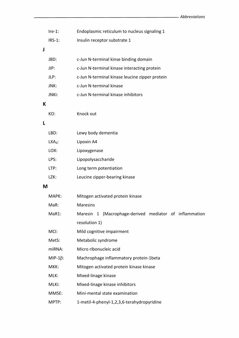
(Figure 2). As a result of anomalous protein burden, an interaction between Ire1
(endoplasmic reticulum to nucleus signaling 1) and presenilin 1 (PS1) has been proposed
to enable the activation of JNK thus leading to pro-apoptotic signaling activation (Shoji
et al., 2000). In addition, it has been suggested that ER stress activates apoptosis-signal
regulating kinase 1 (ASK1) through IRE1/TRAF2/ASK1 pathway. ASK1 is included in
MAP3K group, which subsequently triggers JNK signalling (Viana et al., 2012). Direct
modulation of JNK-activation by the cdk5/p35 complex has also been described,
although the underlying mechanisms that lead to this molecular phenomenon are
unclear (Otth et al., 2003).
Figure 2. Simplified diagram showing mechanisms involved in activation of the JNK pathway. Different stress conditions might activate JNK signalling via scaffold proteins. UPR and an interaction between IRE1 and PS1 have also been described as potential activators of JNK. Aβ: β-amyloid; HPK: hematopoietic progenitor kinase 1; Ire1: endoplasmic reticulum to nucleus signalling 1; JIP: JNK interacting protein; MKK: mitogen activated protein kinase kinase; MLK: mixed-linage kinase; ROS, radical oxygen species; UPR: unfolded protein response.
JNK
PP
P
UPR Ire1
PS1
?
JIP
MLK(MAP3K)
MKK4/MKK7(MAP2K)
JNK
HPK
ROS, Aβ, kainic acid, ischemia, hypoxia,
deprivationof neurotrophic factors
JNK
PP
P
Introduction
7
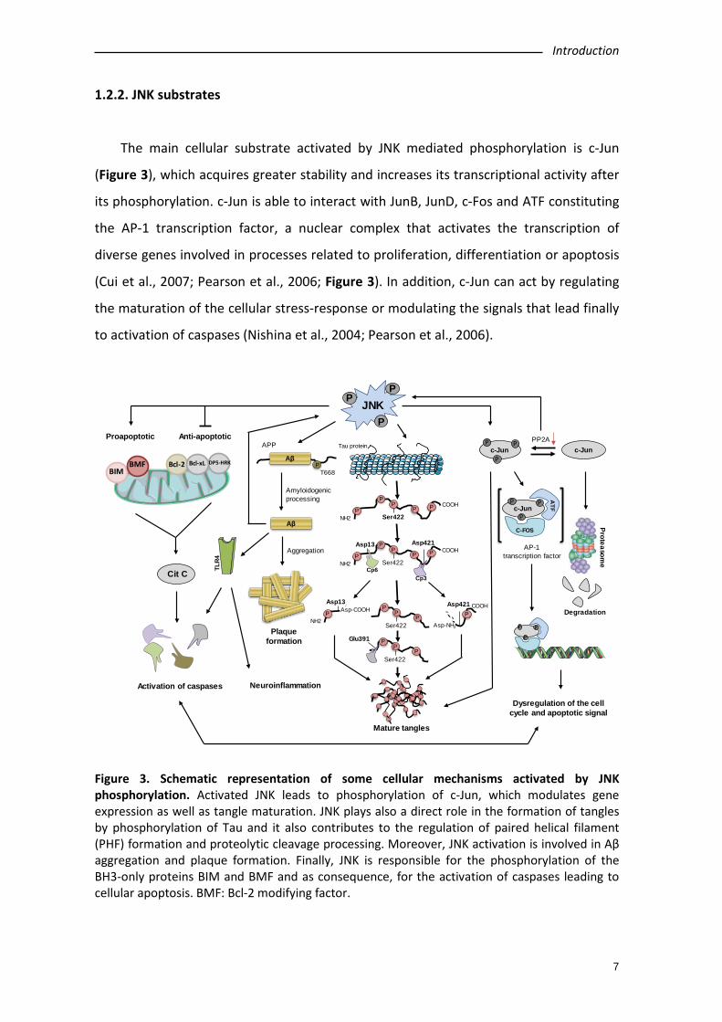
1.2.2. JNK substrates
The main cellular substrate activated by JNK mediated phosphorylation is c-Jun
(Figure 3), which acquires greater stability and increases its transcriptional activity after
its phosphorylation. c-Jun is able to interact with JunB, JunD, c-Fos and ATF constituting
the AP-1 transcription factor, a nuclear complex that activates the transcription of
diverse genes involved in processes related to proliferation, differentiation or apoptosis
(Cui et al., 2007; Pearson et al., 2006; Figure 3). In addition, c-Jun can act by regulating
the maturation of the cellular stress-response or modulating the signals that lead finally
to activation of caspases (Nishina et al., 2004; Pearson et al., 2006).
Figure 3. Schematic representation of some cellular mechanisms activated by JNK phosphorylation. Activated JNK leads to phosphorylation of c-Jun, which modulates gene expression as well as tangle maturation. JNK plays also a direct role in the formation of tangles by phosphorylation of Tau and it also contributes to the regulation of paired helical filament (PHF) formation and proteolytic cleavage processing. Moreover, JNK activation is involved in Aβ aggregation and plaque formation. Finally, JNK is responsible for the phosphorylation of the BH3-only proteins BIM and BMF and as consequence, for the activation of caspases leading to cellular apoptosis. BMF: Bcl-2 modifying factor.
JNKP
P
P
BIMBMF Bcl-2 Bcl-xL DP5-HRK
Cit C
Activation of caspases
PAsp421
NH2
COOHP
PP PP
Ser422
Asp13 Asp421Asp-COOH
Tau protein
NH2
COOHP
PP PP
Ser422
NH2
COOHP
PP PP
Ser422
Asp13 Asp421
Cp6Cp3
PP
PSer422 Asp-NH2
PP
PSer422
Glu391
Mature tangles
Proapoptotic Anti-apoptotic
Neuroinflammation
Plaque formation
c-Junc-JunP
P
P PP2A
Proteasome
Degradation
c-JunP
P
P
ATF
C-FOS
P
P
P
Dysregulation of the cell cycle and apoptotic signal
APP
AβP
T668
Aβ
Amyloidogenicprocessing
Aggregation
TLR4
COOH
AP-1 transcription factor
Introduction
8
Given the cytosolic nature of active JNK, several cytoskeletal components and
intracellular transport proteins are found within JNK substrates such as Tau or other
proteins associated with microtubules (Zeke et al., 2016). Moreover, JNK is able to
phosphorylate and activate directly apoptosis-related proteins such as BIM (homologous
to BAX) and BMF, both pro-apoptotic proteins resulting in activation of caspases. JNK
also phosphorylates DP5-HRK, Bcl-2 and Bcl-xL (Cui et al., 2007; Okazawa & Estus, 2002),
which are anti-apoptotic proteins inhibited by phosphorylation of JNK (Figure 3). In
summary, JNK activation induces an imbalance between the pro-apoptotic and anti-
apoptotic members of the Bcl-2 family that regulate mitochondrial control of apoptosis
and, thus, leading to mitochondrial cytochrome C release, caspases activation and,
finally, apoptotic death (Coffey, 2014).
Furthermore, it has also been described that JNK might exert its effects via
microRNA (miRNA) mechanisms or regulation of histone H3 acetylation, as reviewed by
Bogoyevitch et al., (2010).
2. JNK AND ALZHEIMER’S DISEASE
2.1. Alzheimer’s disease
Alzheimer’s disease (AD) is the main cause of dementia and it is considered a global
priority in the field of health-care. According to the World Health Organization (WHO),
more than 47 million people worldwide have dementia and there are near 10 million of
new cases every year, being AD the major contributor with a prevalence close to 70% of
the cases (WHO, 2017).
AD is an age-related neurodegenerative disorder clinically characterized by
progressive deterioration of cognitive functions that leads to an irreversible memory
loss and executive dysfunction interfering with daily life activities due to a progressive
neuronal deterioration. Although the clinical signs and symptoms are widely
characterized, AD etiology remains unclear and treatment strategies are only effective
in the early disease stages (Thal et al., 2006). It has a variable duration presenting a
Introduction
9
deterioration of the cognitive state that leads to death about 10 years after diagnosis
(Querfurth & Laferla, 2010).
Within AD, two subtypes are distinguished: familiar AD and sporadic AD. The
prevalence of familiar AD is lower (5% of AD cases) than sporadic. In familiar AD, there is
a mutated gene that causes AD so manifestations start much earlier than in sporadic AD
due to its genetic origin. Several mutations in different genes, such as APP, presenilin 1
or presenilin 2, have been identified. Among them, several autosomal dominant
mutations have been described in APP gene that lead to the stimulation of the
amyloidogenic processing of the APP such as the Swedish, London, Artic or Indiana
mutation.
In sporadic AD (95% of AD cases), the symptoms usually appear after 65 years old.
Its etiology is attributed to several risk factors: age, gender, vascular factors, metabolic
factors and other risk factors related to lifestyle such as stress, tobacco or alcohol. The
age is the most important risk factor to suffer AD; indeed, its incidence and prevalence
increase exponentially as patients get older (Prince et al., 2013). Women show a higher
probability to suffer from AD (Carter et al., 2012). Furthermore, any disease related to
vascular events, such as hypertension, cardiovascular or cerebrovascular disease,
increases the risk of suffering from AD (Newman et al., 2005). Moreover, some authors
refer to AD as diabetes mellitus type 3 because of the relationship between
hyperglycemia, insulin resistance, loss of grey matter and brain atrophy. In this line, it
has been probed that there is a decrease of insulin levels in CNS of AD patients (Gil-Bea
et al. 2010b). Moreover, hypercholesterolemia and obesity are associated with
neurodegeneration and cognitive impairment (Popp et al., 2013). Special attention must
be paid to the implication of obesity in AD. Several studies reported a negative
correlation between obesity and hippocampus and frontal cortex atrophy in non-
demented people (for review see Miller & Spencer, 2014). Given the importance of
these brain regions sizes in the cognitive function performance, this fact might
contribute to a cognitive decline in obese individuals (Miller & Spencer, 2014).
Furthermore, it has been shown that obese population presents worse cognitive state
compared with normal weight cases, categorized by body mass index (BMI) (Trakas et al.
2001).
Introduction
10
There are different AD clinical stages depending on the disease development.
Normally, the diagnosis is made in the later stages when the symptoms are evident; but,
fortunately, increasing attention is now paid to early-onset AD, where AD biomarkers
can contribute to the early diagnosis (Scheltens et al., 2016).
At cellular level, major neuropathological lesions of AD include extracellular
deposits of Aβ peptides leading to formation of senile/neuritic plaques and intracellular
neurofibrillary tangles (NFTs) which are paired helical filaments (PHF) of hyper-
phosphorylated Tau proteins (Haas, 2012). According to the amyloid hypothesis, Aβ
peptides are produced by amyloidogenic processing of amyloid precursor protein (APP)
that is a ubiquitous transmembrane protein. β-amyloid peptides originate from
proteolysis of the amyloid precursor protein by the sequential enzymatic actions of
beta-site amyloid precursor protein–cleaving enzyme 1 (BACE-1), a β-secretase, and γ-
secretase, a protein complex with presenilin 1 at its catalytic core. An imbalance
between the Aβ peptides production and clearance results in aggregation, accumulation
and Aβ oligomerization into insoluble fibrils and subsequent Aβ deposition establishing
the primary factor that propitiates AD.
On the other hand, the main component of NFTs is an abnormally
hyperphosphorylated and aggregated form of Tau. Under physiological conditions, Tau
binds to tubulin, among other proteins, stabilizing the axonal microtubules, which
confers a very important role in the maintenance of the neuronal structure and in the
transport of proteins through the axons. In physiological conditions Tau is responsible
not only for the stabilization of neuronal cytoskeleton by its binding to tubulin
monomers but also for many intra and extracellular signaling processes (Kolarova et al.,
2012). Hyperphosphorylated Tau lacks affinity for microtubules and becomes to
aggregate into PHF, leading to microtubule destabilization (Querfurth & Laferla, 2010).
Synaptic dysfunction and intra- and extracellular accumulation of abnormal
proteins, as NFTs and senile plaques, are the main pathological hallmarks of AD.
Synaptic neuronal loss has been observed in AD, which is related with the level of
cognitive deficit (Terry, 2000). Synapsis degeneration in AD is characterized by
progressive terminal loss, lower expression of pre- and post-synaptic proteins,
alterations and loss of dendritic spines (Scheff et al., 2007). Aβ peptide accumulation
seems to be one of the main mechanisms that produce synaptic dysfunction in AD.

Introduction
11
Indeed, correlation between synaptic dysfunction and alterations of learning and
memory in different animal models of AD has been observed. For example, it has been
demonstrated that Aβ-related synaptic dysfunction can lead to learning deficits in APP
transgenic mice (Westerman et al., 2002).
The etiology of AD remains elusive, but the nosogenic basis of AD seems to be
related to neuron apoptosis and loss of synaptic terminals within the central nervous
system’s parenchyma. Thus, the increased concentration of reactive oxygen
intermediates (ROI) and superoxide dismutase, both as markers of cellular stress, and
increased intracellular calcium in AD are congruent with an underlying activation of
apoptotic mechanisms via mitochondrial dysfunction. However, the molecular
mechanisms that lead to the activation of apoptotic signals are not fully understood.
2.2. JNK and pathological markers of Alzheimer’s disease
It has been shown an increased expression of phosphorylated JNK (pJNK) in human
post-mortem brain samples from AD patients and a positive co-localization with Aβ
(Killick et al., 2014; Zhu et al., 2001). In particular, JNK3 is highly expressed and activated
in brain tissue and cerebrospinal fluid from patients with AD and statistically correlates
with the rate of cognitive decline (Gourmaud et al., 2015).
It has been described that Aβ peptides might be able to induce JNK activation, as it
has been found in vitro that pJNK increases after treatment with Aβ in primary cortical
and hippocampal cultures from C57BL/6 mice, in primary cortical cell cultures from
Wistar rat and in SH-SY5Y neuroblastoma cells (Morishima et al., 2001; Suwanna et al.,
2014; Xu et al., 2015). Some reports confirmed that JNK3-mediated phosphorylation
regulated APP cleavage by inducing the amyloidogenic processing of the protein, while
JNK inhibition reduced amyloidogenic processing in favour of the non-amyloidogenic
route in vitro by blocking APP phosphorylation (Colombo et al., 2009; Morishima et al.,
2001; Savage et al., 2002). Interestingly, Yoon et al. (2012) demonstrated that JNK3 is
the major kinase for APP phosphorylation at T668, a phosphorylation site that favours
the amyloidogenic processing. In fact, genetic depletion of JNK3 in transgenic AD mice

Introduction
12
resulted in a dramatic reduction in Aβ42 peptide levels and overall plaque loads as well
as in an increased number of neurons and improved cognition (Yoon et al., 2012).
In experimental models of AD, research using a mouse model of AD that
incorporates the Swedish APP mutation and a mutant presenilin-1 (PS1) -Tg2576/PS1-
has demonstrated that JNK activation is associated with increased levels of senile
plaques and Tau phosphorylation (Savage et al., 2002; Figure 4). However, in contrast
with these data, no significant differences were found in pJNK levels in the triple
transgenic mice (3xTg mice, Feld et al. 2014).
Figure 4. Simplified schematic of cellular mechanisms relevant for AD activated by JNK phosphorylation. Activated JNK (pJNK) leads to tangle maturation by phosphorylation of Tau as well as c-Jun activation. JNK activation contributes to Aβ aggregation and plaque formation. The amyloidogenic processing of APP also induces a positive feedback increasing JNK activation.
Research has also been conducted in experimental models of AD based on well-
known risk factors contributing to the development of AD, such as stress or insulin
resistance (Dhikav & Anand, 2007; Martisova et al., 2013). In mice subjected to chronic
mild stress (CMS) known to increase Tau misprocessing and amyloidogenic processing,
JNK phosphorylation is increased (Solas et al. 2013a). The intracerebroventricular (ICV)
administration of subdiabetogenic doses of streptozotocin (STZ) induced cognitive and
brain cholinergic deficits, oxidative stress, insulin resistant brain state and high levels of
pJNK (Giuliani et al. 2013; Salkovic-Petrisic et al. 2013; Xiong et al. 2013). It is to be
noted that JNK may also directly induce insulin resistance, as JNK phosphorylates insulin

Introduction
13
receptor substrate 1 (IRS1), in an inhibitory site, blocking the transduction signal
produced by the insulin receptor (Sabio et al., 2008; Solas et al., 2013b).
c-Jun has been identified to play other possible roles in AD, e.g., phosphorylated c-
Jun burdens within the structure of NFTs may play an indirect regulatory role in tangle
maturation in AD, mostly regulated by its phosphorylation by JNK. Due to the imbalance
established between decreased PPA2 (protein phosphatase 2) expression and JNK
mediated phosphorylation of c-Jun, phospho-c-Jun levels are preponderant over the
non-phosphorylated form. As a matter of fact, phospho-c-Jun shows a lesser tendency
for its degradation via proteasomes, leading to its accumulation within NFTs and, thus,
contributing to tangle maturation process (Pearson et al., 2006; Figure 4).
JNK also modulates directly the formation of NFTs (Figure 4) by direct
phosphorylation of Tau (Lagalwar et al., 2006). In vitro phosphorylation experiments
show that JNK3 isoform can strongly autophosphorylate itself and contribute to Tau
hyperphosphorylation (Vogel et al., 2009). JNK was identified to phosphorylate Tau at
Ser422, and concretely, JNK3 has the highest affinity towards phosphorylation at Ser422
(Yoshida et al., 2004). In fact, phosphorylation at Ser422 has proved to protect against
caspase hydrolysis (Guillozet-Bongaarts et al., 2005; Guillozet-Bongaarts et al., 2006;
Kolarova et al., 2012).
2.3. Apoptosis regulation mediated by JNK activation
JNK plays a key role in the balance between cell survival and apoptosis. There are
two pathways capable of initiating apoptosis and are classified as extrinsic pathway
activated by death receptors such as TNF-α and targeted at the nuclear events, and
intrinsic pathway stimulated by mitochondrial dysfunction. Thus, JNK is involved in both
processes, as it is able to activate apoptotic signaling by its translocation to the nucleus
and upregulation of pro-apoptotic genes. Furthermore, it is able to directly modulate
the mitochondrial activity, through phosphorylation of pro-apoptotic proteins (Zeke et
al., 2016).
In AD, Aβ accumulation, ROS release and oxidative stress cause JNK activation
leading to c-Jun phosphorylation, which allows formation of the AP-1 complex that

Introduction
14
induces the transcription of pro-apoptotic genes such as Tnfα, Fas, Puma or Bak. In
addition, activated JNK translocates to mitochondria and phosphorylates the Bcl-2
family members in order to antagonize the anti-apoptotic activity of Bcl-2 and Bcl-xL.
Ultimately, cytochrome C release and caspases activation is induced leading to neuronal
death, as explained above (Akhter et al., 2015; Dhanasekaran & Reddy, 2008).
It has been suggested that neurodegeneration in early age of AD patients could be a
result of an increased vulnerability of neurons through activation of different apoptotic
pathways as a consequence of elevated levels of oxidative stress, and that these effects
could be mediated by JNK activation (Marques et al., 2003; Sahara et al., 2008).
Furthermore, JNK has been involved in Aβ triggered down regulation of the anti-
apoptotic Bcl-w (Yao et al., 2005) and activation of toll-like receptor 4 (TLR4) signaling
(Figure 3). Neurons from TLR4 mutant mice exhibit reduced JNK and caspase-3
activation and protection against Aβ induced apoptosis (Tang et al., 2008).
2.4. Role of JNK in neuroinflammation
Inflammation is a protective mechanism in the body, as its main function is to repair
and protect against infections, injury or disease. Neuroinflammation is defined as the
natural immune system activation at CNS. Initially, neuroinflammation is a protective
response in the brain, but a chronic neuroinflammatory response might play an
important role in the onset and progression of neurodegenerative disease such as AD,
Parkinson’s disease or multiple sclerosis (Kempuraj et al., 2016; Zhang & Jiang, 2015).
The regulation of the inflammatory cascade is under the control of intracellular
signalling pathways, including MAPK signalling pathway. It has been suggested that Aβ
peptide induces neuroinflammation mediated by JNK pathway activation resulting in c-
Jun phosphorylation. There is evidence that Aβ induces translational block leading to
activation of JNK (Yoon et al., 2012), which in turn, results in neuroinflammation and
neurodegeneration. Activated c-Jun leads to stimulation of AP-1 that up-regulated the
expression of inflammatory genes. Interestingly, some studies have reported that JNK
inhibitor SP600125 attenuates the neuroinflammatory response induced by Aβ (Lin et
al., 2013; Vukic et al., 2009; Zhang & Jiang, 2015).

Introduction
15
In the elderly and specially in AD patients, there are changes in blood perfusion,
blood-brain barrier (BBB) disruption, together with Aβ aggregation and NFT that result
in ATP synthesis and electrolytes imbalance, leading to oxidative stress and ROS
production which cause overproduction of pro-inflammatory molecules such as
interleukin 1 and 6 (IL-1 and IL-6), TNFα, cluster of differentiation-14 (CD14) or toll like
receptor 2 and/or 4 (TLR2 or TLR4). Stabilization of this inflammatory cascade induces
an irreversible neuronal damage (Bagyinszky et al., 2017).
HPA axis dysregulation in AD leads to glucocorticoids (GC) excess (Gil-Bea et al.
2010a) and decreased insulin sensitivity, trough TLR4, JNK and nuclear factor-kappa B
(NF-κB) pathways. Given that hippocampus contains the highest concentrations of
glucocorticoids receptors (GR) in the brain, altered GC levels causes hippocampal
inflammation and disruption that is likely to lead to cognitive dysfunction. However,
inflammation induced hypothalamic outputs are not only targeted to hippocampus, but
also to other brain regions, such as cortical tissue and amygdala (Miller & Spencer, 2014;
Williams, 2012).
3. NOVEL COMPOUNDS TARGETING JNK INHIBITION
Inhibition of JNKs is an attractive therapeutic strategy that has been investigated
with considerable recent effort from both the pharmaceutical industry and academia.
The development of JNK inhibitors prior to 2010 has been extensively reviewed (Siddiqui
& Reddy, 2010). Within the last years, several patents claiming inhibitors of all JNK
isoforms have been published, but few JNK inhibitors have entered clinical trials for
different indications such as cancer, lupus erythematosus or stroke (Gehringer et al.,
2015), but none for the treatment of AD (Bogoyevitch & Arthur, 2008).
Current compounds under evaluation are: bentamapimod for the treatment of
inflammatory endometriosis, CC-930 (tanzisertib) for the treatment of idiopathic
pulmonary fibrosis and discoid lupus erythematous and D-JNKi1 for the treatment of
inflammation and stroke (as reviewed by Koch et al. 2015). In the following sections, and
shown in Tables 1 and 2 and Figure 5, current knowledge of the JNK inhibitors will be
described.
Introduction
16
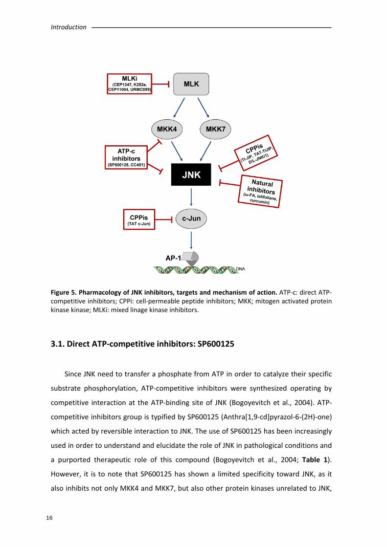
Figure 5. Pharmacology of JNK inhibitors, targets and mechanism of action. ATP-c: direct ATP-competitive inhibitors; CPPi: cell-permeable peptide inhibitors; MKK; mitogen activated protein kinase kinase; MLKi: mixed linage kinase inhibitors.
3.1. Direct ATP-competitive inhibitors: SP600125
Since JNK need to transfer a phosphate from ATP in order to catalyze their specific
substrate phosphorylation, ATP-competitive inhibitors were synthesized operating by
competitive interaction at the ATP-binding site of JNK (Bogoyevitch et al., 2004). ATP-
competitive inhibitors group is typified by SP600125 (Anthra[1,9-cd]pyrazol-6-(2H)-one)
which acted by reversible interaction to JNK. The use of SP600125 has been increasingly
used in order to understand and elucidate the role of JNK in pathological conditions and
a purported therapeutic role of this compound (Bogoyevitch et al., 2004; Table 1).
However, it is to note that SP600125 has shown a limited specificity toward JNK, as it
also inhibits not only MKK4 and MKK7, but also other protein kinases unrelated to JNK,
Introduction
17
such as SGK, p70 ribosomal protein S6 kinase (S6K1), AMpk, CDk2, CK1d, and DYRK1A
(Bain et al., 2003).
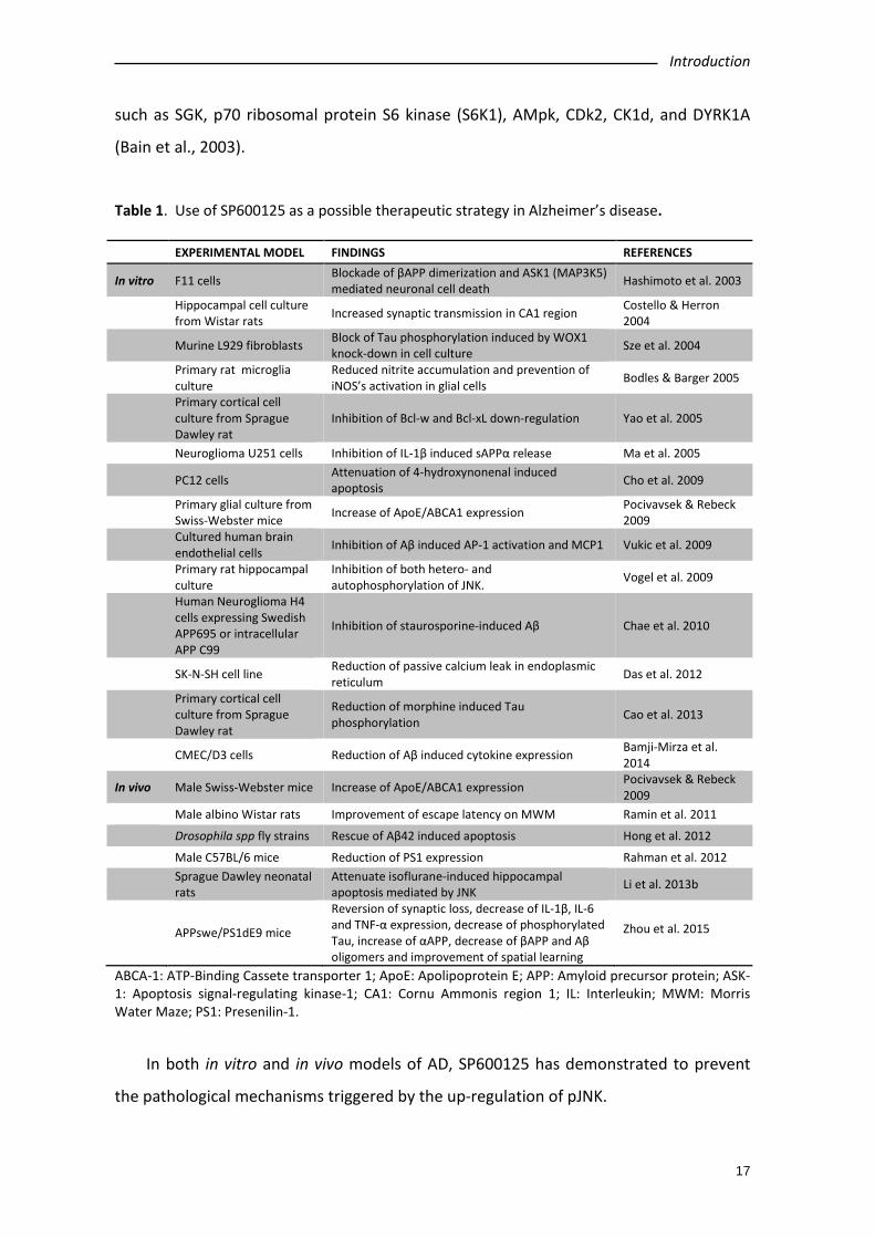
Table 1. Use of SP600125 as a possible therapeutic strategy in Alzheimer’s disease. EXPERIMENTAL MODEL FINDINGS REFERENCES
In vitro F11 cells Blockade of βAPP dimerization and ASK1 (MAP3K5) mediated neuronal cell death Hashimoto et al. 2003
Hippocampal cell culture from Wistar rats Increased synaptic transmission in CA1 region Costello & Herron
2004 Murine L929 fibroblasts Block of Tau phosphorylation induced by WOX1
knock-down in cell culture Sze et al. 2004
Primary rat microglia culture
Reduced nitrite accumulation and prevention of iNOS’s activation in glial cells Bodles & Barger 2005
Primary cortical cell culture from Sprague Dawley rat
Inhibition of Bcl-w and Bcl-xL down-regulation Yao et al. 2005
Neuroglioma U251 cells Inhibition of IL-1β induced sAPPα release Ma et al. 2005
PC12 cells Attenuation of 4-hydroxynonenal induced apoptosis Cho et al. 2009
Primary glial culture from Swiss-Webster mice Increase of ApoE/ABCA1 expression Pocivavsek & Rebeck
2009 Cultured human brain
endothelial cells Inhibition of Aβ induced AP-1 activation and MCP1 Vukic et al. 2009
Primary rat hippocampal culture
Inhibition of both hetero- and autophosphorylation of JNK. Vogel et al. 2009
Human Neuroglioma H4 cells expressing Swedish APP695 or intracellular APP C99
Inhibition of staurosporine-induced Aβ Chae et al. 2010
SK-N-SH cell line Reduction of passive calcium leak in endoplasmic reticulum Das et al. 2012
Primary cortical cell culture from Sprague Dawley rat
Reduction of morphine induced Tau phosphorylation Cao et al. 2013
CMEC/D3 cells Reduction of Aβ induced cytokine expression Bamji-Mirza et al. 2014
In vivo Male Swiss-Webster mice Increase of ApoE/ABCA1 expression Pocivavsek & Rebeck 2009
Male albino Wistar rats Improvement of escape latency on MWM Ramin et al. 2011 Drosophila spp fly strains Rescue of Aβ42 induced apoptosis Hong et al. 2012 Male C57BL/6 mice Reduction of PS1 expression Rahman et al. 2012
Sprague Dawley neonatal rats
Attenuate isoflurane-induced hippocampal apoptosis mediated by JNK Li et al. 2013b
APPswe/PS1dE9 mice Reversion of synaptic loss, decrease of IL-1β, IL-6 and TNF-α expression, decrease of phosphorylated Tau, increase of αAPP, decrease of βAPP and Aβ oligomers and improvement of spatial learning
Zhou et al. 2015
ABCA-1: ATP-Binding Cassete transporter 1; ApoE: Apolipoprotein E; APP: Amyloid precursor protein; ASK-1: Apoptosis signal-regulating kinase-1; CA1: Cornu Ammonis region 1; IL: Interleukin; MWM: Morris Water Maze; PS1: Presenilin-1.
In both in vitro and in vivo models of AD, SP600125 has demonstrated to prevent
the pathological mechanisms triggered by the up-regulation of pJNK.
Introduction
18
In vitro, SP600125 has demonstrated that this inhibitor prevents βAPP induced
neuronal cell death as well as down-regulation of ASK1 in F11 cell-lines (Hashimoto et
al., 2003). Interestingly, this study did not find neuroprotection against βAPP induced
neuroapoptosis when exposing cultures to a p38 inhibitor, highlighting the importance
of JNK within this process. Other in vitro experiments showed decrease of Aβ-induced
cytokine expression (IL6, IL8, MIP1β, TNF-α, Gro-α, GM-CSF) (Bamji-Mirza et al., 2014).
In vivo studies have shown that ICV administration of SP600125 improved escape
latency in the Morris water maze (MWM) (Ramin et al., 2011). In this line, in an AD
transgenic mouse model (APPxPS1), administration of SP600125 improved spatial
learning impairment in the MWM, and reduced pTau and Aβ oligomeric burden (Zhou et
al., 2015).
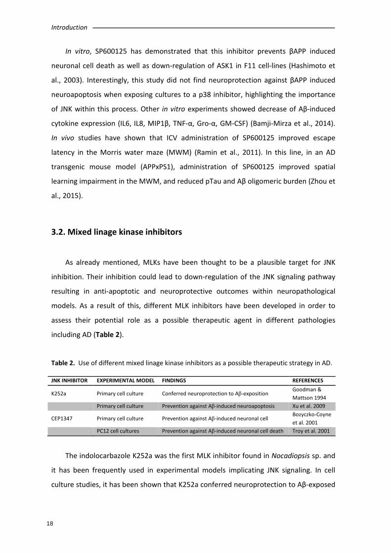
3.2. Mixed linage kinase inhibitors
As already mentioned, MLKs have been thought to be a plausible target for JNK
inhibition. Their inhibition could lead to down-regulation of the JNK signaling pathway
resulting in anti-apoptotic and neuroprotective outcomes within neuropathological
models. As a result of this, different MLK inhibitors have been developed in order to
assess their potential role as a possible therapeutic agent in different pathologies
including AD (Table 2).
Table 2. Use of different mixed linage kinase inhibitors as a possible therapeutic strategy in AD. JNK INHIBITOR EXPERIMENTAL MODEL FINDINGS REFERENCES
K252a Primary cell culture Conferred neuroprotection to Aβ-exposition Goodman & Mattson 1994
Primary cell culture Prevention against Aβ-induced neuroapoptosis Xu et al. 2009
CEP1347 Primary cell culture Prevention against Aβ-induced neuronal cell Bozyczko-Coyne et al. 2001
PC12 cell cultures Prevention against Aβ-induced neuronal cell death Troy et al. 2001
The indolocarbazole K252a was the first MLK inhibitor found in Nocadiopsis sp. and
it has been frequently used in experimental models implicating JNK signaling. In cell
culture studies, it has been shown that K252a conferred neuroprotection to Aβ-exposed

Introduction
19
hippocampal cells (Goodman & Mattson, 1994) and prevented Aβ-induced
neuroapoptosis (Xu et al., 2009) which could be of potential benefit in AD.
The compound CEP1347 derives from K252a by the addition of two ethylthiomethyl
groups (Saporito et al., 2002) and acts over MLK1, MLK2, MLK3, DLK (dual leucine zipper
kinase) and leucine zipper-bearing kinase (LZK, Bogoyevitch et al. 2004). CEP1347
reached clinical phase studies (Parkinson Study Group, 2004; Parkinson Study Group
PRECEPT Investigators, 2007; Schwid et al., 2010) for the treatment of Parkinson’s
disease (Wang et al., 2004). Unfortunately, the results were disappointing. Regarding
AD, CEP1347 has been shown to prevent Aβ-induced neuronal cell death and it reduced
caspase-3 activity (Bozyczko-Coyne et al., 2001; Troy et al., 2001).
CEP11004 is another carbazole-derived MLK inhibitor that has proved to be useful
in PD models, as this compound prevented 6-hydroxydopamine-induced neuroapoptosis
in neurons of the substantia nigra (Ganguly et al., 2004) and it also appeared as a good
inhibitor of the JNK cascade in a MPTP-induced cellular stress model (De Girolamo &
Billett, 2006). Further studies will be needed to evaluate the effects of CEP11004 in AD
experimental models for its possible relation towards the AD-related pathophysiologic
mechanisms explained above.
URMC099 is a novel MLK inhibitor with good BBB penetrating properties which has
already been proved to be useful in reducing inflammatory response both in vivo and in
vitro models (Goodfellow et al., 2013; Marker et al., 2013). However, no studies have
been performed up to date to evaluate the effects of URMC099 in neurodegenerative
models.
In summary, although the use of MLK inhibitors has been limited in the AD field,
further studies are expected to come.
3.3. Cell-permeable peptide inhibitors
Cell-permeable peptide inhibitors of JNK (CPPis) are peptide sequences that
specifically bind to the JNK binding domain (JBD) leading to its inhibition (Bogoyevitch et
al., 2004; Borsello & Bonny, 2004). Their characterization came primarily from studies
that confirm the interaction of highly expressed JIP1 with JNK, showing that high
Introduction
20
concentrations of JIP1 are able to induce inhibition of JNK and down-regulation of JNK
substrates (Dickens et al., 1997). A conserved 21 amino acid long sequence was firstly
identified within JNK’s primary protein conformation at position 143-163 (Barr et al.,
2002; Bogoyevitch et al., 2004). This region is widely known to be the JNK binding
domain where JIP1 mediates down-modulation over JNK, leading to its inhibition.
Purification of the 143-163 region and synthesis of the polypeptide out of this sequence,
named I-JIP, showed the capacity of triggering inhibition of JNK. Moreover, a shorter
polypeptide obtained from the sequence specified in-between 153-163 demonstrated
to exert the minimal inhibitory effect on JNK. This compound receives the name of TI-JIP
(Barr et al., 2002; Bogoyevitch & Arthur, 2008).
However, the disadvantages that result from the relative non-permeability of TI-JIP
need to be solved. In order to achieve this aim, it has been proposed to couple TI-JIP to
a cell-penetrating peptide (CPP, Bogoyevitch et al., 2004). CPPs are small peptides
(typically 5-25 amino acids) which are used to facilitate the delivery of normally non-
permeable cargos such as other peptides, proteins, nucleic acids, or drugs into cells
(Meloni et al., 2014). Hence Borsello & Bonny, (2004) observed that attaching CPPs,
such as TAT 48-57 or antannapedia, to TI-JIP facilitates the diffusion of peptides through
membranes in order to exert their action over the desired targets.
JNK-interacting protein derived compounds have been studied for their possible
role in preventing neurodegenerative pathways in which JNK has been proved to be
implicated. TAT-TIJIP was obtained linking TI-JIP (truncated form of I-JIP) to TAT 48-57
(CPP) which is a transporter sequence of 10 amino acids derived from the human
immunodeficiency virus TAT protein (Borsello et al., 2003). TAT-TIJIP has been
demonstrated to be able to prevent neuronal apoptosis via JNK inhibition and
effectively prevented cell death by interfering with several processes that have been
identified as leading to cell death by necrosis. In particular, reactive oxygen species
production was reduced and the increase in cytosolic calcium following the excitotoxic
insult was attenuated. These neuroprotective properties of JNK peptide inhibitors likely
reflect their abilities to prevent cell death by necrosis as well as apoptosis (Arthur et al.,
2007). Interestingly, a peptide inhibitor of c-Jun has also been synthesized, the TAT-c-
Jun peptide (Antoniou et al., 2011; Holzberg et al., 2003).
Introduction
21
It is noteworthy that CPPs show themselves as promising molecules for targeting
JNK, as these compounds have the advantage of specificity, showing little activity over
other kinases (Barr et al., 2002). In fact, one of the most important disadvantages shown
by other synthetic JNK inhibitors, such as SP600125 or MLK inhibitor, is their lack of
specificity towards their target (Bogoyevitch et al., 2004). Moreover, a study showed the
neuroprotective efficacy of four CPPs, namely TAT, penetratin, Ar-9 and Pep-1 in a
glutamic acid, kainic acid and in vitro ischemia injury model (Meloni et al., 2014). AP-1
inhibitory peptides (both full-length and truncated) have also shown neuroprotective
efficacy in kainic acid and glutamate neuronal excitotoxicity models (Meade et al.,
2010). However it is to note that TAT-like peptides and other non-related CPPs possess
intrinsic neuroprotective properties (Meloni et al., 2014) and pose the question of the
contribution of the CPP versus cargo in the neuroprotective effect.
In this way, different post-modifications of TAT-TIJIP were performed that led to the
synthesis of JNK inhibitors (JNKi). Furthermore, the in vitro synthesis of JNKi using pure
D-isomers with the intention of preserving protein functionality and avoiding proteolytic
instability, led to the obtaining of D-JNKi1 and its L-isomer (L-JNKi1, Borsello & Bonny
2004). D-JNKi1 is the most frequently used inhibitor in experimental neurodegenerative
models. It has been proved useful utility to reverse ischemia-induced neuronal damage
(Borsello et al., 2003). It has been demonstrated that D-JNKi1 is able to decrease levels
of APP in human neuroglioma H4 cell lines with the consequent reduction of βAPP levels
and Aβ burdens, and it also shifted APP processing toward the non-amyloidogenic
pathway (Colombo et al., 2009). Again, these events are of high interest as they are
directly related to the central pathogenesis of AD.
Regarding AD models (Table 3), different studies have confirmed the potential
therapeutic benefit of these inhibitors for their capacity to interact within a wide variety
of molecular signaling processes implicated in this pathology.
Sclip et al. pointed out the efficacy of D-JNKi1 in a murine AD model, TgCRND8, in
which D-JNKi1 demonstrated to prevented JNK action leading to decreased APP
phosphorylation at Thr-668 and reduced amyloidogenic cleavage of APP and Aβ
oligomers (Sclip et al., 2011). Tran et al. demonstrated that D-JNKi1 mediated down-
regulation of JNK induced Tau phosphorylation in an AD transgenic model
(PS1xAPPxTau) (Tran et al., 2012). D-JNKi1 has also proved beneficial effects rescuing

Introduction
22
synaptic loss and potentiating LTP in TgCRND8 (Sclip et al., 2014). The increase in pTau
levels and neuronal cell death shown in a stress model of AD was also reversed by the
administration of D-JNKi1 (Solas et al. 2013b). In this scenario, peptide inhibitors could
represent a good therapeutic option for the continuously widening therapeutic
armamentarium in AD.
Table 3. Use of different peptide inhibitors as a possible therapeutic strategy in AD. JNK INHIBITOR EXPERIMENTAL MODEL FINDINGS REFERENCES TAT-TIJIP Primary cell culture Prevention against neuronal apoptosis Meade et al. 2010
Primary cell culture Decrease of neuronal degenerarion and dendrite loss
Meloni et al. 2014
D-JNKi TgCRND8 mice Decrease of APP phosphorylation. Improvement of memory
Sclip et al. 2011
3xTg-AD mice with traumatic brain injury
Prevention of Tau phosphorylation Tran et al. 2012
SAMP8 mice Decrease of Tau phosphorylation. Improvement of memory in MWM task
Orejana et al., 2013
C57BL/6J mice + corticosterone regimen
Reversion of insulin resistance. Improvement of cognitive function in NORT task
Solas et al., 2013b
TgCRND8 mice Decrease of synaptic loss and preventing synaptic dysfunction
Sclip et al. 2014
APP: amyloid precursor protein; MWM: Morris water maze; NORT: novel object recognition test.
3.4. Natural inhibitors
Three different types of compounds can be mentioned in this section: latifolians,
curcumin and ω-fatty acids (ω-FAs).
Latifolians A and B are natural compounds isolated from the stem bark of the Papua
New Guinean vine Gnetum latifolium that have been identified as inhibitors of JNK3
(Rochfort et al., 2005). However, no studies have been performed concerning the
possible use of latifolians as neuroprotective agents in neurodegenerative models.
Curcumin is a natural compound that resides in the Zingiberaceae spp. family. Aside
for its implications as an anti-inflammatory and antioxidant agent, curcumin has also
proved to play a direct role in the modulation of the JNK pathway (Chen & Tan, 1998).
Indeed, it has been proposed an underlying role of curcumin toward the inhibition of
JNK, demonstrating its capacity to ameliorate MPTP (1-methyl-4-phenyl-1,2,3,6-
Introduction
23
tetrahydropyridine) and MPP+ (1-methyl-4-phenylpyridnium) induced neuronal loss
models both in vivo and in vitro (Yu et al., 2010). It also promotes an increase in the
expression of HSPs (heat shock proteins) that are centrally implicated in preserving the
functionality of the proteasome-mediated degradation of abnormally misfolded
proteins (Maiti et al., 2014).
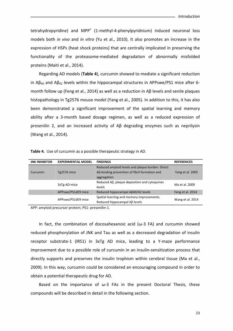
Regarding AD models (Table 4), curcumin showed to mediate a significant reduction
in Aβ40 and Aβ42 levels within the hippocampal structures in APPswe/PS1 mice after 6-
month follow up (Feng et al., 2014) as well as a reduction in Aβ levels and senile plaques
histopathology in Tg2576 mouse model (Yang et al., 2005). In addition to this, it has also
been demonstrated a significant improvement of the spatial learning and memory
ability after a 3-month based dosage regimen, as well as a reduced expression of
presenilin 2, and an increased activity of Aβ degrading enzymes such as neprilysin
(Wang et al., 2014).
Table 4. Use of curcumin as a possible therapeutic strategy in AD. JNK INHIBITOR EXPERIMENTAL MODEL FINDINGS REFERENCES
Curcumin Tg2576 mice Reduced amyloid levels and plaque burden. Direct Aβ-binding prevention of fibril formation and aggregation
Yang et al. 2005
3xTg-AD-mice Reduced Aβ, plaque deposition and cytoquines levels
Ma et al. 2009
APPswe/PS1dE9 mice Reduced hippocampal Aβ40/42 levels Feng et al. 2014
APPswe/PS1dE9 mice Spatial learning and memory improvements. Reduced hippocampal Aβ levels
Wang et al. 2014
APP: amyloid precursor protein; PS1: presenilin-1.
In fact, the combination of docosahexanoic acid (ω-3 FA) and curcumin showed
reduced phosphorylation of JNK and Tau as well as a decreased degradation of insulin
receptor substrate-1 (IRS1) in 3xTg AD mice, leading to a Y-maze performance
improvement due to a possible role of curcumin in an insulin-sensitization process that
directly supports and preserves the insulin trophism within cerebral tissue (Ma et al.,
2009). In this way, curcumin could be considered an encouraging compound in order to
obtain a potential therapeutic drug for AD.
Based on the importance of ω-3 FAs in the present Doctoral Thesis, these
compounds will be described in detail in the following section.
Introduction
24
4. ω-3 DERIVATIVES AND ALZHEIMER’S DISEASE
The implication of polyunsaturated fatty acids (PUFAs) in neurodegenerative
diseases is currently widely accepted. Albeit, some controversy regarding the benefit of
ω-3 FAs in mild to moderate AD patients have been reported and the effects on people
with other type of dementia remain unclear (Burckhardt et al., 2016). Therefore, all
above mentioned suggest the importance of further studies that could confirm the
beneficial outcomes of the use of ω-FAs in the onset and progression of AD.
4.1. ω-3 polyunsaturated fatty acid
The organism can receive two different types of fat through diet: saturated and
unsaturated fat. Saturated fats can be found naturally in food, mainly from animal
sources, such as meat and dairy products, and have been related with higher risk of
heart disease (de Souza et al., 2015). On the other hand, unsaturated fats are
predominantly found in food derived from fish or plants, such as olive oil, nuts and
seeds. These unsaturated fats can be differentiated in monounsaturated and
polyunsaturated fats.
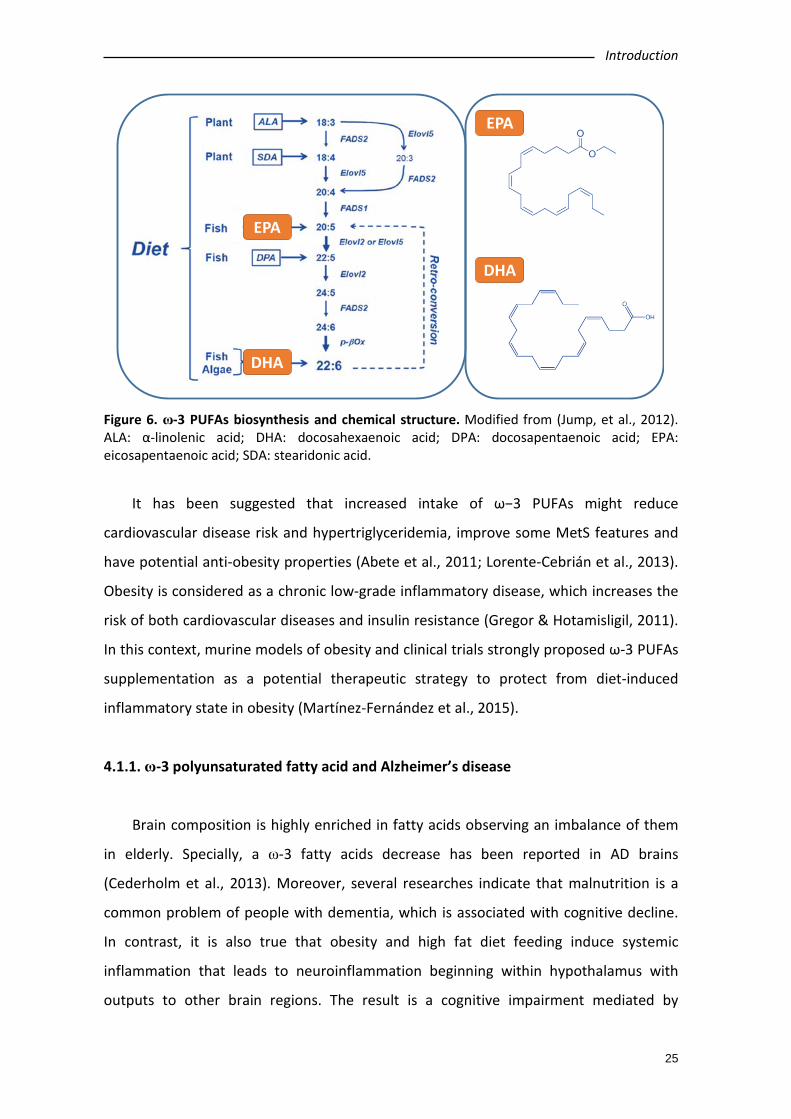
In this sense, the long chain omega-3 polyunsaturated fatty acids (ω-3 PUFAs) are
derived from marine or vegetal sources (Figure 6). Among marine ω-3 PUFAs, the most
significant ones are Eicosapentaenoic acid (EPA, ω-3, 20:5) and Docosahexaenoic acid
(DHA, ω-3, 22:6). EPA and DHA can be found mainly in oily fish including salmon, tuna,
mackerel and anchovy among others (for review see Lorente-Cebrián et al., 2013;
Lorente-Cebrián et al., 2015). On the other hand, the most relevant ω-3 PUFA derived
from plants is the α-linolenic acid (ALA, 18:3). Nevertheless, mammals can obtain ALA
through EPA and DHA processing into organism (Burdge et al., 2002; Burdge & Wootton,
2002).
Marine ω-3 PUFAs have been widely recognised to exert favourable anti-
inflammatory actions on inflammatory-related diseases such as cardiovascular diseases,
atherosclerosis, AD, asthma, arthritis, colitis, obesity or metabolic syndrome (MetS) (for
review see Calder, 2015).
Introduction
25
Figure 6. ω-3 PUFAs biosynthesis and chemical structure. Modified from (Jump, et al., 2012). ALA: α-linolenic acid; DHA: docosahexaenoic acid; DPA: docosapentaenoic acid; EPA: eicosapentaenoic acid; SDA: stearidonic acid.
It has been suggested that increased intake of ω−3 PUFAs might reduce
cardiovascular disease risk and hypertriglyceridemia, improve some MetS features and
have potential anti-obesity properties (Abete et al., 2011; Lorente-Cebrián et al., 2013).
Obesity is considered as a chronic low-grade inflammatory disease, which increases the
risk of both cardiovascular diseases and insulin resistance (Gregor & Hotamisligil, 2011).
In this context, murine models of obesity and clinical trials strongly proposed ω-3 PUFAs
supplementation as a potential therapeutic strategy to protect from diet-induced
inflammatory state in obesity (Martínez-Fernández et al., 2015).
4.1.1. ω-3 polyunsaturated fatty acid and Alzheimer’s disease
Brain composition is highly enriched in fatty acids observing an imbalance of them
in elderly. Specially, a ω-3 fatty acids decrease has been reported in AD brains
(Cederholm et al., 2013). Moreover, several researches indicate that malnutrition is a
common problem of people with dementia, which is associated with cognitive decline.
In contrast, it is also true that obesity and high fat diet feeding induce systemic
inflammation that leads to neuroinflammation beginning within hypothalamus with
outputs to other brain regions. The result is a cognitive impairment mediated by
EPA
DHA
EPA
DHA
Introduction
26
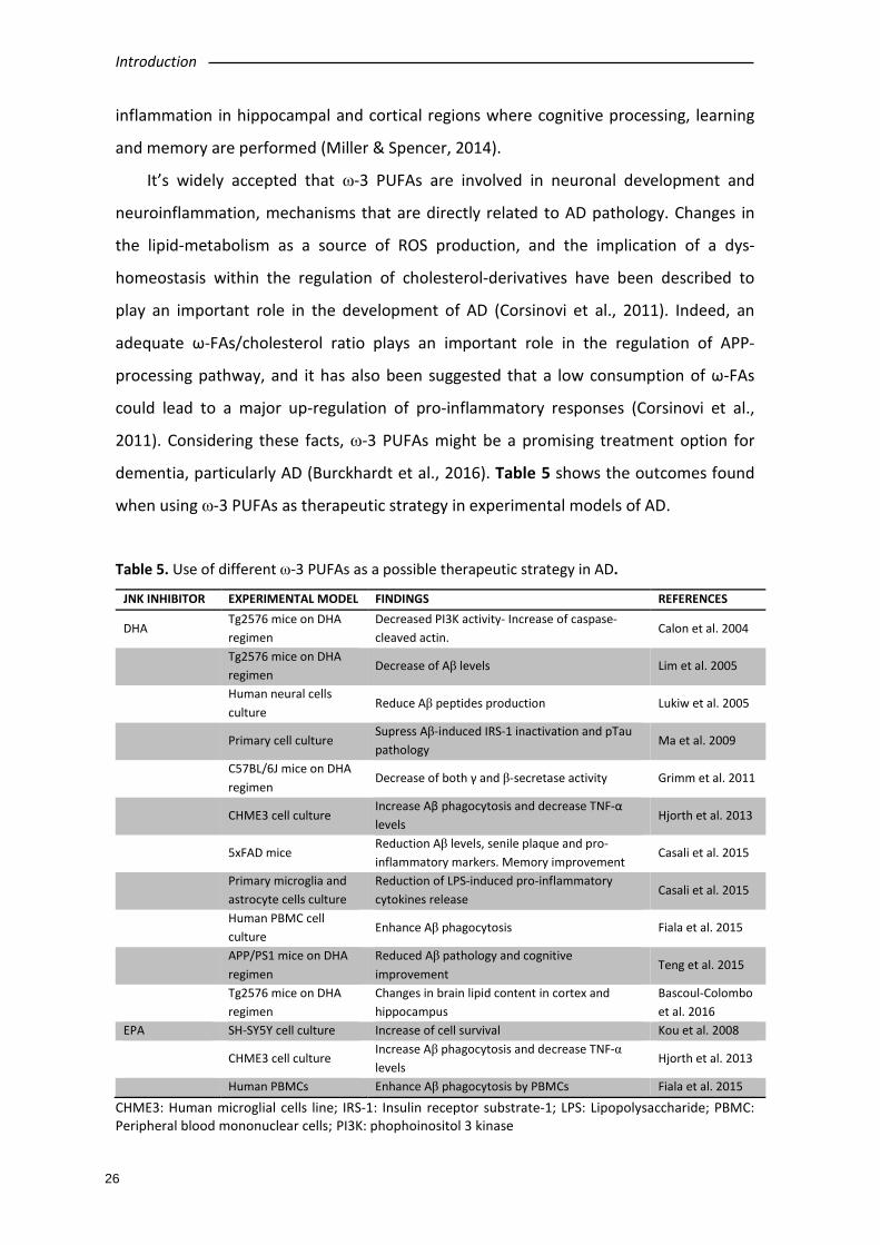
inflammation in hippocampal and cortical regions where cognitive processing, learning
and memory are performed (Miller & Spencer, 2014).
It’s widely accepted that ω-3 PUFAs are involved in neuronal development and
neuroinflammation, mechanisms that are directly related to AD pathology. Changes in
the lipid-metabolism as a source of ROS production, and the implication of a dys-
homeostasis within the regulation of cholesterol-derivatives have been described to
play an important role in the development of AD (Corsinovi et al., 2011). Indeed, an
adequate ω-FAs/cholesterol ratio plays an important role in the regulation of APP-
processing pathway, and it has also been suggested that a low consumption of ω-FAs
could lead to a major up-regulation of pro-inflammatory responses (Corsinovi et al.,
2011). Considering these facts, ω-3 PUFAs might be a promising treatment option for
dementia, particularly AD (Burckhardt et al., 2016). Table 5 shows the outcomes found
when using ω-3 PUFAs as therapeutic strategy in experimental models of AD.
Table 5. Use of different ω-3 PUFAs as a possible therapeutic strategy in AD.
JNK INHIBITOR EXPERIMENTAL MODEL FINDINGS REFERENCES
DHA Tg2576 mice on DHA regimen
Decreased PI3K activity- Increase of caspase- cleaved actin.
Calon et al. 2004
Tg2576 mice on DHA regimen
Decrease of Aβ levels Lim et al. 2005
Human neural cells culture
Reduce Aβ peptides production Lukiw et al. 2005
Primary cell culture Supress Aβ-induced IRS-1 inactivation and pTau pathology
Ma et al. 2009
C57BL/6J mice on DHA regimen
Decrease of both γ and β-secretase activity Grimm et al. 2011
CHME3 cell culture Increase Aβ phagocytosis and decrease TNF-α levels
Hjorth et al. 2013
5xFAD mice Reduction Aβ levels, senile plaque and pro-inflammatory markers. Memory improvement
Casali et al. 2015
Primary microglia and astrocyte cells culture
Reduction of LPS-induced pro-inflammatory cytokines release
Casali et al. 2015
Human PBMC cell culture
Enhance Aβ phagocytosis Fiala et al. 2015
APP/PS1 mice on DHA regimen
Reduced Aβ pathology and cognitive improvement
Teng et al. 2015
Tg2576 mice on DHA regimen
Changes in brain lipid content in cortex and hippocampus
Bascoul-Colombo et al. 2016
EPA SH-SY5Y cell culture Increase of cell survival Kou et al. 2008
CHME3 cell culture Increase Aβ phagocytosis and decrease TNF-α levels
Hjorth et al. 2013
Human PBMCs Enhance Aβ phagocytosis by PBMCs Fiala et al. 2015
CHME3: Human microglial cells line; IRS-1: Insulin receptor substrate-1; LPS: Lipopolysaccharide; PBMC: Peripheral blood mononuclear cells; PI3K: phophoinositol 3 kinase
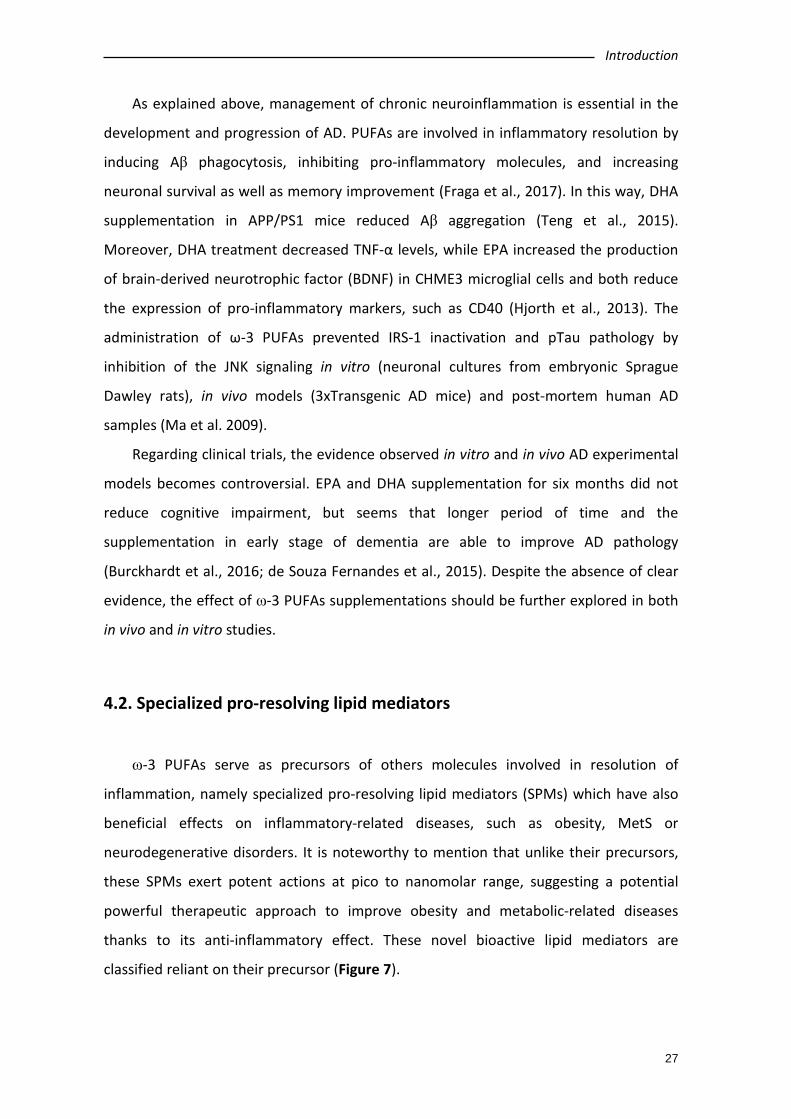
Introduction
27
As explained above, management of chronic neuroinflammation is essential in the
development and progression of AD. PUFAs are involved in inflammatory resolution by
inducing Aβ phagocytosis, inhibiting pro-inflammatory molecules, and increasing
neuronal survival as well as memory improvement (Fraga et al., 2017). In this way, DHA
supplementation in APP/PS1 mice reduced Aβ aggregation (Teng et al., 2015).
Moreover, DHA treatment decreased TNF-α levels, while EPA increased the production
of brain-derived neurotrophic factor (BDNF) in CHME3 microglial cells and both reduce
the expression of pro-inflammatory markers, such as CD40 (Hjorth et al., 2013). The
administration of ω-3 PUFAs prevented IRS-1 inactivation and pTau pathology by
inhibition of the JNK signaling in vitro (neuronal cultures from embryonic Sprague
Dawley rats), in vivo models (3xTransgenic AD mice) and post-mortem human AD
samples (Ma et al. 2009).
Regarding clinical trials, the evidence observed in vitro and in vivo AD experimental
models becomes controversial. EPA and DHA supplementation for six months did not
reduce cognitive impairment, but seems that longer period of time and the
supplementation in early stage of dementia are able to improve AD pathology
(Burckhardt et al., 2016; de Souza Fernandes et al., 2015). Despite the absence of clear
evidence, the effect of ω-3 PUFAs supplementations should be further explored in both
in vivo and in vitro studies.
4.2. Specialized pro-resolving lipid mediators
ω-3 PUFAs serve as precursors of others molecules involved in resolution of
inflammation, namely specialized pro-resolving lipid mediators (SPMs) which have also
beneficial effects on inflammatory-related diseases, such as obesity, MetS or
neurodegenerative disorders. It is noteworthy to mention that unlike their precursors,
these SPMs exert potent actions at pico to nanomolar range, suggesting a potential
powerful therapeutic approach to improve obesity and metabolic-related diseases
thanks to its anti-inflammatory effect. These novel bioactive lipid mediators are
classified reliant on their precursor (Figure 7).
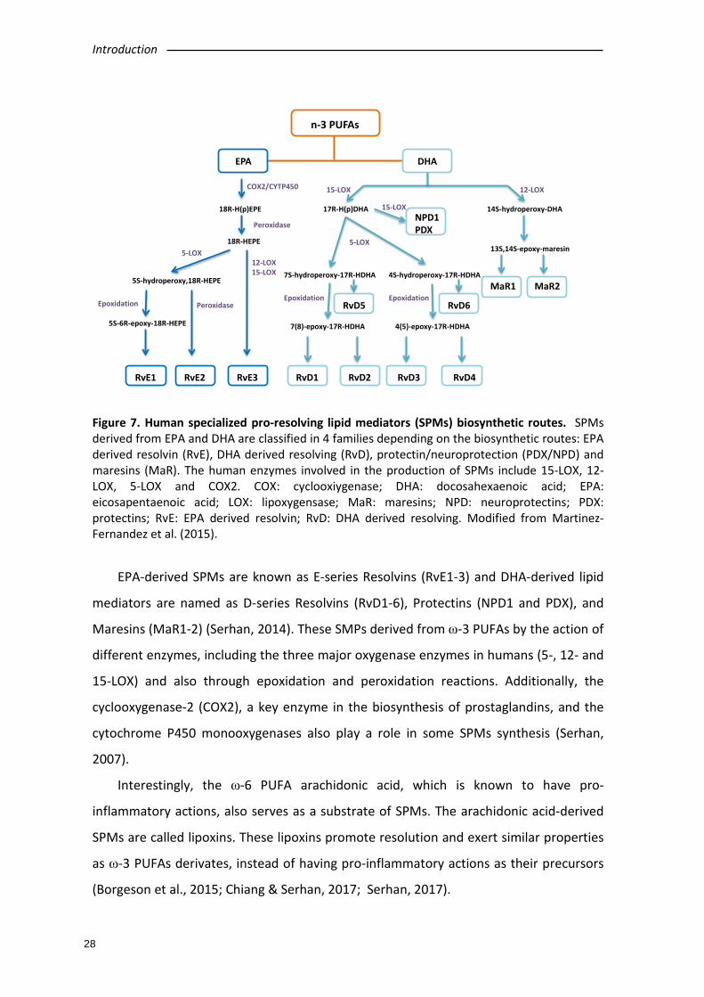
Introduction
28
Figure 7. Human specialized pro-resolving lipid mediators (SPMs) biosynthetic routes. SPMs derived from EPA and DHA are classified in 4 families depending on the biosynthetic routes: EPA derived resolvin (RvE), DHA derived resolving (RvD), protectin/neuroprotection (PDX/NPD) and maresins (MaR). The human enzymes involved in the production of SPMs include 15-LOX, 12-LOX, 5-LOX and COX2. COX: cyclooxiygenase; DHA: docosahexaenoic acid; EPA: eicosapentaenoic acid; LOX: lipoxygensase; MaR: maresins; NPD: neuroprotectins; PDX: protectins; RvE: EPA derived resolvin; RvD: DHA derived resolving. Modified from Martinez-Fernandez et al. (2015).
EPA-derived SPMs are known as E-series Resolvins (RvE1-3) and DHA-derived lipid
mediators are named as D-series Resolvins (RvD1-6), Protectins (NPD1 and PDX), and
Maresins (MaR1-2) (Serhan, 2014). These SMPs derived from ω-3 PUFAs by the action of
different enzymes, including the three major oxygenase enzymes in humans (5-, 12- and
15-LOX) and also through epoxidation and peroxidation reactions. Additionally, the
cyclooxygenase-2 (COX2), a key enzyme in the biosynthesis of prostaglandins, and the
cytochrome P450 monooxygenases also play a role in some SPMs synthesis (Serhan,
2007).
Interestingly, the ω-6 PUFA arachidonic acid, which is known to have pro-
inflammatory actions, also serves as a substrate of SPMs. The arachidonic acid-derived
SPMs are called lipoxins. These lipoxins promote resolution and exert similar properties
as ω-3 PUFAs derivates, instead of having pro-inflammatory actions as their precursors
(Borgeson et al., 2015; Chiang & Serhan, 2017; Serhan, 2017).
n-3 PUFAs
EPA DHA
18R-H(p)EPE
5S-hydroperoxy,18R-HEPE
5S-6R-epoxy-18R-HEPE
RvE1 RvE2
14S-hydroperoxy-DHA17R-H(p)DHA
13S,14S-epoxy-maresin
MaR1 MaR2
NPD1PDX
7S-hydroperoxy-17R-HDHA 4S-hydroperoxy-17R-HDHA
7(8)-epoxy-17R-HDHA 4(5)-epoxy-17R-HDHA
RvD1 RvD2 RvD3 RvD4
COX2/CYTP450
Epoxidation
5-LOX5-LOX
15-LOX
Epoxidation
12-LOX15-LOX
18R-HEPE
Peroxidase
RvD5Epoxidation
RvD6Peroxidase
RvE3
12-LOX15-LOX
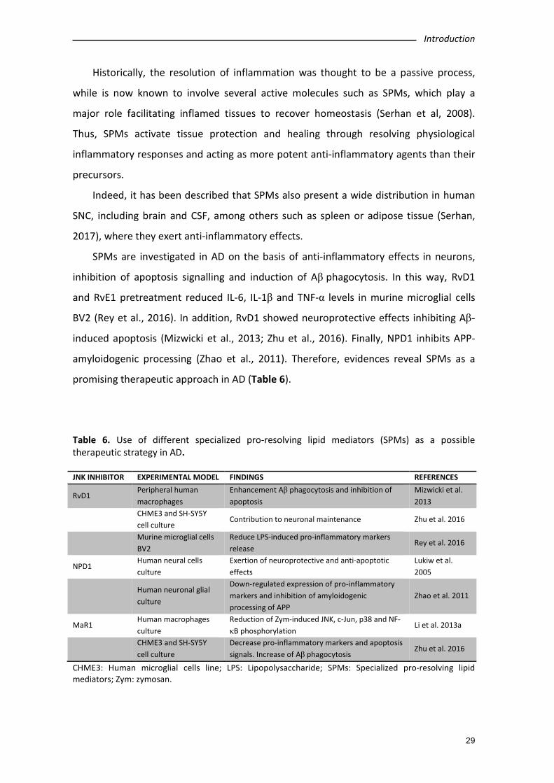
Introduction
29
Historically, the resolution of inflammation was thought to be a passive process,
while is now known to involve several active molecules such as SPMs, which play a
major role facilitating inflamed tissues to recover homeostasis (Serhan et al, 2008).
Thus, SPMs activate tissue protection and healing through resolving physiological
inflammatory responses and acting as more potent anti-inflammatory agents than their
precursors.
Indeed, it has been described that SPMs also present a wide distribution in human
SNC, including brain and CSF, among others such as spleen or adipose tissue (Serhan,
2017), where they exert anti-inflammatory effects.
SPMs are investigated in AD on the basis of anti-inflammatory effects in neurons,
inhibition of apoptosis signalling and induction of Aβ phagocytosis. In this way, RvD1
and RvE1 pretreatment reduced IL-6, IL-1β and TNF-α levels in murine microglial cells
BV2 (Rey et al., 2016). In addition, RvD1 showed neuroprotective effects inhibiting Aβ-
induced apoptosis (Mizwicki et al., 2013; Zhu et al., 2016). Finally, NPD1 inhibits APP-
amyloidogenic processing (Zhao et al., 2011). Therefore, evidences reveal SPMs as a
promising therapeutic approach in AD (Table 6).
Table 6. Use of different specialized pro-resolving lipid mediators (SPMs) as a possible therapeutic strategy in AD. JNK INHIBITOR EXPERIMENTAL MODEL FINDINGS REFERENCES
RvD1 Peripheral human macrophages
Enhancement Aβ phagocytosis and inhibition of apoptosis
Mizwicki et al. 2013
CHME3 and SH-SY5Y cell culture
Contribution to neuronal maintenance Zhu et al. 2016
Murine microglial cells BV2
Reduce LPS-induced pro-inflammatory markers release
Rey et al. 2016
NPD1 Human neural cells culture
Exertion of neuroprotective and anti-apoptotic effects
Lukiw et al. 2005
Human neuronal glial culture
Down-regulated expression of pro-inflammatory markers and inhibition of amyloidogenic processing of APP
Zhao et al. 2011
MaR1 Human macrophages culture
Reduction of Zym-induced JNK, c-Jun, p38 and NF-κB phosphorylation
Li et al. 2013a
CHME3 and SH-SY5Y cell culture
Decrease pro-inflammatory markers and apoptosis signals. Increase of Aβ phagocytosis
Zhu et al. 2016
CHME3: Human microglial cells line; LPS: Lipopolysaccharide; SPMs: Specialized pro-resolving lipid mediators; Zym: zymosan.

Introduction
30
4.2.1. Maresin 1 in Alzheimer’s disease
The “Macrophage mediator in resolving inflammation” (Maresins) are a family of
lipid mediators biosynthesized from DHA via 12-LOX through macrophages action.
Maresin 1 (7R,14S-dihydroxy-docosa-4Z,8E,10E,12Z,16Z,19Z-hexaenoic acid or MaR1),
was the first member of this family identified. The biosynthesis of MaR1 takes place
during inflammation resolution. In fact, one of the intermediate in MaR1 biosynthesis,
14S-hydroperoxydocosa-4Z,7Z,10Z,12E,16Z,19Z-hexaenoic acid (14-HpDHA) derived
from endogenous DHA has been reported to be accumulated in the exudates of mouse
peritonitis during resolution. Then, 14-HpDHA is converted to 13S,14S-epoxy-maresin
through epoxidation (Dalli et al. 2016). Finally, an enzymatic hydrolysis through
hydrolases converts 13S,14S-epoxy-maresin into MaR1. Maresins have been
predominantly detected as products synthetized by monocytes/macrophages upon
acute inflammatory response. MaR1 has been suggested to exert strong anti-
inflammatory effects and potent pro-resolutive actions (Serhan, 2009). Additionally, it
has been related to have pro-healing properties at similar extent to other members of
the ω-3 PUFAs-derived SPMs (Serhan et al., 2012; Tang et al., 2013).
Deficiencies in SPMs levels, particularly MaR1, in AD brain turns into a decrease in
the resolution of inflammation and deprivation of protective and restorative signals in
neurons, being able to contribute to AD pathogenesis. Decreased levels of MaR1 have
been observed in hippocampus and entorhinal cortex of AD cases (Zhu et al., 2016).
Importantly, as described above, MaR1 effects have been described related to AD
pathology (Table 6). MaR1 reduced neuronal apoptosis and improved cell survival in SH-
SY5Y neuroblastoma cells. Moreover, Aβ–induced microglial activation was
downregulated by MaR1, because pro-inflammatory markers decline, such as CD40 or
CD11. Additionally, MaR1 induced Aβ phagocytosis (Wang et al., 2015; Zhu et al., 2016).
The mechanism of action of MaR1 has not still been established, but its similar
structure to PD1 and RvD1 suggests that MaR1 might bind to peroxisome proliferator
associated receptor (PPAR-γ). PPAR-γ is a receptor for several different PUFAs and SPMs,
including DHA or RvD1, which was shown to be involved in neuroprotection. On the
other hand, supporting this idea, a study on human macrophages described that MaR1
and RvD1 decreased in zym-induced phosphorylation of JNK and p38, an effect

Introduction
31
mediated by NF-κB inhibition (Li et al. 2013a). Keeping in mind that Aβ increases the
phosphorylation of JNK and p38 in CHME3 microglial cells and taking into account that
ω-3 PUFAs are considered natural inhibitors of JNK, it is suggested that MaR1 could be a
potential compound for AD treatment (Table 6).


HYPOTHESIS AND AIMS


Hypothesis and aims
35
1. JUSTIFICATION FOR THE STUDY
AD is the main cause of dementia and there is currently no treatment. The
different clinical trials performed so far have failed and none of them have been able
to reverse or halt dementia in patients. For this reason, it is essential to propose new
strategies or therapeutic approaches for the disease (Scheltens et al., 2016).
In last years, different studies have demonstrated the involvement of MAPK
pathways in the mechanisms of neuronal death and neuroinflammation,
characteristics of neurodegenerative diseases, such as AD (Ferrer et al., 2001; Lin et al.,
2013). In this context, chronic neuroinflammation induces neuronal damage through
the activation of different signaling pathways. Among them, JNK activation could play
important role in neurodegenerative diseases. Changes in the brain of AD patients,
such as rupture of the BBB or Aβ aggregation, have been reported to induce JNK
activation (Zhang & Jiang, 2015).
Activation of JNK could lead to maturation of NFTs, formation of senile plaques
and mitochondrial stress. In addition pJNK produces neuroinflammation and activation
of caspases that induce apoptosis. Therefore, inhibition of JNK is postulated as a
promising therapeutic strategy that should be investigated in AD.
2. HYPOTHESIS
Based on all the observations previously mentioned, the hypothesis of the present
study is that an over-activation of JNK may be contributing to the neuropathology in
AD, and that JNK inhibition could constitute a new therapeutic target in AD.
3. AIMS
The main aim of the present study is to validate JNK as a biological target for the
diagnosis and treatment of AD, as well as to determine the ability of ω-3 PUFAs (DHA,
EPA and MaR1) to reverse the functional alterations derived from JNK activation in
animal models of AD.
Hypothesis and aims
36
To achieve the main objective, the proposed specific aims are:
1. To study the implication of JNK in the pathological processes of AD.
1.1. Analysis of pJNK levels in mouse models of AD.
Levels of activated JNK (pJNK) will be measured in a familiar type AD mouse
model (Tg2576) and a sporadic type AD mouse model (SAMP8).
1.2. Measurement of pJNK in AD and other dementias.
Levels of pJNK will be measured in post-mortem human samples of AD. The
selectivity of this activation will be assessed by studying pJNK levels in other
dementias (Lewy body dementia, frontotemporal dementia and vascular
dementia).
1.3. Characterization of pJNK as a potential new AD biomarker.
To achieve this aim, levels of pJNK will be analyzed in cerebrospinal fluid (CSF)
of control, mild cognitive impairment (MCI), progressive mild cognitive
impairment (PMCI) and AD.
2. To investigate the relationship between pJNK and Aβ in AD.
2.1. Measurement of Aβ levels and its relation to pJNK in AD murine models
To this end the same mouse models as in objective 1.1. will be used.
2.2. Assessment of Aβ levels in AD and other dementias.
To perform this aim Aβ levels and its relation to pJNK will be analyzed in the
same postmortem brain tissue described in aim 1.2.
2.3. Study of the co-localization of Aβ and pJNK.
To this end immunohistochemistry analysis will be performed in murine models
of AD and postmortem brain tissue.
Hypothesis and aims
37
2.4. Investigate the causal relationship between Aβ increase and pJNK
activation.
To achieve this aim Aβ42 will be ICV injected to wild type mice in order to study
the subsequently JNK activation.
3. Validate JNK inhibition as a treatment for AD.
3.1. Three JNK inhibitors of ω-3 PUFAs group (DHA, EPA and MaR1) will be
studied in order to assess in their ability to reverse the cognitive impairment
showed by the mouse model of sporadic AD (SAMP8).
3.2. To demonstrate the plausible mechanism underlying the beneficial effects
of ω-3 PUFAs on cognition. To achieve this aim, Tau hyperphosphorylation and
endoplasmic reticulum (ER) stress markers will be analyzed.


EXPERIMENTAL DESIGN AND METHODS

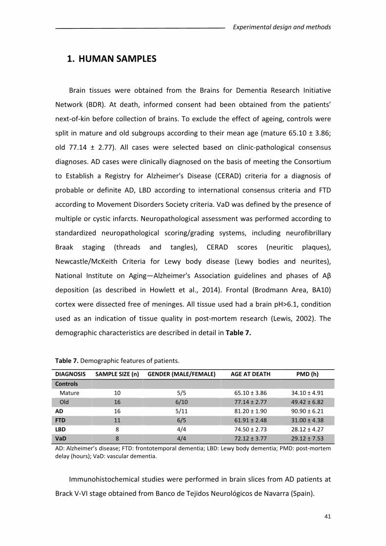
Experimental design and methods
41
1. HUMAN SAMPLES
Brain tissues were obtained from the Brains for Dementia Research Initiative
Network (BDR). At death, informed consent had been obtained from the patients’
next-of-kin before collection of brains. To exclude the effect of ageing, controls were
split in mature and old subgroups according to their mean age (mature 65.10 ± 3.86;
old 77.14 ± 2.77). All cases were selected based on clinic-pathological consensus
diagnoses. AD cases were clinically diagnosed on the basis of meeting the Consortium
to Establish a Registry for Alzheimer's Disease (CERAD) criteria for a diagnosis of
probable or definite AD, LBD according to international consensus criteria and FTD
according to Movement Disorders Society criteria. VaD was defined by the presence of
multiple or cystic infarcts. Neuropathological assessment was performed according to
standardized neuropathological scoring/grading systems, including neurofibrillary
Braak staging (threads and tangles), CERAD scores (neuritic plaques),
Newcastle/McKeith Criteria for Lewy body disease (Lewy bodies and neurites),
National Institute on Aging—Alzheimer's Association guidelines and phases of Aβ
deposition (as described in Howlett et al., 2014). Frontal (Brodmann Area, BA10)
cortex were dissected free of meninges. All tissue used had a brain pH>6.1, condition
used as an indication of tissue quality in post-mortem research (Lewis, 2002). The
demographic characteristics are described in detail in Table 7.
Table 7. Demographic features of patients.
DIAGNOSIS SAMPLE SIZE (n) GENDER (MALE/FEMALE) AGE AT DEATH PMD (h) Controls Mature 10 5/5 65.10 ± 3.86 34.10 ± 4.91 Old 16 6/10 77.14 ± 2.77 49.42 ± 6.82 AD 16 5/11 81.20 ± 1.90 90.90 ± 6.21 FTD 11 6/5 61.91 ± 2.48 31.00 ± 4.38 LBD 8 4/4 74.50 ± 2.73 28.12 ± 4.27 VaD 8 4/4 72.12 ± 3.77 29.12 ± 7.53 AD: Alzheimer’s disease; FTD: frontotemporal dementia; LBD: Lewy body dementia; PMD: post-mortem delay (hours); VaD: vascular dementia.
Immunohistochemical studies were performed in brain slices from AD patients at
Brack V-VI stage obtained from Banco de Tejidos Neurológicos de Navarra (Spain).
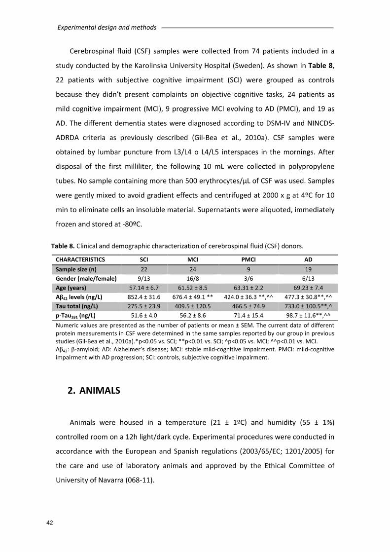
Experimental design and methods
42
Cerebrospinal fluid (CSF) samples were collected from 74 patients included in a
study conducted by the Karolinska University Hospital (Sweden). As shown in Table 8,
22 patients with subjective cognitive impairment (SCI) were grouped as controls
because they didn’t present complaints on objective cognitive tasks, 24 patients as
mild cognitive impairment (MCI), 9 progressive MCI evolving to AD (PMCI), and 19 as
AD. The different dementia states were diagnosed according to DSM-IV and NINCDS-
ADRDA criteria as previously described (Gil-Bea et al., 2010a). CSF samples were
obtained by lumbar puncture from L3/L4 o L4/L5 interspaces in the mornings. After
disposal of the first milliliter, the following 10 mL were collected in polypropylene
tubes. No sample containing more than 500 erythrocytes/µL of CSF was used. Samples
were gently mixed to avoid gradient effects and centrifuged at 2000 x g at 4ºC for 10
min to eliminate cells an insoluble material. Supernatants were aliquoted, immediately
frozen and stored at -80ºC.
Table 8. Clinical and demographic characterization of cerebrospinal fluid (CSF) donors.
CHARACTERISTICS SCI MCI PMCI AD Sample size (n) 22 24 9 19 Gender (male/female) 9/13 16/8 3/6 6/13 Age (years) 57.14 ± 6.7 61.52 ± 8.5 63.31 ± 2.2 69.23 ± 7.4 Aβ42 levels (ng/L) 852.4 ± 31.6 676.4 ± 49.1 ** 424.0 ± 36.3 **,^^ 477.3 ± 30.8**,^^ Tau total (ng/L) 275.5 ± 23.9 409.5 ± 120.5 466.5 ± 74.9 733.0 ± 100.5**,^ p-Tau181 (ng/L) 51.6 ± 4.0 56.2 ± 8.6 71.4 ± 15.4 98.7 ± 11.6**,^^ Numeric values are presented as the number of patients or mean ± SEM. The current data of different protein measurements in CSF were determined in the same samples reported by our group in previous studies (Gil-Bea et al., 2010a).*p<0.05 vs. SCI; **p<0.01 vs. SCI; ^p<0.05 vs. MCI; ^^p<0.01 vs. MCI. Aβ42: β-amyloid; AD: Alzheimer’s disease; MCI: stable mild-cognitive impairment. PMCI: mild-cognitive impairment with AD progression; SCI: controls, subjective cognitive impairment.
2. ANIMALS
Animals were housed in a temperature (21 ± 1ºC) and humidity (55 ± 1%)
controlled room on a 12h light/dark cycle. Experimental procedures were conducted in
accordance with the European and Spanish regulations (2003/65/EC; 1201/2005) for
the care and use of laboratory animals and approved by the Ethical Committee of
University of Navarra (068-11).

Experimental design and methods
43
2.1. Tg2576 mice
In the present study, 9- and 16-month-old Tg2576 AD transgenic mice with the
Swedish mutation and wild-type (WT) mice with the same background (C57BL/6J) were
used. The sample size was 34 mice distributed in four different groups: 9-months-old
WT mice (n=10), 9-months-old Tg2576 mice (n=9), 16-months-old WT mice (n=7) and
16-months-old Tg2576 mice (n=8). This AD mouse model over-expresses the human
APP, thus Aβ accumulation is exponentially associated to ageing. This mouse model
has a normal development, but age-associated cognitive deficits are found. Studies
have reported an impaired spatial learning and working memory by 9 months of age
(Hsiao et al., 1996). This fact coincides with Aβ deposition and formation of senile
plaques in time (Foley et al., 2015). However, NFTs and neuronal loss is absent in this
model (Irizarry et al., 1997). Gliosis is another hallmark of this murine model: microglial
density is increased in the hippocampus and frontal and occipital cortex from 10
months old ( Figure 8; Frautschy et al., 1998).
2.2. SAMP8 mice
As sporadic AD murine model, the SAMP8 mouse model developed by professor
Takeda from the department of pathology of Kyoto’s University has been chosen. The
Jackson Laboratory (Bar Harbor, Maine, USA) donated several pairs of AKR/J mice to
professor Takeda, some of which showed features of an early senescence after sister-
brother mating continued: loss of activity, alopecia and lack of glossiness, skin
coarseness, periophthalmic lesions, increased lordokyphosis of the spine and a
shortened life span, despite the relatively low incidence of thymic lymphoma. These
litters were selected as progenitors of the “senescence-prone” (P series) and the rest
of the litters were selected as “senescence-resistant” (R series) progenitors. Eleven
senescence-prone subtypes of mice and seven senescence-resistant strains were
obtained from the different P and R series, respectively. Life spam from the SAMP
ranges from 10 to 17 months and is markedly shorter tan SAMR, which ranges from 19
months to 21 months. Due to its phenotypical characteristics, SAMP8 mice have
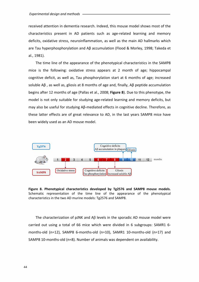
Experimental design and methods
44
received attention in dementia research. Indeed, this mouse model shows most of the
characteristics present in AD patients such as age-related learning and memory
deficits, oxidative stress, neuroinflammation, as well as the main AD hallmarks which
are Tau hyperphosphorylation and Aβ accumulation (Flood & Morley, 1998; Takeda et
al., 1981).
The time line of the appearance of the phenotypical characteristics in the SAMP8
mice is the following: oxidative stress appears at 2 month of age; hippocampal
cognitive deficit, as well as, Tau phosphorylation start at 6 months of age; increased
soluble Aβ , as well as, gliosis at 8 months of age and, finally, Aβ peptide accumulation
begins after 12 months of age (Pallas et al., 2008; Figure 8). Due to this phenotype, the
model is not only suitable for studying age-related learning and memory deficits, but
may also be useful for studying Aβ-mediated effects in cognitive decline. Therefore, as
these latter effects are of great relevance to AD, in the last years SAMP8 mice have
been widely used as an AD mouse model.
Figure 8. Phenotypical characteristics developed by Tg2576 and SAMP8 mouse models. Schematic representation of the time line of the appearance of the phenotypical characteristics in the two AD murine models: Tg2576 and SAMP8.
The characterization of pJNK and Aβ levels in the sporadic AD mouse model were
carried out using a total of 66 mice which were divided in 6 subgroups: SAMR1 6-
months-old (n=12), SAMP8 6-months-old (n=10), SAMR1 10-months-old (n=17) and
SAMP8 10-months-old (n=8). Number of animals was dependent on availability.

Experimental design and methods
45
In order to study the ability of ω-3 PUFAs (DHA, EPA and MaR1) to reverse the
functional alterations derived from JNK activation, 9-months-old SAMP8 and SAMR1
mice were chosen, divided in different groups depending on the treatment: SAMR1
Saline (n=10), SAMP8 Saline (n=8), SAMP8 MaR1 (n=8), SAMP8 DHA (n=8) and SAMP8
EPA (n=8).
Mice were orally treated with 200 mg/kg of DHA, 200 mg/kg of EPA and 25 µg/kg
of MaR1 for 20 days. The drugs were purchased from Cayman Chemical (Michigan,
USA). Behavioral test carried out with these animals are described in section 4.
3. Aβ INTRACEREBROVENTRICULAR INJECTION IN WT MICE
ICV injection of Aβ was performed in 14 WT (C57BL/6N) mice of 9-months-old
divided in two groups (Sham and Aβ injected) in order to study the causal relationship
between pJNK and amyloidogenesis.
Aβ42 peptide was purchased by Bachem laboratories (Bubendorf, Switzerland). For
assuring monomerization, 1 mg of Aβ42 protein was dissolved to 1 mM in
hexafluoroisopropanol (HFIP) and aliquoted into microcentrifuge tubes, then the HFIP
was evaporated, and the peptides were stored at −20°C until use. For oligomeric
assembly, concentrated peptides were suspended in DMSO and then diluted to 100
μM in PBS and incubated at 4°C for 24 h. ICV injection of Aβ42 (300 pmol in 3 µL sterile
dH2O containing 10% DMSO) was stereotaxically performed in both lateral ventricles of
9 month-old-male WT mice (anterior–posterior, +0.3 mm; lateral, 1.0 mm; horizontal,
3.0 mm from the bregma). Sham animals received equivalent amounts of sterile
phosphate buffer saline. Mice were sacrificed 7 days after the injection.
4. BEHAVIORAL TEST
Behavioral experiments were conducted between 09:00 h and 13:00 h. Observers
were blind to the treatment. All behavioral tests were carried out in the same cohort
of animals (Figure 9).
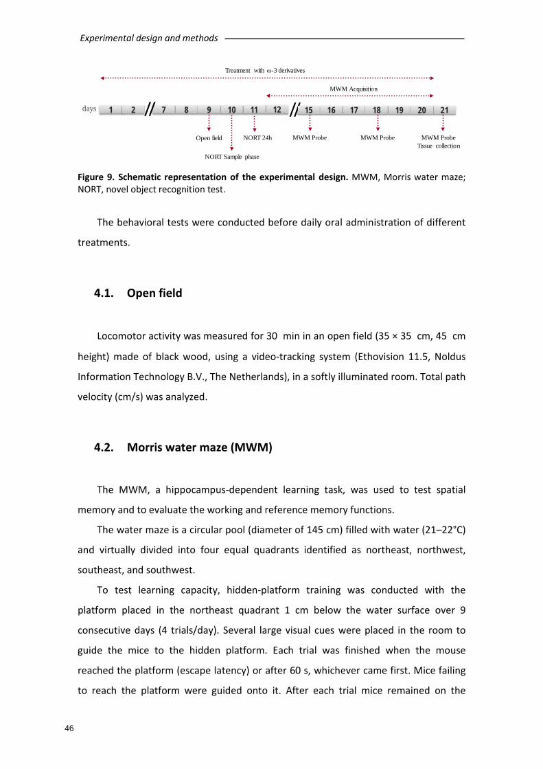
Experimental design and methods
46
Figure 9. Schematic representation of the experimental design. MWM, Morris water maze; NORT, novel object recognition test.
The behavioral tests were conducted before daily oral administration of different
treatments.
4.1. Open field
Locomotor activity was measured for 30 min in an open field (35 × 35 cm, 45 cm
height) made of black wood, using a video-tracking system (Ethovision 11.5, Noldus
Information Technology B.V., The Netherlands), in a softly illuminated room. Total path
velocity (cm/s) was analyzed.
4.2. Morris water maze (MWM)
The MWM, a hippocampus-dependent learning task, was used to test spatial
memory and to evaluate the working and reference memory functions.
The water maze is a circular pool (diameter of 145 cm) filled with water (21–22°C)
and virtually divided into four equal quadrants identified as northeast, northwest,
southeast, and southwest.
To test learning capacity, hidden-platform training was conducted with the
platform placed in the northeast quadrant 1 cm below the water surface over 9
consecutive days (4 trials/day). Several large visual cues were placed in the room to
guide the mice to the hidden platform. Each trial was finished when the mouse
reached the platform (escape latency) or after 60 s, whichever came first. Mice failing
to reach the platform were guided onto it. After each trial mice remained on the
1 2 217 8 9 10 11 16 17days 18
NORT Sample phase
Open field NORT 24h
// 19 20
MWM ProbeTissue collection
MWM Acquisition
12 15
MWM Probe MWM Probe
//
Treatment with ω-3 derivatives

Experimental design and methods
47
platform for 15 s. To test memory, probe trials were performed at the 4th, 7th and last
day of the test (10th day). In the probe trials the platform was removed from the pool
and mice were allowed to swim for 60 s. The percentage of time spent in the target
quadrant was recorded. All trials were monitored by a video camera set above the
center of the pool and connected to a video tracking system (Ethovision 11.5; Noldus
Information Technology B.V, Wageningen, Netherlands).
4.3. Novel object recognition test (NORT)
The open field consisted of a square divided into four sections (35 cm × 35 cm ×
45 cm each) with black walls. On the previous day to the experiment, animals were
familiarized with the square for 30 min. The test consists of 2 trials of 5 min: sample
phase and 24 h trial. During the first trial, two identical objects were placed inside the
cubicle, and the mice were allowed to explore. The second task took place 24 h later in
which one object was replaced by another and the exploration time was recorded for 5
min. Results were expressed as percentage of time spent exploring the new object
with respect to the total exploration time (discrimination index). It is important to
highlight that the exploration was considered complete when the nose of the mouse
was oriented within 2 cm of the object. This behavioral test was carried out using
video-tracking system (Ethovision 11.5; Noldus Information Technology B.V,
Wageningen, Netherlands).
5. BIOCHEMICAL MEASUREMENTS
5.1. Tissue collection
Mice were sacrificed by decapitation between 09:00–12:00 h. Brains were
removed and dissected on ice to obtain the hippocampus and frontal cortex and
stored at −80°C. For immunohistochemistry assays, left hemispheres from 5 mice per
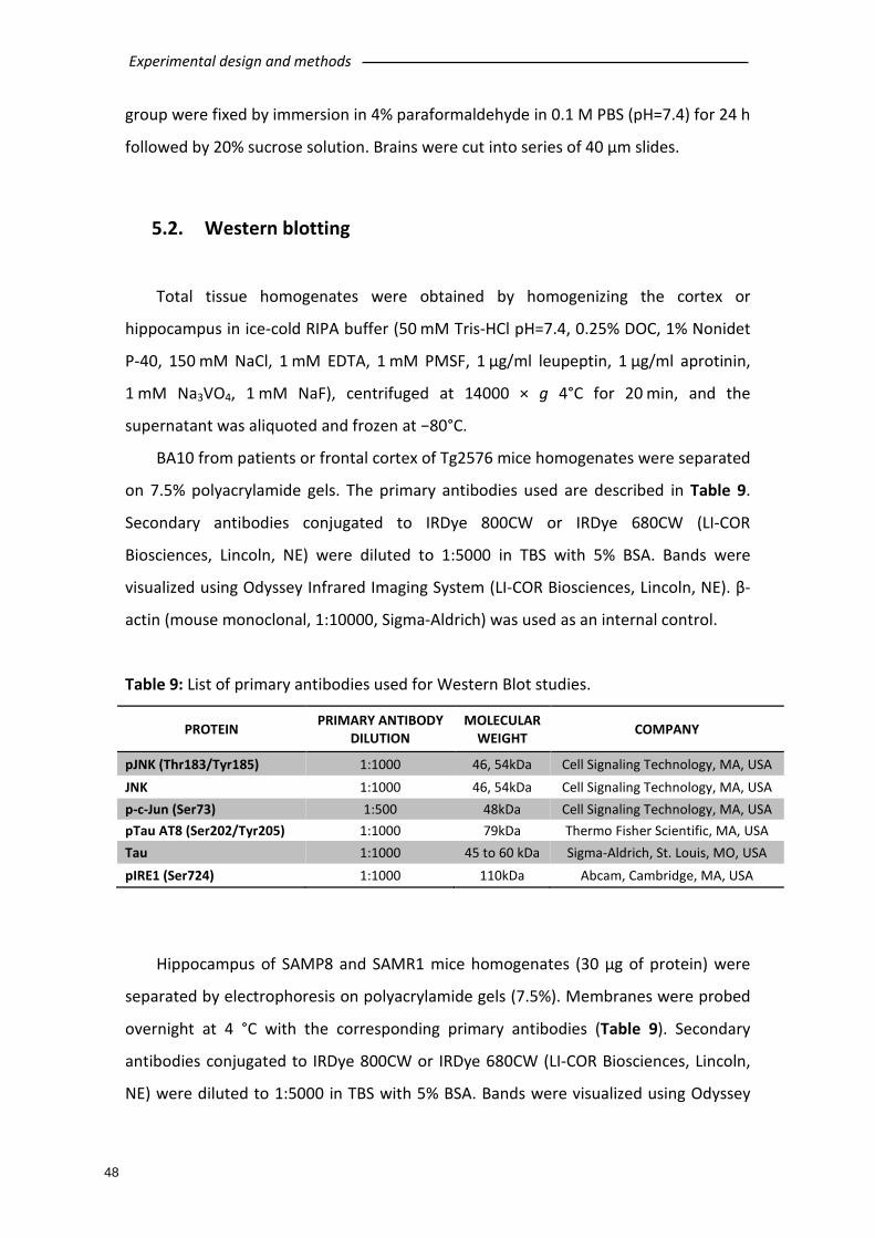
Experimental design and methods
48
group were fixed by immersion in 4% paraformaldehyde in 0.1 M PBS (pH=7.4) for 24 h
followed by 20% sucrose solution. Brains were cut into series of 40 μm slides.
5.2. Western blotting
Total tissue homogenates were obtained by homogenizing the cortex or
hippocampus in ice-cold RIPA buffer (50 mM Tris-HCl pH=7.4, 0.25% DOC, 1% Nonidet
P-40, 150 mM NaCl, 1 mM EDTA, 1 mM PMSF, 1 μg/ml leupeptin, 1 μg/ml aprotinin,
1 mM Na3VO4, 1 mM NaF), centrifuged at 14000 × g 4°C for 20 min, and the
supernatant was aliquoted and frozen at −80°C.
BA10 from patients or frontal cortex of Tg2576 mice homogenates were separated
on 7.5% polyacrylamide gels. The primary antibodies used are described in Table 9.
Secondary antibodies conjugated to IRDye 800CW or IRDye 680CW (LI-COR
Biosciences, Lincoln, NE) were diluted to 1:5000 in TBS with 5% BSA. Bands were
visualized using Odyssey Infrared Imaging System (LI-COR Biosciences, Lincoln, NE). β-
actin (mouse monoclonal, 1:10000, Sigma-Aldrich) was used as an internal control.
Table 9: List of primary antibodies used for Western Blot studies.
PROTEIN PRIMARY ANTIBODY DILUTION
MOLECULAR WEIGHT COMPANY
pJNK (Thr183/Tyr185) 1:1000 46, 54kDa Cell Signaling Technology, MA, USA JNK 1:1000 46, 54kDa Cell Signaling Technology, MA, USA p-c-Jun (Ser73) 1:500 48kDa Cell Signaling Technology, MA, USA pTau AT8 (Ser202/Tyr205) 1:1000 79kDa Thermo Fisher Scientific, MA, USA Tau 1:1000 45 to 60 kDa Sigma-Aldrich, St. Louis, MO, USA pIRE1 (Ser724) 1:1000 110kDa Abcam, Cambridge, MA, USA
Hippocampus of SAMP8 and SAMR1 mice homogenates (30 μg of protein) were
separated by electrophoresis on polyacrylamide gels (7.5%). Membranes were probed
overnight at 4 °C with the corresponding primary antibodies (Table 9). Secondary
antibodies conjugated to IRDye 800CW or IRDye 680CW (LI-COR Biosciences, Lincoln,
NE) were diluted to 1:5000 in TBS with 5% BSA. Bands were visualized using Odyssey
Experimental design and methods
49
Infrared Imaging System (LI-COR Biosciences, Lincoln, NE). β-actin (mouse monoclonal,
1:10000, Sigma-Aldrich) was used as an internal control.
For the visualization of ICV injected Aβ oligomers, increasing concentrations (1,
2.5, 5, 8 and 10 μM) of previously aggregated Aβ42 solution was subjected to SDS-PAGE
electrophoresis in non-termal denatured conditions (samples were not boiled before
loading). The separated proteins were transferred to nitrocellulose membranes for
determining the presence of different Aβ aggregates with 6E10 as primary antibody
(Abcam, Cambridge, MA, USA). Blots were incubated and revealed under the
conditions previously described.
5.3. Measurement of Aβ levels
In human samples and SAMP8 mice samples, Aβ42 levels were determined using a
commercially available high-sensitive ELISA kit (Wako Pure Chemical Industries, Tokyo,
Japan) following manufacturer instructions.
In Tg2576 mice samples, human Aβ42 levels were measured using a commercially
available ultra-sensitive ELISA kit (Thermo Fisher Scientific Waltham, MA, USA)
following manufacturer instructions.
5.4. Measurement of pJNK levels in cerebrospinal fluid (CSF)
pJNK levels were determined in CSF of 74 patients using a commercially available
JNK (pT183/Y185 + Total) ELISA kit (Abcam, Cambridge, UK) with several modifications
implemented by our group in order to reach the detection limits.
In order to obtain detectable pJNK levels, CSF samples were concentrated 3 times
by centrifugal vacuum. Savant Speed Vac is a concentrator equipment that removes
solvents from the samples leading to higher sample concentration. For our
experiment, 60 μL of CSF samples were concentrated during 55 min at 43ºC avoiding
protein denaturation due to high temperatures. Concentrated CSF samples were
subjected to the ELISA kit above mentioned, following manufacturer instructions.

Experimental design and methods
50
5.5. Immunofluorescence staining
For immunofluorescence, free-floating brain sections were washed (3 × 10 min)
with PBS 0.1 M (pH=7.4) and incubated in blocking solution (PBS containing 0.3%
Triton X-100, 0.1% BSA, and 2% normal donkey serum) for 2 h at room temperature.
For 6E10 immunostaining, sections were incubated in 70% formic acid for 7 min to
expose the epitope. Primary and secondary antibodies were diluted in the blocking
solution. Sections were incubated with the primary antibody overnight at 4°C, washed
with PBS and incubated with the secondary antibody for 2 h at room temperature,
protected from light. The primary antibodies used were anti-pJNK (rabbit monoclonal,
1:250, Cell Signaling Technology, Beverly, MA) and 6E10 (mouse monoclonal, 1:250,
Covance, Princeton, NJ). Secondary antibodies used were Alexa Fluor 488 Donkey anti-
rabbit and Alexa Fluor 546 Donkey anti-mouse (1:200, Invitrogen-Molecular Probes,
Eugene, OR).
In order to develop the human brain sections immunofluorescence, our group set
up the paraffin embedded brain sections staining. Slides were dewaxed, washing with
xilol, decreasing concentrations of ethanol (100%, 90%, 70%) and water during 5 min
every time. In order to block the endogenous peroxidase activity, sections were
washed with hydrogen peroxide 3% during 5 min at 37ºC and, then, immersed in dH2O
(2 x 5 min) and 0,1M PBS pH=7,4 (2 x 5min).
With the purpose of antigen retrieval, slides were treated with 0,01 M citrate
buffer pH=6, microwaved 2 min and, then, washed with dH2O (2 x 5 min) and 0,1 M
PBS pH=7.4 (2 x 5 min). After, they were incubated with blocking solution (PBS
containing 0.3% Triton X-100 0.1% BSA and 2% normal donkey serum) for 1 h at room
temperature. Primary and secondary antibodies were diluted in the blocking solution.
Sections were incubated with the primary antibody overnight at 4ºC, washed with PBS
and incubated with the secondary antibody for 2 h at room temperature, protected
from light. Finally, black sudan treatment was applied on brain sections and they were
washed with 70% ethanol 1 min and dH2O (3 x 5 min).
The primary antibodies employed were anti-pJNK (rabbit monoclonal, 1:250, Cell
Signaling Technology, Beverly, MA) and 6E10 (mouse monoclonal, 1:250, Covance,
Princeton, NJ). Secondary antibodies used were Alexa Fluor 488 Donkey anti-rabbit
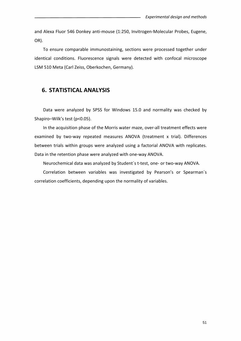
Experimental design and methods
51
and Alexa Fluor 546 Donkey anti-mouse (1:250, Invitrogen-Molecular Probes, Eugene,
OR).
To ensure comparable immunostaining, sections were processed together under
identical conditions. Fluorescence signals were detected with confocal microscope
LSM 510 Meta (Carl Zeiss, Oberkochen, Germany).
6. STATISTICAL ANALYSIS
Data were analyzed by SPSS for Windows 15.0 and normality was checked by
Shapiro–Wilk’s test (p<0.05).
In the acquisition phase of the Morris water maze, over-all treatment effects were
examined by two-way repeated measures ANOVA (treatment x trial). Differences
between trials within groups were analyzed using a factorial ANOVA with replicates.
Data in the retention phase were analyzed with one-way ANOVA.
Neurochemical data was analyzed by Student´s t-test, one- or two-way ANOVA.
Correlation between variables was investigated by Pearson’s or Spearman´s
correlation coefficients, depending upon the normality of variables.


RESULTS

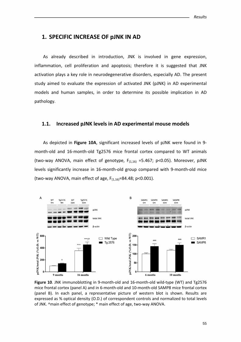
Results
55
1. SPECIFIC INCREASE OF pJNK IN AD
As already described in introduction, JNK is involved in gene expression,
inflammation, cell proliferation and apoptosis; therefore it is suggested that JNK
activation plays a key role in neurodegenerative disorders, especially AD. The present
study aimed to evaluate the expression of activated JNK (pJNK) in AD experimental
models and human samples, in order to determine its possible implication in AD
pathology.
1.1. Increased pJNK levels in AD experimental mouse models
As depicted in Figure 10A, significant increased levels of pJNK were found in 9-
month-old and 16-month-old Tg2576 mice frontal cortex compared to WT animals
(two-way ANOVA, main effect of genotype, F(1,16) =5.467; p<0.05). Moreover, pJNK
levels significantly increase in 16-month-old group compared with 9-month-old mice
(two-way ANOVA, main effect of age, F(1,16)=84.48; p<0.001).
Figure 10. JNK immunoblotting in 9-month-old and 16-month-old wild-type (WT) and Tg2576 mice frontal cortex (panel A) and in 6-month-old and 10-month-old SAMP8 mice frontal cortex (panel B). In each panel, a representative picture of western blot is shown. Results are expressed as % optical density (O.D.) of correspondent controls and normalized to total levels of JNK. ^main effect of genotype; * main effect of age, two-way ANOVA.

Results
56
In the other mouse model analyzed, a significant increase in pJNK levels in SAMP8
mice in all ages was observed (two-way ANOVA, main effect of genotype, F(1,28)= 14.08;
p<0.001; Figure 10B).
Therefore, pJNK is increased in both AD experimental mouse models used.
1.2. Specific increases in pJNK levels in AD human samples
In order to confirm that the increase in JNK activation observed in both AD mouse
models is also observed in AD patients, pJNK levels were measured in frontal cortex
(BA10) of post-mortem samples.
The demographic characteristics of brain samples used were detailed in Table 7.
There were significant differences between age at death among the different
pathological conditions (one-way ANOVA; F(3,42)=11.065, p<0.001), being AD cases
older than the rest of the groups (p<0.01). In addition, it is to note that in controls,
pJNK expression correlated with age (Pearson’s, r=0.435; p<0.05, n=26). In order to
match for age in all pathological conditions, two subset of controls were used, one
being considered mature controls (age at death=65.1±3.86, n=10), being these the
samples used as controls for the FTD, VaD and LBD samples, and the other group was
named old controls (age at death=77.14±2.77, n=16) and was used as control for AD
samples. There were no statistical differences regarding sex or post-mortem delay
between groups.
Significant increases in pJNK levels were seen (Student t test, p<0.05) in the frontal
cortex of patients with AD compared with controls (Figure 11A). Total levels of JNK did
not differ between controls and AD group. pJNK levels in AD did not differ by sex
(male/female, Student t test, p>0.05).
Based in the literature, increases in pJNK levels could be considered a common
denominator for all types of dementia. However, our results do not support this
possible hypothesis because pJNK levels were similar (one-way ANOVA; F(3,31)= 1.210;
p>0.05) in all dementia groups (LBD, FTD or VaD) compared to control samples (Figure
11B). Total levels of JNK did not differ from controls in any of the pathological groups.
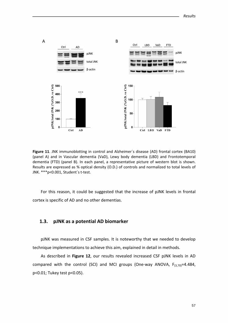
Results
57
Figure 11. JNK immunoblotting in control and Alzheimer´s disease (AD) frontal cortex (BA10) (panel A) and in Vascular dementia (VaD), Lewy body dementia (LBD) and Frontotemporal dementia (FTD) (panel B). In each panel, a representative picture of western blot is shown. Results are expressed as % optical density (O.D.) of controls and normalized to total levels of JNK. ***p<0.001, Student´s t-test.
For this reason, it could be suggested that the increase of pJNK levels in frontal
cortex is specific of AD and no other dementias.
1.3. pJNK as a potential AD biomarker
pJNK was measured in CSF samples. It is noteworthy that we needed to develop
technique implementations to achieve this aim, explained in detail in methods.
As described in Figure 12, our results revealed increased CSF pJNK levels in AD
compared with the control (SCI) and MCI groups (One-way ANOVA, F(3,70)=4.484,
p<0.01; Tukey test p<0.05).
Results
58
Figure 12. CSF levels of pJNK in patients diagnosed with clinical and prodromal stages of AD. SCI: Controls, subjective cognitive impairment; MCI: Stable mild- cognitive impairment; PMCI: Mild-cognitive impairment with AD progression; AD: Alzheimer’s disease. Data are expressed as total levels of pJNK as optical density (O.D.). *p<0.05 vs. SCI, ^p<0.05 vs. MCI; one-way ANOVA; Tukey p<0.05.
2. RELATIONSHIP BETWEEN pJNK AND Aβ LEVELS IN AD
2.1. Aβ levels in mice models of AD
As it has been described before, both AD mouse models (Tg2576 and SAMP8)
assessed in the present work show most of the characteristics present in AD patients,
such as Aβ peptide accumulation, one of the main AD hallmarks. Aβ peptide originates
from the proteolysis of APP and it has the ability to accumulate in Tg2576 model while
it remains soluble in SAMP8 mice without aggregation.
As depicted in Figure 13A, significant increases in Aβ42 levels were observed in 9-
month-old and 16-month-old Tg2576 mice frontal cortex compared to wild-type
animals (two-way ANOVA, main effect of genotype, F(1,22) =181.2; p<0.001). Moreover,
it was observed that this increase follows an age fashion as Aβ42 levels are significantly
higher in 16-month-old mice with respect to 9-month-old mice (two-way ANOVA, main
effect of age, F(1,22) =12.04; p<0.01). Aβ levels in 9-months-old WT were 7.18 ± 1.13
pg/mL.

Results
59
Figure 13. Aβ levels in mouse models of AD. In panel A, Aβ42 levels in 9-month-old and 16-month-old wild-type (WT) and Tg2576 mice frontal cortex. ^main effect of genotype; *main effect of age; two-way ANOVA. In panel B, Aβ42 levels in 6-month-old and 10-month-old SAMR1 and SAMP8 mice frontal cortex. ^vs. 6-month-old SAMR1; *vs. 6-month-old SAMP8; two-way ANOVA; Tukey p<0.05. Levels of Aβ42 are expressed as % versus its corresponding control.
Interestingly, regarding Aβ42 levels in sporadic mouse model of AD, significant
interaction between age and strain was observed (two-way ANOVA, F(1,27) =11.41;
p<0.01; Figure 13B). Further analysis showed increased levels of Aβ peptides in 10-
month-old SAMP8 mice compared to the rest of the groups (Tukey, p<0.001) and 6-
month-old SAMP8 mice in comparison with 6-month-old SAMR1 mice (Tukey, p<0.01).
Aβ levels in 6-month-old SAMR1 were 6.40 ± 2.02 pg/mL.
Therefore, the increase observed in pJNK and Aβ levels present a very similar
pattern in both AD mouse models, reinforcing the possible relationship between pJNK
and Aβ in AD pathology.
2.2. Aβ levels in human samples of AD and other dementias
Aβ42 levels were significantly increased in AD cases compared to controls (Student
t-test p<0.05; Figure 14A) and not in other dementias (one-way ANOVA; F(3,36)=1.210,
p>0.05; Figure 14B).
There was a significant correlation between enhanced pJNK expression in BA10
and Aβ42 levels (Spearman´s rho = 0.733, p<0.05, n=16) in AD. Interestingly, no
correlation was found between pJNK expression in BA10 and Aβ42 levels in any other

Results
60
type of dementia (Spearman´s rho = 0.143, p>0.05, Spearman´s rho = -0.143, p>0.05
and Spearman´s rho = 0.405, p>0.05 for LBD, FTD and VaD, respectively).
Figure 14. Aβ levels in Alzheimer´s disease (AD) cases (panel A) and in Vascular dementia (VaD), Lewy body dementia (LBD) and Frontotemporal dementia (FTD) (panel B). Levels of Aβ42 are expressed as % versus its corresponding controls (Ctrl). *p<0.05, Student´s t-test.
These observations allow us to propose the positive correlation among pJNK levels
and Aβ as a characteristic exclusive of the pathology of AD, underlining the similarity
between the experimental mouse models and the pathogenesis in patients.
2.3. pJNK and Aβ co-localize in AD human samples and in Tg2576
mouse model
In order to deepen in the understanding of the positive correlation between pJNK
and Aβ levels, co-localization studies of both compounds in frontal cortex from
patients and AD mouse model brains were performed. Co-localization studies were not
performed in SAMP8 mouse model because murine Aβ does not accumulate in plaques
and are not detectable in immunohistochemical analysis.
Our inmunohistochemical results revealed that pJNK in Tg2576 mice brains was
detected around the amyloid plaque. In matched aged control mice, pJNK
immunolabelling was not seen (Figure 15).
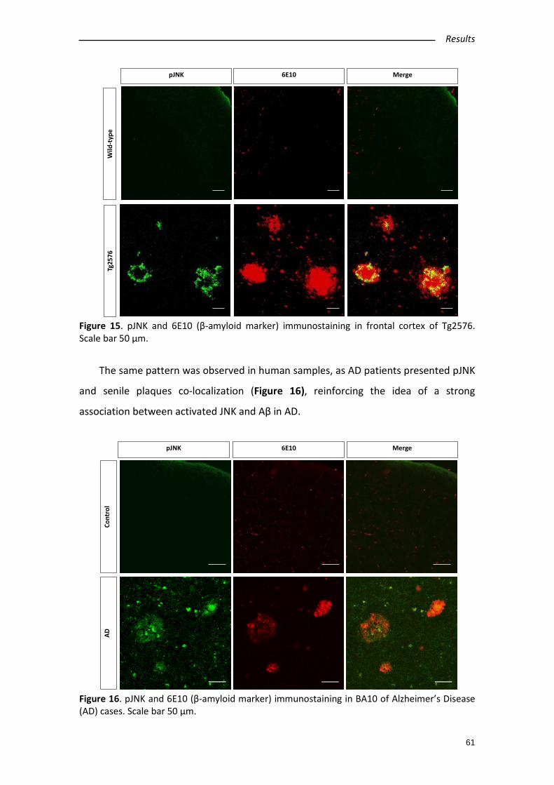
Results
61
Figure 15. pJNK and 6E10 (β-amyloid marker) immunostaining in frontal cortex of Tg2576. Scale bar 50 µm.
The same pattern was observed in human samples, as AD patients presented pJNK
and senile plaques co-localization (Figure 16), reinforcing the idea of a strong
association between activated JNK and Aβ in AD.
Figure 16. pJNK and 6E10 (β-amyloid marker) immunostaining in BA10 of Alzheimer’s Disease (AD) cases. Scale bar 50 µm.
pJNK 6E10 Merge
Tg25
76W
ild-t
ype
6E10pJNK Merge
Cont
rol
AD
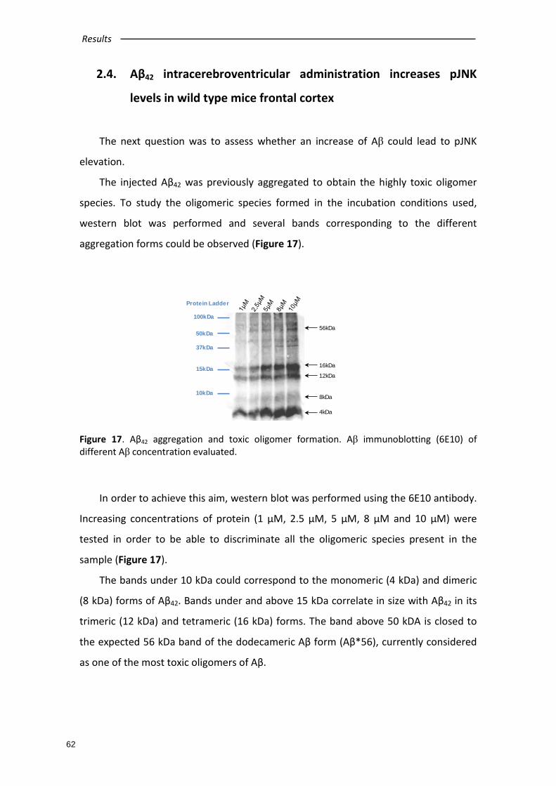
Results
62
Protein Ladder
10kDa
15kDa
37kDa
50kDa
100kDa
4kDa
8kDa
12kDa
16kDa
56kDa
2.4. Aβ42 intracerebroventricular administration increases pJNK
levels in wild type mice frontal cortex
The next question was to assess whether an increase of Aβ could lead to pJNK
elevation.
The injected Aβ42 was previously aggregated to obtain the highly toxic oligomer
species. To study the oligomeric species formed in the incubation conditions used,
western blot was performed and several bands corresponding to the different
aggregation forms could be observed (Figure 17).
Figure 17. Aβ42 aggregation and toxic oligomer formation. Aβ immunoblotting (6E10) of different Aβ concentration evaluated.
In order to achieve this aim, western blot was performed using the 6E10 antibody.
Increasing concentrations of protein (1 μM, 2.5 μM, 5 μM, 8 μM and 10 μM) were
tested in order to be able to discriminate all the oligomeric species present in the
sample (Figure 17).
The bands under 10 kDa could correspond to the monomeric (4 kDa) and dimeric
(8 kDa) forms of Aβ42. Bands under and above 15 kDa correlate in size with Aβ42 in its
trimeric (12 kDa) and tetrameric (16 kDa) forms. The band above 50 kDA is closed to
the expected 56 kDa band of the dodecameric Aβ form (Aβ*56), currently considered
as one of the most toxic oligomers of Aβ.
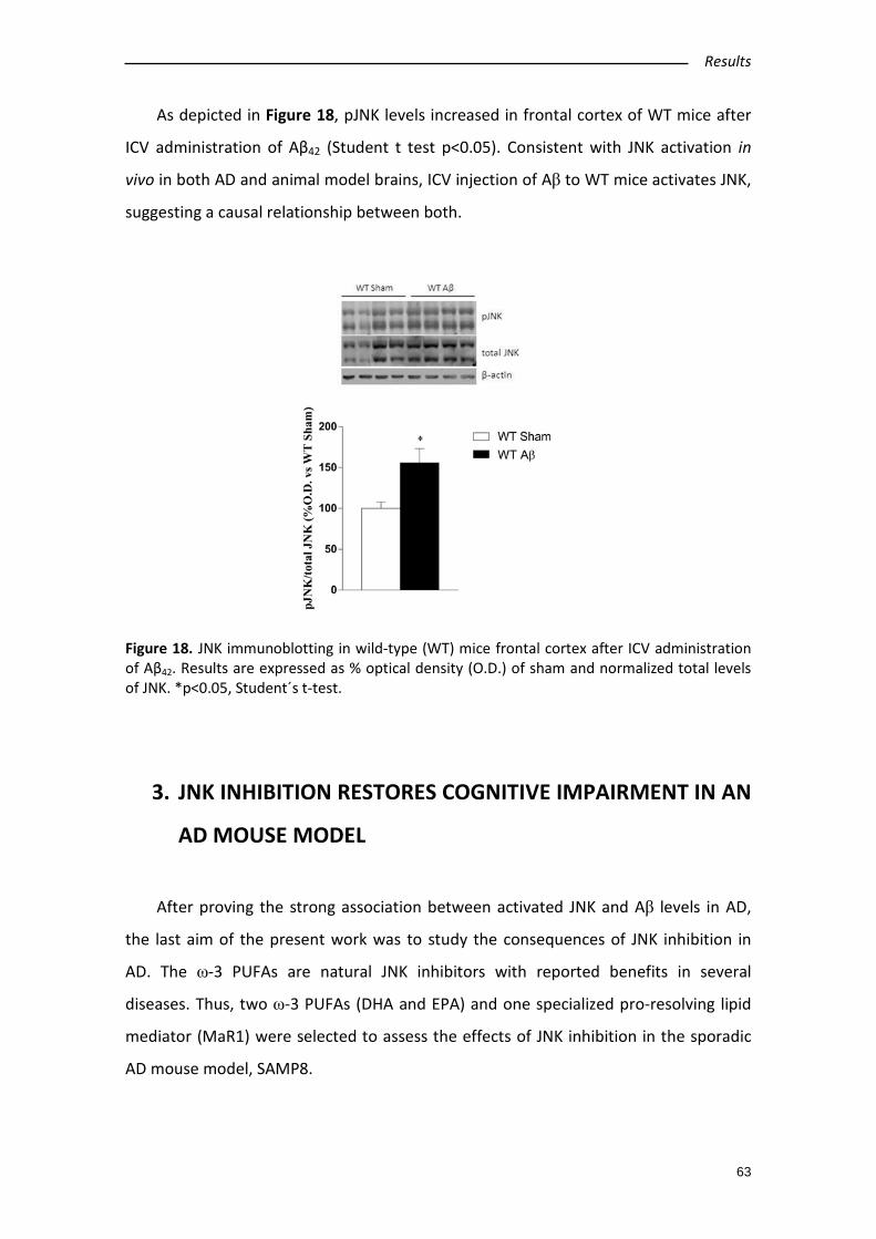
Results
63
As depicted in Figure 18, pJNK levels increased in frontal cortex of WT mice after
ICV administration of Aβ42 (Student t test p<0.05). Consistent with JNK activation in
vivo in both AD and animal model brains, ICV injection of Aβ to WT mice activates JNK,
suggesting a causal relationship between both.
Figure 18. JNK immunoblotting in wild-type (WT) mice frontal cortex after ICV administration of Aβ42. Results are expressed as % optical density (O.D.) of sham and normalized total levels of JNK. *p<0.05, Student´s t-test.
3. JNK INHIBITION RESTORES COGNITIVE IMPAIRMENT IN AN
AD MOUSE MODEL
After proving the strong association between activated JNK and Aβ levels in AD,
the last aim of the present work was to study the consequences of JNK inhibition in
AD. The ω-3 PUFAs are natural JNK inhibitors with reported benefits in several
diseases. Thus, two ω-3 PUFAs (DHA and EPA) and one specialized pro-resolving lipid
mediator (MaR1) were selected to assess the effects of JNK inhibition in the sporadic
AD mouse model, SAMP8.
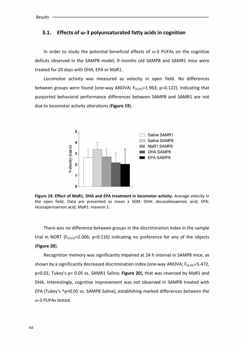
Results
64
3.1. Effects of ω-3 polyunsaturated fatty acids in cognition
In order to study the potential beneficial effects of ω-3 PUFAs on the cognitive
deficits observed in the SAMP8 model, 9 months old SAMP8 and SAMR1 mice were
treated for 20 days with DHA, EPA or MaR1.
Locomotor activity was measured as velocity in open field. No differences
between groups were found (one-way ANOVA; F(4,41)=1.963; p=0.122), indicating that
purported behavioral performance differences between SAMP8 and SAMR1 are not
due to locomotor activity alterations (Figure 19).
Figure 19. Effect of MaR1, DHA and EPA treatment in locomotor activity. Average velocity in the open field. Data are presented as mean ± SEM. DHA: docosahexaenoic acid; EPA: eicosapentaenoic acid; MaR1: maresin 1.
There was no difference between groups in the discrimination index in the sample
trial in NORT (F(4,41)=2.006; p=0.116) indicating no preference for any of the objects
(Figure 20).
Recognition memory was significantly impaired at 24 h interval in SAMP8 mice, as
shown by a significantly decreased discrimination index (one-way ANOVA; F(4,41)=5.472,
p<0.01; Tukey’s p< 0.05 vs. SAMR1 Saline; Figure 20), that was reversed by MaR1 and
DHA. Interestingly, cognitive improvement was not observed in SAMP8 treated with
EPA (Tukey’s ^p<0.05 vs. SAMP8 Saline), establishing marked differences between the
ω-3 PUFAs tested.

Results
65
Figure 20. Effect of MaR1, DHA and EPA treatment in cognition on NORT. Sample phase and 24 h interval of NORT. Data are presented as discrimination index (percentage of time exploring the new object/total exploration time). *vs. SAMR1 Saline, ^vs. SAMP8 Saline; Tukey’s test, p<0.05; one-way ANOVA. NORT: Novel object recognition test; DHA: docosahexaenoic acid; EPA: eicosapentaenoic acid; MaR1: maresin 1.
In the MWM task, no significant differences among groups were found during the
days of visible-platform training, indicating that all groups are able to perform the test
correctly (data not shown).
In the acquisition phase of the test, significant difference between strains was
found (repeated measurements ANOVA; F(4,37)=22.20; p<0.001) where SAMP8 mice
showed higher scape latency compared to SAMR1 mice, indicating a cognitive
impairment characteristic of this mouse model (Figure 21A). Interestingly, when
SAMP8 mice were treated with DHA or MaR1, lower scape latency was observed,
indicating a cognitive performance improvement. However, this improvement was not
enough to reach SAMR1 scape latency (Dunnet p<0.05, vs. SAMR1 Saline group; Figure
21A).
In the retention phase, no significant differences were found among groups in the
first probe trial (day 3 of the MWM task) (one-way ANOVA; F(4,41)=0.219; p=0.926).
Nevertheless, on the second probe trial (Day 7), SAMP8 saline group showed a
statistically significant decrease compared with SAMR1 saline group in the time swam
in the quadrant were the platform used to be located, indicative of a memory deficit of
this AD mouse model. Interestingly, DHA was the only treatment able to reverse the
observed cognitive impairment (one-way ANOVA, F(4,41)=5.150, p<0.01). The same
results were observed in the last probe trial (Day 10), i.e. the memory deficit observed
in SAMP8 mice was completely reversed by DHA (one-way ANOVA, F(4,41)=6.146,

Results
66
p<0.001). These results indicates that DHA administered for 20 days is able to restore
the cognitive function of SAMP8 mice in the MWM test (Tukey’s p<0.05 in all cases;
Figure 21B).
Figure 21. Effect of MaR1, DHA and EPA treatment in cognition. In panel A and B, cognitive performance assessed by MWM acquisition phase and retention phase, respectively. Data are presented as mean ± SEM. In panel A, *vs. SAMR1 Saline; repeated measure two-way ANOVA; Dunnet test p<0.05. In panel B, *vs. SAMR1 Saline, ^vs. SAMP8 Saline; Tukey’s test, p<0.05; one-way ANOVA. MWM: Morris water maze; DHA: docosahexaenoic acid; EPA: eicosapentaenoic acid; MaR1: maresin 1.
3.2. Involvement of pJNK on cognitive improvement
Recently, ω-3 PUFAs have been suggested as JNK inhibitors. Therefore, we next
aimed to evaluate the effect of MaR1, DHA and EPA on pJNK levels in SAMP8 mice
hippocampus, the main brain region involved in learning and memory.

Results
67
In order to confirm this data and study a possible pJNK inhibition induced by
PUFAs, activated and total JNK levels were measured by immunoblotting analysis.
According with previous data, pJNK levels were significantly increased in SAMP8 mice
compared to SAMR1 mice. Only DHA treatment was able to significantly reverse pJNK
levels (one-way ANOVA, F(4,41)=5.501, p<0.01; Tukey’s p<0.05; Figure 22).
Figure 22. JNK immunoblotting in MaR1- DHA- and EPA-treated SAMP8 and SAMR1 mice hippocampus. A representative picture of western blot is shown. Results are expressed as % optical density (O.D.) of correspondent controls (SAMR1 mice) and normalized to total levels of JNK. *vs. SAMR1 Saline, ^vs. SAMP8 Saline; one-way ANOVA, Tukey p<0.05.
This result confirms the possible effects of DHA as JNK inhibitor, but also indicates
that this is not a general characteristic of ω-3 PUFAs, since EPA and MaR1 do not
achieve the same effect.
Interestingly, there was a significant and negative correlation between pJNK levels
and cognitive decline, i.e. the reduction of pJNK levels in SAMP8 hippocampus and
improvement in cognitive function in 24 h interval NORT (Spearman´s rho = -0.601,
p<0.01, n=40) and last day of retention probe in MWM (Day 10) (Spearman´s rho =
-0.560, p<0.01, n=40).

Results
68
3.3. Mechanisms and consequences of JNK inhibition by ω-3
derivatives
The mechanism by which ω-3 derivatives (DHA) could inhibit JNK activation would
involve ERS, direct inhibition of the enzyme or any other step (see Figure 2). The
involvement of ERS can be checked by measuring the pIRE1α, a marker of ERS.
Significant increase of pIRE1α was shown in SAMP8 mice compared to SAMR1 group.
However, no effect of treatment was observed in any case (one-way ANOVA,
F(4,41)=5.917, p<0.001; Tukey’s p<0.05; Figure 23). These results point to a direct
inhibition of JNK activation by ω-3 derivatives.
Figure 23. Activated IRE1α immunoblotting in MaR1- DHA- and EPA-treated SAMP8 and SAMR1 mice hippocampus. A representative picture of western blot is shown. Results are expressed as % optical density (O.D.) of correspondent controls (SAMR1 mice) and normalized to total levels of JNK. *vs. SAMR1 Saline; one-way ANOVA, Tukey p<0.05.
In order to assess the biochemical outcomes after JNK inhibition by DHA, activated
c-Jun (p-c-Jun) was measured. c-Jun is a nuclear transcriptional factor and one of the
main JNK substrates. DHA treatment was able to reverse the increase of p-c-Jun levels
observed in SAMP8 strain in comparison with SAMR1 group (one-way ANOVA,
F(4,41)=2.639, p<0.05; Tukey’s p<0.05; Figure 24).

Results
69
Figure 24. Activated c-Jun immunoblotting in MaR1- DHA- and EPA-treated SAMP8 and SAMR1 mice hippocampus. A representative picture of western blot is shown. Results are expressed as % optical density (O.D.) of correspondent controls (SAMR1 mice) and normalized to total levels of β-actin. *vs. SAMR1 Saline, ^vs. SAMP8 Saline; one-way ANOVA, Tukey p<0.05.
As it is described in the introduction, activated JNK induces Tau
hyperphosphorylation, contributing to decline in AD pathology. Regarding our results,
a significant increase in AT8 was observed in SAMP8 animals when compared to
control SAMR1 strain. However, unlike to our expectations, AT8 phosphorylation was
not reversed after DHA treatment (one-way ANOVA, F(4,41)=3.131, p<0.05; Tukey’s
p<0.05; Figure 25).
Figure 25. Phosphorylated Tau (AT8) immunoblotting in MaR1- DHA- and EPA-treated SAMP8 and SAMR1 mice hippocampus. A representative picture of western blot is shown. Results are expressed as % optical density (O.D.) of correspondent controls (SAMR1 mice) and normalized to total levels of Tau. *vs. SAMR1 Saline; one-way ANOVA, Tukey p<0.05.


DISCUSSION

Discussion
73
Many studies have pointed out the emerging role of JNK in the development of
neurodegenerative processes due to its implication in stress-triggered response (Cui et
al., 2007; Pearson et al., 2006), neuronal apoptosis (Antoniou et al., 2011), caspase
activation (Nishina et al., 2004; Pearson et al., 2006), mitochondrial oxidative burst,
gene modulation (Cui et al., 2007) or its involvement in maturing process of NFTs
(Kolarova et al., 2012; Mondragon-Rodriguez et al., 2008; Reynolds et al., 2000;
Stoothoff & Johnson, 2005). Moreover, many different molecules and biological
mediators associated to markers of neurodegeneration have proved to directly
activate the JNK-c-Jun cascade such as cytokines, reactive oxygen intermediates or Aβ
peptide (Marques et al., 2003; Okazawa & Estus, 2002; Sahara et al., 2008; Tamagno et
al., 2003). Therefore, JNK has been proposed a promising target in the field of
neurodegenerative disorders (Yarza et al., 2016).
Increases in Aβ levels remain a clear pathological mark, albeit unspecific, in the
pathological development of AD, which has been clearly related to neuronal stress and
subsequent pathological perpetuator (Bloom, 2014). In vitro discoveries revealed that
pJNK increases after treatment with Aβ in primary cortical and hippocampal cultures
from C57BL/6 mice, in primary cortical cell cultures from Wistar rat and in SH-SY5Y
neuroblastoma cells (Morishima et al., 2001; Suwanna et al., 2014; Xu et al., 2015). In
AD experimental models, research using a mouse model of AD that incorporates the
Swedish APP mutation and a mutant presenilin-1 (PS1) –Tg2576- has demonstrated
that JNK activation is associated with increased levels of senile plaques (Savage et al.,
2002). Regarding SAMP8 mouse model, it is widely characterized that this mouse
model shows most of the characteristics present in AD patients such as elevated Aβ
peptide (Orejana et al., 2015) and interestingly, this Aβ over-production might be
associated with enhanced pJNK levels (Orejana et al., 2013; Tajes et al., 2010).
According with these data in the present study it has been demonstrated both Aβ and
pJNK increases in the two AD mouse models used: the familiar AD model Tg2576 and
the sporadic AD model SAMP8. Based in the above mentioned literature and according
to our results, it is tempting to speculate that Aβ accumulation could be the cause of
elevated pJNK levels observed in those mice, therefore, the next step was to
investigate this question.
Discussion
74
There is extensive evidence that Aβ induces the activation of JNK in familiar AD
mouse models (Guglielmotto et al., 2011; Hwang et al., 2004; Savage et al., 2002).
Moreover, JNK3 activation is involved in a positive feedback loop of Aβ42 production
(Yoon et al., 2012). In accordance with these data, it has been described that ICV Aβ42
injection induces astroglial and microglial activation, and as consequence,
neuroinflammation and neurocognitive impairment (Frozza et al., 2013). In this line,
our study reported a significant increase of pJNK levels after ICV Aβ42 injection in
healthy mice, according with the published literature (Bicca et al., 2015; Frozza et al.,
2013) and suggesting that pJNK activation is the consequence rather than the cause of
Aβ accumulation. However, some other studies reported the decrease in Aβ42 brain
levels after JNK inhibition, which might suggest that the activation of JNK triggers the
Aβ cascade leading to neuronal death (Ebenezer et al., 2010; Mazzitelli et al., 2011).
Moreover, it has been described that Aβ42 is able to increase BACE-1 expression
through JNK activation, which leads to establish a vicious cycle between JNK and Aβ
(Guglielmotto et al., 2011). Although the mutual feedback between JNK and Aβ42 in AD
has been extensively probed, it remains unclear which of either JNK or Aβ appears first
in the disease onset. The results observed in CSF samples may shed some light to this
dichotomy.
Thank to basic research studies, AD is progressively more characterized. However,
there are still few clinical trials sponsored by pharmaceuticals industry, which
highlights the need to develop clinical tests to diagnose the disease before the
manifestation of the symptoms and thus improve the life quality of the patients. Since
the alteration of Aβ42 and Tau levels in CSF of AD patients were discovered, there is a
clear need to develop new biomarkers that are minimally invasive and that reflect
other pathological mechanisms of the disease such as neurodegeneration,
neuroinflammation or synaptic dysfunction (Mattsson et al., 2015). To date, many CSF
biomarkers have been described in AD: VLP-1, GAP-43, SANP-25, IL-3, IL-6, IL-1β or
YKL-40 (Alcolea et al., 2015; Blennow et al., 2010; Craig-Schapiro et al., 2010; Killick et
al., 2014; Lee et al., 2008; Tang et al., 2008; Zetterberg, 2015). As a matter of fact,
several biomarkers have been measured in our CSF samples, as shown in Table 8.
When determining the levels of pJNK in CSF in patients diagnosed with AD or
Discussion
75
prodromal stages of AD, it has been presently demonstrated a significant increase of
pJNK levels in AD group compared with control and MCI groups. Interestingly, PMCI
levels are higher, although not significantly, than both MCI and control groups,
pointing to JNK as a plausible prodromal biomarker of AD. This result suggests the
need of increasing the number of participants in this group, as it is presently rather
small.
In contrast to our findings, a previous work reported undetectable CSF levels of
pJNK in AD patients (Gourmaud et al., 2015). Indeed, in the present work, the
manufacturer’s protocol needed to be adapted to reach detectable JNK concentration.
To this end, CSF samples were concentrated in a step prior to the performance of the
enzyme-immunoassay, being this concentration of the samples a crucial step in the
technique development. This previous step can be useful for any other CSF
measurement as it allows preserving the chemical and physical characteristics of the
samples. It should be noted that the sensitivity and selectivity is an essential
requirement in the procedures carried out. As future perspectives, it might be possible
to propose the validation of this modified method, consolidating pJNK measurement in
CSF as AD biomarker.
One of the aims of the present work was to study if the activation of JNK is a
central feature not only in AD but also other types of dementias, i.e. VaD, LBD and FTD.
Confirming previously published works, we found increased expression of pJNK in
human post-mortem brain samples from AD patients and a positive correlation with
Aβ levels (Killick et al., 2014; Zhu et al., 2001). Interestingly this increase of pJNK
appeared to be specific of AD, as no alteration in this kinase was observed in the other
dementias. This reinforces the idea of a direct link between pJNK and Aβ, i.e. all our
experiments suggest that increased Aβ levels in AD could lead to an increased
activation of JNK, which in turn could result in neuroinflammation and
neurodegeneration.
The discovery of a lack of involvement of pJNK in VaD is interesting and novel.
There are still several open questions regarding the pathophysiology of VaD and the
possible role of Aβ in the pathophysiology of the disease. Indeed, whether expression
of Aβ and the co-existence of underlying AD-related pathology play a crucial role in the

Discussion
76
pathogenesis of VaD remains still controversial. Several reports suggest that there are
no significant differences in serum and CSF levels of Aβ42 when comparing VaD
patients with the corresponding controls (Jia et al., 2005; Paraskevas et al., 2009; Uslu
et al., 2012). In fact, cortical microinfarcts that are frequently found in AD, with
particular predilection for the occipital lobe, show amyloid burden whereas VaD-
associated subcortical infarcts do not express amyloid (Okamoto et al., 2009).
Consistent with these observations, in the present work it was not found increases in
amyloid expression beyond the limits observed in normal elderly brains. However, it
has been proposed that, in late stages, VaD patients may develop AD pathology that
could reflect an overlap between histopathological features of both VaD and AD (Lewis
et al. 2006).
It is widely accepted that the dementia spectrum varies from pure forms of AD
and LBD to intermediate forms of both, sharing either Lewy body pathology or AD-
related pathology. Although in our hands we did not found Aβ elevation in LBD
samples, it has been described that between 50 and 90% of the LBD cases show in
some extent increased Aβ production without other AD pathological markers such as
NFTs and other AD-related histopathological changes (Halliday et al., 2011; Kovari et
al., 2009). On the other hand, the results obtained in the present work showing the
lack of activation of JNK in samples from LBD are in agreement with a previous study
(Ferrer et al., 2001). In fact, some authors have suggested the possible role of α-
synuclein aggregates in preventing JNK activation and, therefore, inhibiting JNK-
mediated signaling (Hashimoto et al. 2002). Interestingly, another study found
activated JNK within LBD samples when they also show AD-related pathology (i.e.
increases in Aβ and Tau levels), showing that pJNK is exclusively present in areas
affected by AD-related pathology and no neurons affected by Lewy bodies without AD-
markers showed pJNK staining (Ferrer et al., 2001). These results highlight a
presumable specificity of pJNK towards AD and concomitant AD-related pathology in
mixed type dementias.
Regarding FTD, different classifications have been proposed according to clinical
and histopathological features expressed. Three different subtypes are distinguished
according to the predominant inclusions found in the pathological examination

Discussion
77
(Koedam et al., 2013). The Tau positive variant shows Tau pathology similar to AD
cases whereas the other subtypes (TDP-43 and DPR subtypes) do not. As a matter of
fact, Tau subtype shows increased production and expression of amyloid leading to
formation of Aβ although plaques are absent (Vitali et al., 2004). No literature
published so far supports an underlying production of Aβ in the other FTD subtypes,
which is congruent with the results of our study, where no increases in amyloid
production were found. In fact, imaging studies using 18F-Florbetapir (an amyloid
labeling radiotracer) showed a lower expression of cortical Aβ-labeling in FTD
compared to controls (Kobylecki et al., 2015). In this way, the presence of Aβ within
the Tau positive variant in FTD could be the representation of a mixed variant of FTD
that includes AD-related pathology. Opposite to our data, some studies have shown
positive staining for pJNK in the Tau positive variant of FTD (Atzori et al., 2001; Ferrer
et al., 2001; Lagalwar et al., 2007) and in the TDP-43 variant (Parker et al., 2012). It is
important to note that TDP-43 inclusions are not specifically expressed in FTD variants,
as production of this misfolded-protein subtype has also been described in other
dementia sub-entities such as AD (Arai et al., 2009; Jung et al., 2014) and LBD (Higashi
et al., 2007). In fact, some reports describe an expression-frequency of TDP-43
inclusions that varies from 20 to 57% in AD cases (Amador-Ortiz et al., 2007; Josephs et
al., 2014). Moreover, in regard to TDP-43 inclusions, most frequently they are present
mainly in both temporal and limbic structures (Hu et al., 2008), whereas aggregates
are rarely seen within frontal cortex (Josephs et al., 2014). Therefore, the lack of
activation of JNK found in this study could be related to the region studied, as our
study was conducted in the frontal cortex (BA10).
Co-localization studies were performed to further support the relationship
between Aβ and pJNK. Literature published so far (Mehan et al., 2011; Yarza et al.,
2016) highlights the hypothesis that increased expression of pJNK could underlie the
AD pathology, as it has been demonstrated the co-localization of pJNK with the main
AD histopathological markers, i.e., senile plaques and NFTs (Killick et al., 2014; Zhu et
al., 2001) and even with TDP-43 inclusions, which, as mentioned before, seem to be
present in a significant percentage of AD cases (Amador-Ortiz et al., 2007; Josephs et
al., 2014).
Discussion
78
Supporting the tight and specific relation between pJNK and Aβ in AD, this work
showed the co-localization between pJNK with the senile plaque in the BA10 region of
AD patients. In addition, immunohistochemistry in the frontal cortex of Tg2576 mice,
an amyloidogenic transgenic model, reproduced the co- localization of pJNK and Aβ
with a pattern in which pJNK appears to be located around the senile plaque. These
data supports the idea of a tight link between Aβ peptide accumulation and the
neuroinflammation present in AD, in accordance with previous studies where it has
been also reported the co-localization of pJNK with the amyloid deposits (Savage et al.,
2002). The pattern showed by pJNK staining is another key point, as the co-localization
of activated JNK not only appears inside the senile plaques in Tg2576 mice (Savage et
al., 2002), but also co-localizes with dystrophic neurites coinciding with the damage of
neuritic processes and neuronal death (Yoon et al., 2012). This suggests a possible role
of pJNK in the inflammation surrounding the plaque and the cell death that occurs
within that area. Moreover, the similar staining pattern observed in our hands both in
animals and human brain samples indicates the similarity and the validity of Tg2576 as
AD murine model.
Although, SAMP8 mouse model does not show Aβ accumulation in form of senile
plaques, the current project demonstrated the enhanced pJNK expression in SAMP8
strain that mirrors the elevation in Aβ levels. The disadvantages of this model rely on
the fact that the produced Aβ peptides does not accumulate in form of senile plaques
(and therefore it is not possible to perform plaques immunohistochemistry) and that
they only develop certain features of the disease. Indeed, in the SAMP8 mouse it is still
unknown the final cause by which progressive cognitive decline and
neurodegenerative changes are developed. In spite of this, the two selected models
showed increased pJNK levels indicating that are adequate tools to try to understand
the implication of this kinase in AD onset and development.
After our characterization about the specific increase of pJNK levels in AD and its
association with Aβ deposition, it was next proposed to study JNK inhibition as
therapeutic strategy for the treatment of AD. As described in the introduction of this
work, the ω-3 fatty acids (EPA and DHA) are considered natural JNK inhibitors and they
have demonstrated beneficial effects in experimental models of neurodegenerative
Discussion
79
diseases. However, most clinical trials did not meet the expectations and did not
reproduce the good results observed in preclinical stages (Eckert et al. 2013). The
quality of evidence from clinical trials and the duration of supplementation with ω-3
PUFAs has been questioned in relation to the cognitive function of patients included in
those studies (Burckhardt et al., 2016). In addition, it has been shown that ω-3 PUFAs
are capable of being metabolized in vivo to a novel series of bioactive lipid mediators
known as resolvins (Rv), protectins (PDX) and maresines (MaR) (Serhan, 2014). These
lipid mediators not only have potent anti-inflammatory effects, but also actively
participate in the resolution of tissue inflammation, and may contribute to the
beneficial actions of ω-3 PUFAs in AD. Recent studies have revealed that the
administration of some of these SPMs as RvD1 and NPD1 are found to be beneficial in
counteracting the AD pathology (Rey et al., 2016; Zhao et al., 2011; Zhu et al., 2016).
However, the metabolic actions of MaR1 and its possible therapeutic actions in
neurodegeneration have been poorly studied. For these reason, it was decided to
assess the effects of EPA and DHA together with MaR1, a novel DHA metabolite which
has not been previously tested for the treatment of AD, in SAMP8 mice.
It has been already described that EPA pre-treatment induces neuroprotective
actions against irradiation and LPS-induced dysfunction in rat hippocampus through
JNK and caspases activation reduction and a subsequent apoptosis inhibition
(Lonergan et al., 2002; Lonergan et al., 2004). In the same way, pre-treatment with
EPA exhibited neuroprotective effects by inhibiting the mitochondrial apoptotic
pathway in PC12 cells. Moreover, EPA-enriched diet during 3 months restored
cognitive function and reduced anxiety in SAMP8 mice, evaluated in MWM and open
field test, respectively (Wu et al., 2014).
In contrast to previous publications (Zhang & Jiang, 2015), in our hands, EPA did
not show beneficial effects on SAMP8 memory deficits. Supporting our data, recent
studies discovered that EPA decreases cell viability in a dose-dependent way inducing
apoptosis in HepG2 cells, a human hepatoma cell line. The study showed that EPA-
induced ROS production which caused cytoplasmic calcium concentration elevation
and JNK activation leading to mitochondrial-dysfunction triggered by Cyt C release and
caspase activation (Zhang & Jiang, 2015). Notably, this hypothesis could explain the
Discussion
80
different effects of EPA and DHA over the risk to suffer AD observed in a clinical
randomized trial, where it has been shown that only DHA and not EPA is able to
prevent AD development in elderly population (Morris et al., 2003).
On the other hand, DHA promotes cell survival through the induction of anti-
apoptotic and neuroprotective gene expression. Certain evidence has already related
ω-3 fatty acids with Aβ, i.e. DHA limits the production of APP avoiding the production
and accumulation of Aβ (Cole et al. 2009). Moreover, several studies reported the
beneficial impact of DHA diet on synaptic plasticity by increase of pre and postsynaptic
markers in Tg2576 mice leading to memory improvement in MWM (Calon et al., 2004;
Lim et al., 2005). Aged 3x-Tg-AD mice that accumulate human Aβ showed improved
cognition and less dysfunction of entorhinal cortex neurons after daily DHA feeding for
8-10 months (Arsenault et al., 2011). Finally, SAMP8 mice fed with ω-3 FA-enriched fish
oil during 2 months demonstrated memory improvement in a passive avoidance test
and increased survival in SAMP8 strain which presented shortened life-span.
Specifically, these beneficial effects were attributed to DHA and not to other dietary
fatty acids (EPA, DPA or ALA) since it was the only fatty acid that appeared increased in
the brain lipid composition of these animals (Petursdottir et al., 2008; Ueda et al.,
2011). Indeed, age-dependant decrease of DHA in SAMP8 hippocampus was detected
in comparison with SAMR1 which is in agreement with several clinical trials that
reported a positive association between decreases in ω-3 fatty acid brain content and
cognitive decline (Petursdottir et al. 2007; Cole et al. 2009).
Albeit MaR1 is not proved in AD mice model or patients, its anti-inflammatory
effects are widely accepted and it has been proposed as a potential therapeutic tool in
neurodegenerative diseases due to its higher potency compared with its upstream
precursor DHA. MaR1 improved cell survival in SH-SY5Y culture and reduced Aβ levels,
which is a clear AD hallmark (Wang et al. 2015; Zhu et al. 2016).
As expected, in our hands, SAMP8 mice showed cognitive deficit in the NORT and
MWM test, and only treatment with DHA was able to counteract these deficits.
Surprisingly, MaR1 was ineffective. Maybe a higher dose and/or longer treatment
would be essential to detect a positive effect of MaR1 (mice were treated only for 20
days). These results were quite surprising because being MaR1 the active metabolite

Discussion
81
of DHA, a stronger effect that the one observed after DHA treatment was expected.
However, MaR1 could be acting at other levels such as decreasing neutrophil
infiltration, increasing macrophage phagocytosis, inhibiting NF-κB as well as limiting
pro-inflammatory cytokines production among others (Martínez-Fernández et al.,
2017).
Therefore, our data demonstrates that DHA improves cognitive function in SAMP8
mice, remaining controversial the effect of MaR1 on memory. Based on these
evidences, the next step was to try to understand the mechanisms underlying the
cognitive improvement observed in DHA-treated SAMP8 mice.
As ω-3 fatty acids have been proposed as natural JNK inhibitors, the first aim in
the mechanism seeking was to explore the involvement of JNK in these actions.
Regarding our results, SAMP8 mice showed significant increase of pJNK levels (Orejana
et al., 2013). This increase was reversed only after DHA administration. Interestingly,
higher pJNK levels correlate negatively with mice cognitive status, while a decrease in
pJNK expression after DHA treatment is associated with cognitive improvement. On
the contrary, pJNK levels were not reversed after EPA and MaR1 treatment and their
cognitive impairment persisted.
It has been postulated that JNK can be activated intracellularly by ERS; however,
the exact mechanism is still unknown. ERS has been implicated in abnormal protein
processing and neuronal death in cognitive dysfunction. The UPR response starts,
when three ERS sensors located on the ER membrane are phosphorylated: IRE1α, PERK
and ATF6. It has been suggested that misfolded proteins accumulation such Aβ
initiates IRE1α activation inducing UPR which is the adaptive response to ERS. The
effect of IRE1 pathways results in JNK activation through IRE1-TNF receptor associated
factor 2 (TRAF2)-ASK signaling or due to the calcium dysregulation in response to ERS
modulation (Shah et al., 2017). Notably, it has been proposed that DHA protected SH-
SY5Y cell from thapsigargin induced ERS that resulted in decrease of mitochondrial
membrane potential representing an early hallmark of apoptosis (Eckert et al., 2011).
Activated IRE1α measurement aimed to assess the ERS implication in JNK
activation in our AD experimental model and its plausible inhibition after ω-3 fatty
acids treatment. According to a recent study, SAMP8 strain presents elevation in ERS
Discussion
82
markers in comparison with SAMR1 mice, assessed as higher IRE1α phosphorylation
(Zhang et al., 2017). However, in the present study, DHA treatment did not reverse the
increase in pIRE1α levels, suggesting that DHA is a direct JNK inhibitor and it is not able
to act in an upstream level in the JNK pathway (Figure 26). According with these
results, Torres et al. did not find significant changes in pIRE1α levels in SH-SY5Y cells
after DHA derivate metabolite treatment (Torres et al., 2015).
Upon its activation, JNK may act at the cytosolic level, since it is its subcellular
location, but it may also perform a translocation to the nucleus in order to carry out its
kinase activity in this cell compartment (Zeke et al., 2016). c-Jun has been described as
the first and main substrate of JNK (Dérijard et al., 1994). c-Jun is a transcription factor
that is phosphorylated by JNK activity. Previous studies have shown that cell stress
produces an increase in p-c-Jun and that JNK inhibitors succeed in decreasing c-Jun
phosphorylation induced by cellular stress (Barros-Miñones et al., 2013; Reddy et al.,
2013). JNK also acts on other transcription factors such as ATF2, FOXO3, FOXO4 or
Stat3, among others (Zeke et al., 2016). In this way, JNK can regulate the transcription
of many target genes involved in differentiation, growth or apoptosis, altering the
balance between survival and neuronal death. Indeed, c-Jun activation promotes
neuronal apoptosis and DHA was able to reverse the increased p-c-Jun levels in SAMP8
mice suggesting the mechanism of action through which DHA can exert its beneficial
effects on cognition.
At cytosolic level, it has been observed that pJNK leads to hyperphosphorylation of
Tau and results in neurofibrillary tangles maturation, a characteristic event in AD. It
has been proposed that the onset of amyloidosis induces Tau hyperphosphorylation,
thereby suggesting that pathological changes consequent to amyloid deposition such
as neuroinflammation or oxidative stress may be potential events involved in
abnormal Tau processing, where JNK activation might play a key role (Zhang et al.
2016; Ma et al. 2009).
Tau can be phosphorylated at multiple sites by several kinases, but in particular,
JNK is known to phosphorylate Tau at Ser202/Thr205 and Ser422 and these two sites
are strictly associated with AD pathogenesis. Phosphorylation at Ser202/Thr205
residues of Tau, recognized by AT8 antibody, displays at early stage of the disease,
Discussion
83
while phosphorylation at Ser422 is related with tangle formation at the end-stage of
neurodegeneration (Ploia et al. 2011; Ma et al. 2009). Some authors also related
Ser396/Ser404 Tau phosphorylation, recognized by PHF-1 antibody, with JNK
activation in AD or other tautopathies (Bellucci et al., 2007; Tatebayashi et al., 2006).
Regarding our AD mouse model, it is widely accepted that the brains of the SAMP8
strains presents hyperphosphorylation of Tau protein (Morley et al., 2012). Specifically,
these animals are characterized by an increase in Tau phosphorylation at
Ser202/Thr205 (AT8 epitope). In this line it has been described a reduction of pJNK-
induced Tau phosphorylation (AT8) by D-JNKi-1 treatment, a well-standardized JNK
inhibitor (Orejana et al., 2013). Following this evidences, in the present study, AT8
antibody was chosen for the assessment of Tau aberrant phosphorylation, as it has
been demonstrated to be the most appropriated antibody for the measurement of p-
JNK-induced Tau phosphorylation (Ploia et al., 2011). Unexpectedly, although our data
reveals that SAMP8 mice presented increased levels of AT8 in concordance with
previous studies, DHA induced JNK inhibition did not restore this effect.
It could be proposed that other Tau phosphorylation (Ser422 or PHF-1) could be
restored after DHA induced JNK inhibition in SAMP8 mice. Indeed, Ploia et al. did not
observe alteration of Ser422 Tau phosphorylation in TgCRND8 after D-JNKi-1
administration, but Ser422 Tau phosphorylation was reduced by the same treatment
in AD human fibroblasts (Ploia et al., 2011). In addition, the JNK inhibitor SP600125
significantly inhibits Tau phosphorylation at PHF-1 site in SH-SY5Y cells (Tatebayashi et
al., 2006). Interestingly, it has been described that after pretreatment of hippocampal
cultured neurons with DHA during 48h, the Aβ-induced Ser422 Tau phosphorylation
was suppressed trough JNK inhibition. DHA showed a trend for suppression of Ser422
Tau phosphorylation and Ser616 IRS-1 phosphorylation in JNK-dependent pathway
when it is oral administrated during 4 months to HFD-fed 3xTg-AD mice (Ma et al.
2009). Therefore, further experiments are required to study the implication of JNK
inhibition in Tau hyperphosphorylation in AD.

Discussion
84
Concluding remarks
In summary (Figure 26), based on our results, a clear relationship between Aβ and
pJNK is hereby reported highlighting the direct role of JNK within AD pathogenesis.
Even though the etiology of AD remains elusive, the nosogenic basis of AD seems to be
related to neuron apoptosis and loss of synaptic terminals that results in
neuroinflammation and neurodegeneration (Yarza et al., 2016).
The JNK cascade could be understood as an axis in the molecular development of
AD and other neurodegenerative pathologies. Its implication at different stages of the
disease makes clear its importance within neuronal dysregulation, metabolic
disruption as well as in development of its neuropathological hallmarks.
It has been suggested that elevated levels of oxidative stress can activate different
apoptotic pathways mediated by JNK activation (Marques et al., 2003; Sahara et al.,
2008). Our study showed a significant increase in levels of pJNK and Aβ specific for AD,
which is not observed in all other dementias, with a positive correlation between these
levels. In addition, an age- and genotype-dependent increase in pJNK and Aβ levels is
observed in AD transgenic mouse models. Moreover, the positive strong correlation
between both pathological processes is supported by the co-localization of pJNK and
Aβ in both human and mice brains. Finally, the increase of activated JNK in WT mice
after ICV Aβ administration further supports the close relation between them.
Considering all the above mentioned results, the increase of pJNK levels could be a
direct consequence of Aβ increase in AD. Moreover, the specificity of increasing levels
of pJNK in AD allows us to propose it as a possible CSF biomarker for the detection of
the disease.
Several studies and clinical investigations have suggested beneficial effects of ω-3
PUFAs on neurodegenerative diseases, such as AD. The beneficial effects of DHA have
been demonstrated in AD experimental models, but the therapeutic effect in clinical
trials is controversial (Casali et al., 2015; Hjorth et al., 2013; Teng et al., 2015). In the
present work, it has been proposed that the therapeutic actions of DHA could be
related to JNK inhibition. As described in Figure 26, thanks to lipid nature of DHA, it is
able to cross BBB and to disseminate into the neuron. At cytosolic level, DHA exerts its
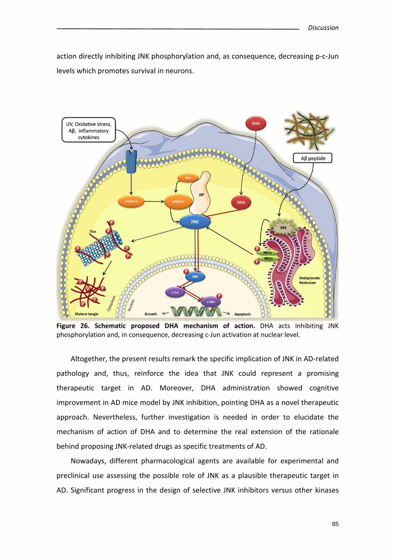
Discussion
85
action directly inhibiting JNK phosphorylation and, as consequence, decreasing p-c-Jun
levels which promotes survival in neurons.
Figure 26. Schematic proposed DHA mechanism of action. DHA acts inhibiting JNK phosphorylation and, in consequence, decreasing c-Jun activation at nuclear level.
Altogether, the present results remark the specific implication of JNK in AD-related
pathology and, thus, reinforce the idea that JNK could represent a promising
therapeutic target in AD. Moreover, DHA administration showed cognitive
improvement in AD mice model by JNK inhibition, pointing DHA as a novel therapeutic
approach. Nevertheless, further investigation is needed in order to elucidate the
mechanism of action of DHA and to determine the real extension of the rationale
behind proposing JNK-related drugs as specific treatments of AD.
Nowadays, different pharmacological agents are available for experimental and
preclinical use assessing the possible role of JNK as a plausible therapeutic target in
AD. Significant progress in the design of selective JNK inhibitors versus other kinases

Discussion
86
has been achieved within the past years. However, directed inhibition of JNK isoforms
in specific tissues is still an open task.
The fact that JNK3 is specifically expressed in the CNS and its activation by stress-
stimuli renders is an attractive and potential target for treating AD. It is possible to
speculate that JNK3 specific inhibition will reduce the possible side-effects of a
systemic JNK inhibition. Although there is no consensus in the literature if there is a
need for whether isoform selectivity is needed for the treatment of AD, the answer to
this question can only be obtained when such compounds are available.

CONCLUSIONS


Conclusions
89
From the results obtained in the present Doctoral Thesis it can be concluded that:
1. Age- and genotype-increased activated JNK (pJNK) levels were observed in
experimental mouse models of AD: Tg2576, a transgenic amyloidogenic model and
SAMP8, a sporadic AD based on accelerated senescence. This increase parallels the
enhanced Aβ levels.
2. Specific increase of pJNK levels in AD postmortem and not in other dementias was
found.
3. Significantly increased pJNK levels were observed in CSF of AD patients compared
with subject cognitive impairment (controls) and stable mild cognitive impairment
groups.
4. Aβ levels were increased in human frontal cortex samples of AD patients, and
these Aβ levels positively correlated to pJNK levels.
5. Aβ and pJNK co-localize in frontal cortex of Alzheimer’s disease brains. Aβ and pJNK
also co-localize in frontal cortex of Tg2576 mice. In both cases the same
immunolabelling pattern was observed which shown activated JNK around the
senile plaque.
6. Intracerebroventricular administration of β-amyloid increased JNK expression in
wild-type mice.
7. SAMP8 mice showed cognitive deficits in NORT and MWM test that were only
reversed by docosahexaenoic acid (DHA), and not other ω-3 polyunsaturated fatty
acids, treatment.
8. Increased pJNK levels in SAMP8 mice were only reversed by DHA treatment.
As general conclusion, pJNK expression is selectively increased in AD and not in
other dementias, probably as a consequence of increased levels of Aβ in this type of
dementia. JNK could be considered as a new AD biomarker and pharmacological
inhibition of JNK with DHA, an ω-3 fatty acid derivative, could be a new therapeutic
approach for the treatment of AD.


REFERENCES


References
93
Abete, I., Goyenechea, E., Zulet, M. A., & Martínez, J. A. (2011). Obesity and metabolic
syndrome: Potential benefit from specific nutritional components. Nutrition,
Metabolism and Cardiovascular Diseases, 21, B1–B15.
https://doi.org/10.1016/j.numecd.2011.05.001
Akhter, R., Sanphui, P., Das, H., Saha, P., & Biswas, S. C. (2015). The regulation of p53
up-regulated modulator of apoptosis by JNK/c-Jun pathway in β-amyloid-induced
neuron death. Journal of Neurochemistry, 134(6), 1091–103.
https://doi.org/10.1111/jnc.13128
Alcolea, D., Martínez-Lage, P., Sánchez-Juan, P., Olazarán, J., Antúnez, C., Izagirre, A., et
al. (2015). Amyloid precursor protein metabolism and inflammation markers in
preclinical Alzheimer disease. Neurology, 85(7), 626–633.
https://doi.org/10.1212/WNL.0000000000001859
Amador-Ortiz, C., Lin, W.-L., Ahmed, Z., Personett, D., Davies, P., Duara, R., et al.
(2007). TDP-43 immunoreactivity in hippocampal sclerosis and Alzheimer’s
disease. Annals of Neurology, 61(5), 435–45. https://doi.org/10.1002/ana.21154
Antoniou, X., Falconi, M., Di Marino, D., & Borsello, T. (2011). JNK3 as a therapeutic
target for neurodegenerative diseases. Journal of Alzheimer’s Disease : JAD, 24(4),
633–42. https://doi.org/10.3233/JAD-2011-091567
Arai, T., Mackenzie, I. R. A., Hasegawa, M., Nonoka, T., Niizato, K., Tsuchiya, K., et al.
(2009). Phosphorylated TDP-43 in Alzheimer’s disease and dementia with Lewy
bodies. Acta Neuropathologica, 117(2), 125–36. https://doi.org/10.1007/s00401-
008-0480-1
Arsenault, D., Julien, C., Tremblay, C., & Calon, F. (2011). DHA Improves Cognition and
Prevents Dysfunction of Entorhinal Cortex Neurons in 3xTg-AD Mice. PLoS ONE,
6(2), e17397. https://doi.org/10.1371/journal.pone.0017397
Arthur, P. G., Matich, G. P., Pang, W. W., Yu, D.-Y., & Bogoyevitch, M. A. (2007).
Necrotic death of neurons following an excitotoxic insult is prevented by a
peptide inhibitor of c-jun N-terminal kinase. Journal of Neurochemistry, 102(1),
65–76. https://doi.org/10.1111/j.1471-4159.2007.04618.x
Atzori, C., Ghetti, B., Piva, R., Srinivasan, A. N., Zolo, P., Delisle, M. B., et al. (2001).
Activation of the JNK/p38 pathway occurs in diseases characterized by tau protein
pathology and is related to tau phosphorylation but not to apoptosis. Journal of

References
94
Neuropathology and Experimental Neurology, 60(12), 1190–7.
Bagyinszky, E., Giau, V. Van, Shim, K., Suk, K., An, S. S. A., & Kim, S. (2017). Role of
inflammatory molecules in the Alzheimer’s disease progression and diagnosis.
Journal of the Neurological Sciences, 376, 242–254.
https://doi.org/10.1016/j.jns.2017.03.031
Bain, J., McLauchlan, H., Elliott, M., & Cohen, P. (2003). The specificities of protein
kinase inhibitors: an update. The Biochemical Journal, 371(Pt 1), 199–204.
https://doi.org/10.1042/BJ20021535
Bamji-Mirza, M., Callaghan, D., Najem, D., Shen, S., Hasim, M. S., Yang, Z., et al. (2014).
Stimulation of insulin signaling and inhibition of JNK-AP1 activation protect cells
from amyloid-β-induced signaling dysregulation and inflammatory response.
Journal of Alzheimer’s Disease : JAD, 40(1), 105–22. https://doi.org/10.3233/JAD-
131949
Barr, R. K., Kendrick, T. S., & Bogoyevitch, M. A. (2002). Identification of the critical
features of a small peptide inhibitor of JNK activity. The Journal of Biological
Chemistry, 277(13), 10987–97. https://doi.org/10.1074/jbc.M107565200
Barros-Miñones, L., Orejana, L., Goñi-Allo, B., Suquía, V., Hervías, I., Aguirre, N., et al.
(2013). Modulation of the ASK1-MKK3/6-p38/MAPK signalling pathway mediates
sildenafil protection against chemical hypoxia caused by malonate. British Journal
of Pharmacology, 168(8), 1820–34. https://doi.org/10.1111/bph.12071
Bascoul-Colombo, C., Guschina, I. A., Maskrey, B. H., Good, M., O’Donnell, V. B., &
Harwood, J. L. (2016). Dietary DHA supplementation causes selective changes in
phospholipids from different brain regions in both wild type mice and the Tg2576
mouse model of Alzheimer’s disease. Biochimica et Biophysica Acta (BBA) -
Molecular and Cell Biology of Lipids, 1861(6), 524–537.
https://doi.org/10.1016/j.bbalip.2016.03.005
Bellucci, A., Rosi, M. C., Grossi, C., Fiorentini, A., Luccarini, I., & Casamenti, F. (2007).
Abnormal processing of tau in the brain of aged TgCRND8 mice. Neurobiology of
Disease, 27(3), 328–338. https://doi.org/10.1016/j.nbd.2007.06.008
Bevilaqua, L. R. M., Kerr, D. S., Medina, J. H., Izquierdo, I., & Cammarota, M. (2003).
Inhibition of hippocampal Jun N-terminal kinase enhances short-term memory
but blocks long-term memory formation and retrieval of an inhibitory avoidance

References
95
task. The European Journal of Neuroscience, 17(4), 897–902.
Bicca, M. A., Costa, R., Loch-Neckel, G., Figueiredo, C. P., Medeiros, R., & Calixto, J. B.
(2015). B2 receptor blockage prevents Aβ-induced cognitive impairment by
neuroinflammation inhibition. Behavioural Brain Research, 278, 482–491.
https://doi.org/10.1016/j.bbr.2014.10.040
Blennow, H. K., Zetterberg, Blennow, K., Blennow, K., Hampel, H., Weiner, M., et al.
(2010). Cerebrospinal fluid and plasma biomarkers in Alzheimer disease. Nature
Publishing Group, 6(16104), 131–144. https://doi.org/10.1038/nrneurol.2010.4
Bloom, G. S. (2014). Amyloid-β and tau: the trigger and bullet in Alzheimer disease
pathogenesis. JAMA Neurology, 71(4), 505–8.
https://doi.org/10.1001/jamaneurol.2013.5847
Bodles, A. M., & Barger, S. W. (2005). Secreted beta-amyloid precursor protein
activates microglia via JNK and p38-MAPK. Neurobiology of Aging, 26(1), 9–16.
https://doi.org/10.1016/j.neurobiolaging.2004.02.022
Bogoyevitch, M. A., & Arthur, P. G. (2008). Inhibitors of c-Jun N-terminal kinases: JuNK
no more? Biochimica et Biophysica Acta, 1784(1), 76–93.
https://doi.org/10.1016/j.bbapap.2007.09.013
Bogoyevitch, M. A., Boehm, I., Oakley, A., Ketterman, A. J., & Barr, R. K. (2004).
Targeting the JNK MAPK cascade for inhibition: basic science and therapeutic
potential. Biochimica et Biophysica Acta, 1697(1–2), 89–101.
https://doi.org/10.1016/j.bbapap.2003.11.016
Bogoyevitch, M. A., Ngoei, K. R. W., Zhao, T. T., Yeap, Y. Y. C., & Ng, D. C. H. (2010). c-
Jun N-terminal kinase (JNK) signaling: Recent advances and challenges. Biochimica
et Biophysica Acta (BBA) - Proteins and Proteomics, 1804(3), 463–475.
https://doi.org/10.1016/j.bbapap.2009.11.002
Borgeson, E., Johnson, A. M. F., Lee, Y. S., Till, A., Syed, G. H., Ali-Shah, S. T., et al.
(2015). Lipoxin A4 Attenuates Obesity-Induced Adipose Inflammation and
Associated Liver and Kidney Disease. Cell Metabolism, 22(1), 125–137.
https://doi.org/10.1016/j.cmet.2015.05.003
Borsello, T., & Bonny, C. (2004). Use of cell-permeable peptides to prevent neuronal
degeneration. Trends in Molecular Medicine, 10(5), 239–244.
https://doi.org/10.1016/j.molmed.2004.03.008

References
96
Borsello, T., Clarke, P. G. H., Hirt, L., Vercelli, A., Repici, M., Schorderet, D. F., et al.
(2003). A peptide inhibitor of c-Jun N-terminal kinase protects against
excitotoxicity and cerebral ischemia. Nature Medicine, 9(9), 1180–1186.
https://doi.org/10.1038/nm911
Bozyczko-Coyne, D., O’Kane, T. M., Wu, Z. L., Dobrzanski, P., Murthy, S., Vaught, J. L., et
al. (2001). CEP-1347/KT-7515, an inhibitor of SAPK/JNK pathway activation,
promotes survival and blocks multiple events associated with Abeta-induced
cortical neuron apoptosis. Journal of Neurochemistry, 77(3), 849–63.
Brecht, S., Kirchhof, R., Chromik, A., Willesen, M., Nicolaus, T., Raivich, G., et al. (2005).
Specific pathophysiological functions of JNK isoforms in the brain. European
Journal of Neuroscience, 21(2), 363–377. https://doi.org/10.1111/j.1460-
9568.2005.03857.x
Burckhardt, M., Herke, M., Wustmann, T., Watzke, S., Langer, G., & Fink, A. (2016).
Omega-3 fatty acids for the treatment of dementia. In M. Burckhardt (Ed.),
Cochrane Database of Systematic Reviews (Vol. 4, p. CD009002). Chichester, UK:
John Wiley & Sons, Ltd. https://doi.org/10.1002/14651858.CD009002.pub3
Burdge, G. C., Jones, A. E., & Wootton, S. A. (2002). Eicosapentaenoic and
docosapentaenoic acids are the principal products of α-linolenic acid metabolism
in young men. British Journal of Nutrition, 88(4), 355.
https://doi.org/10.1079/BJN2002662
Burdge, G. C., & Wootton, S. A. (2002). Conversion of α-linolenic acid to
eicosapentaenoic, docosapentaenoic and docosahexaenoic acids in young
women. British Journal of Nutrition, 88(4), 411.
https://doi.org/10.1079/BJN2002689
Calder, P. C. (2015). Marine omega-3 fatty acids and inflammatory processes: Effects,
mechanisms and clinical relevance. Biochimica et Biophysica Acta (BBA) -
Molecular and Cell Biology of Lipids, 1851(4), 469–484.
https://doi.org/10.1016/j.bbalip.2014.08.010
Calon, F., Lim, G. P., Yang, F., Morihara, T., Teter, B., Ubeda, O., et al. (2004).
Docosahexaenoic Acid Protects from Dendritic Pathology in an Alzheimer’s
Disease Mouse Model. Neuron, 43(5), 633–645.
https://doi.org/10.1016/j.neuron.2004.08.013
References
97
Cao, M., Liu, F., Ji, F., Liang, J., Liu, L., Wu, Q., et al. (2013). Effect of c-Jun N-terminal
kinase (JNK)/p38 mitogen-activated protein kinase (p38 MAPK) in morphine-
induced tau protein hyperphosphorylation. Behavioural Brain Research, 237, 249–
255. https://doi.org/10.1016/j.bbr.2012.09.040
Carter, C. L., Resnick, E. M., Mallampalli, M., & Kalbarczyk, A. (2012). Sex and Gender
Differences in Alzheimer’s Disease: Recommendations for Future Research.
Journal of Women’s Health, 21(10), 1018–1023.
https://doi.org/10.1089/jwh.2012.3789
Casali, B. T., Corona, A. W., Mariani, M. M., Karlo, J. C., Ghosal, K., & Landreth, G. E.
(2015). Omega-3 Fatty Acids Augment the Actions of Nuclear Receptor Agonists in
a Mouse Model of Alzheimer’s Disease. Journal of Neuroscience, 35(24), 9173–
9181. https://doi.org/10.1523/JNEUROSCI.1000-15.2015
Cederholm, T., Salem, N., & Palmblad, J. (2013). Omega-3 Fatty Acids in the Prevention
of Cognitive Decline in Humans. Advances in Nutrition: An International Review
Journal, 4(6), 672–676. https://doi.org/10.3945/an.113.004556
Chae, S. S., Yoo, C. B., Jo, C., Yun, S.-M., Jo, S. A., & Koh, Y. H. (2010). Caspases-2 and -8
are involved in the presenilin1/γ-secretase-dependent cleavage of amyloid
precursor protein after the induction of apoptosis. Journal of Neuroscience
Research, 88(9), NA-NA. https://doi.org/10.1002/jnr.22356
Chen, J.-T., Lu, D.-H., Chia, C.-P., Ruan, D.-Y., Sabapathy, K., & Xiao, Z.-C. (2005).
Impaired long-term potentiation in c-Jun N-terminal kinase 2-deficient mice.
Journal of Neurochemistry, 93(2), 463–73. https://doi.org/10.1111/j.1471-
4159.2005.03037.x
Chen, Y.-R., & Tan, T.-H. (1998). Inhibition of the c-Jun N-terminal kinase (JNK) signaling
pathway by curcumin. Oncogene, 17(2), 173–178.
https://doi.org/10.1038/sj.onc.1201941
Chiang, N., & Serhan, C. N. (2017). Structural elucidation and physiologic functions of
specialized pro-resolving mediators and their receptors. Molecular Aspects of
Medicine. https://doi.org/10.1016/j.mam.2017.03.005
Cho, E. S., Jang, Y. J., Kang, N. J., Hwang, M. K., Kim, Y. T., Lee, K. W., et al. (2009).
Cocoa procyanidins attenuate 4-hydroxynonenal-induced apoptosis of PC12 cells
by directly inhibiting mitogen-activated protein kinase kinase 4 activity. Free

References
98
Radical Biology & Medicine, 46(10), 1319–27.
https://doi.org/10.1016/j.freeradbiomed.2009.02.010
Chuang, J. Y., Huang, Y. L., Yen, W. L., Chiang, I. P., Tsai, M. H., & Tang, C. H. (2014).
Syk/JNK/AP-1 Signaling Pathway Mediates Interleukin-6-Promoted Cell Migration
in Oral Squamous Cell Carcinoma. International Journal of Molecular Sciences,
15(1), 545–559. https://doi.org/10.3390/ijms15010545
Coffey, E. T. (2014). Nuclear and cytosolic JNK signalling in neurons. Nature Reviews.
Neuroscience, 15(5), 285–99. https://doi.org/10.1038/nrn3729
Cole, G. M., Ma, Q.-L., & Frautschy, S. A. (2009). Omega-3 fatty acids and dementia.
Prostaglandins, Leukotrienes and Essential Fatty Acids, 81(2–3), 213–221.
https://doi.org/10.1016/j.plefa.2009.05.015
Colombo, A., Bastone, A., Ploia, C., Sclip, A., Salmona, M., Forloni, G., et al. (2009). JNK
regulates APP cleavage and degradation in a model of Alzheimer’s disease.
Neurobiology of Disease, 33(3), 518–525.
https://doi.org/10.1016/j.nbd.2008.12.014
Corsinovi, L., Biasi, F., Poli, G., Leonarduzzi, G., & Isaia, G. (2011). Dietary lipids and
their oxidized products in Alzheimer’s disease. Molecular Nutrition & Food
Research, 55 Suppl 2(S2), S161-72. https://doi.org/10.1002/mnfr.201100208
Costello, D. A., & Herron, C. E. (2004). The role of c-Jun N-terminal kinase in the Aβ-
mediated impairment of LTP and regulation of synaptic transmission in the
hippocampus. Neuropharmacology, 46(5), 655–662.
https://doi.org/10.1016/j.neuropharm.2003.11.016
Craig-Schapiro, R., Perrin, R. J., Roe, C. M., Xiong, C., Carter, D., Cairns, N. J., et al.
(2010). YKL-40: A novel prognostic fluid biomarker for preclinical Alzheimer’s
disease. Biological Psychiatry, 68(10), 903–912.
https://doi.org/10.1016/j.biopsych.2010.08.025
Cui, J., Zhang, M., Zhang, Y.-Q., & Xu, Z.-H. (2007). JNK pathway: diseases and
therapeutic potential. Acta Pharmacologica Sinica, 28(5), 601–8.
https://doi.org/10.1111/j.1745-7254.2007.00579.x
Das, H. K., Tchedre, K., & Mueller, B. (2012). Repression of transcription of presenilin-1
inhibits γ-secretase independent ER Ca2+ leak that is impaired by FAD mutations.
Journal of Neurochemistry, 122(3), 487–500. https://doi.org/10.1111/j.1471-
References
99
4159.2012.07794.x
Davis, R. J. (2000). Signal transduction by the JNK group of MAP kinases. Cell, 103(2),
239–52. https://doi.org/doi: 10.1016/S0092-8674(00)00116-1
De Girolamo, L. A., & Billett, E. E. (2006). Role of extracellular-regulated kinase and c-
jun NH2-terminal kinase in 1-methyl-4-phenyl-1,2,3,6-tetrahydropyridine-induced
neurofilament phosphorylation. Journal of Neuroscience Research, 83(4), 680–
693. https://doi.org/10.1002/jnr.20765
de Souza, R. J., Mente, A., Maroleanu, A., Cozma, A. I., Ha, V., Kishibe, T., et al. (2015).
Intake of saturated and trans unsaturated fatty acids and risk of all cause
mortality, cardiovascular disease, and type 2 diabetes: systematic review and
meta-analysis of observational studies. BMJ (Clinical Research Ed.), 351, h3978.
de Souza Fernandes, D. P., Canaan Rezende, F. A., Pereira Rocha, G., De Santis
Filgueiras, M., Silva Moreira, P. R., & Gonçalves Alfenas, R. de C. (2015). Effect of
eicosapentaenoic acd and docosahexaenoic acid supplementatations to control
cognitive decline in dementia and Alzheimer’s disease: a systematic review.
Nutricion Hospitalaria, 32(2), 528–33. https://doi.org/10.3305/nh.2015.32.2.9111
Dérijard, B., Hibi, M., Wu, I. H., Barrett, T., Su, B., Deng, T., et al. (1994). JNK1: a protein
kinase stimulated by UV light and Ha-Ras that binds and phosphorylates the c-Jun
activation domain. Cell, 76(6), 1025–37.
Dhanasekaran, D. N., & Reddy, E. P. (2008). JNK signaling in apoptosis. Oncogene,
27(48), 6245–51. https://doi.org/10.1038/onc.2008.301
Dhikav, V., & Anand, K. S. (2007). Glucocorticoids may initiate Alzheimer’s disease: A
Potential therapeutic role for mifepristone (RU-486). Medical Hypotheses, 68(5),
1088–1092. https://doi.org/10.1016/j.mehy.2006.09.038
Dickens, M., Rogers, J. S., Cavanagh, J., Raitano, A., Xia, Z., Halpern, J. R., et al. (1997). A
cytoplasmic inhibitor of the JNK signal transduction pathway. Science, 277(5326),
693–6.
Ebenezer, P. J., Weidner, A. M., LeVine, H., Markesbery, W. R., Murphy, M. P., Zhang,
L., et al. (2010). Neuron specific toxicity of oligomeric amyloid-β: role for JUN-
kinase and oxidative stress. Journal of Alzheimer’s Disease : JAD, 22(3), 839–48.
https://doi.org/10.3233/JAD-2010-101161
Eckert, G. P., Chang, S., Eckmann, J., Copanaki, E., Hagl, S., Hener, U., et al. (2011).
References
100
Liposome-incorporated DHA increases neuronal survival by enhancing non-
amyloidogenic APP processing. Biochimica et Biophysica Acta, 1808(1), 236–43.
https://doi.org/10.1016/j.bbamem.2010.10.014
Eckert, G. P., Lipka, U., & Muller, W. E. (2013). Omega-3 fatty acids in
neurodegenerative diseases: Focus on mitochondria. Prostaglandins, Leukotrienes
and Essential Fatty Acids (PLEFA), 88(1), 105–114.
https://doi.org/10.1016/j.plefa.2012.05.006
Eminel, S., Roemer, L., Waetzig, V., & Herdegen, T. (2008). c-Jun N-terminal kinases
trigger both degeneration and neurite outgrowth in primary hippocampal and
cortical neurons. Journal of Neurochemistry, 104(4), 957–69.
https://doi.org/10.1111/j.1471-4159.2007.05101.x
Engstrom, W., Ward, A., & Moorwood, K. (2010). The role of scaffold proteins in JNK
signalling. Cell Proliferation, 43(1), 56–66. https://doi.org/10.1111/j.1365-
2184.2009.00654.x
Feld, M., Krawczyk, M. C., Sol Fustiñana, M., Blake, M. G., Baratti, C. M., Romano, A., et
al. (2014). Decrease of ERK/MAPK overactivation in prefrontal cortex reverses
early memory deficit in a mouse model of Alzheimer’s disease. Journal of
Alzheimer’s Disease : JAD, 40(1), 69–82. https://doi.org/10.3233/JAD-131076
Feng, H.-L., Fan, H., Dang, H.-Z., Chen, X.-P., Ren, Y., Yang, J.-D., et al. (2014).
Neuroprotective effect of curcumin to Aβ of double transgenic mice with
Alzheimer’s disease. China Journal of Chinese Materia Medica, 39(19), 3846–9.
Ferrer, I., Blanco, R., Carmona, M., & Puig, B. (2001). Phosphorylated mitogen-
activated protein kinase (MAPK/ERK-P), protein kinase of 38kDa (p38-P), stress-
activated protein kinase (SAPK/JNK-P), and calcium/calmodulin-dependent kinase
II (CaM kinase II) are differentially expressed in tau deposits in neurons. Journal of
Neural Transmission, 108(12), 1397–1415.
https://doi.org/10.1007/s007020100016
Fiala, M., Terrando, N., & Dalli, J. (2015). Specialized Pro-Resolving Mediators from
Omega-3 Fatty Acids Improve Amyloid-β Phagocytosis and Regulate Inflammation
in Patients with Minor Cognitive Impairment. Journal of Alzheimer’s Disease,
48(2), 293–301. https://doi.org/10.3233/JAD-150367
Flood, J. F., & Morley, J. E. (1998). Learning and memory in the SAMP8 mouse.

References
101
Neuroscience and Biobehavioral Reviews, 22(1), 1–20.
Foley, A. M., Ammar, Z. M., Lee, R. H., & Mitchell, C. S. (2015). Systematic review of the
relationship between amyloid-β levels and measures of transgenic mouse
cognitive deficit in Alzheimer’s disease. Journal of Alzheimer’s Disease : JAD,
44(3), 787–95. https://doi.org/10.3233/JAD-142208
Fraga, V. G., Carvalho, M. das G., Caramelli, P., de Sousa, L. P., & Gomes, K. B. (2017).
Resolution of inflammation, n-3 fatty acid supplementation and Alzheimer
disease: A narrative review. Journal of Neuroimmunology, 310, 111–119.
https://doi.org/10.1016/j.jneuroim.2017.07.005
Frautschy, S. A., Horn, D. L., Sigel, J. J., Harris-White, M. E., Mendoza, J. J., Yang, F., et
al. (1998). Protease inhibitor coinfusion with amyloid beta-protein results in
enhanced deposition and toxicity in rat brain. The Journal of Neuroscience : The
Official Journal of the Society for Neuroscience, 18(20), 8311–21.
Frozza, R. L., Bernardi, A., Hoppe, J. B., Meneghetti, A. B., Matté, A., Battastini, A. M.
O., et al. (2013). Neuroprotective Effects of Resveratrol Against Aβ Administration
in Rats are Improved by Lipid-Core Nanocapsules. Molecular Neurobiology, 47(3),
1066–1080. https://doi.org/10.1007/s12035-013-8401-2
Funakoshi-Tago, M., Nagata, T., Tago, K., Tsukada, M., Tanaka, K., Nakamura, S., et al.
(2012). Fullerene derivative prevents cellular transformation induced by JAK2
V617F mutant through inhibiting c-Jun N-terminal kinase pathway. Cellular
Signalling, 24(11), 2024–2034. https://doi.org/10.1016/j.cellsig.2012.06.014
Ganguly, A., Oo, T. F., Rzhetskaya, M., Pratt, R., Yarygina, O., Momoi, T., et al. (2004).
CEP11004, a novel inhibitor of the mixed lineage kinases, suppresses apoptotic
death in dopamine neurons of the substantia nigra induced by 6-
hydroxydopamine. Journal of Neurochemistry, 88(2), 469–80.
Gehringer, M., Muth, F., Koch, P., & Laufer, S. A. (2015). c-Jun N-terminal kinase
inhibitors: a patent review (2010 – 2014). Expert Opinion on Therapeutic Patents,
25(8), 849–872. https://doi.org/10.1517/13543776.2015.1039984
Gil-Bea, F., Aisa, B., Solomon, A., Solas, M., Mugueta, C., Winblad, B., et al. (2010a).
HPA Axis Dysregulation Associated to Apolipoprotein E4 Genotype in Alzheimer’s
Disease. Journal of Alzheimer’s Disease, 22(3), 829–838.
https://doi.org/10.3233/JAD-2010-100663

References
102
Gil-Bea, F. J., Solas, M., Solomon, A., Mugueta, C., Winblad, B., Kivipelto, M., et al.
(2010b). Insulin levels are decreased in the cerebrospinal fluid of women with
prodomal Alzheimer’s disease. Journal of Alzheimer’s Disease : JAD, 22(2), 405–
13. https://doi.org/10.3233/JAD-2010-100795
Giuliani, D., Ottani, A., Zaffe, D., Galantucci, M., Strinati, F., Lodi, R., et al. (2013).
Hydrogen sulfide slows down progression of experimental Alzheimer’s disease by
targeting multiple pathophysiological mechanisms. Neurobiology of Learning and
Memory, 104, 82–91. https://doi.org/10.1016/j.nlm.2013.05.006
Goodfellow, V. S., Loweth, C. J., Ravula, S. B., Wiemann, T., Nguyen, T., Xu, Y., et al.
(2013). Discovery, Synthesis, and Characterization of an Orally Bioavailable, Brain
Penetrant Inhibitor of Mixed Lineage Kinase 3. Journal of Medicinal Chemistry,
56(20), 8032–8048. https://doi.org/10.1021/jm401094t
Goodman, Y., & Mattson, M. P. (1994). Staurosporine and K-252 compounds protect
hippocampal neurons against amyloid beta-peptide toxicity and oxidative injury.
Brain Research, 650(1), 170–4.
Gourmaud, S., Paquet, C., Dumurgier, J., Pace, C., Bouras, C., Gray, F., et al. (2015).
Increased levels of cerebrospinal fluid JNK3 associated with amyloid pathology:
Links to cognitive decline. Journal of Psychiatry and Neuroscience, 40(3), 151–161.
https://doi.org/10.1503/jpn.140062
Gregor, M. F., & Hotamisligil, G. S. (2011). Inflammatory mechanisms in obesity.
Annual Review of Immunology, 29(1), 415–45. https://doi.org/10.1146/annurev-
immunol-031210-101322
Grimm, M. O. W., Kuchenbecker, J., Grösgen, S., Burg, V. K., Hundsdörfer, B., Rothhaar,
T. L., et al. (2011). Docosahexaenoic acid reduces amyloid beta production via
multiple pleiotropic mechanisms. The Journal of Biological Chemistry, 286(16),
14028–39. https://doi.org/10.1074/jbc.M110.182329
Guglielmotto, M., Monteleone, D., Giliberto, L., Fornaro, M., Borghi, R., Tamagno, E., et
al. (2011). Amyloid-β₄₂ activates the expression of BACE1 through the JNK
pathway. Journal of Alzheimer’s Disease : JAD, 27(4), 871–83.
https://doi.org/10.3233/JAD-2011-110884
Guillozet-Bongaarts, A. L., Cahill, M. E., Cryns, V. L., Reynolds, M. R., Berry, R. W., &
Binder, L. I. (2006). Pseudophosphorylation of tau at serine 422 inhibits caspase
References
103
cleavage: in vitro evidence and implications for tangle formation in vivo. Journal
of Neurochemistry, 97(4), 1005–14. https://doi.org/10.1111/j.1471-
4159.2006.03784.x
Guillozet-Bongaarts, A. L., Garcia-Sierra, F., Reynolds, M. R., Horowitz, P. M., Fu, Y.,
Wang, T., et al. (2005). Tau truncation during neurofibrillary tangle evolution in
Alzheimer’s disease. Neurobiology of Aging, 26(7), 1015–1022.
https://doi.org/10.1016/j.neurobiolaging.2004.09.019
Haas, C. (2012). Strategies, development, and pitfalls of therapeutic options for
Alzheimer’s disease. Journal of Alzheimer’s Disease, 28(2), 241–281.
https://doi.org/10.3233/JAD-2011-110986
Halliday, G. M., Holton, J. L., Revesz, T., & Dickson, D. W. (2011). Neuropathology
underlying clinical variability in patients with synucleinopathies. Acta
Neuropathologica, 122(2), 187–204. https://doi.org/10.1007/s00401-011-0852-9
Hashimoto, M., Hsu, L. J., Rockenstein, E., Takenouchi, T., Mallory, M., & Masliah, E.
(2002). α-synuclein protects against oxidative stress via inactivation of the c-Jun
N-terminal kinase stress-signaling pathway in neuronal cells. Journal of Biological
Chemistry, 277(13), 11465–11472. https://doi.org/10.1074/jbc.M111428200
Hashimoto, Y., Niikura, T., Chiba, T., Tsukamoto, E., Kadowaki, H., Nishitoh, H., et al.
(2003). The Cytoplasmic Domain of Alzheimer’s Amyloid- Protein Precursor
Causes Sustained Apoptosis Signal-Regulating Kinase 1/c-Jun NH2-Terminal
Kinase-Mediated Neurotoxic Signal via Dimerization. Journal of Pharmacology and
Experimental Therapeutics, 306(3), 889–902.
https://doi.org/10.1124/jpet.103.051383
Higashi, S., Iseki, E., Yamamoto, R., Minegishi, M., Hino, H., Fujisawa, K., et al. (2007).
Concurrence of TDP-43, tau and alpha-synuclein pathology in brains of
Alzheimer’s disease and dementia with Lewy bodies. Brain Research, 1184(1),
284–294. https://doi.org/10.1016/j.brainres.2007.09.048
Hjorth, E., Zhu, M., Toro, V. C., Vedin, I., Palmblad, J., Cederholm, T., et al. (2013).
Omega-3 fatty acids enhance phagocytosis of Alzheimer’s disease-related
amyloid-β42 by human microglia and decrease inflammatory markers. Journal of
Alzheimer’s Disease : JAD, 35(4), 697–713. https://doi.org/10.3233/JAD-130131
Holzberg, D., Knight, C. G., Dittrich-Breiholz, O., Schneider, H., Dörrie, A., Hoffmann, E.,
References
104
et al. (2003). Disruption of the c-JUN-JNK Complex by a Cell-permeable Peptide
Containing the c-JUN δ Domain Induces Apoptosis and Affects a Distinct Set of
Interleukin-1-induced Inflammatory Genes. Journal of Biological Chemistry,
278(41), 40213–40223. https://doi.org/10.1074/jbc.M304058200
Hong, Y. K., Lee, S., Park, S. H., Lee, J. H., Han, S. Y., Kim, S. T., et al. (2012). Inhibition of
JNK/dFOXO pathway and caspases rescues neurological impairments in
Drosophila Alzheimer’s disease model. Biochemical and Biophysical Research
Communications, 419(1), 49–53. https://doi.org/10.1016/j.bbrc.2012.01.122
Howlett, D. R., Whitfield, D., Johnson, M., Attems, J., O’Brien, J. T., Aarsland, D., et al.
(2014). Regional Multiple Pathology Scores Are Associated with Cognitive Decline
in Lewy Body Dementias. Brain Pathology, 25, 401–408.
https://doi.org/10.1111/bpa.12182
Hsiao, K., Chapman, P., Nilsen, S., Eckman, C., Harigaya, Y., Younkin, S., et al. (1996).
Correlative memory deficits, Abeta elevation, and amyloid plaques in transgenic
mice. Science, 274(5284), 99–102.
Hu, W. T., Josephs, K. A., Knopman, D. S., Boeve, B. F., Dickson, D. W., Petersen, R. C.,
et al. (2008). Temporal lobar predominance of TDP-43 neuronal cytoplasmic
inclusions in Alzheimer disease. Acta Neuropathologica, 116(2), 215–220.
https://doi.org/10.1007/s00401-008-0400-4
Hwang, D. Y., Cho, J. S., Lee, S. H., Chae, K. R., Lim, H. J., Min, S. H., et al. (2004).
Aberrant expressions of pathogenic phenotype in Alzheimer’s diseased transgenic
mice carrying NSE-controlled APPsw. Experimental Neurology, 186(1), 20–32.
https://doi.org/10.1016/j.expneurol.2003.09.021
Irizarry, M. C., McNamara, M., Fedorchak, K., Hsiao, K., & Hyman, B. T. (1997). APPSw
transgenic mice develop age-related A beta deposits and neuropil abnormalities,
but no neuronal loss in CA1. Journal of Neuropathology and Experimental
Neurology, 56(9), 965–73.
Jia, J. P., Meng, R., Sun, Y. X., Sun, W. J., Ji, X. M., & Jia, L. F. (2005). Cerebrospinal fluid
tau, Aβ1-42 and inflammatory cytokines in patients with Alzheimer’s disease and
vascular dementia. Neuroscience Letters, 383(1–2), 12–16.
https://doi.org/10.1016/j.neulet.2005.03.051
Josephs, K. A., Murray, M. E., Whitwell, J. L., Parisi, J. E., Petrucelli, L., Jack, C. R., et al.
References
105
(2014). Staging TDP-43 pathology in Alzheimer’s disease. Acta Neuropathologica,
127(3), 441–450. https://doi.org/10.1007/s00401-013-1211-9
Jump, D. B., Depner, C. M., & Tripathy, S. (2012). Omega-3 fatty acid supplementation
and cardiovascular disease. Journal of Lipid Research, 53(12), 2525–45.
https://doi.org/10.1194/jlr.R027904
Jung, Y., Dickson, D. W., Murray, M. E., Whitwell, J. L., Knopman, D. S., Boeve, B. F., et
al. (2014). TDP-43 in Alzheimer’s disease is not associated with clinical FTLD or
Parkinsonism. Journal of Neurology, 261(7), 1344–1348.
https://doi.org/10.1007/s00415-014-7352-5
Kempuraj, D., Thangavel, R., Natteru, P. A., Selvakumar, G. P., Saeed, D., Zahoor, H., et
al. (2016). Neuroinflammation Induces Neurodegeneration. Journal of Neurology,
Neurosurgery and Spine, 1(1).
Killick, R., Ribe, E. M., Al-Shawi, R., Malik, B., Hooper, C., Fernandes, C., et al. (2014).
Clusterin regulates β-amyloid toxicity via Dickkopf-1-driven induction of the wnt-
PCP-JNK pathway. Molecular Psychiatry, 19(1), 88–98.
https://doi.org/10.1038/mp.2012.163
Kim, J.-H., Chae, M., Choi, A.-R., Sik Kim, H., & Yoon, S. (2014). SP600125 overcomes
antimitotic drug-resistance in cancer cells by increasing apoptosis with
independence of P-gp inhibition. European Journal of Pharmacology, 723, 141–
147. https://doi.org/10.1016/j.ejphar.2013.11.026
Kobylecki, C., Langheinrich, T., Hinz, R., Vardy, E. R. L. C., Brown, G., Martino, M.-E., et
al. (2015). 18F-Florbetapir PET in Patients with Frontotemporal Dementia and
Alzheimer Disease. Journal of Nuclear Medicine, 56(3), 386–391.
https://doi.org/10.2967/jnumed.114.147454
Koch, P., Gehringer, M., & Laufer, S. A. (2015). Inhibitors of c-Jun N-Terminal Kinases:
An Update. Journal of Medicinal Chemistry, 58(1), 72–95.
https://doi.org/10.1021/jm501212r
Koedam, E. L. G. E., van der Vlies, A. E., van der Flier, W. M., Verwey, N. A., Koene, T.,
Scheltens, P., et al. (2013). Cognitive correlates of cerebrospinal fluid biomarkers
in frontotemporal dementia. Alzheimer’s & Dementia : The Journal of the
Alzheimer’s Association, 9(3), 269–75. https://doi.org/10.1016/j.jalz.2011.12.007
Kolarova, M., García-Sierra, F., Bartos, A., Ricny, J., & Ripova, D. (2012). Structure and
References
106
pathology of tau protein in Alzheimer disease. International Journal of Alzheimer’s
Disease, 2012. https://doi.org/10.1155/2012/731526
Kou, W., Luchtman, D., & Song, C. (2008). Eicosapentaenoic acid (EPA) increases cell
viability and expression of neurotrophin receptors in retinoic acid and brain-
derived neurotrophic factor differentiated SH-SY5Y cells. European Journal of
Nutrition, 47(2), 104–13. https://doi.org/10.1007/s00394-008-0703-1
Kovari, E., Horvath, J., & Bouras, C. (2009). Neuropathology of Lewy body disorders.
Brain Research Bulletin, 80(4–5), 203–210.
https://doi.org/10.1016/j.brainresbull.2009.06.018
Kuan, C. Y., Yang, D. D., Samanta Roy, D. R., Davis, R. J., Rakic, P., & Flavell, R. A. (1999).
The Jnk1 and Jnk2 protein kinases are required for regional specific apoptosis
during early brain development. Neuron, 22(4), 667–76.
Lagalwar, S., Berry, R. W., & Binder, L. I. (2007). Relation of hippocampal phospho-
SAPK/JNK granules in Alzheimer’s disease and tauopathies to granulovacuolar
degeneration bodies. Acta Neuropathologica, 113(1), 63–73.
https://doi.org/10.1007/s00401-006-0159-4
Lagalwar, S., Guillozet-Bongaarts, A. L., Berry, R. W., & Binder, L. I. (2006). Formation of
Phospho-SAPK/JNK Granules in the Hippocampus Is an Early Event in Alzheimer
Disease. Journal of Neuropathology & Experimental Neurology, 65(5), 455–464.
https://doi.org/10.1097/01.jnen.0000229236.98124.d8
Lee, H. mi, Kim, K.-S., & Kim, J. (2014). A comparative study of the effects of inhibitory
cytokines on human natural killer cells and the mechanistic features of
transforming growth factor-beta. Cellular Immunology, 290(1), 52–61.
https://doi.org/10.1016/j.cellimm.2014.05.001
Lee, J. M., Blennow, K., Andreasen, N., Laterza, O., Modur, V., Olander, J., et al. (2008).
The brain injury biomarker VLP-1 is increased in the cerebrospinal fluid of
Alzheimer disease patients. Clinical Chemistry, 54(10), 1617–1623.
https://doi.org/10.1373/clinchem.2008.104497
Lewis, D. A. (2002). The human brain revisited: Opportunities and challenges in
postmortem studies of psychiatric disorders. Neuropsychopharmacology, 26(2),
143–154. https://doi.org/10.1016/S0893-133X(01)00393-1
Lewis, H., Beher, D., Cookson, N., Oakley, A., Piggott, M., Morris, C. M., et al. (2006).

References
107
Quantification of Alzheimer pathology in ageing and dementia: Age-related
accumulation of beta-amyloid(42) peptide in vascular dementia. Neuropathology
and Applied Neurobiology, 32(2), 103–118. https://doi.org/10.1111/j.1365-
2990.2006.00696.x
Li, Y., Dalli, J., Chiang, N., Baron, R. M., Quintana, C., & Serhan, C. N. (2013a). Plasticity
of leukocytic exudates in resolving acute inflammation is regulated by MicroRNA
and proresolving mediators. Immunity, 39(5), 885–98.
https://doi.org/10.1016/j.immuni.2013.10.011
Li, Y., Wang, F., Liu, C., Zeng, M., Han, X., Luo, T., et al. (2013b). JNK pathway may be
involved in isoflurane-induced apoptosis in the hippocampi of neonatal rats.
Neuroscience Letters, 545, 17–22. https://doi.org/10.1016/j.neulet.2013.04.008
Lim, G. P., Calon, F., Morihara, T., Yang, F., Teter, B., Ubeda, O., et al. (2005). A Diet
Enriched with the Omega-3 Fatty Acid Docosahexaenoic Acid Reduces Amyloid
Burden in an Aged Alzheimer Mouse Model. Journal of Neuroscience, 25(12),
3032–3040. https://doi.org/10.1523/JNEUROSCI.4225-04.2005
Lin, W., Ding, M., Xue, J., & Leng, W. (2013). The role of TLR2/JNK/NF-κB pathway in
amyloid β peptide-induced inflammatory response in mouse NG108-15 neural
cells. International Immunopharmacology, 17(3), 880–884.
https://doi.org/10.1016/j.intimp.2013.09.016
Lonergan, P. E., Martin, D. S. D., Horrobin, D. F., & Lynch, M. A. (2002).
Neuroprotective effect of eicosapentaenoic acid in hippocampus of rats exposed
to gamma-irradiation. The Journal of Biological Chemistry, 277(23), 20804–11.
https://doi.org/10.1074/jbc.M202387200
Lonergan, P. E., Martin, D. S. D., Horrobin, D. F., & Lynch, M. A. (2004).
Neuroprotective actions of eicosapentaenoic acid on lipopolysaccharide-induced
dysfunction in rat hippocampus. Journal of Neurochemistry, 91(1), 20–9.
https://doi.org/10.1111/j.1471-4159.2004.02689.x
Lorente-Cebrián, S., Costa, A. G. V., Navas-Carretero, S., Zabala, M., Laiglesia, L. M.,
Martínez, J. A., et al. (2015). An update on the role of omega-3 fatty acids on
inflammatory and degenerative diseases. Journal of Physiology and Biochemistry,
71(2), 341–349. https://doi.org/10.1007/s13105-015-0395-y
Lorente-Cebrián, S., Costa, A. G. V., Navas-Carretero, S., Zabala, M., Martínez, J. A., &
References
108
Moreno-Aliaga, M. J. (2013). Role of omega-3 fatty acids in obesity, metabolic
syndrome, and cardiovascular diseases: a review of the evidence. Journal of
Physiology and Biochemistry, 69(3), 633–651. https://doi.org/10.1007/s13105-
013-0265-4
Lukiw, W. J., Cui, J.-G., Marcheselli, V. L., Bodker, M., Botkjaer, A., Gotlinger, K., et al.
(2005). A role for docosahexaenoic acid-derived neuroprotectin D1 in neural cell
survival and Alzheimer disease. Journal of Clinical Investigation, 115(10), 2774–
2783. https://doi.org/10.1172/JCI25420
Ma, G., Chen, S., Wang, X., Ba, M., Yang, H., & Lu, G. (2005). Short-term interleukin-
1(beta) increases the release of secreted APP(alpha) via MEK1/2-dependent and
JNK-dependent alpha-secretase cleavage in neuroglioma U251 cells. Journal of
Neuroscience Research, 80(5), 683–92. https://doi.org/10.1002/jnr.20515
Ma, Q.-L., Yang, F., Rosario, E. R., Ubeda, O. J., Beech, W., Gant, D. J., et al. (2009).
Beta-Amyloid Oligomers Induce Phosphorylation of Tau and Inactivation of Insulin
Receptor Substrate via c-Jun N-Terminal Kinase Signaling: Suppression by Omega-
3 Fatty Acids and Curcumin. Journal of Neuroscience, 29(28), 9078–9089.
https://doi.org/10.1523/JNEUROSCI.1071-09.2009
Maiti, P., Manna, J., Veleri, S., & Frautschy, S. (2014). Molecular Chaperone
Dysfunction in Neurodegenerative Diseases and Effects of Curcumin. BioMed
Research International, 2014, 1–14. https://doi.org/10.1155/2014/495091
Marker, D. F., Tremblay, M.-E., Puccini, J. M., Barbieri, J., Gantz Marker, M. A., Loweth,
C. J., et al. (2013). The New Small-Molecule Mixed-Lineage Kinase 3 Inhibitor
URMC-099 Is Neuroprotective and Anti-Inflammatory in Models of Human
Immunodeficiency Virus-Associated Neurocognitive Disorders. Journal of
Neuroscience, 33(24), 9998–10010. https://doi.org/10.1523/JNEUROSCI.0598-
13.2013
Marques, C. A., Keil, U., Bonert, A., Steiner, B., Haass, C., Muller, W. E., et al. (2003).
Neurotoxic mechanisms caused by the alzheimer’s disease-linked Swedish
amyloid precursor protein. Mutation oxidative stress, caspases, and the JNK
pathway. Journal of Biological Chemistry, 278(30), 28294–28302.
https://doi.org/10.1074/jbc.M212265200
Martínez-Fernández, L., González-Muniesa, P., Laiglesia, L. M., Sáinz, N., Prieto-

References
109
Hontoria, P. L., Escoté, X., et al. (2017). Maresin 1 improves insulin sensitivity and
attenuates adipose tissue inflammation in ob/ob and diet-induced obese mice.
The FASEB Journal, 31(5), 2135–2145. https://doi.org/10.1096/fj.201600859R
Martínez-Fernández, L., Laiglesia, L. M., Huerta, A. E., Martínez, J. A., & Moreno-Aliaga,
M. J. (2015). Omega-3 fatty acids and adipose tissue function in obesity and
metabolic syndrome. Prostaglandins & Other Lipid Mediators, 121(Pt A), 24–41.
https://doi.org/10.1016/j.prostaglandins.2015.07.003
Martisova, E., Aisa, B., Guereñu, G., & Ramírez, M. J. (2013). Effects of early maternal
separation on biobehavioral and neuropathological markers of Alzheimer’s
disease in adult male rats. Current Alzheimer Research, 10(4), 420–32.
Mattsson, N., Carrillo, M. C., Dean, R. A., Devous Sr, M. D., Nikolcheva, T., Pesini, P., et
al. (2015). Revolutionizing Alzheimer’s disease and clinical trials through
biomarkers. Alzheimer’s & Dementia: Diagnosis, Assessment & Disease
Monitoring, 1(4), 1–8. https://doi.org/10.1016/j.dadm.2015.09.001
Mazzitelli, S., Xu, P., Ferrer, I., Davis, R. J., & Tournier, C. (2011). The Loss of c-Jun N-
Terminal Protein Kinase Activity Prevents the Amyloidogenic Cleavage of Amyloid
Precursor Protein and the Formation of Amyloid Plaques In Vivo. Journal of
Neuroscience, 31(47), 16969–16976. https://doi.org/10.1523/JNEUROSCI.4491-
11.2011
Meade, A. J., Meloni, B. P., Mastaglia, F. L., Watt, P. M., & Knuckey, N. W. (2010). AP-1
inhibitory peptides attenuate in vitro cortical neuronal cell death induced by
kainic acid. Brain Research, 1360, 8–16.
https://doi.org/10.1016/j.brainres.2010.09.007
Mehan, S., Meena, H., Sharma, D., & Sankhla, R. (2011). JNK: A stress-activated protein
kinase therapeutic strategies and involvement in Alzheimer’s and various
neurodegenerative abnormalities. Journal of Molecular Neuroscience, 43(3), 376–
390. https://doi.org/10.1007/s12031-010-9454-6
Meloni, B. P., Craig, A. J., Milech, N., Hopkins, R. M., Watt, P. M., & Knuckey, N. W.
(2014). The Neuroprotective Efficacy of Cell-Penetrating Peptides TAT, Penetratin,
Arg-9, and Pep-1 in Glutamic Acid, Kainic Acid, and In Vitro Ischemia Injury Models
Using Primary Cortical Neuronal Cultures. Cellular and Molecular Neurobiology,
34(2), 173–181. https://doi.org/10.1007/s10571-013-9999-3

References
110
Miller, A. A., & Spencer, S. J. (2014). Obesity and neuroinflammation: A pathway to
cognitive impairment. Brain, Behavior, and Immunity, 42, 10–21.
https://doi.org/10.1016/j.bbi.2014.04.001
Mizwicki, M. T., Liu, G., Fiala, M., Magpantay, L., Sayre, J., Siani, A., et al. (2013). 1α,25-
dihydroxyvitamin D3 and resolvin D1 retune the balance between amyloid-β
phagocytosis and inflammation in Alzheimer’s disease patients. Journal of
Alzheimer’s Disease : JAD, 34(1), 155–70. https://doi.org/10.3233/JAD-121735
Mondragon-Rodriguez, S., Basurto-Islas, G., Santa-Maria, I., Mena, R., Binder, L. I.,
Avila, J., et al. (2008). Cleavage and conformational changes of tau protein follow
phosphorylation during Alzheimer’s disease. International Journal of Experimental
Pathology, 89(2), 81–90. https://doi.org/10.1111/j.1365-2613.2007.00568.x
Morishima, Y., Gotoh, Y., Zieg, J., Barrett, T., Takano, H., Flavell, R., et al. (2001). Beta-
amyloid induces neuronal apoptosis via a mechanism that involves the c-Jun N-
terminal kinase pathway and the induction of Fas ligand. The Journal of
Neuroscience, 21(19), 7551–7560. https://doi.org/21/19/7551
Morley, J. E., Farr, S. A., Kumar, V. B., & Armbrecht, H. J. (2012). The SAMP8 mouse: a
model to develop therapeutic interventions for Alzheimer’s disease. Current
Pharmaceutical Design, 18(8), 1123–30.
Morris, M. C., Evans, D. A., Bienias, J. L., Tangney, C. C., Bennett, D. A., Wilson, R. S., et
al. (2003). Consumption of Fish and n-3 Fatty Acids and Risk of Incident Alzheimer
Disease. Archives of Neurology, 60(7), 940.
https://doi.org/10.1001/archneur.60.7.940
Mudher, A., Chapman, S., Richardson, J., Asuni, A., Gibb, G., Pollard, C., et al. (2001).
Dishevelled regulates the metabolism of amyloid precursor protein via protein
kinase C/mitogen-activated protein kinase and c-Jun terminal kinase. The Journal
of Neuroscience : The Official Journal of the Society for Neuroscience, 21(14),
4987–95.
Newman, A. B., Fitzpatrick, A. L., Lopez, O., Jackson, S., Lyketsos, C., Jagust, W., et al.
(2005). Dementia and Alzheimer’s Disease Incidence in Relationship to
Cardiovascular Disease in the Cardiovascular Health Study Cohort. Journal of the
American Geriatrics Society, 53(7), 1101–1107. https://doi.org/10.1111/j.1532-
5415.2005.53360.x
References
111
Nishina, H., Wada, T., & Katada, T. (2004). Physiological roles of SAPK/JNK signaling
pathway. Journal of Biochemistry, 136(2), 123–126.
https://doi.org/10.1093/jb/mvh117
Okada, M., Sato, A., Shibuya, K., Watanabe, E., Seino, S., Suzuki, S., et al. (2014). JNK
contributes to temozolomide resistance of stem-like glioblastoma cells via
regulation of MGMT expression. International Journal of Oncology, 44(2), 591–
599. https://doi.org/10.3892/ijo.2013.2209
Okamoto, Y., Ihara, M., Fujita, Y., Ito, H., Takahashi, R., & Tomimoto, H. (2009). Cortical
microinfarcts in Alzheimer’s disease and subcortical vascular dementia.
Neuroreport, 20(11), 990–6. https://doi.org/10.1097/WNR.0b013e32832d2e6a
Okazawa, H., & Estus, S. (2002). The JNK / c-Jun cascade and Alzheimer ’ s disease.
American Journal of Alzheimer’s Disease and Other Dementias, 17(2).
Orejana, L., Barros-Miñones, L., Aguirre, N., & Puerta, E. (2013). Implication of JNK
pathway on tau pathology and cognitive decline in a senescence-accelerated
mouse model. Experimental Gerontology, 48(6), 565–571.
https://doi.org/10.1016/j.exger.2013.03.001
Orejana, L., Barros-Miñones, L., Jordan, J., Cedazo-Minguez, A., Tordera, R. M., Aguirre,
N., et al. (2015). Sildenafil Decreases BACE1 and Cathepsin B Levels and Reduces
APP Amyloidogenic Processing in the SAMP8 Mouse. The Journals of Gerontology.
Series A, Biological Sciences and Medical Sciences, 70(6), 675–85.
https://doi.org/10.1093/gerona/glu106
Otth, C., Mendoza-Naranjo, A., Mujica, L., Zambrano, A., Concha, I. I., & Maccioni, R. B.
(2003). Modulation of the JNK and p38 pathways by cdk5 protein kinase in a
transgenic mouse model of Alzheimer’s disease. Neuroreport, 14(18), 2403–9.
https://doi.org/10.1097/01.wnr.0000099988.54721.3c
Pallas, M., Camins, A., Smith, M. A., Perry, G., Lee, H., & Casadesus, G. (2008). From
aging to Alzheimer’s disease: unveiling “the switch” with the senescence-
accelerated mouse model (SAMP8). Journal of Alzheimer’s Disease : JAD, 15(4),
615–24.
Paraskevas, G. P., Kapaki, E., Papageorgiou, S. G., Kalfakis, N., Andreadou, E., Zalonis, I.,
et al. (2009). CSF biomarker profile and diagnostic value in vascular dementia.
European Journal of Neurology, 16(2), 205–211. https://doi.org/10.1111/j.1468-
References
112
1331.2008.02387.x
Parker, S. J., Meyerowitz, J., James, J. L., Liddell, J. R., Crouch, P. J., Kanninen, K. M., et
al. (2012). Endogenous TDP-43 localized to stress granules can subsequently form
protein aggregates. Neurochemistry International, 60(4), 415–424.
https://doi.org/10.1016/j.neuint.2012.01.019
Parkinson Study Group. (2004). The safety and tolerability of a mixed lineage kinase
inhibitor (CEP-1347) in PD. Neurology, 62(2), 330–2.
Parkinson Study Group PRECEPT Investigators. (2007). Mixed lineage kinase inhibitor
CEP-1347 fails to delay disability in early Parkinson disease. Neurology, 69(15),
1480–1490. https://doi.org/10.1212/01.wnl.0000277648.63931.c0
Pearson, A. G., Byrne, U. T. E., MacGibbon, G. A., Faull, R. L. M., & Dragunow, M.
(2006). Activated c-Jun is present in neurofibrillary tangles in Alzheimer’s disease
brains. Neuroscience Letters, 398(3), 246–250.
https://doi.org/10.1016/j.neulet.2006.01.031
Petursdottir, A. L., Farr, S. A., Morley, J. E., Banks, W. A., & Skuladottir, G. V. (2007).
Lipid peroxidation in brain during aging in the senescence-accelerated mouse
(SAM). Neurobiology of Aging, 28(8), 1170–1178.
https://doi.org/10.1016/j.neurobiolaging.2006.05.033
Petursdottir, A. L., Farr, S. A., Morley, J. E., Banks, W. A., & Skuladottir, G. V. (2008).
Effect of dietary n-3 polyunsaturated fatty acids on brain lipid fatty acid
composition, learning ability, and memory of senescence-accelerated mouse. The
Journals of Gerontology. Series A, Biological Sciences and Medical Sciences,
63(11), 1153–60.
Ploia, C., Antoniou, X., Sclip, A., Grande, V., Cardinetti, D., Colombo, A., et al. (2011).
JNK plays a key role in tau hyperphosphorylation in Alzheimer’s disease models.
Journal of Alzheimer’s Disease : JAD, 26(2), 315–29. https://doi.org/10.3233/JAD-
2011-110320
Pocivavsek, A., & Rebeck, G. W. (2009). Inhibition of c-Jun N-terminal kinase increases
apoE expression in vitro and in vivo. Biochemical and Biophysical Research
Communications, 387(3), 516–520. https://doi.org/10.1016/j.bbrc.2009.07.048
Popp, J., Meichsner, S., Kölsch, H., Lewczuk, P., Maier, W., Kornhuber, J., et al. (2013).
Cerebral and extracerebral cholesterol metabolism and CSF markers of

References
113
Alzheimer’s disease. Biochemical Pharmacology, 86(1), 37–42.
https://doi.org/10.1016/j.bcp.2012.12.007
Prince, M., Bryce, R., Albanese, E., Wimo, A., Ribeiro, W., & Ferri, C. P. (2013). The
global prevalence of dementia: A systematic review and metaanalysis. Alzheimer’s
& Dementia, 9(1), 63–75.e2. https://doi.org/10.1016/j.jalz.2012.11.007
Querfurth, H. W., & Laferla, F. M. (2010). Alzheimer’s Disease. The New England
Journal of Medicine, 329–344. https://doi.org/10.1016/B978-0-12-803699-
0.00045-1
Rahman, M., Zhang, Z., Mody, A. A., Su, D. M., & Das, H. K. (2012). Intraperitoneal
injection of JNK-specific inhibitor SP600125 inhibits the expression of presenilin-1
and Notch signaling in mouse brain without induction of apoptosis. Brain
Research, 1448, 117–128. https://doi.org/10.1016/j.brainres.2012.01.066
Ramin, M., Azizi, P., Motamedi, F., Haghparast, A., & Khodagholi, F. (2011). Inhibition
of JNK phosphorylation reverses memory deficit induced by β-amyloid (1–42)
associated with decrease of apoptotic factors. Behavioural Brain Research, 217(2),
424–431. https://doi.org/10.1016/j.bbr.2010.11.017
Reddy, C. E., Albanito, L., De Marco, P., Aiello, D., Maggiolini, M., Napoli, A., et al.
(2013). Multisite phosphorylation of c-Jun at threonine 91/93/95 triggers the
onset of c-Jun pro-apoptotic activity in cerebellar granule neurons. Cell Death and
Disease, 4(10), e852. https://doi.org/10.1038/cddis.2013.381
Rey, C., Nadjar, A., Buaud, B., Vaysse, C., Aubert, A., Pallet, V., et al. (2016). Resolvin D1
and E1 promote resolution of inflammation in microglial cells in vitro. Brain,
Behavior, and Immunity, 55, 249–259. https://doi.org/10.1016/j.bbi.2015.12.013
Reynolds, C. H., Betts, J. C., Blackstock, W. P., Nebreda, A. R., & Anderton, B. H. (2000).
Phosphorylation sites on tau identified by nanoelectrospray mass spectrometry:
Differences in vitro between the mitogen-activated protein kinases ERK2, c-Jun N-
terminal kinase and P38, and glycogen synthase kinase- 3β Journal of
Neurochemistry, 74(4), 1587–1595. https://doi.org/10.1046/j.1471-
4159.2000.0741587.x
Rochfort, S. J., Towerzey, L., Carroll, A., King, G., Michael, A., Pierens, G., et al. (2005).
Latifolians A and B, Novel JNK3 Kinase Inhibitors from the Papua New Guinean
Plant Gnetum Latifolium. Journal of Natural Products, 68(7), 1080–1082.

References
114
https://doi.org/10.1021/np049616i
Sabio, G., Das, M., Mora, A., Zhang, Z., Jun, J. Y., Ko, H. J., et al. (2008). A Stress
Signaling Pathway in Adipose Tissue Regulates Hepatic Insulin Resistance. Science,
322(5907), 1539–1543. https://doi.org/10.1126/science.1160794
Sahara, N., Murayama, M., Lee, B., Park, J.-M. M., Lagalwar, S., Binder, L. I., et al.
(2008). Active c-jun N-terminal kinase induces caspase cleavage of tau and
additional phosphorylation by GSK-3-beta is required for tau aggregation.
European Journal of Neuroscience, 27(11), 2897–2906.
https://doi.org/10.1111/j.1460-9568.2008.06258.x
Salkovic-Petrisic, M., Knezovic, A., Hoyer, S., & Riederer, P. (2013). What have we
learned from the streptozotocin-induced animal model of sporadic Alzheimer’s
disease, about the therapeutic strategies in Alzheimer’s research. Journal of
Neural Transmission, 120(1), 233–252. https://doi.org/10.1007/s00702-012-0877-
9
Saporito, M. S., Hudkins, R. L., & Maroney, A. C. (2002). Discovery of CEP-1347/KT-
7515, an inhibitor of the JNK/SAPK pathway for the treatment of
neurodegenerative diseases. Progress in Medicinal Chemistry, 40, 23–62.
Savage, M. J., Lin, Y.-G., Ciallella, J. R., Flood, D. G., & Scott, R. W. (2002). Activation of
c-Jun N-terminal kinase and p38 in an Alzheimer’s disease model is associated
with amyloid deposition. Journal of Neuroscience, 22(9), 3376–3385.
https://doi.org/20026352
Scheff, S. W., Price, D. A., Schmitt, F. A., DeKosky, S. T., & Mufson, E. J. (2007). Synaptic
alterations in CA1 in mild Alzheimer disease and mild cognitive impairment.
Neurology, 68(18), 1501–1508.
https://doi.org/10.1212/01.wnl.0000260698.46517.8f
Scheltens, P., Blennow, K., Breteler, M. M. B., de Strooper, B., Frisoni, G. B., Salloway,
S., et al. (2016). Alzheimer’s disease. The Lancet, 388(10043), 505–517.
https://doi.org/10.1016/S0140-6736(15)01124-1
Schwid, S. R., Bausch, J., Oakes, D., Schuchter, L., Tanner, C., Forrest, M., et al. (2010).
Cancer incidence in a trial of an antiapoptotic agent for Parkinson’s disease.
Movement Disorders, 25(12), 1801–1808. https://doi.org/10.1002/mds.23006
Sclip, A., Antoniou, X., Colombo, A., Camici, G. G., Pozzi, L., Cardinetti, D., et al. (2011).

References
115
c-Jun N-terminal Kinase Regulates Soluble Aβ Oligomers and Cognitive
Impairment in AD Mouse Model. Journal of Biological Chemistry, 286(51), 43871–
43880. https://doi.org/10.1074/jbc.M111.297515
Sclip, A., Tozzi, A., Abaza, A., Cardinetti, D., Colombo, I., Calabresi, P., et al. (2014). c-
Jun N-terminal kinase has a key role in Alzheimer disease synaptic dysfunction in
vivo. Cell Death and Disease, 5(1), e1019. https://doi.org/10.1038/cddis.2013.559
Serhan, C. N. (2007). Resolution Phase of Inflammation: Novel Endogenous Anti-
Inflammatory and Proresolving Lipid Mediators and Pathways. Annual Review of
Immunology, 25(1), 101–137.
https://doi.org/10.1146/annurev.immunol.25.022106.141647
Serhan, C. N. (2009). Systems approach to inflammation resolution: identification of
novel anti-inflammatory and pro-resolving mediators. Journal of Thrombosis and
Haemostasis, 7, 44–48. https://doi.org/10.1111/j.1538-7836.2009.03396.x
Serhan, C. N. (2014). Pro-resolving lipid mediators are leads for resolution physiology.
Nature, 510(7503), 92–101. https://doi.org/10.1038/nature13479
Serhan, C. N. (2017). Discovery of specialized pro-resolving mediators marks the dawn
of resolution physiology and pharmacology. Molecular Aspects of Medicine.
https://doi.org/10.1016/j.mam.2017.03.001
Serhan, C. N., Chiang, N., & Van Dyke, T. E. (2008). Resolving inflammation: dual anti-
inflammatory and pro-resolution lipid mediators. Nature Reviews Immunology,
8(5), 349–361. https://doi.org/10.1038/nri2294
Serhan, C. N., Dalli, J., Karamnov, S., Choi, A., Park, C.-K., Xu, Z.-Z., et al. (2012).
Macrophage proresolving mediator maresin 1 stimulates tissue regeneration and
controls pain. The FASEB Journal, 26(4), 1755–1765.
https://doi.org/10.1096/fj.11-201442
Shah, S. Z. A., Zhao, D., Hussain, T., & Yang, L. (2017). The Role of Unfolded Protein
Response and Mitogen-Activated Protein Kinase Signaling in Neurodegenerative
Diseases with Special Focus on Prion Diseases. Frontiers in Aging Neuroscience, 9,
120. https://doi.org/10.3389/fnagi.2017.00120
Shoji, M., Iwakami, N., Takeuchi, S., Waragai, M., Suzuki, M., Kanazawa, I., et al. (2000).
JNK activation is associated with intracellular beta-amyloid accumulation. Brain
Research. Molecular Brain Research, 85(1–2), 221–33.
References
116
Siddiqui, M. A., & Reddy, P. A. (2010). Small Molecule JNK (c-Jun N-Terminal Kinase)
Inhibitors. Journal of Medicinal Chemistry, 53(8), 3005–3012.
https://doi.org/10.1021/jm9003279
Solas, M., Aisa, B., Tordera, R. M., Mugueta, C., & Ramírez, M. J. (2013a). Stress
contributes to the development of central insulin resistance during aging:
implications for Alzheimer’s disease. Biochimica et Biophysica Acta, 1832(12),
2332–9. https://doi.org/10.1016/j.bbadis.2013.09.013
Solas, M., Gerenu, G., Gil-Bea, F. J., & Ramírez, M. J. (2013b). Mineralocorticoid
receptor activation induces insulin resistance through c-Jun N-terminal kinases in
response to chronic corticosterone: cognitive implications. Journal of
Neuroendocrinology, 25(4), 350–6. https://doi.org/10.1111/jne.12006
Stoothoff, W. H., & Johnson, G. V. W. (2005). Tau phosphorylation: Physiological and
pathological consequences. Biochimica et Biophysica Acta - Molecular Basis of
Disease, 1739(2), 280–297. https://doi.org/10.1016/j.bbadis.2004.06.017
Suwanna, N., Thangnipon, W., & Soi-ampornkul, R. (2014). Neuroprotective effects of
diarylpropionitrile against beta-amyloid peptide-induced neurotoxicity in rat
cultured cortical neurons. Neuroscience Letters, 578, 44–49.
https://doi.org/10.1016/j.neulet.2014.06.029
Sze, C.-I., Su, M., Pugazhenthi, S., Jambal, P., Hsu, L.-J., Heath, J., et al. (2004). Down-
regulation of WW domain-containing oxidoreductase induces Tau
phosphorylation in vitro. A potential role in Alzheimer’s disease. The Journal of
Biological Chemistry, 279(29), 30498–506.
https://doi.org/10.1074/jbc.M401399200
Tajes, M., Gutierrez-Cuesta, J., Folch, J., Ortuño-Sahagun, D., Verdaguer, E., Jiménez,
A., et al. (2010). Neuroprotective role of intermittent fasting in senescence-
accelerated mice P8 (SAMP8). Experimental Gerontology, 45(9), 702–710.
https://doi.org/10.1016/j.exger.2010.04.010
Takeda, T., Hosokawa, M., Takeshita, S., Irino, M., Higuchi, K., Matsushita, T., et al.
(1981). A new murine model of accelerated senescence. Mechanisms of Ageing
and Development, 17(2), 183–94.
Tamagno, E., Robino, G., Obbili, A., Bardini, P., Aragno, M., Parola, M., et al. (2003).
H2O2 and 4-hydroxynonenal mediate beta amyloid-induced neuronal apoptosis

References
117
by activating JNKs and p38MAPK. Experimental Neurology, 180(2), 144–155.
https://doi.org/10.1016/S0014-4886(02)00059-6
Tang, S. C., Lathia, J. D., Selvaraj, P. K., Jo, D. G., Mughal, M. R., Cheng, A., et al. (2008).
Toll-like receptor-4 mediates neuronal apoptosis induced by amyloid beta-
peptide and the membrane lipid peroxidation product 4-hydroxynonenal.
Experimental Neurology, 213(1), 114–121.
https://doi.org/10.1016/j.expneurol.2008.05.014
Tang, Y., Zhang, M. J., Hellmann, J., Kosuri, M., Bhatnagar, A., & Spite, M. (2013).
Proresolution therapy for the treatment of delayed healing of diabetic wounds.
Diabetes, 62(2), 618–27. https://doi.org/10.2337/db12-0684
Tatebayashi, Y., Planel, E., Chui, D.-H., Sato, S., Miyasaka, T., Sahara, N., et al. (2006). c-
jun N-terminal kinase hyperphosphorylates R406W tau at the PHF-1 site during
mitosis. The FASEB Journal, 20(6), 762–4. https://doi.org/10.1096/fj.05-4362fje
Teng, E., Taylor, K., Bilousova, T., Weiland, D., Pham, T., Zuo, X., et al. (2015). Dietary
DHA supplementation in an APP/PS1 transgenic rat model of AD reduces
behavioral and Aβ pathology and modulates Aβ oligomerization. Neurobiology of
Disease, 82, 552–560. https://doi.org/10.1016/j.nbd.2015.09.002
Terry, R. D. (2000). Cell death or synaptic loss in Alzheimer disease. Journal of
Neuropathology and Experimental Neurology, 59(12), 1118–9.
Thal, L. J., Kantarci, K., Reiman, E. M., Klunk, W. E., Weiner, M. W., Zetterberg, H., et al.
(2006). The role of biomarkers in clinical trials for Alzheimer disease. Alzheimer
Disease and Associated Disorders, 20(1), 6–15.
https://doi.org/10.1097/01.wad.0000191420.61260.a8
Torres, M., Marcilla-Etxenike, A., Fiol-deRoque, M. A., Escribá, P. V., & Busquets, X.
(2015). The unfolded protein response in the therapeutic effect of hydroxy-DHA
against Alzheimer’s disease. Apoptosis, 20(5), 712–724.
https://doi.org/10.1007/s10495-015-1099-z
Trakas, K., Oh, P. I., Singh, S., Risebrough, N., & Shear, N. H. (2001). The health status
of obese individuals in Canada. International Journal of Obesity and Related
Metabolic Disorders : Journal of the International Association for the Study of
Obesity, 25(5), 662–8. https://doi.org/10.1038/sj.ijo.0801591
Tran, H. T., Sanchez, L., & Brody, D. L. (2012). Inhibition of JNK by a peptide inhibitor
References
118
reduces traumatic brain injury-induced tauopathy in transgenic mice. Journal of
Neuropathology and Experimental Neurology, 71(2), 116–29.
https://doi.org/10.1097/NEN.0b013e3182456aed
Troy, C. M., Rabacchi, S. A., Xu, Z., Maroney, A. C., Connors, T. J., Shelanski, M. L., et al.
(2001). beta-Amyloid-induced neuronal apoptosis requires c-Jun N-terminal
kinase activation. Journal of Neurochemistry, 77(1), 157–64.
Ueda, Y., Wang, M.-F., Irei, A. V., Sarukura, N., Sakai, T., et al. (2011). Effect of dietary
lipids on longevity and memory in the SAMP8 mice. Journal of Nutritional Science
and Vitaminology, 57(1), 36–41. https://doi.org/10.3177/jnsv.57.36
Uslu, S., Akarkarasu, Z. E., Ozbabalik, D., Ozkan, S., Ç Olak, O., Demirkan, E. S., et al.
(2012). Levels of amyloid beta-42, interleukin-6 and tumor necrosis factor-alpha
in alzheimer’s disease and vascular dementia. Neurochemical Research, 37(7),
1554–1559. https://doi.org/10.1007/s11064-012-0750-0
Viana, R. J. S., Nunes, A. F., & Rodrigues, C. M. P. (2012). Endoplasmic reticulum
enrollment in Alzheimer’s disease. Molecular Neurobiology, 46(2), 522–34.
https://doi.org/10.1007/s12035-012-8301-x
Vitali, A., Piccini, A., Borghi, R., Fornaro, P., Siedlak, S. L., Smith, M. et al. (2004).
Soluble amyloid beta-protein is increased in frontotemporal dementia with tau
gene mutations. Journal of Alzheimer’s Disease : JAD, 6(1), 45–51.
Vogel, J., Anand, V. S., Ludwig, B., Nawoschik, S., Dunlop, J., & Braithwaite, S. P. (2009).
The JNK pathway amplifies and drives subcellular changes in tau phosphorylation.
Neuropharmacology, 57(5–6), 539–50.
https://doi.org/10.1016/j.neuropharm.2009.07.021
Volk, A., Li, J., Xin, J., You, D., Zhang, J., Liu, X., et al. (2014). Co-inhibition of NF-κB and
JNK is synergistic in TNF-expressing human AML. The Journal of Experimental
Medicine, 211(6), 1093–1108. https://doi.org/10.1084/jem.20130990
Vukic, V., Callaghan, D., Walker, D., Lue, L.-F., Liu, Q. Y., Couraud, P.-O., et al. (2009).
Expression of inflammatory genes induced by beta-amyloid peptides in human
brain endothelial cells and in Alzheimer’s brain is mediated by the JNK-AP1
signaling pathway. Neurobiology of Disease, 34(1), 95–106.
https://doi.org/10.1016/j.nbd.2008.12.007
Waetzig, V., Zhao, Y., & Herdegen, T. (2006). The bright side of JNKs—Multitalented
References
119
mediators in neuronal sprouting, brain development and nerve fiber
regeneration. Progress in Neurobiology, 80(2), 84–97.
https://doi.org/10.1016/j.pneurobio.2006.08.002
Wang, L. H., Besirli, C. G., & Johnson, E. M. (2004). MIXED-LINEAGE KINASES : A Target
for the Prevention of Neurodegeneration. Annual Review of Pharmacology and
Toxicology, 44(1), 451–474.
https://doi.org/10.1146/annurev.pharmtox.44.101802.121840
Wang, P., Su, C., Li, R., Wang, H., Ren, Y., Sun, H., et al. (2014). Mechanisms and effects
of curcumin on spatial learning and memory improvement in APPswe/PS1dE9
mice. Journal of Neuroscience Research, 92(2), 218–231.
https://doi.org/10.1002/jnr.23322
Wang, X., Zhu, M., Hjorth, E., Cortés-Toro, V., Eyjolfsdottir, H., Graff, C., et al. (2015).
Resolution of inflammation is altered in Alzheimer’s disease. Alzheimer’s &
Dementia, 11(1), 40–50.e2. https://doi.org/10.1016/j.jalz.2013.12.024
Westerman, M. A., Cooper-Blacketer, D., Mariash, A., Kotilinek, L., Kawarabayashi, T.,
Younkin, L. H., et al. (2002). The relationship between Abeta and memory in the
Tg2576 mouse model of Alzheimer’s disease. The Journal of Neuroscience : The
Official Journal of the Society for Neuroscience, 22(5), 1858–67.
WHO. (2017). Health topic | Dementia. Retrieved September 13, 2017, from
http://www.who.int/mediacentre/factsheets/fs362/en/
Williams, L. M. (2012). Hypothalamic dysfunction in obesity. Proceedings of the
Nutrition Society, 71(4), 521–533. https://doi.org/10.1017/S002966511200078X
Wu, F. J., Xue, Y., Liu, X. F., Xue, C. H., Wang, J. F., Du, L., et al. (2014). The protective
effect of eicosapentaenoic acid-enriched phospholipids from sea cucumber
Cucumaria frondosa on oxidative stress in PC12 cells and SAMP8 mice.
Neurochemistry International, 64(1), 9–17.
https://doi.org/10.1016/j.neuint.2013.10.015
Xiong, H., Zheng, C., Wang, J., Song, J., Zhao, G., Shen, H., et al. (2013). The
neuroprotection of liraglutide on Alzheimer-like learning and memory impairment
by modulating the hyperphosphorylation of tau and neurofilament proteins and
insulin signaling pathways in mice. Journal of Alzheimer’s Disease : JAD, 37(3),
623–35. https://doi.org/10.3233/JAD-130584
References
120
Xu, N., Xiao, Z., Zou, T., & Huang, Z. (2015). Induction of GADD34 Regulates the
Neurotoxicity of Amyloid beta. American Journal of Alzheimer’s Disease and Other
Dementias, 30(3), 313–319. https://doi.org/10.1177/1533317514545616 [doi]
Xu, Y., Hou, X.-Y., Liu, Y., & Zong, Y.-Y. (2009). Different protection of K252a and N-
acetyl-L-cysteine against amyloid-beta peptide-induced cortical neuron apoptosis
involving inhibition of MLK3-MKK7-JNK3 signal cascades. Journal of Neuroscience
Research, 87(4), 918–27. https://doi.org/10.1002/jnr.21909
Yang, D. D., Kuan, C.-Y., Whitmarsh, A. J., Rinócn, M., Zheng, T. S., Davis, R. J., et al.
(1997). Absence of excitotoxicity-induced apoptosis in the hippocampus of mice
lacking the Jnk3 gene. Nature, 389(6653), 865–870.
https://doi.org/10.1038/39899
Yang, F., Lim, G. P., Begum, A. N., Ubeda, O. J., Simmons, M. R., Ambegaokar, S. S., et
al. (2005). Curcumin Inhibits Formation of Amyloid β Oligomers and Fibrils, Binds
Plaques, and Reduces Amyloid in Vivo. Journal of Biological Chemistry, 280(7),
5892–5901. https://doi.org/10.1074/jbc.M404751200
Yao, M., Nguyen, T., & Pike, C. (2005). Beta-Amyloid-Induced Neuronal Apoptosis
Involves c-Jun N-Terminal Kinase-Dependent Downregulation of Bcl-w. Journal of
Neuroscience, 25(5), 1149–1158. https://doi.org/10.1523/JNEUROSCI.4736-
04.2005
Yarza, R., Vela, S., Solas, M., & Ramirez, M. J. (2016). c-Jun N-terminal Kinase (JNK)
Signaling as a Therapeutic Target for Alzheimer’s Disease. Frontiers in
Pharmacology, 6(January), 1–12. https://doi.org/10.3389/fphar.2015.00321
Yoon, S. O., Park, D. J., Ryu, J. C., Ozer, H. G., Tep, C., Shin, Y. J., et al. (2012). JNK3
Perpetuates Metabolic Stress Induced by Aβ Peptides. Neuron, 75(5), 824–837.
https://doi.org/10.1016/j.neuron.2012.06.024
Yoshida, H., Hastie, C. J., McLauchlan, H., Cohen, P., & Goedert, M. (2004).
Phosphorylation of microtubule-associated protein tau by isoforms of c-Jun N-
terminal kinase (JNK). Journal of Neurochemistry, 90(2), 352–358.
https://doi.org/10.1111/j.1471-4159.2004.02479.x
Yu, S., Zheng, W., Xin, N., Chi, Z.-H., Wang, N.-Q., Nie, Y.-X., et al. (2010). Curcumin
Prevents Dopaminergic Neuronal Death Through Inhibition of the c-Jun N-
Terminal Kinase Pathway. Rejuvenation Research, 13(1), 55–64.

References
121
https://doi.org/10.1089/rej.2009.0908
Zeke, A., Misheva, M., Reményi, A., & Bogoyevitch, M. A. (2016). JNK Signaling:
Regulation and Functions Based on Complex Protein-Protein Partnerships.
Microbiology and Molecular Biology Reviews : MMBR, 80(3), 793–835.
https://doi.org/10.1128/MMBR.00043-14
Zetterberg, H. (2015). Cerebrospinal fluid biomarkers for Alzheimer’s disease: current
limitations and recent developments. Current Opinion Pyschiatry, 8(5), 402–409.
https://doi.org/10.3233/JAD-2009-1177
Zhang, F., & Jiang, L. (2015). Neuroinflammation in Alzheimer’s disease.
Neuropsychiatric Disease and Treatment, 11, 243.
https://doi.org/10.2147/NDT.S75546
Zhang, S.-J., Xu, T.-T., Li, L., Xu, Y.-M., Qu, Z.-L., Wang, X.-C., et al. (2017). Bushen-Yizhi
formula ameliorates cognitive dysfunction through SIRT1/ER stress pathway in
SAMP8 mice. Oncotarget, 8(30), 49338–49350.
https://doi.org/10.18632/oncotarget.17638
Zhang, X., Tang, S., Zhang, Q., Shao, W., Han, X., Wang, Y., et al. (2016). Endoplasmic
reticulum stress mediates JNK-dependent IRS-1 serine phosphorylation and
results in Tau hyperphosphorylation in amyloid β oligomer-treated PC12 cells and
primary neurons. Gene, 587(2), 183–193.
https://doi.org/10.1016/j.gene.2016.05.018
Zhao, Y., Calon, F., Julien, C., Winkler, J. W., Petasis, N. A., Lukiw, W. J., et al. (2011).
Docosahexaenoic Acid-Derived Neuroprotectin D1 Induces Neuronal Survival via
Secretase- and PPARγ-Mediated Mechanisms in Alzheimer’s Disease Models. PLoS
ONE, 6(1), e15816. https://doi.org/10.1371/journal.pone.0015816
Zhou, Q., Wang, M., Du, Y., Zhang, W., Bai, M., Zhang, Z., et al. (2015). Inhibition of c-
Jun N-terminal kinase activation reverses Alzheimer disease phenotypes in
APPswe/PS1dE9 mice. Annals of Neurology, 77(4), 637–654.
https://doi.org/10.1002/ana.24361
Zhu, M., Wang, X., Hjorth, E., Colas, R. A., Schroeder, L., Granholm, A.-C., et al. (2016).
Pro-Resolving Lipid Mediators Improve Neuronal Survival and Increase Aβ42
Phagocytosis. Molecular Neurobiology, 53(4), 2733–49.
https://doi.org/10.1007/s12035-015-9544-0
References
122
Zhu, X., Raina, A. K., Rottkamp, C. A., Aliev, G., Perry, G., Boux, H., et al. (2001).
Activation and redistribution of c-Jun N-terminal kinase/stress activated protein
kinase in degenerating neurons in Alzheimer’s disease. Journal of Neurochemistry,
76(2), 435–441. https://doi.org/10.1046/j.1471-4159.2001.00046.x