Embed Size (px)
Citation preview

Factors Effecting the Morphology of Eudragit S-100 BasedMicrosponges Bearing Dicyclomine for Colonic Delivery
VIKAS JAIN, DEEPIKA JAIN, RANJIT SINGH
School of Pharmaceutical Sciences, Shobhit University, Meerut, Uttar Pradesh 250110, India
Received 24 July 2010; revised 3 September 2010; accepted 6 September 2010
Published online 19 October 2010 in Wiley Online Library (wileyonlinelibrary.com). DOI 10.1002/jps.22360
ABSTRACT: The purpose of this study was to design microsponge-based novel colon-specificdrug delivery system bearing dicyclomine. Eudragit S-100-based microsponges containing thedrug in varying amount were prepared using quasi-emulsion solvent diffusion method. Themicrosponges were prepared by optimizing various process parameters. Differential scanningcalorimetry and Fourier transform infrared studies indicated compatibility and stability ofthe drug in various formulations. Shape and surface morphology of the microsponges wereexamined using scanning electron microscopy. The formulations were subjected to in vitrorelease studies, and the results were evaluated kinetically and statistically. In vitro releasedata showed a biphasic pattern with an initial burst effect. In the first hour, drug releasefrom microsponges was found to be between 17% and 31%. The cumulative percent release atthe end of eighth hour was noted to be between 53% and 83%. The release kinetics showedthat the data followed Higuchi model and the main mechanism of drug release was diffusion.The colon-specific tablets were prepared by compressing the microsponges followed by coat-ing with pectin:hydroxypropylmethylcellulose mixture. In vitro release studies exhibited thatcompression-coated colon-specific formulations started releasing the drug at the sixth hourcorresponding to the arrival time at colon. The study presents a new approach for colon-specific drug delivery. © 2010 Wiley-Liss, Inc. and the American Pharmacists AssociationJ Pharm Sci 100:1545–1552, 2011Keywords: colonic drug delivery; diffusion; calorimetry (DSC); FTIR; gastrointestinaltransit
INTRODUCTION
Irritable bowel syndrome (IBS) is a mild intestinalchronic disorder associated with abdominal pain, al-tered bowel motility resulting in either diarrhea orconstipation, and increased visceral hypersensitivityand pain.1 IBS is one of the most common functionalgastrointestinal (GI) disorders. It has an estimatedprevalence of 8%–22% in the general population.2 Anumber of biological triggers have been proposed forthe onset of IBS, including bacterial gastroenteritisand alterations of gut microflora. Conventional ther-apy is not very effective for the treatment of IBS, asthe drug molecule does not reach the target site attherapeutic concentration. Therefore, effective treat-ment of IBS by conventional therapy requires rela-
Correspondence to: Vikas Jain (Telephone: +91-9219610427;Fax: +91-121-2575724; E-mail: [email protected])Journal of Pharmaceutical Sciences, Vol. 100, 1545–1552 (2011)© 2010 Wiley-Liss, Inc. and the American Pharmacists Association
tively large dose to compensate for drug loss duringpassage through the upper GI tract. These large dosesmay be associated with undue side effects. This canbe overcome by site-specific delivery of drug moleculeto the colon.3 A colon-specific drug delivery systemshould prevent drug release in the stomach and smallintestine, and affect an abrupt onset of drug releaseupon entry into the colon.4
As a site for drug delivery, colon offers a near neu-tral pH, reduced digestive enzymatic activity, a longtransit time, and an increased responsiveness to ab-sorption enhancers.5 This had led to the developmentof various systems for targeting drug to the colon,6
these include covalent linkage of drug with a carrier,coating with pH-sensitive polymers, formulation oftime-released systems, exploitation of carriers thatare degraded specifically colonic bacteria, bioadhe-sive systems, and osomotically controlled drug deliv-ery systems.7 Every system has advantages as wellas shortcomings. Prodrug is being consider as a newchemical entity from regulatory perspective.8 Minor
JOURNAL OF PHARMACEUTICAL SCIENCES, VOL. 100, NO. 4, APRIL 2011 1545

1546 JAIN, JAIN, AND SINGH
variation in pH between the small intestine and colonmakes the pH-dependent system less specific in termsof targeted release in colon; time-dependent formula-tion predominantly depends on the transit time ofdelivery system in the GI tract.9 Disadvantage of en-teric coated is that a substantial amount of drug mayrelease in the small intestine before the delivery sys-tem arrives in the colon.
As the human colon contains a large number ofcomplex bacteria, which are essentially absent fromthe stomach or the small intestine, and the colonicmicroflora can produce a large number of degradingenzymes; therefore, a microbially controlled deliverysystem is the most appealing among the approachesproposed. Microflora-activated systems formulatedby using nonstarch polysaccharides that are highlypromising because the polysaccharide remains undi-gested in the stomach and the small intestine canonly be degraded by the vast anaerobic microflora ofthe colon.8,10 Furthermore, this strategy exploitingthe abrupt increase of the bacteria population accom-plish greater site specificity of initial drug release.The polysaccharides are inexpensive, naturally occur-ring, and easily available.11
Microsponges are polymeric delivery systems com-posed of porous microspheres. They are tiny sponge-like spherical particles that consist of myriad of in-terconnecting voids within a noncollapsible structurewith large porous surface.12 Moreover, they may en-hance stability, reduce side effect, and modify drugrelease favorably.
The present study was undertaken to developmicrosponge-based novel colon-specific drug deliv-ery system containing dicyclomine using naturalpolysacchrides (pectin) for the treatment of IBS. Themicrosponges of dicyclomine were prepared usingpH-sensitive polymer (Eudragit S-100) and formu-lated as colon-specific tablets by compression coatingof microsponges with pectin:hydroxypropylmethyl-cellulose (HPMC) mixture and subjected to in vitrocharacterization for various attributes.
MATERIALS AND METHODS
Materials
Dicyclomine (drug) was purchased from JacksonLaboratories Pvt. Ltd. (Amritsar, Punjab, India).Eudragit S-100 was gifted by Evonic India Pvt.Ltd. (Mumbai, Maharashtra, India). Polyvinyl al-cohol (PVA) (30,000–70,000), triethylcitrate (TEC),and HPMC (100,000 cps) were purchased from Sig-ma–Aldrich (St Louis, Missouri). Pectinex Ultra SP-L(26,000 FDU/mL), pectin (from citrus fruits, methoxycontent 9.4%), and sodium carboxymethyl cellulose(Na-CMC) were procured from Sigma. All chemicalsused for analysis were of analytical grade.
Table 1. Composition of Various Microsponge Formulations
Formulations
Components FDS1 FDS2 FDS3 FDS4
Dicyclomine (mg) 600 1200 1800 2400Eudragit S-100 (mg) 200 200 200 200Triethylcitrate (%, w/v) 1 1 1 1Dichloromethane (mL) 5 5 5 5Polyvinyl alcohol (0.5%, w/v) 0.5 0.5 0.5 0.5
Methods
Preparation of Microsponges with Eudragit S-100
Dicyclomine microsponges were prepared by quasi-emulsion solvent diffusion method. The internalphase was consisted of Eudragit S-100 (200 mg) andTEC (1%, w/v) dissolved in 5 mL dichloromethane.TEC was used as plasticizer. The drug was addedgradually with stirring (1000 rpm). The resulting so-lution was then poured into 0.5% (w/v) of PVA (30,000–70,000) solution in water (external phase). After 8 hof stirring, the microsponges were formed due to re-moval of dichloromethane from the system. They werefiltered and dried at 40◦C for 12 h. The compositionsof microsponge formulation are given in Table 1.
Optimization of Formulation and Process Parameters
Effect of Drug to Polymer Ratio. The drug andpolymer were taken in the ratios 3:1, 6:1, 9:1,and 12:1 to prepare different microsponge formu-lations. In every formulation, the amounts of poly-mer, dichloromethane, and PVA were kept constantat 200 mg, 5 mL, and 0.5% (w/v), respectively. Theformulations were prepared using a stirring rate of1000 rpm for 8 h (Remi RQ1217-D, REMI LaboratoryInstruments, Mumbai, India).
Effect of the Volume of Internal Phase. The effect ofvolume of internal phase on the microsponge formu-lation (FDS1) was studied by taking 5—10 mL of thedichloromethane.
Effect of Stirring Rate. The effect of stirring rate onthe microsponge’s properties was studied using stir-ring speeds of 500 and 1000 rpm for the formulationFDS1.
Determination of Loading Efficiency andProduction Yield
The percent loading efficiency was calculated usingfollowing formula.
Loading Efficiency
=(
actual drug content in microspongeintial drug content
)
× 100
JOURNAL OF PHARMACEUTICAL SCIENCES, VOL. 100, NO. 4, APRIL 2011 DOI 10.1002/jps
DICYCLOMINE-LOADED MICROSPONGES-BASED COLONIC DELIVERY 1547
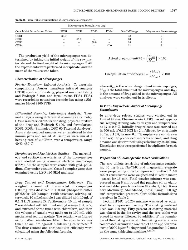
Table 2. Core Tablet Formulations of Dicyclomine Microsponges
Microsponges Formulations (mg)
Core Tablet Formulation Codes FDS1 FDS2 FDS3 FDS4 Na-CMC (mg) Magnesium Stearate (mg)
CDS1 60.0 – – – 18 2CDS2 – 55.0 – – 23 2CDS3 – – 50.0 – 28 2CDS4 – – – 47.0 31 2
The production yield of the microsponges was de-termined by taking the initial weight of the raw ma-terials and the final weight of the microsponges.13 Allthe experiments were performed in triplicate, and themean of the values was taken.
Characterization of Microsponges
Fourier Transform Infrared Analysis. To ascertaincompatibility Fourier transform infrared analysis(FTIR) spectra of the drug, physical mixture of drugand Eudragit S-100, and formulations FDS1–FDS4were recorded in potassium bromide disc using a Shi-madzu Model 8400 FTIR.
Differential Scanning Calorimetry Analysis. Ther-mal analysis using differential scanning calorimetry(DSC) was carried out for the drug, physical mixtureof the drug and Eudragit S-100, and formulationsFDS1–FDS4 (Shimadzu DSC-60 Thermal Analyzer).Accurately weighed samples were transferred to alu-minum pans and sealed. All samples were run at aheating rate of 20◦C/min over a temperature range40◦C–430◦C.
Morphology and Particle Size Studies. The morphol-ogy and surface characteristics of the microspongeswere studied using scanning electron microscope(SEM). All the samples were coated with gold–palla-dium alloy under vacuum. Coated samples were thenexamined using LEO 430 SEM analyzer.
Drug Content and Encapsulation Efficiency. Theweighed amount of drug-loaded microsponges(100 mg) was dissolved in 100 mL phosphate bufferpH 6.8 for 12 h (sample 1) with occasional stirring. Af-terwards, 10 mL of sample 1 was diluted with 10 mL of0.1 N HCl (sample 2). Furthermore, 10 mL of sample2 was diluted with 50 mL of methyl orange (1%, w/v)and extracted three times with chloroform, and thenthe volume of sample was made up to 100 mL withmethylated sodium acetate. The solution was filteredusing 0.45-m membrane filter. The absorbance wastaken at 420 nm against blank using colorimeter.14
The drug content and encapsulation efficiency werecalculated using the following formula.
Actual drug content(%) =(
Mact
Mms
)× 100
Encapsulation efficiency(%) =(
Mact
Mthe
)× 100
where Mact is the actual drug content in microsponges,Mms is the total amount of the microsponges, and Mtheis the amount of drug added to the microsponges. Allanalyses were carried out in triplicate.
In Vitro Drug Release Studies of MicrospongeFormulations
In vitro drug release studies were carried out inUnited States Pharmacopeia (USP) basket appara-tus keeping stirring rate at 50 rpm and temperatureat 37 ± 0.5◦C. Initially drug release was carried outin 900 mL of 0.1N HCl for 2 h followed by phosphatebuffer, pH 6.8, for next 6 h.15 Samples were withdrawnafter regular predecided intervals of time, and drugamount was determined using colorimetry at 420 nm.Dissolution tests were performed in triplicate for eachsample.
Preparation of Colon-Specific Tablet Formulations
The core tablets consisting of microsponges contain-ing 40 mg drug, Na-CMC and magnesium stearatewere prepared by direct compression method.11 Alltablet constituents were weighed and mixed in motor–passel for 15 min. Final powder mixture was com-pressed using 8 mm round flat punches on an eight-station tablet punch machine (Kambert, D-8, Kam-bert Machinery, Ahmedabad, India) using 1000 kg/cm2 compression pressure. Core tablet formulationsare given in Table 2.
Pectin:HPMC (80:20) mixture was used as outershell for compression coating. The coating materialused was 200 mg. Fifty percent of coating materialwas placed in the die cavity, and the core tablet wasplaced in center followed by addition of the remain-der of the coating material. The coating material wascompressed around the core tablet at an applied pres-sure of 2000 kg/cm2 using round flat punches (12 mm)on the same tabletting machine.8,15,16
DOI 10.1002/jps JOURNAL OF PHARMACEUTICAL SCIENCES, VOL. 100, NO. 4, APRIL 2011

1548 JAIN, JAIN, AND SINGH
In Vitro Drug Release Studies of Colon-SpecificFormulations
In vitro drug release studies of colon-specific formu-lations were carried out according to Souder andEllenbogen17 extraction technique using USP basketapparatus with stirring rate 50 rpm at 37 ± 0.5◦C.
The scheme of using the simulated fluids at differ-ent pH was as follows:
At first hour, simulated gastric fluid of pH 1.2 wasused followed by second and third hour mixture ofsimulated gastric and intestinal fluid of pH 4.5, fourthand fifth hours simulated intestinal fluid of pH 6.8and sixth hour simulated intestinal fluid of pH 7.5 wasused. Additionally, Pectinex Ultra SP-L was added tothe dissolution medium at sixth hour in order to simu-late the enzymatic action of the colonic bacteria. Sam-ples were withdrawn at regular intervals of time (i.e.,1 h) and compensate with equal volume of fresh dis-solution medium to maintain the sink condition. Thesamples were analyzed for drug content by measuringabsorbance at 420 nm using colorimetry.
RESULTS AND DISCUSSION
Because of its simplicity and reproducibility, quasi-emulsion solvent diffusion method was used forpreparation of microsponges. Moreover, it has ad-vantage of avoiding solvent toxicity. The drug andpolymer in the ratios 3:1, 6:1, 9:1, and 12:1 weretaken to prepare different microsponge formula-tions, namely FDS1, FDS2, FDS3, and FDS4, respec-tively. In each formulation, the amounts of polymer(200 mg), dichloromethane (5 mL), and PVA (0.5%,w/v) were kept constant. The microsponge formula-tions were prepared using mechanical stirrer (RemiRQ1217-D) at a stirring rate of 1000 rpm for 8 h. Thecomposition of various microsponge formulations aredepicted in Table 1.
Optimization of Formulation and Process Parameters
Effect of Drug–Polymer Ratio on Microsponges
The morphology of the microsponges was studiedby SEM. The representative photographs of the mi-crosponges are shown in Figure 1. The microspongeswere observed to be spherical and uniform with nodrug crystals on the surface. Figure 1 shows thatdrug–polymer ratio has considerable effect on themorphology and size of microsponges. It was observedthat as the ratio of drug to polymer was increased,the particle size decreased. This could probably bedue to the fact that in high drug–polymer ratios, theamount of polymer available per microsponge wascomparatively less. Probably in high drug–polymerratios, less polymer amounts surround, the drug andmicrosponges with smaller size were obtained. The re-
sult supported the claim made by Nokhodchi et al.12
that as the drug–polymer ratio increases, particlesize decreases. The mean particle size of formulationsFDS1–FDS4 is presented in Table 3. The mean parti-cle diameters for the ratios of 3:1 and 12:1 were foundto be 59.54 ± 5.24 and 39.43 ± 4.99 :m, respectively.
Effect of Stirring Rate on the Morphology and Yieldof Microsponges
The effect of stirring rate on the morphology of mi-crosponges is shown in Figure 2. The formulation withthe lower drug to polymer ratio (i.e., 3:1) was chosento investigate the effect of stirring rate on the mor-phology of microsponges. The stirring rate was variedin the range of 500–1000 rpm. The dispersion of thedrug and polymer into the aqueous phase was foundto be dependant on the agitation speed. As the speedincreased, the size of microsponges was reduced, andthe microsponges were found to be spherical and uni-form. When the rate of stirring was increased up to1000 rpm, the spherical microsponges were formedwith mean particle size of 59.54 ± 5.24 :m.
The result was found to be in agreement with thestudied performed by Orlu et al.8 and Perumal.18
However, at higher stirring rates, the production yieldwas found to be decreased, possibly at the higher stir-ring rates, the polymer adhered to paddle due to theturbulence created within the external phase, andhence production yield decreased.
Effect of Volume of Internal Phase on the Productionof Microsponges
It was observed that on increasing the volume of in-ternal phase (dichloromethane) from 5 to 10 mL, mi-crosponges were not formed. This may be due to thedecrease in viscosity of internal phase as discussed byYang et al.19 At increased volume of dichloromethane,the finely dispersed spherical quasi-emulsion dropletswere observed in solvent under the agitation, butas the agitation was discontinued, emulsion dropletsadhered together and coalesce. Consequently, mi-crosponges could not be formed. The result suggeststhat the amount of dichloromethane need to be con-trolled within an appropriate range to effect not onlythe formation of quasi-emulsion droplets at the initialstage but also the solidification of drug and polymerin the droplets.
Effect of Amount of Emulsifying Agent on theProduction Yield and Particle Size of Microsponges
An increase in amount of emulsifying agent resultedin decreased production yield. As the emulsifier isnonionic in nature, it develops some hydrophobic re-gion and dissolves some of the drug and polymer. Also,
JOURNAL OF PHARMACEUTICAL SCIENCES, VOL. 100, NO. 4, APRIL 2011 DOI 10.1002/jps

DICYCLOMINE-LOADED MICROSPONGES-BASED COLONIC DELIVERY 1549
Figure 1. (a-h) Scanning electron microscopy photograph of microscope formulation (drug:eudragit S-100). The photograph coded ‘A’ represents whole image ‘B’ represents surface pho-tographs.
the increase in the amount of emulsifying agent in-creased the particle size. This could be due to theincreased viscosity wherein larger emulsion dropletsformed in larger microsponges.20 The results of theeffect of emulsifying agent on production yield andmean particle size are shown in Table 4. An increasedamount of emulsifying agent decreased the produc-tion yield from 73% to 67% and increased the meanparticle size from 59 to 62 :m.
Characterization of Microsponges
FTIR spectra were recorded to assess the compat-ibility of the drug and excipients.21 FTIR spectraof the drug, physical mixture of drug and EudragitS-100, and formulations FDS1–FDS4 are given inFigure 3. In FTIR spectra of the drug, characteristicC O stretching band was observed at 1716.53 cm−1
(reported peak 1725–1700 cm−1). Eudragit S-100
Table 3. Production Yield, Actual Drug Content, Encapsulation Efficacy, and Mean Particle Size of Various Microsponges Formulations(n = 3)
Formulation Drug–Polymer RatioProduction
Yield (% ± SD)Theoretical Drug
Content (%)Actual Drug
Content (% ± SD)Encapsulation
Efficiency (% ± SD)Mean Particle
Size (:m ± SD)
FDS1 3:1 73.06 ± 0.21 75.00 67.12 ± 0.034 89.49 ± 0.01 59.54 ± 5.24FDS2 6:1 72.30 ± 0.03 85.71 72.39 ± 0.78 84.45 ± 0.34 52.78 ± 4.89FDS3 9:1 68.45 ± 0.002 90.00 78.92 ± 0.41 87.68 ± 0.56 46.76 ± 5.21FDS4 12:1 77.30 ± 0.007 92.30 83.42 ± 0.34 90.37 ± 0.90 39.43 ± 4.99
DOI 10.1002/jps JOURNAL OF PHARMACEUTICAL SCIENCES, VOL. 100, NO. 4, APRIL 2011

1550 JAIN, JAIN, AND SINGH
Figure 2. Scanning electron microscopy photograph of drug:eudragit S-100 microspongesprepared at different stirring rates of (a) 500 rpm; (b) 1000 rpm.
Figure 3. FTIR spectra of dicyclomine, physical mixtureof drug and eudragit S-100 FDSI-FDS4 microsponges for-mulations.
showed carbonyl stretching at 1718.46 cm−1 andbond characteristic to carboxylic group in the range2437–3473 cm−1. All characteristic peaks of the drugwere observed in the FTIR spectra of FDS1–FDS4formulations. These results indicated compatibility ofthe drug with excipients used for microsponge prepa-ration.
Figure 4. Differential scanning calorimetry thermogen ofdicyclomine, physical mixture of drug and Eudragit S-100,and FDSI-FDS4 microsponges formulations.
DSC studies were carried out to confirmcompatibility.22 The thermal behavior of the drug,physical mixture of drug and Eudragit S-100 (1:1),and formulations FDS1–FDS4 are given in Figure 4.The thermograms showed a sharp endothermic peakat 174.23◦C, corresponding to the melting point ofdrug in the crystalline form. The DSC curve of phys-ical mixture of drug and polymers and formulationsFDS1–FDS4 exhibited the characteristic peaks of thedrug. The result is indicative of compatibility between
Table 4. The Effect of Emulsifying Agent on Microsponges Formulations
Internal Phase External Phase
Formulation Code Drug (mg) Polymer (mg) Dichloromethane (mL) Water (mL) PVA (%, w/v) Yield (%) Mean Diameter (:m ± SD)
FDS1 (a) 600 200 5 100 0.5 73.06 ± 0.21 59.54 ± 5.24FDS1 (b) 600 200 5 100 1.0 66.54 ± 5.24 62.43 ± 4.65
JOURNAL OF PHARMACEUTICAL SCIENCES, VOL. 100, NO. 4, APRIL 2011 DOI 10.1002/jps

DICYCLOMINE-LOADED MICROSPONGES-BASED COLONIC DELIVERY 1551
Figure 5. In vitro release profile of dicyclomine from dif-ferent microsponges formulations (FDS1-FDS4).
drug and polymers, and the suitability of preparationprocess.
Production yield, actual drug content, encapsula-tion efficiency, and mean particle size of formulationsFDS1–FDS4 are presented in Table 3. The produc-tion yield, actual drug content, and encapsulation ef-ficiency of FDS1–FDS4 formulations was found to bebetween 73% and 77%, 67% and 83% 89% and 90%,respectively.
The data obtained for various formulations with re-spect to production yield, actual drug content, and en-capsulation efficiency were subjected to t-test at 95%level of significance. No significant difference in rela-tion to these parameters was observed among variousformulations at P < 0.05.
In Vitro Release Studies of the MicrospongeFormulations
The microsponge formulations were subjected to invitro release studies using USP XX1V dissolution as-sembly at the stirring rate at 50 rpm and temperatureat 37 ± 0.5◦C. Initially, drug release was carried outin of 0.1 N hydrochloric acid for 2 h followed by phos-phate buffer, pH 6.8, for next 6 h.
The release profiles obtained for the formulationsFDS1–FDS4 are presented in Figure 5. It was ob-served that the drug release decreased with increasein the amount of polymer for each formulation. This
may be due to the fact that the release of drug from thepolymer matrix takes place after complete swelling ofthe polymer, and as the amount of polymer in theformulation increases, the time required to swell alsoincreases. The release showed a biphasic pattern withan initial burst effect. In the first hour, drug releasewas found to be 17%–30%. This may be attributedto the drug present in the pores of the microspongesor improper entrapment of drug as reported earlierby Mastiholimath et al.23 The cumulative percent re-lease for FDS1–FDS4 at the end of eighth hour wasfound to be 53%–83%.
In vitro dissolution data were analyzed usingvarious mathematical models and are presented inTable 5. Based on highest regression value, the bestfit was observed for Higuchi matrix. The n value forPeppas model was found to be between 0.5 and 1, in-dicative of non-Fickian diffusion.
The in vitro dissolution data were subjected to sta-tistical analysis using analysis of variance. The Pvalue was found to be 0.5930, indicating no signifi-cant difference in the release behavior (P > 0.05).
In Vitro Dissolution Studies of the Colon-Specific TabletFormulations
In order to prepare the compression-coated tablet for-mulations, core tablets were prepared as the firststep. The homogenous granular characteristic of mi-crosponges is due to their highly porous structure andin these means, microsponges have the compressibil-ity to produce strong tablets and 1000–2000 kgf/cm2
pressure did not cause the structure deformation ofmicrosponges.16In vitro drug release studies of thecolon-specific tablet formulations were carried out us-ing USP basket apparatus with stirring rate of 50 rpmat 37 ± 0.5◦C. The release profiles obtained for theformulations CDS1–CDS4 are presented in Figure 6.No drug was released in the first 6 h. After this lagtime of 6 h, the drug release started at the beginningof seventh hour due to the addition of the PectinexUltra SP-L and continued up to fourteenth hour forCDS1 (72.39%), fourteenth hour for CDS2 (91.36%),thirteenth hour for CDS3 (97.01%), and twelfth hourfor CDS4 (97.59%).
The results of in vitro drug release showed thatthe ratio of pectin:HPMC (80:20) protected the cores
Table 5. In vitro Drug Release Models for Different Microsponges Formulations
Zero Order First Order Higuchi /ModelKorsmeyer-Peppa’s
Model
Code R K (mg/h) R K (h−1) R K (mg/h Polyvinyl Alcohol1/2) R n
FDS1 0.9647 6.59 0.9832 0.0983 0.9863 20.538 0.9657 0.7231FDS2 0.9567 8.4377 0.9790 0.1488 0.9821 26.407 0.9578 0.7135FDS3 0.9607 8.2378 0.9927 0.1534 0.9944 25.994 0.9902 0.6201FDS4 0.9720 9.2613 0.9890 0.2034 0.9921 28.816 0.9920 0.6199
DOI 10.1002/jps JOURNAL OF PHARMACEUTICAL SCIENCES, VOL. 100, NO. 4, APRIL 2011

1552 JAIN, JAIN, AND SINGH
Figure 6. In vitro release profile of dicyclomine from colonspecific formulations (CDS1-CDS4).
up to sixth hour, corresponding to the time to reachthe colon and after that under the influence of theenzyme, the system could be degraded faster and de-liver the drug to the proximal colon that forms themain site of bacterial carbohydrate metabolism. So,the results were in accordance with the triggeringmechanism due to the very active metabolism in theproximal part compared with the distal part of colonand the pectin could be degraded by the bacterial en-zyme in colon.
CONCLUSION
This study presents new approach for the preparationof modified microsponges. The prepared microspongesexhibited characteristics of an ideal delivery systemfor colon targeting. The unique compressibility of mi-crosponges offers a new alternative for producing me-chanically strong tablets. Furthermore, colon-specifictablets based on microsponges could provide effectivelocal action as microsponges may selectively be takenup by the macrophages present in the colon.
ACKNOWLEDGMENTS
The authors are thankful to the Director, Schoolof Pharmaceutical Sciences, Shobhit University,Meerut, for providing necessary facilities.
REFERENCES
1. Kraneveld AD, Rijinierse A, Nijkamp FP, Garssen J. 2008.Neuro-immune interaction in inflammatory bowel disease andirritable bowel syndrome: Future therapeutic targets. Eur JPharmacol 585:361–374.
2. McBeth J, Nicholl BI, Musleh M. 2008. Psychosocial risk mark-ers for new onset irritable syndrome-result of a large prospec-tive population-based study. Pain 137:147–155.
3. Paharia A, Yadav AK, Rai G, Jain SK, Pancholi SS,Agrawal GP. 2007. Eudragit-coated pectin microspheres of5-fluorouracil for colon targating. AAPS Pharma Sci Tech8:E1–E7.
4. Varshosaz J, Dehkordi AJ, Golanfshan S. 2006. Colon-specificdelivery of mesalazine chitosan microspheres. J Microencap-sulation 23: 329–339.
5. Kinget R, Kalale W, Vervoort L, Mooter GV. 1998. Colonic drugdelivery. J Drug Target 6:129–149.
6. Sinha VR, Kumria R. 2003. Coating polymers for colon specificdrug delivery: A comparative in vitro evaluation. Acta Pharm53:41–47.
7. Chourasia MK, Jain SK. 2003. Pharmaceutical approaches tocolon targeted drug delivery systems. J Pharm PharmaceutSci 6:33–56.
8. Orlu M, Cevher E, Araman A. 2006. Design and evaluationof colon specific drug delivery system containing flurbiprofenmicrosponges. Int J Pharm 318: 103–117.
9. Kosaraju SL. 2005. Colon targeted delivery systems: Review ofpolysaccharides for encapsulation and delivery. Crit Rev FoodSci Nutr 45:251–258.
10. Rubinstein A. 2000. Natural polysaccharides as targeting toolsof drugs to the human colon. Drug Dev Res 50:435–443.
11. Vandamme TH, Lenourry A, Charroeau C, Chaumeil JC. 2002.The use of polysaccharides to target drugs to the colon. Carbo-hydr Polym 48:219–231.
12. Nokhodchi A, Jelvehgari M, Siahi MR, Mozafari MR. 2007.Factors affecting the morphology of benzoyl peroxide mi-crosponges. Micron 38:834–840.
13. Jelvehgari M, Siahi-Shadbad MR, Azarmi S, Martin GP,Nokhodchi A. 2006. The microsponge delivery system of ben-zoyl peroxide: Preparation, characterization and release stud-ies. Int J Pharm 308:124–132.
14. Sethi PD. 2008. Quantitative analysis of drugs in pharma-ceutical formulations. New Delhi, India: CBS publishers anddistributors.
15. Kawashima Y, Niwa T, Takeuchi H, Hino T, Ito Y. 1992.Control of prolonged drug release and compression proper-ties of ibuprofen microsponges with acrylic polymer, eudragitRS by changing their intrapartical porosity. Chem Pharm Bul40:196–201.
16. Comoglu T, Gonul N, Baykara T. 2003. Preparation and invitro evaluation of modified release ketoprofen microsponges.Il Farmaco 58:101–106.
17. Chourasia MK, Jain SK. 2004. Design and development ofmultiparticulate system fort targeted drug delivery to colon.Drug Deliv 11: 201–207.
18. Perumal D. 2001. Microencapsulation of ibuprofen andEudragit R© S 100 by the emulsion solvent diffusion technique.Int J Pharm 218:1–11.
19. Yang M, Cui F, You B, Fan Y. 2003. Preparation of sustained-release nitrendipine microspheres with Eudragit RS andAerosil using quasi-emulsion solvent diffusion method. Int JPharm 259: 103–113.
20. Devrim B, Canefe K. 2006. Preparation and evaluation ofmodified release ibuprofen microspheres with acrylic polymers(eudragit R©) by quasi emulsion solvent diffusion method: Effectof variables. Acta Poloniae Pharmaceutica and Drug Res 63:521–534.
21. Mukherjeea B, Mahapatraa S, Guptab R, Patraa B, Tiwarib A,Arora P. 2005. A comparison between povidone-ethylcelluloseand povidone-eudragit transdermal dexamethasone matrixpatches based on in vitro skin permeation. Eur J Pharm Bio-pharm 59:475–483.
22. Ceschel GC, Badiello R, Ronchi C, Maffei P. 2003. Degra-dation of components in drug formulations: A comparisonbetween HPLC and DSC methods. J Pharm Biomed Anal32:1067–1072.
23. Mastiholimath VS, Dandagi PM, Jain SS, Gadad AP, Kulka-rni AR. 2007. Time and pH dependent colon specific, pulsatiledelivery of theophylline for noctural asthma. Int J Pharm328:49–56.
JOURNAL OF PHARMACEUTICAL SCIENCES, VOL. 100, NO. 4, APRIL 2011 DOI 10.1002/jps