Embed Size (px)
Citation preview
Factor VIII and IX Gene Polymorphisms and CarrierAnalysis in Indian Population
Shrimati Shetty, 1 Kanjaksha Ghosh, 1 Anil Pathare, 1 Roshan Colah, 1 Suresh Badakare, 1 andDipika Mohanty 1*
1Institute of Immunohaematology, Indian Council of Medical Research, Bombay, India,2Department of Haematology, King Edward Memorial Hospital, Bombay, India
The efficacy of the three common intra- and extragenic polymorphic sites of the factor VIIIand IX genes has been examined in the Indian population, with an aim to develop astrategy that would be accurate and informative, yet economical. The approach for he-mophilia A carrier detection includes tests for Bcl I, XbaI, and TaqI polymorphic sites forintrons 18 and 22 and the extragenic locus St 14, respectively, whereas for hemophilia B,tests include detection of TaqI, DdeI, and HhaI polymorphic sites for introns 4 and 1, andthe 3 * flanking region of the factor IX gene, respectively. In hemophilia A, the cumulativeefficiency of these three polymorphisms has been found to be 100%, since all 37 testedfamilies were informative for at least one of these three polymorphisms. It is of interestto note that a case of recombination between St 14 and the factor VIII gene was alsoobserved. Of the 47 unrelated X chromosomes examined (normal = 10, factor VIII:Cdeficiency = 37), heterozygosity for Bcl I, XbaI, and St 14 was found to be 47%, 36%, and86%, respectively, in the factor VIII gene. However, when 37 unrelated X chromosomes(normal = 10, factor IX:C = 27) were analyzed for polymorphism with TaqI, DdeI, and HhaI,it was found that the polymorphism detection rate was only 18% for the TaqI site but 45%each for the DdeI and HhaI sites, in the factor IX gene. This indicates a low effectivenessof the TaqI restriction site in carrier analysis of hemophilia B families in our population.Am. J. Hematol. 54:271–275, 1997 © 1997 Wiley-Liss, Inc.
Key words: hemophilia; polymorphism; carrier detection
INTRODUCTION
In a developing country like India, carrier detectionand prenatal diagnosis of hemophilia will go a long wayin reducing the burden of hemophilia care in the society.
Direct detection of mutations would obviously be themethod of choice for carrier detection and prenatal diag-nosis of hemophilia A and B. However, due to the highheterogeneity of mutations in hemophilia, and the sizeand structural complexity of factor VIII and IX genes,direct diagnosis of the molecular defect causing hemo-philia is often difficult, and linkage analysis with restric-tion fragment-length polymorphism (RFLP) is the mostuseful approach.
There are two possible limitations of linkage analysisin prenatal diagnosis: 1) recombination between the mu-tant gene and the polymorphic marker, and 2) the rate ofinformativeness of the marker, i.e., homozygosity ofthe women at the marker site, resulting in failure to
discriminate the mutant gene from the normal gene.The risk of recombination is directly related to the dis-tance between the marker and the mutation, while in-formativeness depends on the prevalence of the poly-morphic site in a given population. Despite these po-tential limitations, the combined use of all available in-tra- and extragenic polymorphic sites makes an earlydiagnosis feasible in the great majority of families at risk,even though confirmation on fetal blood is sometimesnecessary. Furthermore, there is no information avail-able regarding either the pattern of mutation in hemo-philia A and B or the heterozygosity rate in the Indianpopulation.
*Correspondence to: Dipika Mohanty, M.D., Ph.D., Director, Instituteof Immunohaematology, Indian Council of Medical Research, 13thfloor, Multistoreyed Complex, King Edward Memorial Hospital,Parel, Bombay 400 012, India.
Received for publication 12 December 1995; accepted 6 November1996.
American Journal of Hematology 54:271–275 (1997)
© 1997 Wiley-Liss, Inc.

Since the heterozygosity rates for different RFLPs dif-fer in different ethnic groups, we examined the utilityrates of the three common polymorphic sites each of thefactor VIII and IX genes i.e., theBclI, XbaI, and extra-genic TaqI sites of the factor VIII gene, and theTaqI,DdeI, and 38 HhaI sites of the factor IX gene. Our resultssuggest a strategy for providing accurate genotype as-signment for virtually all female relatives of hemophili-acs in an economic and efficient way, which is verymuch essential for our country.
MATERIALS AND METHODS
The study involved 37 unrelated families with hemo-philia A, 27 unrelated families with hemophilia B, and 10unrelated normal families where no family history ofhemophilia or any bleeding disorders could be elicitedover the last four generations.
One female from each of these families was selectedfor study. The females selected from the hemophiliafamilies were obligate carriers and were mothers of theindex patients. Index patients and their fathers were alsostudied.
Routine coagulation tests and relevant factor assayswere done at least twice on each index patient to confirmthe nature and severity of hemophilia. Platelet aggrega-tion with ristocetin and Von Willebrand factor (VWF)
assay on relevant patients were done to rule out VonWillebrand’s disease. DNA was extracted from periph-eral blood leukocytes of the individuals under study, us-ing standard techniques [1].
XbaI/KpnI, TaqI/St 14 RFLPs for the factor VIII geneandTaqI RFLPs for the factor IX gene were studied bySouthern blotting.BclI RFLPs for the factor VIII gene,andDdeI and HhaI RFLPs for the factor IX gene werestudied by polymerase chain reaction (PCR). Three fami-lies with hemophilia B were also studied forMnlI andBamHI RFLPs, because other RFLPs did not give ad-equate information to assign carrier status in these threefamilies.
Southern BlottingTen mg DNA were digested with various restriction
endonucleases according to the manufacturer’s instruc-tions and subjected to electrophoresis for 16 h at 25 voltsin 0.8% agarose gel. DNA fragments were transferred tonylon membranes (Hybond N+, Amersham, Bucking-hamshire, U.K.) by Southern blotting. Probes were gen-erous gifts from Prof. G.G. Brownlee, Sir William DunnSchool of Pathology, Oxford and Dr. J.L. Mandel,INSERM U-184, France.
Polymerase Chain ReactionFive hundredhg DNA were used for PCR amplifica-
tion of (1) intron 18 of the factor VIII gene for theBclI
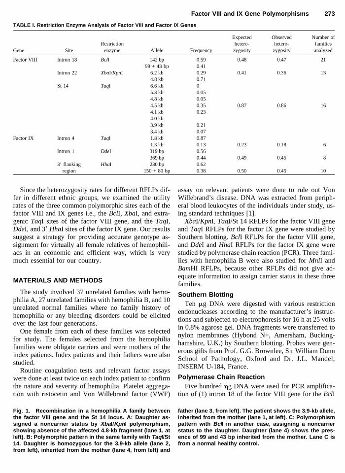
TABLE I. Restriction Enzyme Analysis of Factor VIII and Factor IX Genes
Gene SiteRestriction
enzyme Allele Frequency
Expectedhetero-
zygosity
Observedhetero-
zygosity
Number offamiliesanalyzed
Factor VIII Intron 18 BclI 142 bp 0.59 0.48 0.47 2199 + 43 bp 0.41
Intron 22 XbaI/KpnI 6.2 kb 0.29 0.41 0.36 134.8 kb 0.71
St 14 TaqI 6.6 kb 05.3 kb 0.054.8 kb 0.054.5 kb 0.35 0.87 0.86 164.1 kb 0.234.0 kb3.9 kb 0.213.4 kb 0.07
Factor IX Intron 4 TaqI 1.8 kb 0.871.3 kb 0.13 0.23 0.18 6
Intron 1 DdeI 319 bp 0.56369 bp 0.44 0.49 0.45 8
38 flanking HhaI 230 bp 0.62region 150 + 80 bp 0.38 0.50 0.45 10
Fig. 1. Recombination in a hemophilia A family betweenthe factor VIII gene and the St 14 locus. A: Daughter as-signed a noncarrier status by XbaI/Kpn I polymorphism,showing absence of the affected 4.8-kb fragment (lane 1, atleft). B: Polymorphic pattern in the same family with TaqI/St14. Daughter is homozygous for the 3.9-kb allele (lane 2,from left), inherited from the mother (lane 4, from left) and
father (lane 3, from left). The patient shows the 3.9-kb allele,inherited from the mother (lane 1, at left). C: Polymorphismpattern with Bcl I in another case, assigning a noncarrierstatus to the daughter. Daughter (lane 4) shows the pres-ence of 99 and 43 bp inherited from the mother. Lane C isfrom a normal healthy control.
Factor VIII and IX Gene Polymorphisms 273

polymorphism, and (2) intron 1 and the 38 flanking re-gion of the factor IX gene for theDdeI and HhaI poly-morphisms, respectively. The primer sequences were aspreviously described for various PCR amplifications [2].The reaction was performed in a total incubation volumeof 50 ml, and the PCR conditions and number of cycleswere as described earlier [3–6]. The amplified sampleswere either run in 10% polyacrylamide gel or 2% agarosegel for resolution of amplified fragments.
RESULTS
The polymorphism heterozygosity for the different re-striction sites in the factor VIII and IX genes and thenumber of families analyzed for carrier status by differ-ent polymorphisms are shown in Table I.
Factor VIII Gene
TaqI RFLPs for the factor VIII gene had eight alleles,and the frequency of heterozygosity in our populationwas 86%. This could have been a very useful marker forcarrier detection in hemophilia A, but unfortunately, asthis is an extragenic marker, occasional recombinationcan confound interpretation, as shown in Figure 1A, B,which shows the results from the same family with he-mophilia A.
It is evident from Figure 1A that the male patient hasinherited the 4.8-kb RFLP and the diseased gene from hismother. The daughter has inherited the 6.2-kb RFLPfrom her mother, which is not associated with the dis-ease. Hence, as shown in Figure 1A, the daughter is nota carrier. However, the results of RFLP withTaqI/St 14,as shown in Figure 1B, indicate that the daughter is acarrier, since 3.9 kb from the mother, associated with thedisease, are present in the daughter. This anomaly oc-curred because of recombination between the extragenicTaqI/St 14 restriction site and the healthy factor VIIIgene.
BclI RFLP (intron 18) was heterozygous in 47% of ourpatients, followed byXbaI/KpnI RFLP (intron 22) in36% of our patients. Figure 1C shows a typical RFLP inhemophilia A, usingBclI as the restriction enzyme. In allhemophilia A families studied, either one or the other ofthe three restriction enzyme-related RFLPs were hetero-zygous in the female, and hence informative.
Factor IX Gene
DdeI (intron 1) RFLP was heterozygous in 45% of thefemales, as also were theHhaI RFLPs (38 flanking re-gion). TaqI (intron 4) RFLPs yielded the lowest hetero-zygosity, of 18%, in our population.
DISCUSSION
India has a population of 920 million people. A con-servative estimate indicates 51,000 patients with hemo-
philia A, and 10,000 patients with hemophilia B. Unfor-tunately, most of these patients have no access to high-quality hemophilia care as provided by the Westernnations, mainly due to financial and logistic constraints.
Gene tracking by polymorphism analysis has limita-tions, particularly with regard to apparent new mutations,the possibility of mosaicism, and the question of pater-nity. However, the relative simplicity of the procedureand its application to all hemophiliacs, irrespective ofgenetic background, has made these analyses extremelyuseful in hemophilia genetic studies.
The intron 18BclI restriction site in the factor VIIIgene showed the maximum heterozygosity, i.e., 47% inour population, as compared to other populations re-ported earlier, whereas theXbaI/KpnI restriction siteshowed a slightly reduced heterozygosity as compared toCaucasians, Japanese, and Chinese populations [2]. Inthe present series of 37 families analyzed for carrier sta-tus, all were informative, with at least one of the threepolymorphisms. Carrier status in only three hemophiliaA families in our series could not be confirmed by intra-genic polymorphisms.
A case of recombination between the factor VIII geneand the St 14 locus was observed during the familyanalysis. The consultant was assigned a noncarrier statusbased onBclI and XbaI/KpnI polymorphisms, butTaqI/St 14 analysis indicated that she was a carrier (Fig. 1).This showed the event of recombination between theextragenic locus (St 14) and factor VIII gene. Under suchcircumstances, the results from intragenic RFLPs may berelied upon.
The efficiency of theTaqI restriction site in the factorIX gene has been found to be quite low in our population,i.e., 18%, as compared to Caucasians with a heterozy-gosity of 45% [6]; but it is much higher than in theJapanese (0%) [7], and Chinese and Malays (0.02%)[8,9]. Of the 27 hemophilia B families analyzed withTaqI, DdeI, andHhaI, only three families failed to revealany of the three polymorphic sites. However, they wereassigned carrier status byMnlI and BamHI polymor-phism segregation analysis.MnlI was informative fortwo families, andBamHI for one family.
Thus, from the present work it can be concluded thatthe ability of the above three polymorphic sites of thefactor VIII gene to detect carrier status is almost 100% inour Indian population. Further, it can also be emphasizedthat one should utilize extragenic polymorphism detec-tion when the intragenic polymorphisms are not infor-mative. TheTaqI site in the factor IX gene is probablythe restriction site of last preference in our population,with only 18% heterozygosity. Other intragenic markers,like MnlI or XmnI sites, should be analyzed for hetero-zygosity rating and subsequently carrier analysis.
Recently, inversion analysis of intron 22 [10,11] andminisatellite DNA analysis [2] around the factor VIII
274 Shetty et al.

gene have been increasingly used to study hemophilia Apatients and their families. Major advances have alsobeen made in the elucidation of mutations involving fac-tor IX gene in hemophilia B patients [12]. These tech-niques have yet to find their places in research as well asin clinical practice, in developing countries such as India.
ACKNOWLEDGMENTS
The authors thank Prof. P.M. Mannucci, Director andChairman, the Angela Bianchi Bonomi Hemophilia andThrombosis Center (Milan, Italy) for his generous help incarrying out this work. We acknowledge the secretarialassistance of Mr. C.T. Kulkani at Institute of Immuno-haematology in preparing this manuscript.
REFERENCES
1. Sambrook J, Fritsch EF, Manniatis T: ‘‘Molecular Cloning: A Labo-ratory Manual,’’ Vol. 2. New York Cold Spring Harbor LaboratoryPress, 1989, p. 14.
2. Peak IR, Lillicrap DP, Boulyjenkov V, Briet E, Chan V, Ginter EK,Kraus EM, Ljung R, Mannucci PM, Nicolaides K, Tuddenham EGD:Haemophilia: Strategies for carrier detection and prenatal diagnosis.Bull WHO 71:429, 1993.
3. Kogan SC, Doherty M, Gitschier J: An improved method of prenataldiagnosis of genetic diseases by analysis of amplified DNA sequences.Application to hemophilia A. N Engl J Med 317:985, 1987.
4. Winship PR, Rees DJG, Alkan M: Detection of polymorphisms atcytosine phosphoguanidine dinucleotides and diagnosis of haemo-philia B carriers. Lancet 1:631, 1989.
5. Bowen DJ, Thomas P, Webb CE, Bignell P, Peake IR, Bloom AL:Facile and rapid analysis of three DNA polymorphisms within thehuman factor IX gene using the polymerase chain reaction. Br J Hae-matol 77:559, 1991.
6. Winship PR, Anson DS, Rizza CR, Brownlee GG: Carrier detection inhaemophilia B using two further intragenic restriction fragment lengthpolymorphisms. Nucleic Acids Res 12:8861, 1984.
7. Kojima T, Tanimoto M, Kamiya T, Obata Y, Takahashi T, Ohno R,Kurachi K, Saito H: Possible absence of common polymorphism incoagulation factor IX gene in Japan. Blood 69:349, 1987.
8. Chan V, Yip B, Tong TMF, Chan TPT, Lau K, Yam I, Chan TK:Molecular defects in haemophilia B: Detection by direct restrictionenzyme analysis. Br J Haematol 79:63, 1991.
9. Graham JB, Kunkel GR, Egilmez NK, Wallmark A, Fowlkrs DM,Lord ST: The varying frequencies of five DNA polymorphisms ofX-linked coagulant factor IX in eight ethnic groups. Am J Hum Genet49:537, 1991.
10. Lakich D, Kazazian HH, Antonarakis SE, Gitschier J: Inversions dis-rupting the factor VIII gene are a common cause of severe haemophiliaA. Nat Genet 5:236, 1993.
11. Naylor J, Brinke A, Hassock S, Green P, Gianelli F: CharacteristicmRNA abnormality found in half the patients with severe haemophiliaA is due to large DNA inversions. Hum Mol Genet 2:1773, 1993.
12. Ketterling RP, Vielhaber E, Bottema CDK, Schaid DJ, Cohen MP,Sexaurer CL, Sommer SS: Germ-line origins of mutation in familieswith haemophilia B: The sex ratio varies with the type of mutation.Am J Hum Genet 52:152, 1993.
Factor VIII and IX Gene Polymorphisms 275