Embed Size (px)
Citation preview

Facsimile reproduction of the cover of an original reprint of the 1924 article by HansSpemann and Hilde Mangold, with a handwritten dedication by H. Spemann which
reads "With best regards, H.S."(Courtesy of K. Sander, Freiburg).

The following article, reformatted and copy edited at the IJDB Editorial Office with the help of KlausSander, is Viktor Hamburger´s translation of the original 1924 paper by Hans Spemann and HildeMangold entitled: "Über Induktion von Embryonalanlagen durch Implantation artfremderOrganisatoren", published in Archiv für Mikroskopische Anatomie und Entwicklungsmechanik, 100: 599-638, 1924. This translation first appeared in "Foundations of Experimental Embryology" (B.H. Willierand J.M. Oppenheimer, eds.), Prentice Hall, Inc., Englewood Cliffs, N.J. USA, pp. 146-184, 1964. Theillustrations were taken from the original article in German.

Induction of Embryonic Primordia by Implantationof Organizers from a Different Species
by
HANS SPEMANN and HILDE MANGOLD (Née Pröscholdt)
Freiburg i.B.
With 25 illustrations(Submitted 1 June 1923)
CONTENTS
I. Introduction II. Experimental Analysis
Experiment Triton 1921, Um 8b (Figs. 1-6); Triton 1922, Um 25b (Figs. 7-9); Triton 1922, Um 214;Triton 1922, Um 131b (Figs. 10-15); Triton 1922, Um 83 (Figs. 16-18); Triton 1922, Um 132(Figs. 19-25).
III. Discussion of the Results1. Origin and prospective significance [normal fate] of the organizer
and site of its implantation2. Behavior of the organizer after implantation3. Structure of the secondary embryonic primordium4. The causes for the origin of the secondary embryonic anlage5. The organizer and the organizing center
IV. Summary of Results V. References
I. Introduction
In a Triton embryo, at the beginning of gastrulation, the different areas are not equivalent withrespect to their determination. It is possible to exchange by transplantation parts of the ectoderm atsome distance above the blastopore that in the course of further development would have become neuralplate and parts that would have become epidermis, without disturbing normal development by thisoperation. This is feasible not only between embryos of the same age and of the same species but alsobetween embryos of somewhat different age and even between embryos of different species (Spemann1918, 1921). For instance, presumptive epidermis of Triton cristatus transplanted into the forebrainregion of Triton taeniatus can become brain; and presumptive brain of Triton taeniatus transplantedinto the epidermal region of Triton cristatus can become epidermis. Both pieces develop according totheir new position; however they have the species characteristics with which they are endowed accordingto their origin. O. Mangold (1922, 1923) has extended these findings and has shown that prospectiveepidermis can furnish not only neural plate but even organs of mesodermal origin, such as somites andpronephric tubules. It follows from these experimental facts, on the one hand, that the exchangeablepieces are still relatively indifferent with respect to their future fate; and, on the other hand, thatinfluences of some sort must prevail in the different regions of the embryo that determine the later fateof those pieces that are at first indifferent.
Notes added by the IJDB Editorial Office: 1. The serial number of each experiment, e.g. Um 25, refers to two embryos (a andb), between which transplants were exchanged. Thus "a" usually refers to the donor cristatus embryo while "b" typicallyrepresents the host taeniatus embryo. 2. It is worthwhile noting that all figures in this paper were hand-drawn by HildeMangold. The drawings of histological sections are based on photographic paper prints. On these, each nucleus and cellborder was traced with Indian ink. Thereafter, the silver halogenide grains were removed chemically, after which thedrawing stood out on the white background. This method was described in Spemann (1918, p. 545).
[Abbreviations used in this paper: Bl, blastopore; Oc, optic vesicles; pc, pericardium; pr. Med, primary neural tube; sec. Ch,secondary notochord; sec. D, secondary intestine; sec. Lab, secondary otocyst; sec. Med, secondary neural tube; sec. Mes,secondary mesoderm; sec. Pron, secondary pronephric duct; sec. Uw, secondary somite; Um X, Urmund (meaning "primitivemouth" or blastopore) followed by the serial number "X" of the experiment.]

16 Hans Spemann and Hilde Mangold
A piece from the upper lip of the blastopore behaves quite differently. If it is transplanted into theregion that would later become epidermis, it develops according to its origin; in this region, a smallsecondary embryonic primordium develops, with neural tube, notochord and somites (Spemann 1918).Such a piece therefore resists the determining influences that impinge on it from its new environment,influences that, for instance, would readily make epidermis out of a piece of presumptive neural plate.Therefore, it must already carry within itself the direction of its development; it must be determined.Lewis (1907) had already found this for a somewhat later developmental stage, when he implanted asmall piece from the upper and lateral blastopore lip under the epidermis of a somewhat older embryoand saw it develop there into neural tissue and somites.
It suggested itself from the beginning that effects might emanate from these already determined partsof the embryo that would determine the fate of the still indifferent parts. This could be proved by cuttingthe embryo in half and shifting the halves with respect to each other; in this case, the determined partproved to be decisive for the direction that subsequent development would take. For instance, theanimal half of the gastrula was rotated 90° or 180° with respect to the vegetal half; determination thenspread from the lower vegetal piece, that contained just the upper lip, to the upper animal piece. Or twogastrula halves of the same side, for instance two right ones, were fused together. As a result, the halfblastoporal lips completed themselves from adjacent material of the fused other half, and in this way,whole neural plates were formed (Spemann 1918).
Thus, the concept of the organization center emerged; that is, of a region of the embryo that haspreceded the other parts in determination and thereupon emanates determination effects of a certainquantity in certain directions. The experiments to be presented here are the beginning of the analysisof the organization center.
Such a more deeply penetrating analysis presupposes the possibility of subdividing the organizationcenter into separate parts and of testing their organizing capacities in an indifferent region of theembryo. This experiment has already been performed, and it was precisely this experiment that gavethe first indication that the parts of the embryo are not equivalent at the beginning of gastrulation (1918).However, this intraspecific, homoplastic transplantation did not make it possible to ascertain how thesecondary embryonic anlage that originated at the site of the transplant was constructed, that is, whichpart of it was derived from the material of the implant and which part had been induced by the implantfrom the material of the host embryo. The identification of these two components is made possible byheteroplastic transplantation, as for instance by implantation of organizers from Triton cristatus intoindifferent material of Triton taeniatus.
This experiment, that followed logically from its presuppositions, was performed during the summersof 1921 and 1922 by Hilde Mangold née Pröscholdt. It gave at once the expected result that has alreadybeen reported briefly (Spemann 1921, pp. 551 and 568). In the following, we shall present the basic factin more detail.
II. Experimental Analysis
Fig. 1. Um 8 crist. The cristatus embryo atthe neurula stage. The taeniatus trans-plant is dark and elongated; it is located inthe presumptive neural plate. 20X.
Nothing new need be said concerning the experimental tech-nique; it was the same as in previous experiments (Spemann,1920).
Of the species of Triton available, taeniatus can best toleratethe absence of the egg membrane, from early developmentalstages on and it is the easiest to rear. Hence the organizer that wasto be tested for its capacities was always taken from a cristatusembryo and usually implanted into the presumptive epidermis ofa taeniatus embryo. The place of excision was marked by implan-tation of the piece removed from the taeniaius embryo; that is, thepieces were exchanged.
Experiment Triton 1921, Um 8b. The exchange was madebetween a cristatus embryo with distinctly U-shaped blastoporeand a taeniatus embryo of the same stage. A small circular pieceat some distance above the blastopore was removed from the
Induction of embryonic primordia by implantation of organizers from a different species 17
a few hours later, neural folds appeared, indicating the contour of a future neural plate. The implantwas still distinctly recognizable in the midline of this plate; it extended forward from the blastopore asa long narrow strip, slightly curved, over about two-thirds of the plate (Fig. 3).
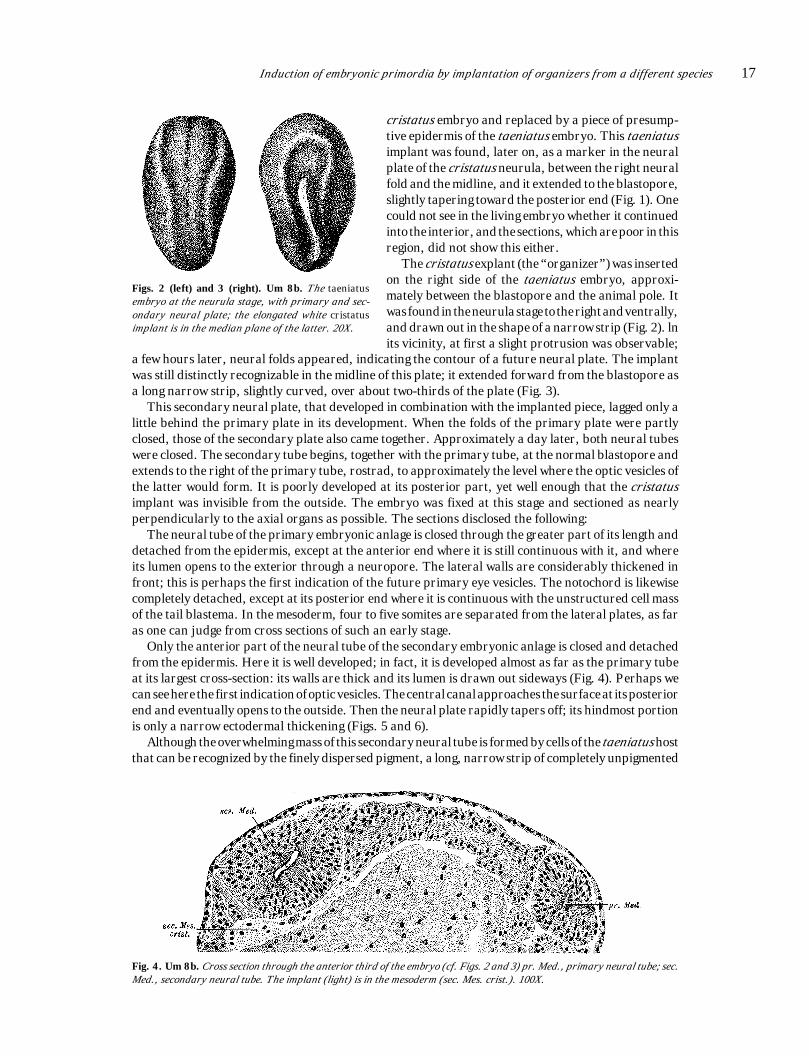
This secondary neural plate, that developed in combination with the implanted piece, lagged only alittle behind the primary plate in its development. When the folds of the primary plate were partlyclosed, those of the secondary plate also came together. Approximately a day later, both neural tubeswere closed. The secondary tube begins, together with the primary tube, at the normal blastopore andextends to the right of the primary tube, rostrad, to approximately the level where the optic vesicles ofthe latter would form. It is poorly developed at its posterior part, yet well enough that the cristatusimplant was invisible from the outside. The embryo was fixed at this stage and sectioned as nearlyperpendicularly to the axial organs as possible. The sections disclosed the following:
The neural tube of the primary embryonic anlage is closed through the greater part of its length anddetached from the epidermis, except at the anterior end where it is still continuous with it, and whereits lumen opens to the exterior through a neuropore. The lateral walls are considerably thickened infront; this is perhaps the first indication of the future primary eye vesicles. The notochord is likewisecompletely detached, except at its posterior end where it is continuous with the unstructured cell massof the tail blastema. In the mesoderm, four to five somites are separated from the lateral plates, as faras one can judge from cross sections of such an early stage.
Only the anterior part of the neural tube of the secondary embryonic anlage is closed and detachedfrom the epidermis. Here it is well developed; in fact, it is developed almost as far as the primary tubeat its largest cross-section: its walls are thick and its lumen is drawn out sideways (Fig. 4). Perhaps wecan see here the first indication of optic vesicles. The central canal approaches the surface at its posteriorend and eventually opens to the outside. Then the neural plate rapidly tapers off; its hindmost portionis only a narrow ectodermal thickening (Figs. 5 and 6).
Although the overwhelming mass of this secondary neural tube is formed by cells of the taeniatus hostthat can be recognized by the finely dispersed pigment, a long, narrow strip of completely unpigmented
Figs. 2 (left) and 3 (right). Um 8b. The taeniatusembryo at the neurula stage, with primary and sec-ondary neural plate; the elongated white cristatusimplant is in the median plane of the latter. 20X.
cristatus embryo and replaced by a piece of presump-tive epidermis of the taeniatus embryo. This taeniatusimplant was found, later on, as a marker in the neuralplate of the cristatus neurula, between the right neuralfold and the midline, and it extended to the blastopore,slightly tapering toward the posterior end (Fig. 1). Onecould not see in the living embryo whether it continuedinto the interior, and the sections, which are poor in thisregion, did not show this either.
The cristatus explant (the “organizer”) was insertedon the right side of the taeniatus embryo, approxi-mately between the blastopore and the animal pole. Itwas found in the neurula stage to the right and ventrally,and drawn out in the shape of a narrow strip (Fig. 2). lnits vicinity, at first a slight protrusion was observable;
Fig. 4. Um 8b. Cross section through the anterior third of the embryo (cf. Figs. 2 and 3) pr. Med., primary neural tube; sec.Med., secondary neural tube. The implant (light) is in the mesoderm (sec. Mes. crist.). 100X.

18 Hans Spemann and Hilde Mangold
Fig. 6. Um 8b. Cross section in the region of the blastopore (Bl.) (cf. Figs. 2 and 3). pr. Med., primary neural tube; sec. Med.,secondary neural tube. The implant (light) has several cells in the secondary neural tube, with its main mass in the mesoderm(sec. Mes. crist.). 100X.
At its posterior end, the cristatus strip reaches the blastopore, and it is continuous with a mass ofcristatus cells that is located between the secondary neural tube and the mesoderm on one side, and theendoderm on the other (Fig. 6). Because of their position one would be inclined to consider these cellsas endoderm; but in size they resemble more the mesoderm of the taeniatus embryo, with which theyare associated. At any rate, this cell mass, which extends a bit farther rostrad, has reached its positionby invagination around the blastoporal lip. There is yet another mass of cristatus cells still fartherrostrad. It has the form of a thin plate underlying the anterior part of the induced neural tube, as faras it is closed; at its anterior end and at its sides, it coincides approximately with the edge of the tube,and at its posterior end, it extends to the ectodermal strip of the implant. This plate is incorporated inthe normal taeniatus mesoderm (Fig. 4). It is not differentiated further into notochord or somites.
Altogether, a rather substantial part of the implant remained in the ectoderm. This portion wasgreatly stretched in length; as a result, the circular white disk that was implanted has become a longnarrow strip that turns inwards around the blastoporal lip. Shifting of cells in the surroundingepidermis may have played a role in these form changes; the extent to which this occurs would have tobe tested by implantation of a marker of indifferent material. A piece from a region near the upper lipof the blastopore could handily be considered as suitable for this purpose. We know from earlierexperiments (Spemann 1918, 1921) that convergence and stretching of the cell material occurs at theposterior part of the neural plate. It is improbable that the cells of the neural plate are entirely passivein this process; rather, they may have an inherent tendency to shift that perhaps has been, together withother characteristics, induced by the underlying endo-mesoderm. This tendency would be retained by
Fig. 5. Um 8b. Cross sec-tion through middle thirdof the embryo (cf. Figs. 2and 3). pr. Med., primaryneural tube; sec. Med., sec-ondary neural tube. Theimplant (light) is in the sec-ondary neural tube.
cells is intercalated in its floor, in sharp contrast to the adjacent regions. This white strip is part of thecristatus implant that was clearly recognizable from the outside in the living embryo before the neuralfolds closed (Fig. 3). The anterior end of this strip is approximately at the point where the thickness ofthe neural tube decreases rather abruptly; it opens to the outside shortly thereafter. The strip is wedge-shaped, with the pointed edge toward the outside; as a result, only the tapering ends of the cells reachthe surface of the embryo (Figs. 5 and 6) or the central canal at the short stretch where they border it.

Induction of embryonic primordia by implantation of organizers from a different species 19
the piece in the foreign environment. In this way we might also explain the fact that the piece gains contactwith the invaginating region of the normal blastoporal lip, although it was originally far distant from it. Onceit has arrived there by active stretching, it could be carried along, at least in part, by local cell shiftings.
Whereas this posterior cell mass is continuous with the cell strip that has remained on the surface,it is separated from the more anterior cristatus cell plate by taeniatus mesoderm. Therefore, thisanterior plate that underlies the neural tube cannot have arrived at its position by invagination aroundthe upper blastoporal lip; it must have been located in the deeper position from the beginning.Undoubtedly it derives from the inner layer of the implant; hence it was originally just under thecristatus cells, some of which are now formed partly in the neural plate as a narrow strip, and othersof which had migrated inside around the blastoporal lip. These displacements carried it along andbrought it forward to such an extent that now its posterior margin is approximately level with theanterior end of the cristatus cell strip in the neural tube.
Although a piece of presumptive neural plate taken from a region a little anterior to the actualtransplant would have become epidermis after transplantation to presumptive epidermis, this implanthas resisted the determinative influences of the surroundings and has developed essentially accordingto its place of origin. Its ectodermal part has become part of the neural plate and the endo-mesodermalpart has placed itself beneath it.
Furthermore, not only did the implant assert itself, but it made the indifferent surroundingssubservient to it and it has supplemented itself from these surroundings. The host embryo has developeda second neural plate out of its own material, that is continuous with the small strip of cristatus cells andunderlain by two cell plates of cristatus origin. This secondary plate would not have arisen at all withoutthe implant, hence it must have been caused, or induced, by it.
There seems to be no possible doubt about this. However, the question remains open as to the wayin which the induction has taken place. In the present case it seems to be particularly plausible to assumea direct influence on the part of the transplant. But even under this assumption, there are still twopossibilities open. The ectodermal component of the transplant could have self-differentiated into thestrip of neural plate, and could have caused the differentiation of ectoderm anterior and lateral to itprogressively to form neural tissue. Or the determination could have emanated from the subjacent partsof the endo-mesoderm and have influenced both the cristatus and taeniatus components of the overlyingectoderm in the same way. And finally, it is conceivable that the subjacent layer is necessary only forthe first determination, which thereafter can spread in the ectoderm alone. A decision between thesepossibilities could be made if it were possible to transplant successfully pure ectoderm, and pure endo-mesoderm from the region of the upper lip of the blastopore, and, finally, such ectoderm which had beenunderlain by the endo-mesoderm. In such experiments, heteroplastic transplantation offers again theinestimable advantage that one can establish afterwards with absolute certainty whether the intendedisolation was successful.
In our case, such a separation of the factors under consideration has not been accomplished.Nevertheless it seems noteworthy that the induced neural plate is poorly developed in its posterior partwhere it is in closest and most extensive contact with the ectodermal part of the transplant; and, incontrast, that it is well developed at its anterior end where it is remote from the cristatus cell strip, butunderlain by the broad cristatus cell plate.
Fig. 7. Um 25b. The taeniatusembryo at the neurula stage.On the right is the primary andon the left the secondary neuraltube. 20X.
We shall discuss later a second possibility of a fundamentally differentnature that is particularly applicable to more completely formed secondaryembryonic primordia.
A second experiment, similar to the first, confirms it in all essentialpoints. They both have in common that the implant remains estodermal toa considerable extent, and therefore later forms part of the neural tube.The situation is different in the following experiment.
Experiment Triton 1922, Um 25b. A median piece of the upperblastoporal lip was taken from a cristatus embryo at the beginning ofgastrulation (sickle-shaped blastopore). It came from directly above themargin of invagination and was implanted into a taeniatus gastrula of thesame stage in the ventral midline at some distance from the future blast-opore. Twenty-two hours later, when the taeniatus embryo had completed

20 Hans Spemann and Hilde Mangold
Fig. 8. Um 25b. Cross section in the middle third of the embryo (cf. Fig. 7). In the figure the secondary neural tube is seento the right of the primary tube. The implant (light) is in the right primary mesoderm (sec. Mes. crist.). 100X.
Fig. 9. Um 25b. Cross section in the posterior third of the embryo (cf. Fig. 7). The secondary neural tube is attached tothe left side (right in the figure) of the primary tube. The implant (light) forms secondary notochord (sec. Ch.). 100X.
its gastrulation, the implant had disappeared from the surface, which looked completely smooth andnormal. Another 24 hours later, the embryo had two neural plates whose folds were about to close. Thesecondary neural plate starts from the same blastopore as the primary one; at first it runs parallel tothe primary plate, adjacent to its left side, and then it bends sharply to the left (Fig. 7). Shortlythereafter, the embryo was fixed; the sections were cut perpendicular to the posterior part of the axialorgans.
The primary neural tube is completely closed and separated from the epidermis; its optic vesicles areprotruding. The notochord is separate down to its posterior end which becomes lost in the indifferentzone. Seven or eight somites are formed.
The secondary neural tube is also closed and separated from the epidermis; anteriorly its walls arebroad and its lumen is transverse (probably an indication of optic vesicles). It decreases in thicknessposteriorly. In its anterior one-third, it is bent sharply to the left and is therefore at some distance fromthe primary neural tube: but more posteriorly, at its posterior two-thirds, it approaches the latter andeventually fuses with it. However, the lumina, as far as they are present, remain separate. Thissecondary neural tube is formed completely by taeniatus cells, that is, by material supplied by the hostembryo. Cristatus cells, that is, material of the organizer, do not participate in its formation.
The implant has moved completely below the surface. Its most voluminous, anterior part is a ratheratypical mass located directly under the secondary neural tube (Fig. 8), between it and the large yolkcells of the intestine. Separate somites cannot be seen, but the contour of a notochord can be delineated;in the anterior sections, where the axial organs curve outward, it is cut longitudinally, but transverselyin the more posterior ones (Fig. 8). Toward its posterior end, the implant tapers off; it forms only thenotochord and a few cells that merge with the endoderm (Fig. 9). Thereafter, the notochord disappearsalso, and the implant lies entirely in the endoderm and forms the upper covering of a secondary intestinallumen that extends over a few sections. In its entire posterior part, the implant is separated from thesecondary neural tube by interposed mesoderm of the taeniatus embryo (Fig. 9). The neural tubeextends considerably farther caudal than the implant.

Induction of embryonic primordia by implantation of organizers from a different species 21
In contrast to the first experiment, the implant in the present case forms a uniform mass; it is notseparated into two sections by intervening mesoderm. This must have something to do with the way inwhich it was shifted to below the surface. However nothing definite can be ascertained concerning thispoint. The fact that the two embryonic anlagen share the remainder of the blastopore proves that theimplant has been invaginated in the normal way around the blastopore. However, it is doubtful whetherthe implant was entirely passive in this process. It comes from a region whose cells normally participateactively in invagination; and in other instances they have retained this capacity after transplantation.For this reason, the situation becomes complicated.
The implant has formed the entire notochord, the greater part of the mesoderm, which however isnot typically segmented, and a small part of the intestinal primordium. It is not clear in the present casewhether it has also exerted an inductive effect on the adjacent mesoderm. However, it has certainlyevoked the formation of the entire secondary neural tube; but in which way this has occurred remainsundecided. A direct influence would be possible in the anterior region where the implant lies directlyunder the neural tube (Fig. 8). However this explanation is improbable farther back where the implantis displaced by host mesoderm (Fig. 9) or is entirely missing. One would have to assume that this
Fig. 10. Um 131a. The cristatus em-bryo at the neurula stage. The taeniatusimplant (dark), in the shape of a trian-gle with unequal sides, lies in the poste-rior dorsal half. 20X.
mesoderm has been altered by the organizer and has, in turn,initiated the formation of the neural plate in the overlying ectoderm.However, it could be that the organizer had exerted its entire effecton the ectoderm before it had moved to the interior.
In summary, it is characteristic of this case that implant cells arecompletely absent in the secondary neural tube, and that thenotochord is formed completely by cells of the implant. The samething is shown, perhaps even more beautifully, in another case(Triton 1922, Um 214), in which the notochord formed by theimplant, and also the induced neural tube, extend almost over theentire length of the host embryo, and are both near the normalaxial organs. But this case again fails to indicate whether theimplant can form somites or induce them in host mesoderm. Thenext case gives information on that point.
Experiment Triton 1922, Um 131b. The exchange of materialwas done in advanced gastrulae, after formation of the yolk plug. A large piece of cristatus, derived fromthe median line directly above the blastopore, was interchanged with a piece of taeniatus whose origincould not be definitely determined.
The taeniatus implant has not participated in invagination in the cristatus embryo; it has caused apeculiar fission (Fig. 10). The neural tube is closed anteriorly; at the point where it meets the taeniatus
Fig. 11. Um 131b. The taeniatus embryoat the neurula stage. The neural folds areclosing. The implant (light), in the middleand posterior third, to the right of thedorsal median plane, is visible throughthe surface layer, and continues into theprotuberance.
piece, it divides into two halves, one to the left and one to the right.At this point, a bit of endoderm comes to the surface, perhaps asthe result of incomplete healing or of a later injury. The cross-sections show a neural tube and notochord in the anterior partback to the point of bifurcation. The two divisions of the neuraltube are still distinct for a few sections, but then they becomeindistinguishable from the surrounding tissue. The same is true,to a greater degree, of the notochord.
The taeniatus embryo has reached the neurula stage 20 hourslater. The implant is located on the right side, somewhat behindthe middle, and next to the right neural fold. Its original anteriorhalf is still on the surface and strongly elevated over the surround-ings; its original posterior half is invaginated and appears as alight area underneath the darker cells of the taeniatus embryo.The piece is stretched lengthwise and directed from posteriorly,and somewhat above, to anteriorly and somewhat downward.Invagination still continues; a half-hour later, a strip of cristatuscells is visible only at the outer margin of invagination. Twenty-five hours later, the neural folds are almost closed; the implant is

22 Hans Spemann and Hilde Mangold
visible to their right as a long, stretched out pale strip shining through the epidermis. At its posterior end,it continues into an elevation above the surface of the embryo that has the shape of a small blunt horn(Fig. 11). After another 22 hours, the neural tube is noteworthy for its breadth. The implant is still visibleat its right side. It apparently participates in the formation of somites; it continues posteriorly into theoutgrowth. The embryo was fixed 11.5 hours later when a small area of disintegration appeared on thehead. The sections were [cut] perpendicular to the longitudinal axis.
We shall consider the axial organs, at first disregarding their different origin, and we begin in themiddle region, where they show the typical appearance of a duplication (Fig. 14). The neural tube isincompletely duplicated; the upper outer walls and the lower inner walls of the two individual partsmerge in such a fashion that their median planes converge dorsally and meet at a right angle ventrally.There is one notochord underneath each of the two halves. There is an outer row of somites lateral toeach notochord, and between them a third row, not quite double in size, that is common to bothembryonic anlagen. Also, the intestine shows a double lumen in this region.
Fig. 13. Um 131b. Cross section in the anterior third of the embryo (cf. Fig. 11). Primary and secondary neural tubes arefused but their lumina are separate. The implant (light) has differentiated into notochord (sec. Ch.). 100X.
Fig. 12. Um 131b. Section through the head (cf. Fig. 11). Primary and secondary neural tubes are fused and their luminaare continuous. Oc., optic vesicles of the primary neural tube. 100X.

Induction of embryonic primordia by implantation of organizers from a different species 23
Fig. 14. Um 131b. Cross section in the middle third of the embryo (cf. Fig. 11). Primary and secondary neural tubes are fusedand their lumina continuous. The implant (light) forms the secondary somite (sec. Uw.) and the secondary notochord, and inaddition the roof of the secondary gut. 100X.
We now follow the different organs forwards and backwards from such a middle section.The left half of the neural tube (at the right of the sections), which already in this middle region is
somewhat larger than the right one, becomes relatively larger more and more anteriorly and continueseventually into a normal brain primordium with primary optic vesicles (Fig. 12). Thus the right halfbecomes reduced to an increasingly more insignificant appendage and terminates finally without
Fig. 15. Um 131b. Cross section at the base of the secondary tail (cf. Fig. 11). The primary and secondary neural tubesare fused; their lumina are separate. The implant (light) is in the floor of the secondary neural tube and forms mesoderm(sec. Mes. crist.) in the secondary tail. 100X.
forming optic vesicles. The two tubes continue to have a common lumen; where it seems to be dividedinto two (as in Fig. 13), we are dealing with a curvature of the tubes resulting in tangential sectionsthrough their walls. Toward the posterior region, the two tubes separate from each other; at first theirlumina separate (Fig. 15), and then also their walls. As far as one can make out, mesoderm intervenedbetween them. The larger left tube (at the right in the sections) continues into the normal tail bud andthe smaller right tube into the secondary tail-bud outgrowth. The greater width of the neural tube hadalready been observed in the living embryo; but in the stage of the open neural plate neither the largersize nor the duplication of the folds, that must have been present, had been noticed.
The left notochord runs medially, in typical fashion, under the left part of the neural tube (Figs. 14and 15, right). The right notochord extends even farther forward than the left one (Fig. 13). It is clearlydelineated (Fig. 14) up to the point where the secondary tail bud begins (Fig. 15). Here, its contourbecomes indistinct and eventually it disappears entirely.

24 Hans Spemann and Hilde Mangold
Of the somites, only the outer left row (Fig. 14, right) is typically developed in its entire length. Theouter right row, which is its symmetrical counterpart in the middle region (Fig. 14, left), anteriorlydecreases in size considerably. Toward the posterior end it becomes symmetrical within itself, so thatthe notochord primordium lies approximately in its median plane (Fig. 15). It fades out eventually inthe secondary tail bud. The middle row of somites seems, in its middle portion, to belong equally to bothsides (Fig. 14). Toward the posterior end, where the right row achieves its own symmetry, the middlerow becomes more and more the mirror image of the left row (Fig. 15). The primary plane of symmetryof the duplication therefore no longer bisects the middle row, as is the case in the middle region, but itpasses between it and the right row.
Parts of these primordia derive from the cristatus cells of the transplant. In the neural tube, thereare only a few cristatus cells in the median floor of the right half (Fig. 15). Furthermore, the entire rightnotochord and the entire outer row of somites are formed by cristatus cells (Figs. 13-15). In the gut,again, there are only a few such cells, located dorsally, forming the border of a small secondary lumenfor a short distance (Fig. 14).
Besides these parts whose material derives from the implant, others have received the stimulus fortheir formation from the implanted organizer. This is certainly the case with respect to the entire rightneural tube. But also the middle row of somites, in its symmetrical portion, has apparently beeninfluenced from both sides, that is, from the normal and the implanted center; and it, in turn, seems tohave affected the outer row of cristatus somites that are symmetrical to it.
The peculiarity of this case lies in the formation of somites from implanted material and, furthermore,in the interference of the implanted organizer with the normal organization center over a long distance.
Fig. 16. Um 83. The alpestris embryo at theneurula stage. Dorsal view. The secondary neu-ral tube branches off laterally from the primarytube and deviates to the right. 20X.
In the next case, this interference is limited to theanterior most parts of the two embryonic primordia.Furthermore, the cristatus organizer was implantedinto the very dark alpestris embryo, and the differencein pigmentation is, in part, very sharp.
Experiment Triton 1922, Um 83. The organizerwas taken from an early gastrula of cristatus, medially,close to the blastopore, and implanted at the animalpole of an alpestris embryo in the blastula stage. Thecristatus embryo disintegrated.
Gastrulation in the alpestris embryo begins after 23hours. The implant is located in the animal half; it islarge and curved inwards. Gastrulation is not yet com-plete after another 23 hours; the implant has disap-peared completely into the interior. In its place, a little
horn composed of alpestris cells protrudes on the dorsal side of the embryo. After another 21 hours,the folds have just begun to form. The little outgrowth is on the right neural fold, at the posterior borderof the broad plate. After another 24 hours, the neural folds are in the process of closure; the little hornhas disappeared. In the position where it had been visible, a small secondary tube branches off theneural tube; it extends obliquely toward the caudal end (Fig. 16). After further development for 24hours, the embryo was preserved and the sections were cut as nearly transverse to the two branches ofthe neural tube as possible.
The primary neural tube is closed and separated from the epidermis for almost its entire length (Fig.18); it is still continuous with the epidermis in the midbrain region where it opens to the outside. The opticvesicles are indicated by compact protrusions of the brain wall.
The primary notochord is delineated in normal fashion for the greatest part of its length (Fig. 18);at the posterior end, it merges with the indifferent tissue of the tail bud.
Of the somites, the left or outer row is normal (Fig. 18, to the right); 7 to 8 somites are separate fromthe lateral plate. The right or inner row (Fig. 18, to the left) seems to be somewhat deranged at theanterior end, in front of the bifurcation, as if dammed up.
The secondary neural tube is closed in its middle portion and separated from the epidermis (Fig. 18).It meets the primary tube anteriorly at an acute angle and fuses with it at approximately the level of thefuture midbrain (Fig. 17); at this point, its lumen opens to the outside. Posteriorly, it becomes lost

Induction of embryonic primordia by implantation of organizers from a different species 25
indistinguishably in the surrounding mesoderm of the secondary embryonic anlage, as it would in anormal tail bud.
The notochord is likewise distinctly delineated in the middle portion (Fig. 18); it lies directly abovethe wall of the intestine. Anteriorly, it passes without clear demarcation into the mesoderm formed bythe implant (Fig. 17), and caudally it merges in the same way with the alpestris mesoderm that it hasinduced.
In the middle region, the secondary somites are symmetrically arranged with respect to the secondarynotochord and neural tube (Fig. 18). Anteriorly, near the bifurcation point, a mesoderm strip of
Fig. 17. Um 83. Cross section in the anterior third of the embryo (cf. Fig. 16). In the upper right of the figure may be seen theprimary neural tube, from which the secondary tube branches off. The implant (light) is in the mesoderm (sec. Mes. crist). 100X.
cristatus cells appears between the somites; it connects the lower edges of the somites and separates thenotochord from the intestine. In the same region the somites become smaller and indistinct, the left(inner) row earlier than the right (outer) row. Farther back, the somites merge with the unidentifiabletissue in which the notochord and neural tube also loose their identity.
The lumen of the intestine in its middle portion is shifted toward the side of the secondary embryonicanlage, so that it comes to lie in the overall median plane of the duplication (Fig. 18). In this case, thecell material of the implant participates only in mesodermal structures. The neural tube is composedpurely of alpestris cells, at least as far as it is delimited from other parts. The notochord, on the otherhand, is formed principally of unpigmented cells derived from the cristatus implant. But, here andthere, distinctly pigmented cells are interspersed along its entire length; they are of the same color as thecells of the neighboring somites (Fig. 18). Since they were never observed in a cristatus notochord, theyundoubtedly derive from the alpestris embryo. Lateral to the notochord, the implant is in an
Fig. 18. Um 83. Cross section in the middle part of the embryo (cf. Fig. 16). The primary axial organs are at the upper rightof the figure and the secondary axial organs are at the upper left. The implant (light) is in the left secondary somite (l. sec. Uw.)and in the secondary notochord (sec. Ch.). 100X.

26 Hans Spemann and Hilde Mangold
asymmetrical position; in its middle portion it appears in the edges of the left somites (Fig. 18, to the rightof the notochord), but in its anterior portion in the right somites [to the left of the notochord, (see Fig.17)]. In addition, the transplant furnishes the mesoderm strip mentioned above that connects the twosides.
The neural tube and somites of the secondary embryo are definitely induced by the transplant, as faras they are composed of alpestris cells. The pigmentation of the primary and secondary neural tubes isequally deep in both. However, it is surprising how dark the secondary somites are, in comparison tothe primary somites (in Fig. 18, however, the difference is exaggerated). It might be assumed that theyare formed of different material, that is, of the deeply pigmented cells of the animal half. Theexperiments of O. Mangold (1922, 1923) have proved that the latter are capable of forming somites. Theimplant would have carried these cells along with it when it invaginated; this would have been facilitatedby the early age of the host embryo (blastula). We shall return to this possibility later. We shall then alsodiscuss the remarkable fact that the implant does not lie in the longitudinal axis of the organs inducedby it, but at an acute angle to it.
Experiment Triton 1922, Um 132. The organizer was taken from a cristatus embryo in advancedgastrulation (medium-sized yolk plug). The median region, directly above the blastopore, was trans-
Fig. 21. Um 132b. The taeniatus em-bryo shown in Figs. 19 and 20, devel-oped further; viewed from the left side.Surface view of the secondary embryo,with tailbud, neural tube, somites, andotocysts. 20X.
Figs. 19 (left) and 20. Um 132. The taeniatus embryo atthe neurula stage; the secondary neural folds are viewedfrom the right side (Fig. 19), and from above (Fig. 20).20X.
planted into a taeniatus embryo of the same stage.The implant moved inward in the shape of a shallowcup. The cristatus embryo, with the exchange im-plant from taeniatus, developed to a larva withprimary optic vesicles; it was lost by accident beforesectioning. In the neurula stage, the implant hadbeen located medially in the posterior part of theneural plate and extended to the blastopore. Closureof the neural folds was delayed and not quite com-plete at the caudal end; it was similar to, but not quiteas abnormal as that in Triton 1922, 131b.
In the neurula stage taeniatus embryo, 19.5hours after the operation, the implant is no longervisible. In its place are two short neural folds sur-rounding a groove. They extend obliquely across the
ventral side of the embryo, from left posterior to right anterior in top view. Twenty-five hours later theneural folds have approached each other (Fig. 20). The two folds mentioned above and the groovebetween them are on the left ventral side of the embryo; they are lengthened, and they approach the
anterior ends of the host neural folds at an acute angle (Figs. 19 and20). After another 22 hours, this secondary embryonic primordiumhas flattened out anteriorly, but posteriorly it projects considerablyabove the surface. In this region, somites seem to form. Approxi-mately 28 hours later, the embryo has primary optic vesicles, oticpits and a tail bud. In the secondary embryo, at least on the rightside, somites can be quite clearly recognized. After another 20hours, paired otocysts are seen at its anterior end; they are at thesame level as those of the primary embryo. The free posterior endhas grown somewhat and is bent toward the primary embryo. Fourhours later, a pronephric duct is visible in the induced anlage. Theembryo was fixed 6 hours later, when a blister appeared on thedorsal surface; the sections were cut transversely.
Immediately before fixation, the living object showed the follow-ing features:
The embryo is stretched lengthwise, but its tail is still bentventrad (Fig. 21). The optic vesicles are strongly expanded, the oticpits distinct, and a large number of somites is formed. The head iscontinuously bent to the left, probably due to the secondary embry-onic anlage which is on the left side. The latter is rather far ventral,

Induction of embryonic primordia by implantation of organizers from a different species 27
and approximately parallel to the primary axial organs, which it approaches anteriorly at an acuteangle. It extends over a considerable part of the length of the primary embryo, from the posterior borderof the left optic vesicle to the level of the anus. Its posterior end is lifted up like a tail bud. The centralcanal of its neural tube is visible through the epidermis, and likewise the lumen of the otocysts and ofthe right somites. The left somites are not recognizable.
The evaluation of the finer structures is facilitated by the almost complete independence of the normaland the induced embryonic primordia, in contrast to the two previously described cases.
Of the axial organs of the primary embryonic anlage, the neural tube, notochord and somites areentirely normally developed; so is the right pronephros. The left pronephros, however, which faces the
Fig. 22. Um 132b. Cross section at the level of the primary pronephros (cf. Fig. 21). The primary axial organs are at the upperleft of the figure and the secondary axial organs at the right. l. sec. Lab., left secondary otocyst; pc., pericardium. 100X.
Fig. 23. Um 132b. Cross section in the anterior third of the embryo (cf. Fig. 21). The primary axial organs are at the leftof the figure and the secondary axial organs at the right. The implant (light) has differentiated into notochord and leftsecondary somite. 100X.

28 Hans Spemann and Hilde Mangold
secondary primordium, shows a minor irregularity. In the brain primordium, the primary optic vesiclesare already transformed into cups, and the lens primordia are recognizable as slight thickenings of theepidermis. The otic pits have closed to form vesicles, but they are not further differentiated, except forthe indication of a ductus endolymphaticus. The notochord is separated from the adjacent partsthroughout almost its entire length. Between 11 and 13 clearly segregated somites can be counted. Neuraltube, notochord and somites pass into undifferentiated tissue at the tip of the tail. The primordium ofthe pronephros consists on each side of two nephrostomes with associated tubules (Figs. 22 and 23).These open into pronephric ducts, in a normal fashion (Figs. 23 and 24). The left duct has a largerdiameter anteriorly than has the right one. The pronephric ducts can be traced far posteriorly, but notto their opening to the outside.
The secondary embryonic anlage also possesses all the axial organs; they are in part very well formed.The neural tube is closed in its entire length and detached from the epidermis. It is sharply delimited
Fig. 25. Um 132b. Cross sectionthrough the secondary axial organs,slightly anterior to the secondary tailbud(cf. Fig. 21). The implant (light) is bothin the floor of the secondary neural tubeand in the left secondary somite, andhas formed the notochord. 100X.
Fig. 24. Um 132b. Cross section through the middle of the embryo (cf. Fig. 21). The primary axial organs are to the left ofthe figure and the secondary axial organs to the right. r. sec. Pron., right secondary pronephric duct. The implant (light) hasformed notochord and part of the right secondary somite. 100X.

Induction of embryonic primordia by implantation of organizers from a different species 29
except for its caudal end where it becomes continuous with the undifferentiated mass of the secondarytail bud. In its middle part, the right side is somewhat more strongly developed than the left side (Fig.24). Toward its anterior end, the diameter increases, and the roof becomes broader and thinner, as ina normal medulla (Fig. 22). At this level, two otocysts are adjacent to it. The right otocyst is shiftedforward; it lies at the level of the anterior end (compare the surface view, Fig. 21), and the left one isslightly more posterior (Fig. 22). They are still attached to the epidermis, and the formation of theendolymphatic duct seems indicated. The notochord extends less far craniad than normally. It is notyet found at the level of the posterior otocyst (Fig. 22); it does not begin until 90 µ behind this section.Otherwise it is well formed, and sharply delimited all the way to its posteriormost part in the tail bud.Somites are formed on both sides; there are more (4 to 6) on the right side facing the primary embryothan on the left side (2 to 3). On the right side, they extend farther forwards (Fig. 24). A pronephric ductis formed on both sides; again, the left one is longer (about 300 µ) than the right one (about 500 µ) [figuresprobably erroneously reversed]. Caudally they are not yet separated from the mesoderm, andanteriorly, tubules and funnels are not formed, or not yet. The two adjacent ducts, namely the left oneof the primary embryo and the right one of the secondary embryo, are in communication with each otherdirectly behind the second pronephric tubule.
Both embryos share the intestine which is mainly directed toward the primary embryo. It cannot beascertained with certainty to what extent the secondary embryo has a share in it in all regions. In thepharynx, primordia of visceral pouches may belong to the secondary embryo (Fig. 22); however, theycould also belong to the primary embryo and merely be shifted slightly by the secondary embryo. Thisholds, at any rate, for the heart primordium (Fig. 22 pc, in section through the posterior end of thepericardium). In contrast, a secondary intestinal lumen is distinctly induced beneath the axial organsof the induced anlage, although it can be traced for only a very short distance (about 60 µ; Fig. 24). Theanus is somewhat expanded, so that the endoderm is exposed; it is also shifted toward the left side.
The secondary embryonic anlage is again a chimera formed by cells of the host and of the implantedorganizer. The two posterior thirds of the neural tube have a ventral strip of cristatus cells (Figs. 24 and25). The notochord is formed entirely of cristatus cells. In the somites, the cristatus contribution is inthe anterior and posterior sections of the left row (Figs. 23 and 25, right) and in the middle part of theright row (Fig. 24, left); there are no somites at all in the middle of the left row (Fig. 24, right). The implanthas remained continuous throughout its length (Figs. 23-25).
All the other structures of the secondary embryo that are not formed by cristatus cells have beenundoubtedly induced in taeniatus material by the organizer.
Hence, in this case the two embryonic anlagen have interfered with each other only to the extent thatsome of the organ primordia are somewhat more strongly developed on the inner side than on the outerside, and that the pronephric ducts are connected with each other. In other respects, the inducedembryonic primordium is entirely independent. This is perhaps one of the main conditions for itscomplete development.
III. Discussion of the Results
1. Origin and prospective significance [normal fate] of the organizer and site of its implantationIn all these experiments, the organizer was taken from a cristatus embryo and inserted into a
taeniatus embryo, except for one case, where alpestris was the host. This combination has proved to beadvantageous. The unpigmented cristatus cells can be clearly distinguished, over a long period of time,from the pigmented taeniatus or alpestris cells, and the part supplied by the organizer can thus besharply delimited from the regions induced by it. Of course, the same would have been true for theimplantation of an organizer from the pigmented taeniatus or alpestris embryo into the unpigmentedcristatus embryo. This reciprocal experiment would have offered the additional advantage that anorganizer could have been implanted into the considerably larger cristatus embryo, thus more easilyavoiding its interference with the primary organization center; and, on the other hand, there would havebeen room for the implantation of several organizers, side by side, and their mutual interference couldhave been tested. However, several considerable disadvantages cancel out these advantages. For one,the cristatus embryos proved to be in general more delicate, as was mentioned above; they seemed,therefore, to be less suitable as host embryos. The larger embryo has probably more difficulty ingastrulation after removal of the vitelline membrane. Furthermore, the neural plate of cristatus does

30 Hans Spemann and Hilde Mangold
not become distinct in early stages, by pigmentation, as is the case in taeniatus and alpestris. Even afterelevation of the neural folds, it is much less conspicuous; and for this reason, the small and even lessdistinct induced neural plates are very difficult to see in the living embryo.
The region from which the organizer is taken can easily be ascertained in early gastrulae because thecrescent-shaped blastopore gives safe points of orientation. Once the blastopore has become circular,a definite orientation is often no longer possible in the undisturbed embryo. Hence, the piece of hostembryo for which the organizer was substituted was implanted in the donor as a marker. This wouldbe an ideal method to determine the normal fate, that is, the prospective significance, of the organizer,if one could be certain that development continues undisturbed despite the operation. As a matter offact, development is probably somewhat altered (once in a while this can be directly observed), in thatgastrulation is impeded. It could be that parts that normally invaginate remain on the surface. Theopposite, that is, that more material invaginates than normally, can be excluded almost with certainty.However, this marker is not useless. Even in the most unfavorable case, it will show the position of theorganizer with respect to the median plane, whether it was in this plane, or lateral to it; and it will show,furthermore, the minimal posterior extent of the organizer. We shall disregard those cases in which amore far-reaching disturbance of development, that is, spina bifida, was caused by the implant.
To judge from these markers, or from direct observation, the organizers were all derived from themedian plane, closely above the invaginating border of the upper blastoporal lip, or at a short distancefrom it. They always belonged to the zone of invagination, at least in their posterior part. Accordingly,probably in some cases they would have formed the posteriormost part of the neural plate, but theywould always certainly have formed notochord and somites. It cannot be said with the same certaintywhether they would have also formed the roof of the intestine. This depends on the lateral extent of thepiece, that is, on its width when it was a median piece.
The age of the host embryos was variable; it ranged from blastula to advanced gastrula with medium-sized yolk plug. Implantation was always into the animal half of the embryo, but at different places,partly within, and partly outside of the zone of invagination.
Although all this could be determined exactly, the same has not been possible, so far, with respect tothe orientation of the implanted pieces, since they are exactly circular as is the opening of themicropipette with which they were punched out. This is a disadvantage that will have to be overcomein future experiments. Several different methods suggest themselves, for example, marking theorganizer by implanting into it some cells with different pigmentation before it is lifted out; or perhapsan implant with a more characteristic contour can be obtained. Only when the organizer has beenimplanted in an exactly determined orientation is it possible to establish with certainty the relationsbetween its structure and the direction in which it exerts its effects on its surroundings.
2. Behavior of the organizer after implantationAll cases observed have in common the fact that the organizer, which is at first on the surface and level
with its surroundings, moves later into the interior, either entirely or in its greater part. The mannerin which this occurs differs according to the site of implantation.
If the implant is within the normal zone of invagination, then it passes inwards around the blastoporallip together with its surroundings. This could be established frequently by direct observation; the piecewas seen moving toward the margin of invagination, or immediately in front of it. In other instances, itcould be deduced from the result of gastrulation.
Such an invagination of implanted pieces has been observed recently by W. Vogt (1922) and O.Mangold (1922, 1923). In the latter experiments, the implant was indifferent material from the animalhemisphere; it demonstrated its capacity for transformation by becoming mesoderm when carriedinside, even though it was presumptive ectoderm. It was also remarkable that an implant taken froma young gastrula seemed to participate more readily in invagination than one from an advanced gastrula(O. Mangold, 1923, p. 286 ff).
Our experiments cannot be compared directly with these experiments because our implants, derivedfrom the upper blastoporal lip, have brought with them their own invagination tendencies which,depending on the orientation of the piece, might affect the invagination [of the host mesoderm] by eitherimpeding or promoting it. Definite conclusions cannot be expected until it is possible to control theorientation of the implant.

Induction of embryonic primordia by implantation of organizers from a different species 31
The implant also moves into the interior if it lies outside the zone of invagination. There can be nodoubt but that this is caused by forces which the piece brings with it from its region of origin, namelythe upper blastoporal lip. Perhaps the first stage in this process is the formation of a depression by theimplant; this occasionally appears immediately after implantation and it is frequently still visible on thefollowing day (see [p. 26]). We have also often observed the gradual disappearance of the implant. Thedetails of this process of independent invagination require more precise investigation. During and afterinvagination, the implant undergoes a stretching which corresponds approximately in amount to thatdemonstrated recently by W. Vogt in reimplanted parts of the upper blastoporal lip (cf. v. Ubisch, 1923,Fig. 9). The remarkable protrusion of the piece, which was observed repeatedly (for instance in 1922,Um 131, see [p. 21]), can probably be ascribed to an obstruction of this invagination which is combinedwith stretching.
Once arrived in the interior, the implant almost always forms a coherent complex. Only in one case(1921, Um 8, see [p.18]) did the mesoderm consist of two portions separated by intercalated host tissue.It was shown that the anterior part probably derived from the deeper layer of the implant.
Even though the process of invagination has to be studied in more detail, the end result is completelyclear; it can be read off, directly, from the sections. Depending on its origin and perhaps also on its placeof insertion, the implant is brought into the interior more or less completely. That is, part of it remainsin the ectoderm and can then be recognized in the neural plate by direct inspection or in sections, whereit is found in the wall of the neural tube; or it is completely sunk into the interior where it forms onlymesoderm and perhaps endoderm.
3. Structure of the secondary embryonic primordiumThe structure of the secondary embryonic primordium is quite complete and can be interpreted most
easily when it does not interfere with the primary one. In such cases as that described above (1922, Um132), all organ primordia, such as neural tube with otocysts, notochord, somites, pronephros, andperhaps also intestine, can be present and relatively well developed. The only deficiencies are in theneural tube, the anterior parts of the brain with the optic vesicles; in the pronephros, the tubules andnephrostomes; in the gut, the anus. It does not seem impossible to expect more nearly complete embryosin the course of continued experimentation.
Part of this secondary embryonic primordium always derives from the implant, which can always besharply distinguished from its surroundings by virtue of its different histological characteristics. Thesize and position of this component are very variable, depending, undoubtedly, on the size and pointof origin of the implant. Host tissue prevails in the neural tube; cristatus cells are either absent (e.g. 1922,Um 25; Figs. 8,9; 1922, Um 83, Fig. 18), or they form only a narrow strip (e.g. 1921, Um 8, Fig. 3; 1922,Um 131, Fig. 15; 1922, Um 132, Figs. 24,25). This strip is of very different length in the individual cases,but as observed so far, it is always in the median plane; this is of theoretical significance. In contrast,implant tissue predominates in the notochord; in fact, the notochord consisted completely of cristatuscells in all cases except one (1922, Um 83), where small cell groups of the host are interspersed (Fig. 18).The somites assume an intermediate position: they can be composed completely of cristatus cells (Fig.14), or completely of host cells (Figs. 18 and 25, left); or they can be chimeric, i.e., composed of both(Figs. 18 and 25 right).
The implant as a whole is not rigidly limited to the median plane; this is again of theoreticalimportance. For instance, in one case (1922, Um 83) its posterior part extends farther to the left (Fig.18, right) and its anterior part farther to the right [with respect to the notochord, cf. Fig. 17]; henceit forms an acute angle with the median plane (see [pp. 25, 33]).
The orientation of these secondary embryonic primordia with respect to the primary axial organs ofthe host embryo varies considerably. They may be almost parallel to them and nowhere contiguous(1922, Um 132, (see [pp. 26 ff]); or they may meet at a more or less acute angle and fuse with them eitherat the tip, or laterally over a long stretch. To the extent that they are not formed by the cristatus cellsof the implant, they must have originated from the parts of the host that either were already on the spot,or that came there under the influence of the organizer. This is quite evident for the neural tube; it isformed of cells which otherwise would have formed epidermis of the lateral body wall. The situation isless simple for the more deeply located parts, that is, notochord, somites and pronephros. Sometimesit seems as if they were carved out, as it were, of the lateral plates of the host (e.g. 1922, Um 132. Figs.
32 Hans Spemann and Hilde Mangold
24 and 25). In one case however (1922, Um 83, see [p. 25]), the secondary somites were so much moredarkly pigmented than the primary ones that the idea suggested itself that they might have been formedby presumptive ectoderm, like the secondary neural tube which they resemble with respect topigmentation. It would then have to be assumed that the organizer had evoked intensive invaginationin the blastula cells of the animal pole where it had been implanted, and had subsequently determinedthem to form somites. The basis for this possibility is undoubtedly provided by the previously mentionedexperiments of O. Mangold. The details of these processes would have to be elucidated by investigationsdirectly aimed at this point.
4. The causes for the origin of the secondary embryonic anlageThe causal relationships in the origin of the secondary embryonic anlage are still completely in the
dark. The only point that is certain is that somehow an induction by the implant occurs. But even thequestion of the stage of development at which this takes place, hence, whether it is a direct, or a moreindirect influence, cannot yet be decided.
It is very probable that the inducing action of the implant already begins very early and that it consistsat first of inducing its new environment to participate actively in the invagination. That something likethis is possible is proved by an earlier experiment (Spernann 1918, pp. 497 ff) in which the bisectedblastopore of a medially split gastrula had been fused with material of a different prospective fate andhad drawn this latter material into invagination.
The inducing action of the implant could have run its course with this instigation of invagination;everything else could be merely the consequence of this secondary gastrulation. It would then have tobe assumed that the general condition imposed on the cells participating in the gastrulation, and byvirtue of this process, would in turn provide the stimulus by which further developments are initiated.The different components of the composite chimeric gastrula would then be subjected to this determi-nation process irrespective of their origin. This is actually the case in those chimeras produced by theimplantation of indifferent material.
But there is another possibility, namely that after the termination of gastrulation the implantcontinues to exert determinative influences on its surroundings. For instance, the long, narrow strip ofcristatus cells in the neural plate could have caused the adjacent cells, which otherwise would havebecome epidermis, to differentiate likewise into neural plate. And if it should turn out that this is notthe correct causal relationship because the development of the neural plate is perhaps evoked by theunderlying endo-mesoderm, it is still conceivable that the mesodermal parts of host origin were formedunder the influence of the implanted parts.
Both explanations are based on the assumption that the implanted parts have become, by and large,what they would have formed in normal development. According to the first notion, their differentiationwould be merely the result of their inherent tendency toward a certain degree of invagination; accordingto the second notion, the transplants were, in addition, determined with respect to their futuredifferentiation tendency, though perhaps only within the range of a certain degree of variation. Thesealready determined parts would then have the capacity to supplement themselves from the surroundingindifferent parts. It is on this point that the experiments would have to focus that could decide betweenthe two possibilities.
The question of whether decisive facts are already available may be left in abeyance; instead, keepingboth possibilities in mind, we shall discuss the factors on which the orientation, the size and thecompleteness of the secondary embryonic primordia depend.
The first question of interest concerns the orientation of the secondary primordium in the hostembryo. These are three possibilities: the orientation could be caused entirely by the host embryo, orentirely by the implant, or by a combination of both.
Assuming the first notion to be correct, then the implant would have to be without structure and tobehave passively during its submersion. Its form and position would be imposed on it entirely by therelations of the host embryo; it would be simply towed along by the cell movements of the latter.Furthermore, the determinative effect would proceed exclusively from this underlying endo-mesoderm;and this effect would be somehow symmetrical with respect to the shape that had been imposed on it fromthe outside. In this instance, it would probably have to be expected that the secondary primordiumwould always be similarly oriented with respect to the primary one, and, more specifically, probably

Induction of embryonic primordia by implantation of organizers from a different species 33
parallel to it; but this obviously is not the case. Furthermore, the capacity of the organizer to invaginateautonomously when implanted outside of the normal invagination zone of the host cannot be reconciledwith lack of structure within the organizer.
According to the second and third assumptions, the implanted organizer would have a definitestructure of its own. On this would depend the direction of invagination and longitudinal stretching andfinally, sooner or later, its determinative effect. In this event, the host embryo, in turn, could be eitherpurely passive, or it could participate in the final form and position of the implant by virtue of its ownstructure or cell movements.
The assumption of an inner structure in the organizer is supported by the fact that the randomorientation of the secondary embryonic primordium with respect to the primary one corresponds to therandom orientation of the implant. A definite decision will not be possible until the orientation of theorganizer can be manipulated at will.
A cooperation of the host embryo seems to be indicated by a peculiarity in the position of theimplant to which attention has already been called: namely, the longitudinal extent of the implantdoes not necessarily coincide exactly with the median plane of the secondary embryonic primor-dium, nor is it necessarily parallel to it; it may form an acute angle with it. This fact would besurprising if the longitudinal stretching of the implant were attributed exclusively to forces residingin it, and if it were assumed at the same time that the implant alone fixes the direction of thedetermination emanating from it. Under these premises the implant would be expected to stretchexactly in its own sagittal plane and then to supplement itself anteriorly and laterally from adjacentmaterial. It would then be expected to lie exactly in the median plane or at least sagittally in theinduced axial organs. The deviation from such a position should probably be attributed to aninfluence of the host embryo. Either the elongation of the implant is influenced by the cell shifts ofthe environment, in which event it would then be the resultant of inherent tendencies and extrinsicforces, or the determination itself could be diverted by an inner structure of the host embryo.
These considerations suggest the experiment of destroying the suspected structure of theorganizer to test whether the latter can then still have a determinative effect. For instance, a pieceof the upper blastoporal lip would have to be crushed, and the attempt would have to be made to placeit between the two germ layers of the gastrula by introducing it into the blastocoel of the blastula.
Obviously, the parts of the upper blastoporal lip possess a definite structure by virtue of whichthey invaginate in a definite direction and perhaps also release stimuli that cause the moreindifferent parts to differentiate further in a specific manner. It is irrelevant whether these parts arenormally adjacent to the blastoporal lip or brought in contact with it by the experiment. Theseindifferent parts may also have a directional structure of their own; however this is by no meanssufficiently fixed to abolish the influence of the organizer or even to modify it decisively. Dependingon the orientation of the implant in the deeper layer of the host embryo, the direction in which itsdeterminative influence pervades the host tissue will differ. For instance, it will pass through theectoderm in a direction oblique to that of the primary neural plate in cases where the secondaryneural plate later forms a more or less acute angle with the primary plate. Whether determinationwithin the induced neural plate, and in the primary as well as the secondary one, is initiated at theposterior or anterior end; that is, whether it progresses cephalad or caudad, as von Ubisch (1923)believes, or whether the entire ectoderm area underlain by organizer is simultaneously affectedcannot yet be decided by definite arguments. It may suffice for now to refer to the noteworthydiscussions by von Ubisch.
The size of the secondary embryonic anlage may depend on several circumstances. The thoughtimmediately comes to mind that it increases with the size of the implant. In addition, its origin, thatis, its prospective significance may be of influence and, in this connection, its shape too. It couldmake a difference whether the implant is short and wide, or long and narrow. Furthermore, the siteof implantation could be of importance; and also the age of the implant, either in itself or in relationto the host embryo. These considerations suggest numerous experiments that are feasible; theypromise much further insight, quite apart from the surprises on which one can always count fromsuch experiments. One very important factor will be pointed out shortly.
The completeness of the secondary embryonic primordium may depend on factors similar to thosethat influence its size. Again, either the conditions in the host embryo or those in the organizer could

34 Hans Spemann and Hilde Mangold
be of primary importance. With respect to the first alternative, there come to mind not only theinstances of a very obvious interference of the primordia, where the development of the secondaryprimordium is impeded by the precocious encounter of its anterior end with that of the primaryprimordium and by its subsequent fusion with it. It could also be that, despite an apparentindependence of the secondary primordium, the completeness of its formation depends on theprimary primordium; or, more precisely, the primary organization center could co-determine themode of action of the implanted secondary center. In this respect, it is noteworthy, for instance, thatin experiment 1922, 132 (Fig. 21), the two otocysts of the secondary primordium are at almost exactlythe same level as the primary otocysts, and that the secondary neural tube ends there, blindly. Thereason for this could be that the primary organization center caused the ectoderm at this level to formthe respective sections of the neural tube and the otocysts. And the reason for the absence of theanterior portion of the secondary neural tube and the optic vesicles could be that the secondaryprimordium did not extend to the level of the optic vesicles of the primary one. Although, accordingto this version, the primary organization center would, in the final analysis, also be responsible forthe degree of completeness of the secondary primordium, the other assumption could also be correct,namely that the defect is to be traced back to deficiency in the implanted organizer. The latter couldhave been deficient in certain parts of the organization center which would be necessary for theinduction of anterior neural plate with eye primordia.
Quite similar considerations had been made previously in the discussion of peculiar defects induplications that originate after a somewhat oblique constriction in early developmental stages (cf.Spemann 1918, pp. 534-536). The neural tube of the deficient anterior end can be so seriouslydefective that it ends blindly at the level of the otocysts, without widening, exactly like the neural tubeof the secondary primordium of the experiment just discussed. It is remarkable that here again thefour otocysts of the two heads are at the same level. The same possibilities, in principle, wereconsidered as an explanation: the new method [i.e. of heteroplastic transplantation] will perhapspermit an exact decision between these possibilities.
Interferences between the two organization centers, the primary one and the implanted second-ary one, are complications that should be avoided for the time being, as far as possible. Once theanalysis has progressed, valuable information concerning the finer details of the mode of action ofthe centers can be expected of them.
Of particular theoretical importance is the question of whether, apart from visible interference,the two embryonic primordia mutually influence, or more precisely, limit each other’s size. Simpleexperimental facts show that this is entirely within the realm of possibility. One could have assumedfrom the beginning that the presumptive neural plate is already determined, in sharp outline, in theectoderm of the beginning gastrula. This, however, is ruled out by its interchangeability withpresumptive epidermis. Then, it could be the size of the organization center which determines thesize of the neural plate by the magnitude of its effect. But this is also refuted by the fact that we canremove the ventral half of the embryo without disturbing the organization center and then the sizeof the neural plate is also reduced to such a degree that it maintains approximately its normalproportion to the reduced whole (Ruud-Spemann, 1923, p. 102 ff). Therefore there must be someretroaction of the whole on the part. We could imagine, for instance, that different primordiarequire a certain specific degree of saturation which is naturally reached earlier in an embryo ofreduced size than in a normal embryo. If something of this sort actually occurs, then we shouldexpect a secondary primordium to exert an inhibitory effect on the first. To test these relationships,more precise measurements would be necessary; these will be tedious but rewarding.
The possibilities that have been discussed presuppose partly one and partly the other of the twobasic concepts concerning the mode of induction. It is therefore necessary to find out whether factsare already available to permit a decision in one direction or the other, and to discuss the type ofexperiments that would have to be designed to bring to light such facts.
It will not be easy to decide by unequivocal experiment whether the process of invagination itself,as the first assumption holds, can create an overall situation which guides further development ina certain direction. We could try to find out whether passive submersion [of presumptive endo-mesoderm] under the surface has the same effect as active invagination. This could be investigatedby implanting endo-mesoderm of a very early gastrula under the ectoderm of another embryo and

Induction of embryonic primordia by implantation of organizers from a different species 35
then observing whether it can produce there the same effect as the endomesoderm of a completedgastrula that has already gone through the process of invagination. However, even if the results wereclearly positive, the main problem, i.e. the harmonious patterning subsequent to gastrulation,would not be brought much closer to its solution.
As to the other assumption mentioned above, which implies that the implant not only invaginatesbut also differentiates further by virtue of its inherent developmental tendencies, a qualifyingremark should be made at this time. The possibility was present from the beginning that theimplanted piece may undergo pure self-differentiation and develop into exactly the same parts whichit would have formed at the place where it came from, and that in order to form a complete whole,it would appropriate from the indifferent surroundings the parts that were missing. However, suchcomplete self-differentiation of the organizer almost certainly does not occur, because the implantwould then have been too large for the smaller secondary primordium. Insofar as it adapts itselfharmoniously to the secondary primordium, its material has been disposed differently than innormal development.
The results of W. Vogt (1922) also argue perhaps against its complete self-differentiation. Hefound that a piece from the neighborhood of the blastopore becomes ectoderm or endo-mesoderm,depending on whether it remains outside or invaginates inside during the process of gastrulation.The most recent experiments of O. Mangold (1922 and 1923) have shown the same very clearly forthe indifferent embryonic areas (i.e. presumptive ectoderm); the experiments of W. Vogt extend thisresult [namely, lack of rigid self-differentiation] to the parts near the blastopore.
But complete self-differentiation does not seem to be necessary for the implant to enable it to exertan inducing influence beyond the stimulus for gastrulation. Definitely directed inherent develop-mental tendency and capacity for regulation are not mutually exclusive. Clarification of this pointcould be achieved by experiments that would test the effects of different regions of the organizationcenter. If, for instance, a piece taken from its lateral margin should be found later to occupy a lateralposition in the embryonic primordium induced by it, then it could be concluded that it was alreadydetermined as lateral at the moment of implantation, and that it retained this characteristic afterimplantation and influenced its surroundings correspondingly. Or, if the degree of completeness ofthe secondary embryonic primordium should differ according to the exact place of origin of thegrafted organizer, this would also indicate differences within the organization center that couldhardly have been transmitted to the induced embryonic primordium by stimulation of gastrulationalone.
This much at least is probable: that the possibility exists of a determining effect progressing fromcell to cell, not only as suggested by the first assumption, during the period shortly after implanta-tion, when the assumption of an effect on the environment can hardly be escaped, but also duringlater developmental stages. Among the most recent experiments of O. Mangold (1922, 1923) alreadymentioned several times, there are some whose continuation could contribute to a decision betweenthe points in question. If presumptive epidermis, after implantation into the zone of invagination ofa beginning gastrula, comes to lie within the somite region, it participates in somite formation. It isnot possible to decide when and how the determination of these indifferent cells took place. Theycould have acquired the characteristics of somites soon after implantation into the upper blastoporallip, and, on the basis of this first determination, could have participated in all the later destinies oftheir surroundings. However, this explanation meets with difficulties in the cases where the implantlater does not seem to fit smoothly into its environment but forms supernumerary structures. Thisgives the direct impression that the determining influence emanated from the somite and determinedthe adjacent indifferent tissue in the same direction. This suggests a new experiment, the implanta-tion of indifferent tissue such as presumptive epidermis of the beginning gastrula, into an olderembryo that has completed gastrulation, so that it reaches its destination without having been partof the blastopore lip. Moreover, the same situation would have prevailed in those cases in which thepresumptive epidermis was implanted in the yolk plug, and then moved first into the floor of thearchenteron, apparently shifting secondarily into the somite region where it was subjected todetermination to form somites (O. Mangold, 1923, p. 258).
If wishful thinking were permissible in questions of research, then we might hope in this case thatthe second of the previously discussed assumptions would prove to be the correct one. For, if

36 Hans Spemann and Hilde Mangold
induction should be limited to a stimulus for gastrulation, then the problem of the harmoniousequipotential system, which had just seemed to become accessible to experimental analysis, wouldright from the start confront us again in all its inaccessibility.
Concerning the means of the determinative influence, no factual clues are yet available. Theexperiment proposed above (implantation between the germ layers of crushed organizer that is thusdeprived of its structure) could lead us further into this subject.
We would have assumed that the species whose embryos can interact with each other should notbe too widely separated in their taxonomic relationship. Triton cristatus, taeniatus, and alpestris,between which mutual induction is feasible, belong at least to the same genus. However, surprisesof great importance seem to be in prospect; Dr. Geinitz in our laboratory has just very recentlysucceeded (May, 1923) in inducing embryonic primordia in Triton by organizers of Bombinatorand Rana. Thus he brought anurans and urodeles into determinative interaction. With thisdiscovery, experimental ideas which seemed to be more dreams than plans (Spemann, 1921, p. 567)have passed into the realm of feasibility.
5. The organizer and the organizing centerThe concept of the organization center is based on the idea that determination proceeds from cell
to cell in the embryo. Such an assumption suggests itself whenever differentiation, that is, thevisible consequence of determination, does not start in all parts simultaneously but, beginning atone place, progresses thence in a definite direction. However, pure observation is by no meanssufficient evidence of progressing determination. We might be dealing merely with a chronologicalsequence in the absence of causal relationship. One way of testing this consists in the interruptionof spatial continuity. If such separation does not result in a disturbance, that is, if development thathad started on one side of a separating transection continued on the other side, then differentiationin the latter would have been independent, at least from the moment of severance.
A clear example of such a situation in the field of amphibian development is the progressiveformation of the blastopore in gastrulation. This begins medially with the formation of the upperblastopore lip; it progresses from there to both sides, and finally reaches the median plane againwhen the circle is closed at the lower blastopore lip. The observer, quite naturally, gets theimpression that the part that is in the process of invagination always draws the adjacent cells of themarginal zone with it. However, if the dorsal half of the embryo including the upper blastopore lipis removed, this does not prevent the formation of the lateral and ventral blastoporal lips, whichis not even perceptibly retarded. This holds not only for frontal bisection at the beginning ofgastrulation, when determination possibly emanating from the upper lip might already havetransgressed the line of transection, but it holds also after frontal ligation in the two-cell stage.Failure of the ventral half to gastrulate would still not have been stringent proof for progressivedetermination. The fact that gastrulation does occur excludes at least the necessity of assumingsuch a causal relation.
Braus (1906) followed the same method, in principle, when he analyzed the skeletal developmentof the pectoral fin of elasmobranch embryos. It is known that the first primordium of fins is a skinfold into which grow muscle buds from the myotomes of the trunk. However, the skeletal rods ofthe fin differentiate from the mesoderm which fills the skin fold; the rods in the middle form first,then differentiation progresses craniad and caudad. If the tissue that is still indifferent is separatedby a cut from the skeletal rods that are already in the process of differentiation, then histologicaldifferentiation of pre-cartilage and cartilage proceeds in the former, but organization into separateskeletal rods does not take place. The spatial and temporal progression of this patterningapparently depends on determination that progresses into the indifferent tissue.
We can call this difference in the degree of differentiation at a given moment a differentiationgradient, as does von Ubisch (1923). A gradient is an obvious presupposition for progressivedifferentiation, although the latter is not a necessary consequence of the former.
This conception of progressive determination leads of necessity back to the conception that thereare points in the developing embryo from which determination emanates. It is therefore notsurprising to find that this idea has been advocated before. For instance, several sentences in the

Induction of embryonic primordia by implantation of organizers from a different species 37
paper of Boveri on the polarity of the sea urchin egg (1901) hint at an idea akin to ours. Bovericonsiders the possibility (op. cit., p. 167) that in the sea urchin embryo
"every region of the blastula is prepared to form mesoderm or to invaginate and the restrictionto one point is effected by the fact that at this point these processes are more readily initiated thanat all other points. Once differentiation has started here, then from this point all other regionsare determined for their fate by a process of regulation. The existence of such a preferential regionis explained by the demonstrable differences in the properties of the cytoplasm in the differentregions of the egg”.
These sentences are qualified later (op. cit., p. 170) in the sense
“that beyond a certain zone in the animal region of the egg, the cytoplasmic quality which isnecessary for gastrulation is not represented at all, or at least not in sufficient quantity”.
Shortly thereafter a similar possibility was considered for the Triton embryo (Spemann, 1903, p.606).
The facts that were known earlier sufficed only to establish the concept of a starting point fordifferentiation, but not to demonstrate the real existence of such centers. To obtain this evidence, it isnot enough to separate the region to be tested, which is believed to be such a center, from its potentialfield of activity. It must be brought into contact with other parts, normally foreign to it, on which it candemonstrate its capacities. This has apparently been done for the first time in the embryonictransplantations at the gastrula stage. In these experiments the organization center was left in its normalposition, and indifferent material was presented to it, so to speak, for further elaboration. A much morepenetrating analysis is made feasible by the transplantation of the organization center itself, and of itsparts, the organizers. The present investigation makes a first beginning of this analysis. The newpossibilities now opened up, particularly in combinations with heteroplastic transplantation, are not yetforeseeable. Several possible approaches to further advances have been indicated in the precedingpages.
For the moment, it is of subordinate significance whether the concepts of organizer and organizationcenter will still prove to be useful when the analysis has advanced further, or whether they are to bereplaced by other terms which would be more exact. We can already state that the concept of theorganizer is the fundamental one, and that the term organization “center” shall be used only to designatethe embryonic area in which the organizers are assembled at a given stage, but not to designate a centerfrom which development is being directed. The designation “organizer" (rather than, perhaps,“determiner”) is supposed to express the idea that the effect emanating from these preferential regionsis not only determinative in a definite restricted direction, but that it possesses all those enigmaticpeculiarities which are known to us only from living organisms.
IV. Summary of Results
A piece taken from the upper blastopore lip of a gastrulating amphibian embryo exerts an organizingeffect on its environment in such a way that, following its transplantation to an indifferent region ofanother embryo, it there causes the formation of a secondary embryo. Such a piece can therefore bedesignated as an organizer.
If the organizer is implanted within the normal zone of invagination, then it participates in thegastrulation of the host embryo and, afterwards, shares the blastopore with it; if transplanted outsidethe zone of invagination, it invaginates autonomously. In this case, part of it may remain on the surfaceand there participate in the formation of the ectoderm and, specifically, of the neural plate; or it maymove altogether into the interior and become endo-mesoderm entirely. In this event it is likely that cellsof the host embryo can also be invaginated along with the transplant. Indeed, this might be consideredalready as a determinative effect of the implant on its environment.
In the host embryo, a secondary embryo originates in connection with the implant; it can showdifferent degrees of differentiation. This depends, in part, on whether it interferes with the primary

38 Hans Spemann and Hilde Mangold
axial organs, or whether it remains completely independent. In one case in the latter category, a neuraltube without brain and eyes, but with otic vesicles, and also notochord, somites, and pronephric ductsdeveloped.
These secondary embryonic primordia are always of mixed origin; they are formed partly by cells ofthe implant and partly by cells of the host embryo. If, in the experiments under discussion, an organizerof another species is used for induction, then the chimeric composition can be established with certaintyand great accuracy. This was demonstrated for most organs, for the neural tube, somites, and even forthe notochord.
There can be no doubt but that these secondary embryonic primordia have somehow been inducedby the organizer; but it cannot yet be decided in what manner this occurs and, above all, when and inwhat way. The inductive effect could be limited to a stimulation to gastrulation, whereupon all else wouldfollow, as in normal development. In this event, the different parts of the secondary zone of gastrulationwould be subjected to determination without regard to their origin. But the induction by the implantcould also continue beyond the stage of gastrulation. In this case, the organizer, by virtue of its intrinsicdevelopmental tendencies, would essentially continue its development along the course which it hadalready started and it would supplement itself from the adjacent indifferent material. This might alsohold for the determination of the neural plate; but it is more likely that the latter is determined by theunderlying endo-mesoderm. But the development of the implant could not be pure self-differentiation;otherwise it could not have been harmoniously integrated with the secondary embryonic primordiumwhich is smaller than the primary primordium. Apparently the inducing part, while in action, wassubjected to a counter-action by the induced part. Such reciprocal interactions may play a large role,in general, in the development of harmonious equipotential systems.
V. References
BOVERI, T.H. (1901). Über die Polarität des Seeigeleies. Verhandl. d. Phys.-Med. Ges. zu Würzburg.N.F. Bd. 34.
BRAUS, H. (1906). Ist die Bildung des Skelettes von den Muskelanlagen abhängig? Morphol. Jahrb.Bd. 35, S. 38 bis 110.
LEWIS, W.H. (1907). Transplantation of the lips of the blastopore in Rana palustris. Americ. Journ.of Anat. Vol. 7. S. 137-143.
MANGOLD, O. (1922). Transplantationsversuche zur Ermittelung der Eigenart der Keimblätter.Verhandl. d. dtsch. zool. Ges. Bd. 27, S. 51-52.
MANGOLD, O. (1923). Transplantationsversuche zur Frage der Spezifität und Bildung der Keimblätterbei Triton. Arch. f. mikrosk. Anat. u. Entwicklungsmech. Bd. 100, S. 198-301.
RUUD, G. and SPEMANN, H. (1923). Die Entwicklung isolierter dorsaler und lateraler Gastrulahälftenvon Triton taeniatus und alpestris, ihre Regulation und Postgeneration. Arch. f. Entwicklungsmech.d. Organismen. Bd. 52, S. 95-165.
[SPEMANN, H. (1903). Entwicklungsphysiologische studien am Tritonei. III Ibid. Bd. 16, S. 551-631.]
SPEMANN, H. (1918). Über die Determination der ersten Organanlagen des Amphibienembryo I-IV.Ibid. Bd. 43, S. 448-555.
SPEMANN, H. (1920). Mikrochirurgische Operationstechnik. Abderhaldens Handb. d. biol.Arbeitsmethoden, 2. Aufl., S. 1-30.
SPEMANN, H. (1921). Über die Erzeugung tierischer Chimären durch heteroplastische embryonaleTransplantation zwischen Triton cristatus und Triton taeniatus. Arch. f. Entwicklungsmech. d.Organismen Bd. 48, S. 533-570.
v. UBISCH, L. (1923). Das Differenzierungsgefälle des Amphibienkörpers und seine Auswirkungen.Ibid. Bd. 52, S. 641-670.
VOGT, W. (1922). Die Einrollung und Streckung der Urmundlippen bei Triton nach Versuchen miteiner neuen Methode embryonaler Transplantation. Verhandl. d. dtsch. zool. Ges. Bd. 27, S. 49-51.