Embed Size (px)
Citation preview

FACIAL EMOTION PROCESSING IN SCHIZOPHRENIA 1
Running head: FACIAL EMOTION PROCESSING IN SCHIZOPHRENIA
Facial Emotion Processing in Paranoid and Non-Paranoid Schizophrenia
Sophie Jacobsson
Bachelor Degree in Cognitive Neuroscience C, 15hp
Spring 2010
School of Humanities and Informatics
The University of Skövde, Sweden
FACIAL EMOTION PROCESSING IN SCHIZOPHRENIA 2
Facial Emotion Processing in Paranoid and Non-Paranoid Schizophrenia
Submitted by Sophie Jacobsson to the University of Skövde as a final year project
towards the degree of B.Sc. in the School of Humanities and Informatics. The project has been
supervised by Katja Valli.
2010-06-07
I hereby certify that all material in this final year project which is not my own work has been
identified and that no work is included for which a degree has already been conferred on me.
Signature: ________________________________

FACIAL EMOTION PROCESSING IN SCHIZOPHRENIA 3
Abstract
The aim of this essay is to investigate how paranoid and non-paranoid schizophrenic patients
differ in the processing of emotional facial expressions from healthy individuals, and how this
could lead to deficits in the area of social cognition. Researchers have conducted many
behavioral and neuroimaging studies on facial emotion processing and emotion recognition in
schizophrenia. Several studies have shown that individuals with schizophrenia have deficits in
recognizing and processing emotional facial stimuli. It is known that patients with different
subtypes of schizophrenia also show differences in facial emotion processing. It has also been
shown that patients with schizophrenia uses different strategies in the processing of emotional
faces compared to healthy individuals.
Key words: Schizophrenia, Paranoid Schizophrenia, Facial processing, Emotion, Social cognition
FACIAL EMOTION PROCESSING IN SCHIZOPHRENIA 4
Table of contents
Abstract
Introduction ...................................................................................................................................... 5
1. Schizophrenia ............................................................................................................................... 6
2. Neural Correlates of Facial Emotion Processing ......................................................................... 8
3.1. Facial Emotion Processing in Schizophrenia .................................................... 12
3.2. Facial Emotion Processing in Paranoid and Non-Paranoid Schizophrenia ....... 14
3.2.1. Processing of Anger .................................................................. 14
3.2.2. Processing of Fear ..................................................................... 15
3.2.3. Processing of Disgust................................................................ 17
3.2.4. Processing of Trustworthiness .................................................. 17
3.3. Cognitive Strategies in Facial Emotion Processing ........................................... 19
3.4. The Role of Facial Emotion Processing in Social Cognition ........................... 22
4. Discussion .................................................................................................................................. 23
5. Limitations ................................................................................................................................. 26
6. Future Research .......................................................................................................................... 28
7. Conclusions ................................................................................................................................ 28
References ...................................................................................................................................... 30

FACIAL EMOTION PROCESSING IN SCHIZOPHRENIA 5
1. Introduction
The aim of this essay is to conduct a theoretical investigation of the deficit in
facial emotion processing in two different subtypes of schizophrenia: Individuals with paranoid
schizophrenia and individuals with non-paranoid schizophrenia and what effect it could have on
social cognition. According to several studies (Doop & Park 2009; Gur et al., 2007;
Michalopoulou et al., 2008) that have been reviewed, schizophrenic patients have difficulties in
facial emotion processing and this may also have an effect on social cognition (Marwick & Hall,
2008). There are several approaches to study social cognition and studies in processing of facial
expressions could be one way to come near the schizophrenics underlying deficits in social
cognition which can give more understanding behind their apparent difficulties in daily life. It is
important to study the underlying cognitive deficits because it could help in right diagnosing of
different subtypes in schizophrenia, leading to right treatment and a possible better prognosis.
Understanding of the neural basis of cognitive deficits in schizophrenia could also be a potential
therapeutic tool for cognitive therapies (Marwick & Hall, 2008) as social cognition and
interaction training (Roberts & Penn, 2008).
Recognizing of facial expressions is importance in social interaction, facial
expressions has a communicative function to observers of them. Our facial expressions and
recognition of them evolved as we became engaged in a more complex nonverbal
communication. The perception and production of facial expressions are cognitive processes and
specific cortical and sub cortical areas are responsible for these processes (Erickson & Schulkin,
2003). According to Erickson and Schulkin (2003) emotions and facial expressions are social and
communicative tools, which serves as an interpreting function of other peoples’ intentions and

FACIAL EMOTION PROCESSING IN SCHIZOPHRENIA 6
goals. Humans and other animals use facial expressions to communicate information to other
members in the species. Facial responses are regulated and modified through interactions with
others. The ability to express and interpret facial expressions leads to increased fitness and
survivability in an evolutionary perspective (Erickson & Schulkin, 2003). If schizophrenics
individuals have deficits in facial emotion processing it means that they misinterpret the feelings
of other people and this leads to deficits in understanding of other people and bad coping of
social interactions. Schizophrenics deficit in facial emotion processing make it more
understandable to see why they have severe difficulties of interactions with other people.
In this review of facial emotion processing in paranoid and non-paranoid
schizophrenia, a presentation of schizophrenia will be given and a description of the differences
between paranoid and non-paranoid schizophrenia. Then, a review of the neural correlates of
general face processing in healthy individuals, studies that have been conducted on facial
emotion processing in schizophrenic patients and differences in facial emotion processing in
paranoid and non-paranoid will be presented. This essay also mentions the possible strategies for
processing of emotional face expressions and how deficits in face emotion processing in
schizophrenia have an influence on social cognition. The last section is devoted to a discussion
about the results of the conducted studies with schizophrenic patients, the importance of
perception and recognition of emotional facial expressions and suggestions for further studies.
2. Schizophrenia
According to the Diagnostic and Statistical Manual of Mental Disorders, Fourth
Edition (DSM-IV), schizophrenia is a common, chronic neuropsychiatric condition with
symptoms including abnormal mental features, in other words, positive symptoms such as
FACIAL EMOTION PROCESSING IN SCHIZOPHRENIA 7
hallucinations and delusions e.g. believing that one has been abducted by aliens and symptoms of
absence in normal mental features, negative symptoms, including deficits in affect and social
skills (American Psychiatric Association, 2000). The disorder is characterized by marked social
and occupational dysfunction, and also difficulties in interpreting the emotions and intentions of
other people (Marwick & Hall, 2008). Schizophrenia appears to have a strong genetic hereditary
and it can be related to abnormalities in the structure and chemistry of the brain. Social factors
such as stress could also be linked to the cause of schizophrenia. Medication is the most common
part of the treatment in schizophrenia and it can reduce the psychotic symptoms. Schizophrenic
patients could become needing of assistance in their daily life, with therapy they can improve
social and occupational skills (American Psychiatric Association, 2000).
Schizophrenia can be divided into several different subtypes and it is known that
patients diagnosed with different subtypes have differences in facial processing (Marwick & Hall,
2008). There are five subtypes in schizophrenia according to DSM-IV: The paranoid type
(295.3), disorganized type (295.1), catatonic type (295.2), undifferentiated type (295.9) and the
residual type (295.6). The paranoid type is suffering from delusions or frequent auditory
hallucinations, this is the only subgroup that has paranoid symptoms and since this subtype have
an additional of mental features (hallucinations and delusions) this means that they have positive
symptoms and no reduced effect on negative symptoms such as inappropriate or flat affect etc.
The disorganized type has affect that is flat or inappropriate, this means that this type have an
absence of mental features since their affect have a negatively effect. This type is also
disorganized in speech or behavior. The catatonic type has symptoms such as motor immobility,
hyperactivity, mutism or negativism and peculiar behavior etc. The undifferentiated type meets
only the basic criteria for schizophrenia, but no criteria for paranoid, disorganized or catatonic
types. The residual type has one time met the criteria for catatonic, disorganized, paranoid or

FACIAL EMOTION PROCESSING IN SCHIZOPHRENIA 8
undifferentiated schizophrenia but has no longer any symptoms from these subtypes. These
patients have either negative symptoms such as reduced affect or characteristic symptoms of
schizophrenia such as hallucination, delusions, disorganized speech or behavior etc. It is
important to remember that all of these subtypes fulfill the basic criteria for schizophrenia but
they are different in relating to each other (American Psychiatric Association, 1994).
In order to measure different subtypes in schizophrenia the most often used measure
is the Maine Scale for paranoid and non-paranoid schizophrenia. Some studies classified
schizophrenics as either paranoid (Paranoid type, 295.3) or non-paranoid (Catatonic, 295.2,
Undifferentiated, 295.9, Disorganized type, 295.3 and Residual type, 295.6) and places all
schizophrenics without paranoid symptoms into one group: the non-paranoid group in other
words, so in this non-paranoid type one can find different schizophrenics subtypes, this means
that there are an overrepresentation of non-paranoid schizophrenics. (Zalewski, Johnson-
Selfridge, Ohriner, Zarrella & Seltzer, 2008).
3. The neural correlates of facial emotion processing
Face perception is essential to our social interactions (Gazzaniga, Ivry & Mangun,
2009b). The human face is the most common and most important visual stimuli we process.
Faces are something that we process daily, and when we see someone’s face we can easily judge
how the person is feeling (Haxby, Hoffman & Gobbini., 2000; Itier & Batty, 2009). Based on
other individuals’ facial expressions we tend to interact with them differently, depending on our
perception of the expression (Itier & Batty, 2009).
It can be difficult to define what an emotion is but Smith and Kosslyn (2007, p.
535) define emotion as “a relatively brief episode of synchronized responses (which can include

FACIAL EMOTION PROCESSING IN SCHIZOPHRENIA 9
bodily responses, facial expression, and subjective evaluation) that indicates the evaluation of an
internal or external event as significant”. It seems that face expressions represent an inner feeling
about the responses to environmental events or internal content.
When a social target has been identified, the next step is to determine if the target is
willing to interact and if it is approachable. This type of social information arises from
changeable aspects of the face, such as the eyes and the mouth. Changes in eye and mouth show
facial expressions and are indicators of different emotions (Pinkham, Penn, Perkins & Lieberman,
2003). The amygdala is responsible for social and emotional stimuli (Adolphs, 2001), since
amygdala is responsible for social and emotional stimuli, it is common that it activates during any
kinds of facial expressions when one perceives a face (Gazzaniga et al., 2009b). The responsible
cortical areas in general facial emotion processing is regions of the occipital and temporal
cortices such as Fusiform Gyrus (FG) (Haxby et al., 2000), a region also called the Fusiform Face
Area (FFA) (Gazzaniga et al., 2009b), and Superior Temporal Sulcus (STS) (Haxby et al., 2000).
Different emotions, in turn, seem to have different responsible brain areas (see
Table 1.). For example, in James Blair’s (1999) functional Magnetic Resonance Imaging (fMRI)
study, the orbitofrontal cortex (OFC) was active when the participants viewed angry, but not sad
faces. The activity of amygdala and right temporal pole is associated with sad faces while
anterior insula and anterior cingulate cortex activity is linked to faces of disgust (Gazzaniga et al.,
2009b). Gazzaniga et al., (2009b) suggest that the limbic system in the brain is responsible for
emotion processing, and Adolphs (2001) suggests that several cortical regions in the temporal
lobe activates when one is perceiving a social stimuli: the amygdala, right somatosensory
cortices, orbitofrontal cortices and cingulate cortices (see figure 1 for activated brain areas).
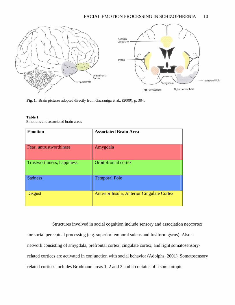
FACIAL EMOTION PROCESSING IN SCHIZOPHRENIA 10
Emotion
Emotion Associated Brain Area
Fear, untrustworthiness Amygdala
Trustworthiness, happiness Orbitofrontal cortex
Sadness Temporal Pole
Disgust Anterior Insula, Anterior Cingulate Cortex
Structures involved in social cognition include sensory and association neocortex
for social perceptual processing (e.g. superior temporal sulcus and fusiform gyrus). Also a
network consisting of amygdala, prefrontal cortex, cingulate cortex, and right somatosensory-
related cortices are activated in conjunction with social behavior (Adolphs, 2001). Somatosensory
related cortices includes Brodmann areas 1, 2 and 3 and it contains of a somatotopic
Fig. 1. Brain pictures adopted directly from Gazzaniga et al., (2009), p. 384.
Table 1
Emotions and associated brain areas

FACIAL EMOTION PROCESSING IN SCHIZOPHRENIA 11
representation of the body and it is also called the sensory homunculus (Gazzaniga et al., 2009b).
Right somatosensory cortices are important in recognizing the feelings of other people. We
recognize an individual’s inner emotional state by having internally somatosensory
representations that tells us how an individual feels when one is perceiving a facial expression.
(Adolphs, Damasio, Tranel, Cooper & Damasio, 2000)
Winston, Strange, Doherty and Dolan (2002) performed an fMRI study on the
perception of trustworthiness of faces. Subjects viewed faces and rated them in trusthworthiness.
Happy faces were rated as significantly more trustworthy than neutral faces, whereas angry and
sad faces were rated as significantly less trustworthy. Significant activation bilaterally in
amygdala was found in the contrast of untrustworthy to trustworthy faces. The more
untrustworthy the subjects rated faces, the greater was the Blood Oxygenation Level Dependent
(BOLD) response evoked in the amygdala. In contrast, orbitofrontal cortex was activated during
processing of trustworthy faces. Gazzaniga et al. (2009b) suggested that the amygdala should
been activated during any kind of facial expression but in this study (Winston et al., 2002) the
amygdala was significant more activated during processing of untrustworthy faces such as angry
and fearful faces, this could mean that negative facial expressions, such as untrustworthy faces,
evoke a greater activation in amygdala than neutral or positive expressions.
In sum, the following areas are actively involved in face processing in healthy
individuals: FG and STS are involved in general face processing. Processing of emotional faces
activates the limbic system in the brain, but specifically the amygdala (Gazzaniga et al., 2009b).
Other important brain areas that activate when one is perceives social stimuli, such as emotional
faces are: right somatosensory cortices, orbitofrontal cortices and cingulate cortices. Social
cognition network includes sensory and association neocortex (e.g. STS and FG), and a network

FACIAL EMOTION PROCESSING IN SCHIZOPHRENIA 12
consisting of amygdala, prefrontal cortex, cingulate cortex, and right somatosensory related
cortices.
3. 1. Facial emotion processing in schizophrenia
Patients with schizophrenia have deficits in recognition and identification of emotional
expressions in faces. It is still unclear whether these deficits result from abnormal processing of
emotional faces or an impaired ability to process complex visual stimuli such as faces (Doop &
Park, 2009). Doop and Park (2009) found a correlation between worse social functioning and
facial emotion matching errors in a study with patients with schizophrenia and a healthy control
group. Patients with schizophrenia showed general deficits in processing of emotional facial
expressions which is associated with reduced social functioning. In a study by Radua (2010) they
found that the neural response to eyes was similar in patients with schizophrenia and healthy
individuals. On the other hand, they detected a lower activation in amygdala and hippocampus
region in schizophrenic patients.
Gur et al. (2007) conducted an event-related fMRI study and they found that
patients with schizophrenia showed reduced limbic activation compared to healthy controls in a
facial emotion identification task. In controls, the analysis showed that greater amygdala
activation was associated with correct identifications of threat-related (anger and fear)
expressions. Analysis showed significant activation for the emotion identification task in a
network of regions which included amygdala, hippocampus, thalamus, FG and frontal and visual
association cortex in healthy controls and schizophrenic patients, however, several regions
showed significantly greater activation in controls. In controls there were significantly different
activation levels in amygdala and other limbic regions related to anger and fear compared to
neutral stimuli. Controls showed greater activation for correct responses to angry faces in inferior

FACIAL EMOTION PROCESSING IN SCHIZOPHRENIA 13
frontal and orbotifrontal regions, in contrast patients with schizophrenia showed less activation
for correct identification of threat-related expressions of anger and fear. The difference between
patients and controls was significant for anger in FG and amygdala, it was also significant for
fear in all regions.
Michalopoulou et al. (2008) used fearful stimuli in an fMRI study. The
participants viewed grey-scaled images showing facial emotional expressions of fear, sadness and
neutral faces. In healthy subjects, the processing of fearful faces showed a significantly greater
activation within the amygdala, right-sided parahippocampal gyrus, FG, superior temporal gyrus,
middle occipital and temporal gyri and right insula than the processing of neutral faces.
Processing of facial fear led to a significantly greater activation of amygdala in healthy subjects
compared to schizophrenic patients, similar to what was found by Gur et al. (2007). Healthy
subjects also had greater activation within the right FG and superior temporal gyrus in response
to fearful faces compared to neutral faces, compatible with the results of the study by Gur et al.
(2007). Participants with schizophrenia showed significantly reduced activation within these
regions during facial fear processing compared to healthy controls. These results may reflect a
deficit in the visual processing of facial fear in people with schizophrenia.
The above-mentioned studies show that patients with schizophrenia have reduced
limbic activation in processing of emotional expressions compared to healthy individuals. Correct
identifications of threat-related expressions, such as anger and fear, were linked to greater
activation of the amygdala and other limbic regions in healthy controls compared to neutral
stimuli. In contrast, patients with schizophrenia had reduced activation for correct identifications
of threat-related expressions. The difference between patients and controls was significant for
anger in FG and amygdala and for fear in all regions (amygdala, hippocampus, thalamus, FG,

FACIAL EMOTION PROCESSING IN SCHIZOPHRENIA 14
frontal and visual association cortex). The conclusion of this is that patients not only have deficits
in emotional processing of faces, they also show a deficit in the processing of faces in general,
since FG activation is lower in schizophrenic patients during processing of emotional expressions
and FG is involved in general face processing (Gazzaniga et al., 2009b).
3.2. Facial emotion processing in paranoid and non-paranoid schizophrenia
Individuals with non-paranoid schizophrenia as opposed to individuals diagnosed
with paranoid schizophrenia have normal levels of brain activation during passive viewing of
emotional facial expressions (Williams et al., 2004). This explains why individuals with non-
paranoid schizophrenia rate faces differently from individuals with paranoid schizophrenia but
similarly to healthy individuals (Pinkham et al., 2008).
One extensive study of several kinds of different facial expressions was made by
Phillips et al. (1999). They compared the neural responses to facial expressions in paranoid, non-
paranoid schizophrenic patients and healthy subjects with fMRI scanning. Subjects viewed black
and white facial expressions of fear, anger and disgust, and also expressions of mild happiness.
All patients were less accurate in identifying emotional expressions and they also showed less
activation to these expressions than healthy controls.
In the following section I will present different studies investigating different
emotional expressions such as threat related emotions, such as anger and fear, and also non
threatening face expressions such as disgust and trustworthy faces in paranoid and non-paranoid
schizophrenia.
3.2.1. Processing of anger
FACIAL EMOTION PROCESSING IN SCHIZOPHRENIA 15
In the study by Phillips et al. (1999), in healthy individuals the inferior frontal
gyrus, anterior cingulated gyrus, putamen, superior temporal gyrus, hippocampus, cerebellum and
FG were activated in response to angry expressions. In healthy individuals, activation in response
to neutral expressions could be seen in the cerebellum, superior temporal gyrus, anterior
cingulated gyrus, medial frontal gyrus and caudate nucleus (deactivation of angry expression).
Activation in response to neutral expressions in the paranoid schizophrenics was shown in the
inferior frontal gyrus and anterior cingulated gyrus and a little activation in response to angry
expressions. Non-paranoid schizophrenics showed activation in the posterior cingulated gyrus,
inferior temporal gyrus, cerebellum and thalamus in response to angry expressions, and little
activation to neutral expressions.
These results shows a reduced neural response in both groups of schizophrenic
patients, but especially in paranoid schizophrenics, in regions activated by angry expressions
compared to healthy individuals. Healthy individuals showed significantly greater activation in
response to angry expressions compared to the schizophrenic patients in the inferior frontal
gyrus, putamen and cerebellum, and also significantly greater activation to neutral expressions in
the superior temporal gyrus, medial frontal gyrus and anterior cingulated gyrus. Non-paranoid
schizophrenics showed significantly greater activation in the cerebellum, thalamus and inferior
temporal gyrus to expressions of anger compared to paranoid schizophrenics. No brain regions
were activated significantly more in the paranoid schizophrenics.
3.2.2. Processing of fear
A study by Phillips et al. (1999) showed that in healthy controls, fearful
expressions activated the limbic system and visual cortical regions: hippocampus, amygdala, the

FACIAL EMOTION PROCESSING IN SCHIZOPHRENIA 16
middle temporal gyrus, the superior temporal gyrus, putamen, cerebellum and the inferior frontal
gyrus. Activation in response to neutral expressions could be seen in superior and middle
temporal gyri. In all schizophrenic patients, fearful expressions activated the postcentral gyrus
and inferior frontal gyrus and activation in response to neutral expressions was demonstrated in
visual areas: the middle temporal gyrus and fusiform gyrus. Paranoid schizophrenics showed
activation in the fusiform gyrus, cerebellum, precentral gyrus and lingual gyrus in response to
fearful expressions, but not in the amygdala. Non-paranoid schizophrenics showed activation in
the middle and superior temporal gyri, also in non-paranoid schizophrenics there was no
activation in the amygdala in response to fearful expressions. Healthy individuals showed
significantly greater activation compared with the schizophrenic patients in response to fearful
expressions in the superior temporal gyrus, amygdala and putamen. No brain regions were
activated significantly more in the schizophrenic patients compared to healthy individuals.
Williams et al. (2004) found, when participants were viewing fearful and neutral
facial expressions, that emotion recognition was significantly impaired in the schizophrenia
patients compared to healthy controls. There were no brain regions that showed greater activity in
schizophrenic patients than in healthy subjects. Regarding differences between paranoid and non-
paranoid schizophrenia, paranoid patients had greater impairment for recognition of fear.
Paranoid patients had also significant reduction in the lateral prefrontal cortex compared to non-
paranoid schizophrenics.
In the same study (Williams et al., 2004) processing of fearful faces in healthy
subjects activated limbic, prefrontal and visual brain regions. Schizophrenic patients showed
significantly reduced activity in the right amygdala, related central gray region, both medial and
lateral prefrontal cortices and bilaterally in fusiform gyrus in comparison with healthy subjects.
FACIAL EMOTION PROCESSING IN SCHIZOPHRENIA 17
Also an additional reduction in the visual region could be observed in both subtypes of
schizophrenia. Patients with paranoid schizophrenia showed significantly reduced activity,
compared to healthy subjects, in the right amygdala, central gray area, both medial and lateral
prefrontal cortices and bilateral fusiform gyri. Paranoid patients showed a reduction in the lateral
prefrontal cortex relative to non-paranoid patients, and together with reductions in amygdala, it
leads to impairment in integration and processing of threat-related signals in this schizophrenic
subtype.
3.2.3. Processing of disgust
In the study by Phillips et al. (1999), the processing of disgust activated the insula
and striatum in healthy individuals. In schizophrenic patients the insula was also activated in
response to facial expressions of disgust. Non-paranoid schizophrenics showed significantly
greater activation in response to expressions of disgust in the superior temporal gyrus, thalamus,
postcentral gyrus and amygdala compared with paranoid schizophrenics (Phillips et al., 1999).
3.2.4. Processing of trustworthiness
Pinkham, Hopfinger, Pelphrey, Piven and Penn (2008) used both paranoid
individuals with schizophrenia and another group with schizophrenia but without any paranoid
tendencies. All groups in the study showed significant activation of the social cognitive network:
amygdala, fusiform face area (FFA), STS and ventrolateral prefrontal cortex (VLPFC) while they
were judging the trustworthiness of faces. Subjects with paranoid schizophrenia showed
significantly reduced neural activation in the right amygdala, FFA and left VLPFC compared to
controls and in the left VLPFC as compared to non-paranoid schizophrenia individuals during the
task. Non-paranoid schizophrenia group also showed significantly less activation in the FFA

FACIAL EMOTION PROCESSING IN SCHIZOPHRENIA 18
compared to healthy control, but in contrast to the other group, non paranoid individuals showed
similar levels of activation to controls in all the other brain regions. The results demonstrated that
the patients with paranoid schizophrenia have significant reductions in neural activation during
tasks involving judgment of complex social stimuli and therefore they have also deficits in
complex social cognition.
Phillips et al. (1999) suggests that the absence of amygdala activation in
schizophrenic patients and their impairment in emotion recognition and emotion processing may
be linked to reduced neural responses in limbic structures. In schizophrenic patients, visual
cortical regions and regions important for visual processing (fusiform, lingual and temporal gyri)
did not fail to activate. Therefore, the reduced neural response in schizophrenic patients was not a
reflection of the schizophrenics’ avoidance of the facial stimuli since the visual regions were
activated. Phillips et al. 1999 suggest that the impaired neural response in the amygdala, insula,
frontal regions and to a lesser extent, visual cortical regions, during emotional stimuli, may be a
result of specific deficits in the attention and processing of emotional visual information.
The conclusion that can be drawn based on these studies is that non-paranoid
individuals have similar levels of activation in the processing of emotional stimuli as healthy
individuals when compared to paranoids. Paranoid individuals had lower levels of activation in
response to angry expressions compared to healthy controls. Non-paranoids had greater
activation than paranoid individuals in processing of angry expressions. Both patient groups had
reduced activation in response to angry expressions compared to healthy individuals. In the
matter of the expressions of fear, paranoids showed activation in FG but not in the amygdala, and
non-paranoids had no amygdala activation in response to expressions of fear. Healthy individuals
showed a greater activation in amygdala in response to this kind of stimuli and they also showed

FACIAL EMOTION PROCESSING IN SCHIZOPHRENIA 19
an activation of limbic, prefrontal and visual regions in the brain, whereas schizophrenics showed
reduced activity in the amygdala, prefrontal cortices and bilateral FG, compared to healthy
individuals. In processing of trustworthy faces, paranoids showed a reduced neural activation in
the right amygdala, FFA and left VLPFC compared to controls and in left VLPFC compared to
non-paranoids. Non-paranoids had also a reduced activation in FFA compared to healthy
individuals but they had similar levels of activation in all the other brain regions (amygdala, STS
and VLPFC).
3.3. Cognitive strategies in facial emotion processing
Matching emotional faces can be done with different strategies. One is to process
the facial expression holistically, in other words, to use a configuration-based strategy which
enables an intuitive and automatic processing of the emotion on the face. Another strategy is to
match common features individually and independently by a feature-based strategy. The
schizophrenic patients use the second one of these strategies, according to Fakra, Salgdao-Pineda,
Delaveau, Hariri and Blin (2008), because in these patients’ brain the regions that are involved in
emotional face processing, such as bilateral amygdala, putamen and inferior frontal cortex
(Williams et al., 2004), fail to activate.
Schizophrenic patients have reduced activation in left FG. FG is a region that is
highly associated with face processing (Haxby et al., 2000) and schizophrenic individuals usually
adopt a feature-based strategy when they process emotional faces. The schizophrenic patients had
longer reaction times in the matching condition compared to healthy subjects in the study by
Fakra et al. (2008). The increased reaction times represent the use of a more time-consuming
cortical pathway compared to automatic or intuitive emotional face processing (Fakra et al.,

FACIAL EMOTION PROCESSING IN SCHIZOPHRENIA 20
2008).
Fakra et al. (2008) investigated the neural bases of different cognitive strategies for
emotional face processing in schizophrenia. The task was to perform facial affect identification
and matching of emotional faces during fMRI scanning. Fourteen schizophrenic patients and
fourteen matched controls participated in the study. The results were that both groups had similar
networks engaged in facial affect processing, but in the schizophrenic patients, regions of the
limbic system, especially the amygdala which is a part of automatic processing in emotions
(Gazzaniga et al., 2009b), failed to activate in response to stimuli. Instead, neocortical areas such
as inferior parietal cortex (IPC), left middle temporal cortex and right precuneus were activated,
which are normally not engaged in emotional judging of stimuli.
The results by Fakra et al. (2008) showed a greater bilateral BOLD response in
amygdala in healthy subjects compared to schizophrenic patients, but in contrast, the
schizophrenic patients showed an increased response in neocortical network.
Psychophysiological interactions (PPI) revealed a functional connectivity between prefrontal
regions and the left amygdala in healthy controls, but the PPI couldn’t reveal such interactions in
schizophrenic patients. Compared to controls the patients showed decreased activation of regions
involved in face processing (FG) and increased activation in brain regions associated with feature
analysis (inferior parietal cortex, left middle temporal lobe and right precuneus). These findings
suggest that schizophrenic patients adopt a different kind of cognitive approach when identifying
facial expressions. The results indicate that patients may use a feature-based rather than
configuration-based processing, and this means that schizophrenic patients may be using a
compensatory strategy due to limbic dysfunction.

FACIAL EMOTION PROCESSING IN SCHIZOPHRENIA 21
Fakra et al. (2008) investigated the neural activity during two different types of emotional
face processing, the matching condition which is based on automatic and intuitive processing,
and the labeling condition which is dependent on higher cognitive processes, such as judgment
and interpretation of emotional facial expressions. They found activation of both cortical and sub-
cortical structures, such as amygdala, anterior and middle cingulate as well as parahippocampal
gyri, putamen, thalamic pulvinar nuclei and right middle temporal gyrus. These areas are
involved in automatic processing of emotional information. During the labeling condition they
observed activation of cortical and sub-cortical regions, such as medial prefrontal regions and
hippocampus nuclei, which are instead involved in the cognitive processing of emotional stimuli.
Both labeling and matching conditions activated inferior occipital and FG, which are involved in
face processing (Gazzaniga et al., 2009b). The subjects with schizophrenia had similar activation
patterns for both matching and labeling conditions as healthy subjects. But during the matching
condition the patients had significantly lower activation in several limbic structures, especially in
the amygdala. The patients with schizophrenia did not show any correlation between activity in
amygdala and other brain regions, and this may be so because the patients may be employing a
different cognitive strategy when they are faced with emotional tasks, rather than to process it
automatically or intuitively.
Neuroimaging studies of schizophrenic patients performing emotion facial tasks
have shown dysfunction of the limbic system, especially in the amygdala and the frontal areas
compared to healthy individuals. Neuroimaging methods demonstrate a failure to activate
amygdala in schizophrenic individuals (Fakra et al., 2008). Fakra et al. (2008) suggest, based on
other studies, that this lack of limbic activation is compensated by an over activation of the PFC
in schizophrenic individuals. This could be a possible answer to why amygdala is under activated
FACIAL EMOTION PROCESSING IN SCHIZOPHRENIA 22
in schizophrenic patients, because they use another strategy to process emotional faces. Instead of
processing faces emotionally they rather process them cognitively. Neuroimaging studies have
pointed out that the amygdala is involved in processing of emotional facial expressions,
particularly negative emotions such as fear and anger (Phillips et al., 1999). Activation of the
amygdala is only apparent when subjects perform automatic or intuitive processing of emotional
stimuli (Fakra et al., 2008). Interestingly, this activation is reduced when subjects are asked to
label the emotion or to attend to non-emotional characteristics of the stimuli. Neuroimaging
studies have also revealed that regions of the neocortex, such as the prefrontal cortex, are
involved in the cognitive appraisal of processing emotional stimuli and the inhibition of
emotional reactions. These findings suggest that different cortical regions are involved when
identifying facial affect depending on the strategy (intuitive or cognitive) (Fakra et al., 2008).
3.4. The role of facial emotion processing in social cognition
It is important to recognize expressions in each face because of the ability to adapt
to the social environment. Schizophrenics often perform poorly in social interactions and they
also tend to misinterpret social cues (Phillips et al., 1999). In the studies that have been presented
in this review, one can see that schizophrenics clearly have deficits in processing of emotional
stimuli and that it has an effect on social cognition.
Social cognition is defined as: ”social cognition is the processing of any
information which culminates in the accurate perception of the dispositions and intentions of
other individuals” (Brothers, 1990, p. 28). The cortex surrounding the STS along with the FG is
associated with social cognition, and these regions are involved in visual perception of socially
relevant stimuli (Adolphs, 2001). As schizophrenics have deficits in social cognition, this may

FACIAL EMOTION PROCESSING IN SCHIZOPHRENIA 23
have an influence on their social functioning (Michalopoulou et al., 2008), which could be linked
to schizophrenics’ deficits in facial emotion processing, according to Doop and Park (2009).
In a review by Marwick and Hall (2008), they discuss the relation between face
processing and social cognition in schizophrenia. Marwick and Hall (2008) suggest that a deficit
in facial identification may lead to social difficulties. Several brain areas are involved in social
cognition: the STS, amygdala and prefrontal cortex. During social judgment of trustworthy faces,
schizophrenics show abnormalities in blood flow, specifically to amygdala (Pinkham et al.,
2008). There is a link between brain regions that are abnormal in schizophrenia and social
judgment from facial expressions, and this could mean that schizophrenic patients have deficits in
several components which are involved in social cognition and this leads to difficulties in
processing of emotional stimuli (Marwick & Hall, 2008).
4. Discussion
Patients with schizophrenia have significant impairments in social cognition and
social functioning (Pinkham et al., 2008). Research suggests that their social dysfunction has a
cause in the neural systems that underlie social cognition. Pinkham et al. (2008) speculates that it
is possible that individuals with paranoid schizophrenia are not applying appropriate emotional
significance to facial stimuli when they are asked to make a complex social judgment, and this
may be an explanation for their social dysfunction. Schizophrenic patients have greater
impairment for perception of negative emotional stimuli compared to positive facial affect,
especially perception of fear (Phillips et al., 1999; Williams et al., 2005).
Neuroimaging studies of facial emotion processing shows that individuals with non-
paranoid schizophrenia are better at facial emotion perception than individuals with paranoid

FACIAL EMOTION PROCESSING IN SCHIZOPHRENIA 24
schizophrenia. Phillips et al. (1999) discuss the underlying causes to paranoid schizophrenics’
difficulties in identifying of threat-related emotions, anger and fear. First, schizophrenics identify
a threat when there is none, but the amygdala has decreased activation during threat-related
stimuli, such as anger and fear, and this is true in both paranoid and non-paranoid individuals.
Second, schizophrenics avoid the threat, but then the visual regions and FFA shouldn’t be
activated, but they are so, this is true according to the study by Phillips et al. (1999). On the other
hand, studies by Michalopoulou et al. (2008) and Williams et al. (2005), comes to different
conclusions regarding activation of the visual regions in Schizophrenic patients. Visual regions
have a reduced activation during processing of fear in the study by Michalopoulou et al. (2008)
and Williams et al. (2005) in both paranoid and non-paranoid schizophrenia, compared to healthy
individuals. These contradictions could be explained by the year of the carrying out studies since
the studies by Michalopoulou et al. (2008) and Williams et al. (2005) is more recent compared to
the study by Phillips et al. (1999). This conveys that the result of the more recent studies is more
reliable than the study by Phillips et al. (1999). The third suggestion from Phillips et al. (1999) of
underlying causes of Schizophrenics deficits in identifying of threat-related emotions is that
schizophrenics have deficits in labeling the correct emotion. Explanation number three that
schizophrenics have deficits in labeling emotions correctly seems to be right together with
explanation two, according to all the reviewed studies here. It also seems that paranoids
especially have troubles with processing of emotional face stimuli. The amygdala has been linked
to recognition of negative emotion perception, especially threat detection. Individuals with
schizophrenia have abnormal amygdala activation (Breiter et al., 1996) and several functional
imaging studies have shown that these individuals are less accurate in identifying emotions and
they also show reduced amygdala activation to fearful expressions (Michalopoulou et al., 2005),
The amygdala is an important area for social cognition (Pinkham, 2003) and since schizophrenic

FACIAL EMOTION PROCESSING IN SCHIZOPHRENIA 25
individuals have abnormal amygdala activation, they also have deficits in social cognition.
The social environment is complex, and it is hard to predict other people’s
behavior. Facial expressions are a cue that informs us how one self should behave (Adolphs,
2001). Social cognition has been shaped by evolution. Early human social groups were smaller
(Adolphs, 2001), and we needed to evolve our social cognition when the social groups were
becoming bigger, in order to understand the complex emotional facial expressions which we
developed. In many situations, individuals must decide whether one should approach someone or
avoid the person, trust or distrust the person (Winston et al., 2002). Thus, Facial emotion
recognition may have an evolutionary basis. Some emotional facial expressions might serve as
reliable signals of threat (Radua et al., 2010). Lesion studies have demonstrated that subjects with
bilateral damage in the amygdala judge people to look more trustworthy than normal subjects do,
the amygdala makes it possible for us to discover possible threats (Adolphs, 2001). When we
meet other people we tend to look on their faces more than their bodies, as the facial expressions
provide the most cues regarding affective states. Facial expression is one example of emotional
behavior, important in both social cognition and basic survival (Erickson & Schulkin, 2003).
Impaired recognition of threat related expressions (anger and fear) may result from
dysfunction of emotional face processing and this could lead to misinterpretations of expressions
as being threatening. Schizophrenics have abnormalities in the social network in the brain.
Schizophrenics also show delayed interpretation of correct identification of emotional
expressions, accurate and fast responses to threat stimuli is critical for basic survival (Green &
Phillips, 2004). Emotions are important to correctly recognize because they prepare our behavior
to the right action, e.g., running when you face a dangerous encounter. People interpret facial
expressions and based on that they decide how to react (Gazzaniga, Heatherton & Halpern,

FACIAL EMOTION PROCESSING IN SCHIZOPHRENIA 26
2009a). It can mean a lot of difficulties if schizophrenics’ don’t have the ability to match the right
expression to the right emotion, as they end up with difficulties in interpreting other people’s
emotions and predicting others behavior. The conclusion could be that schizophrenic individuals,
especially paranoid schizophrenics, would not have the best survivability based on their deficits
in interpreting emotional expressions.
5. Limitations
It can be somewhat difficult to study emotions and emotional face processing
systematically (Gazzaniga et al., 2009b). It is also problematic to come up with an absolute
definition of what an emotion is. Some studies have few participants, such as the study by
Phillips et al. (2004), this could be an explanation to why this study comes to different
conclusions than the studies by Pinkham et al. (2008) and Williams et al. (2004), regarding visual
processing of faces. It is problematic to divide schizophrenia into paranoid and non-paranoid
schizophrenia, since the “subtype” non-paranoid schizophrenia contains all other subtypes in
schizophrenia except the one with paranoid Schizophrenia (American Psychiatric Association,
2000), this means that there could be a high variety in the non-paranoid subtype regarding
processing of emotional faces. There is more problems in dividing schizophrenic patients into a
paranoid or a non-paranoid subtype, one individual can be characterized with paranoid
symptoms, while in earlier life the same individual could be fitted in the non-paranoid subtype,
this would results in that non-paranoid subtypes has longer mean duration of illness (Zalewski et
al., 2008). Studies conducted with schizophrenic patients are also problematic, because the
patients are often on medication, and this may influence the results. Antipsychotic medication
could affect limbic regions and also some regions of neocortex, but, on the other hand, the
literature suggests that antipsychotic medication has little influence on activation patterns

FACIAL EMOTION PROCESSING IN SCHIZOPHRENIA 27
(Phillips et al., 2003); therefore medication cannot be used as treatment option to alleviate
symptoms related to social functioning.
There are also some limitations with this review. It only takes brainimaging studies
in account, excluding behavioral, electrophysiological and neuroanatomical studies due to several
reasons. First: not many studies have investigating the differences in paranoid and non-paranoid
schizophrenia, and the most studies that have been conducted have been used brainimaging
methods. Second: Since not many studies have been conducted with paranoid and non-paranoid
schizophrenia with other methods, this review only take the brainimaging in account when the
deficits in facial emotion processing in schizophrenia is reviewed. Otherwise, it is hard to
compare the studies between healthy, schizophrenia in general and paranoid and non-paranoid
schizophrenia, if this review take other kinds of methods in account and using of behavioral,
electrophysiological and neuroanatomical results when schizophrenia is reviewed, it is
complicating to compare these results with only brainimaging studies of the differences between
paranoid and non-paranoid schizophrenia regarding facial emotion processing. Third: If It had
been conducted more research regarding the differences between paranoid and non-paranoid
Schizophrenia it would, anyway, be too extensiveness to review behavioral, electrophysiological
and neuroanatomical studies. These are the reasons to why this review only takes brainimaging in
account when investigating the differences between paranoid and non-paranoid schizophrenia,
this should be take into consideration when making conclusions of the studies above since other
methods could possibly be revealing different results than results of neuroimaging studies.
6. Future research
More research should be done with different using of methods such as behavioral,

FACIAL EMOTION PROCESSING IN SCHIZOPHRENIA 28
electrophysiological and neuroanatomical studies investigating the differences between paranoid
and non-paranoid schizophrenia and the deficits in facial emotion processing. Researchers in the
field should investigate other kinds of treatment beyond medication. In Roberts and Penn’s
(2009) study they investigate potential training in social cognition and interaction in
schizophrenic patients. Social cognition and interaction (SCIT) is a 20-week group treatment
which is designed to improve social functioning in schizophrenia by improved social cognition.
The treatment contains of several phases: Emotions, which handles dysfunction of emotion
perception, and integration in where the participants take the learned skills into their own lives. In
the study by Roberts & Penn (2008) they used an emotion perception task as a social cognitive
measure, and in the future, we need to investigate not only the schizophrenic patients, but also the
different kind of subtypes in this disorder, regarding facial emotion processing. Processing of
facial emotion recognition could be done by different approaches, for example, by viewing facial
expressions and then matching a similar expression, or identify a facial expression (Roberts &
Penn (2008). The next step could be to use standardized movies of social interactions as a
measure of processing of emotional faces, and investigate how these stimuli could be linked to
social cognition through tests in social functioning. Based on this, further training of social
cognition and interaction should be developed, such as SCIT.
7. Conclusions
This review comes to several conclusions based on the research in facial emotion
processing of schizophrenia: Individuals with schizophrenia have deficits in recognition and
processing facial emotion stimuli compared to healthy individuals. Schizophrenic patients show
greater impairment for perception of negative emotions compared to positive emotions, with the
greatest impairment for the perception of fear. Research has shown that individuals with non-

FACIAL EMOTION PROCESSING IN SCHIZOPHRENIA 29
paranoid schizophrenia are better at recognizing fear and disgust but not anger than paranoid
individuals. Amygdala is one brain area which is involved in the processing of emotional
expressions of faces. The amygdala shows a greater activation in negative facial expressions,
especially fear, than in neutral facial expressions, and it has also been linked to threat detection.
Individuals with schizophrenia are not only less accurate in identifying emotional stimuli, they
also show decreased amygdala activation to fearful expressions, this makes it harder for these
patients to recognize threat-related stimuli. Schizophrenic patients may have difficulties in
recognizing fear due to deficits in the visual network (Williams et al., 2004), and it could be an
explanation to why they troubles with facial emotion processing. Deficits in processing of
emotional stimuli are likely to lead to deficits in social cognition.

FACIAL EMOTION PROCESSING IN SCHIZOPHRENIA 30
References
Adolphs, R., Damasio, H., Tranel, D., Cooper, G., Damasio, A. R. (2000). A role for
somatosensory cortices in the visual recognition of emotion as revealed by three-
dimensional lesion mapping. The Journal of Neuroscience, 20(7), 2683–2690.
Adolphs, R. (2001). The neurobiology of social cognition. Current Opinion of Neurobiology,
11(2), 231-239.
American Psychiatric Association. (2000). Diagnostic and statistic manual of mental disorders
(4th ed.). Washington DC: American Psychiatric Press.
Blair, R . J. R., Morris, J. S., Frith, C. D., Perret, D. I and Dolan R. J. (1999). Dissociable neural
responses to facial expressions of sadness and anger. Brain, 122, 883, 893.
Breiter, H. C., Etcoff., N. L., Whalen, P. J., Kennedy, W. A., Rauch, S. L., Buckner, R. L.,
Strauss, M. M., Hyman, S. E., & Rosen, B. R. (1996). Response and habituation of
the human amygdala during visual processing of facial expression. Neuron, 17, 875-
887.
Brothers, L. The social brain: a project for integrating primate behavior and neurophysiology in a
new domain. Concepts Neurosci, 1990, 1, 27–51.
Couture, S. M., Penn, D. L, & Roberts, D. L. (2006). The functional significance of social
cognition in schizophrenia: A review. Schizophrenia Bulletin, 32(S1), S44-S63.

FACIAL EMOTION PROCESSING IN SCHIZOPHRENIA 31
Doop, M. K & Park, S. (2009). Facial expression and face orientation processing in
schizophrenia. Psychiatry Research, 170(2-3), 103-107.
Erickson, K., & Skulking, J. (2003). Facial expressions of emotion: A cognitive neuroscience
perspective. Brain and Cognition, 52(1), 52-60.
Fakra, E., Salgdao-Pineda, P., Delaveau, P., Hariri, A, R., & Blin, O. (2008). Neural bases of
different cognitive strategies for facial affect in schizophrenia. Schizophrenia
Research, 100(1-3), 191-205.
Gazzaniga, M. S., Heatherton, T. F., Halpern, D. F. (2009a). Psychological science. Norton:
London.
Gazzaniga, M. S., Ivry, R. B., & Mangun, G. R. (2009b). Cognitive neuroscience: The biology of
the mind. Norton: New York.
Green, M. J. & Phillips, M. L. (2004). Social threat perception and the evolution of paranoia.
Neuroscience and Biobehavioral Reviews. 28, 333-342.
Gur, R. E., Loughead, J., Kohler, C. G., Elliot, M. A., Lesko, K., Ruparel, K., Wolf, D. H., Bilker,
W. B., & Gur, R. C. (2007). Limbic activation associated with misidentification of
fearful faces and flat affect in schizophrenia. Archives of General Psychiatry,
64(12), 1356-1366.
Haxby, J. V., Hoffman, E. A., & Gobbini, M. I. (2000). The distributed human neural system for
face perception. Trends in Cognitive Sciences, 4(6), 223-232.

FACIAL EMOTION PROCESSING IN SCHIZOPHRENIA 32
Hooker, C. & Park, S. (2002). Emotion processing and its relationship to social functioning in
Schizophrenia patients. Psychiatry research, 112(1), 41-50.
Itier, R. J., & Batty, M. (2009). Neural bases of eye and gaze processing: The core of social
cognition. Neuroscience and Biobehavioral Reviews, 33(6), 843-863.
Marwick, K., & Hall, J. (2008). Social cognition in schizophrenia: a review of face processing.
British Medical Bulletin, 88, 43-58.
Michalopoulou, P. G., Surguladze, S., Morley L. A., Giampietro, V. P., Murray, R. M., &
Shergill, S., S. (2008). Facial fear processing and psychotic symptoms in
schizophrenia: functional magnetic resonance imaging study. The British Journal of
Psychiatry, 192, 191-196.
Phillips, M. L., Williams, L., Senior, C., Bullmore, B. T., Brammer, M. J., Andrew, C., Williams
S. C, David A. S. (1999). A differential neural response to threatening and non-
threatening negative facial expressions in paranoid and non-paranoid
schizophrenics. Psychiatry Research, 92, 11–31.
Pinkham, A.E., Hopfinger, J.B., Pelphrey, K.A.; Piven, J.; Penn, D.L. (2008). Neural bases for
impaired social cognition in schizophrenia and autism spectrum disorders.
Schizophrenia Research, 99(1-3), 164-175.

FACIAL EMOTION PROCESSING IN SCHIZOPHRENIA 33
Pinkham, A. E., Penn, D. L., Perkins, D. O., & Lieberman, J. (2003). Implications for the neural
basis of social cognition for the study of schizophrenia. The American Journal of
Psychiatry, 160 (5), 815-824.
Radua, J., Phillips, M. L., Russell, T., Lawrence, N., Marshall, N., Kalidindi, S., El-Hage, W.,
McDonald, C., Giampietro, V., Brammer, M. J., David, A. S., Surguladze, S. A.
(2010). Neural response to specific components of fearful faces in healthy and
schizophrenic adults. Neuroimage, 49(1), 939-946.
Reske, M., Habel, U., Kellermann, T., Backes, V., Shah, N, J., Wilmsdorff, M. V., Gaebel, W.,
Zilles, & K., Schneider, F. (2009). Differential brain activation during facial
emotion discrimination in first-episode schizophrenia. Journal of Psychiatric
Research, 43, 592-599.
Roberts, D. L., Penn, D, L. (2009). Social cognition and interaction training (SCIT) for
outpatients with schizophrenia: A preliminary study. Psychiatry Research, 166,
141-147.
Smith, E. E. & Kosslyn, S. M. (2007). Cognitive Psychology: Mind and Brain. Pearson: New
Jersey.
Williams, L. M., Das, P., Harris, A, W. F., Liddell, B. B., Brammer, M, J., Olivieri, G., Skerrett,
D., Phillips, M, L., David, A, S., Peduto, A., & Gordon, E. (2004). Dysregulation of
arousal and amygdala-prefrontal systems in paranoid schizophrenia. American

FACIAL EMOTION PROCESSING IN SCHIZOPHRENIA 34
Journal of Psychiatry, 161(3), 480-490.
Winston, J. A., Strange, B. A., Doherty, J. O., & Dolan, R. J. (2002). Automatic and inventional
brain responses during evaluation of trustworthiness of faces. Nature Neuroscience,
5(3), 277-283.
Zalewski, C., Johnson-Selfridge, M. T., Ohriner, S., Zarrella, K., Seltzer, J. C. (2008). A review
of neuropsychological differences between paranoid and nonparanoid
schizophrenia. Schizophrenia Bulletin, 24(1), 127-145.
.