Embed Size (px)
Citation preview
FABRY DISEASE IN CHILDHOOD
ROBERT J. DESNICK, PHD, MD, AND ROSCOE O. BRADY, MD
Fabry disease, also known as Anderson-Fabry disease or angiokeratoma corporis diffusum universale, is an inborn error ofmetabolism with profound clinical consequences. Patients with Fabry disease have a deficiency of a-galactosidase A (a-GalA), the lysosomal enzyme responsible for the breakdown of globotriaosylceramide and related glycosphingolipids.1 These
glycosphingolipids have a terminal a-galactosyl residue and are components of most cell membranes. The deficiency of a-Gal Aleads to progressive accumulation of globotriaosylceramide in the plasma and in the lysosomes of most cells in the body.
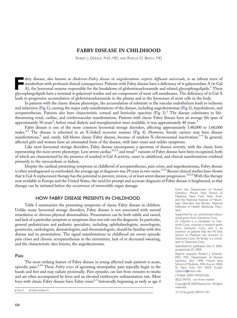
In patients with the classic disease phenotype, the accumulation of substrate in the vascular endothelium leads to ischemiaand infarction (Fig 1), causing the major early manifestations of the disease, including angiokeratomas (Fig 2), hypohidrosis, andacroparesthesias. Patients also have characteristic corneal and lenticular opacities (Fig 3).2 The disease culminates in life-threatening renal, cardiac, and cerebrovascular manifestations. Patients with classic Fabry disease have an average life span ofapproximately 50 years3; before renal dialysis and transplantation were available, it was approximately 40 years.4
Fabry disease is one of the more common lysosomal storage disorders, affecting approximately 1:40,000 to 1:60,000males.2,5 The disease is inherited in an X-linked recessive manner (Fig 4). However, female carriers may have diseasemanifestations,6 and, rarely, full-blown classic Fabry disease, because of random X-chromosomal inactivation.7-9 In general,affected girls and women have an attenuated form of the disease, with later onset and milder symptoms.
Like most lysosomal storage disorders, Fabry disease encompasses a spectrum of disease severity, with the classic formrepresenting the most severe phenotype. Less severe cardiac10,11 and renal12 variants of Fabry disease have been recognized, bothof which are characterized by the presence of residual a-Gal A activity, onset in adulthood, and clinical manifestations confinedprimarily to the myocardium or kidney.
Despite the cardinal presenting symptoms in childhood of acroparesthesias, pain crises, and angiokeratomas, Fabry diseaseis often misdiagnosed or overlooked; the average age at diagnosis was 29 years in two series.5,13 Recent clinical studies have shownthat a-Gal A replacement therapy has the potential to prevent, reverse, or at least arrest disease progression.14,18With this therapynow available in Europe and the United States, the need for prompt and accurate diagnosis of Fabry disease is heightened, so thattherapy can be initiated before the occurrence of irreversible organ damage.
HOW FABRY DISEASE PRESENTS IN CHILDHOODTable I summarizes the presenting symptoms of classic Fabry disease in children.
Unlike many lysosomal storage disorders, Fabry disease is not associated with mentalretardation or obvious physical abnormalities. Presentation can be both subtle and varied,and lack of a particular symptom or symptoms does not rule out the diagnosis. In particular,general pediatricians and pediatric specialists, including ophthalmologists, neurologists,geneticists, cardiologists, dermatologists, and rheumatologists, should be familiar with thisdisease and its presentation. The signal manifestations in childhood are severe episodicpain crises and chronic acroparesthesias in the extremities, lack of or decreased sweating,and the characteristic skin lesions, the angiokeratomas.
Pain
The most striking feature of Fabry disease in young affected male patients is acute,episodic pain.2,19 These Fabry crises of agonizing neuropathic pain typically begin in thehands and feet and may radiate proximally. Pain episodes can last from minutes to weeksand are often accompanied by fever and an elevated erythrocyte sedimentation rate. Mostboys with classic Fabry disease have Fabry crises2,3 historically beginning as early as age 4
From the Department of HumanGenetics, Mount Sinai School ofMedicine, New York, New York;and the National Institute of Neuro-logic Disorders and Stroke, NationalInstitutes of Health, Bethesda, Mary-land.Supported by an unrestricted educa-tional grant from Genzyme Corp.Dr Desnick is a consultant to Gen-zyme Corp, receives a research grantfrom Genzyme Corp, and is aninventor on patents that the Mt SinaiSchool of Medicine has licensed toGenzyme Corp. Dr Brady is a consul-tant to Genzyme Corp.
Submitted for publication Dec 9, 2003;accepted Jan 23, 2004.Reprint requests: Robert J. Desnick,PhD, MD, Department of HumanGenetics, Box 1498, Mount SinaiSchool of Medicine, Fifth Ave at 100thSt, New York, NY 10029. E-mail:[email protected].
J Pediatr 2004;144:S20-S26.0022-3476/$ - see front matter
Copyrightª 2004 Elsevier Inc. All rightsreserved.
10.1016/j.jpeds.2004.01.051a-Gal A a-Galactosidase A
S20
years (mean age of onset, 10 years).3 Pain is often triggered bystress, heat, fatigue, or exercise and is related to the fact thataffected male patients have hypohidrosis and cannot rid theirbodies of excess heat through sweating, causing an increasedcore temperature that triggers the pain. Hypohidrosis alsoleads to heat sensitivity and exercise intolerance, both of whichworsen with age. Affected male patients also have a secondtype of pain, acroparesthesia—chronic burning or tingling painin the extremities. Female carriers may also have ac-roparesthesias, usually tingling in nature and beginning inadolescence.6
Despite their severity, the acroparesthesias in childrenwith undiagnosed Fabry disease are often dismissed as ma-lingering or growing pains. Physical examination may notprovide clues about the diagnosis, particularly if the cutaneousinvolvement is subtle. Electromyography and nerve conduc-tion studies usually fail to detect abnormalities because theneuropathology primarily involves small nerve fibers. Therecurrence of the pain and the lack of a medical explanationhave led to depression, even contemplation of suicide by ado-lescents.20 Pain also has led to misdiagnoses including rheu-matic fever, joint pain, carditis (misdiagnosed mitral murmur),and rash (misdiagnosed angiokeratoma). Other misdiag-noses are listed in Table II.
Gastrointestinal Disturbances
Beginning in childhood, boys with Fabry disease mayhave mild to severe gastrointestinal disturbances, includingdiarrhea, abdominal discomfort, nausea, and vomiting. Female
Fig 1. Electron micrograph showing the vascular endothelium ofa small vessel from a patient with Fabry disease. Note theelectron-dense vesicles (lysosomes) in the endothelium containingundegraded globotriaosylceramide and related glycosphingolipids.The progressive lysosomal glycosphingolipid accumulation inthe vascular endothelium leads to ischemia and infarction of thesevessels. Reprinted with permission.26
Fabry Disease in Childhood
carriers may also have gastrointestinal symptoms, usuallybeginning in adolescence or early adulthood.21,22Acute abdom-inal pain also occurs and can be mistaken for appendicitis.2
Many affected male patients have difficulty gaining weight.23
Dermatologic Manifestations
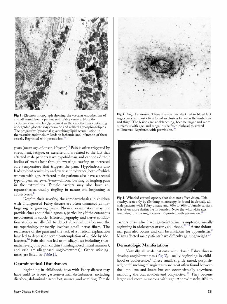
Virtually all male patients with classic Fabry diseasedevelop angiokeratomas (Fig 3), usually beginning in child-hood or adolescence.2 These small, slightly raised, purplish-red, nonblanching telangiectases are most often found betweenthe umbilicus and knees but can occur virtually anywhere,including the oral mucosa and conjunctiva.19 They becomelarger and more numerous with age. Approximately 10% to
Fig 2. Angiokeratomas. These characteristic dark red to blue-blackangiectases are most often found in clusters between the umbilicusand thigh. The lesions are nonblanching, become larger and morenumerous with age, and range in size from pinhead to severalmillimeters. Reprinted with permission.26
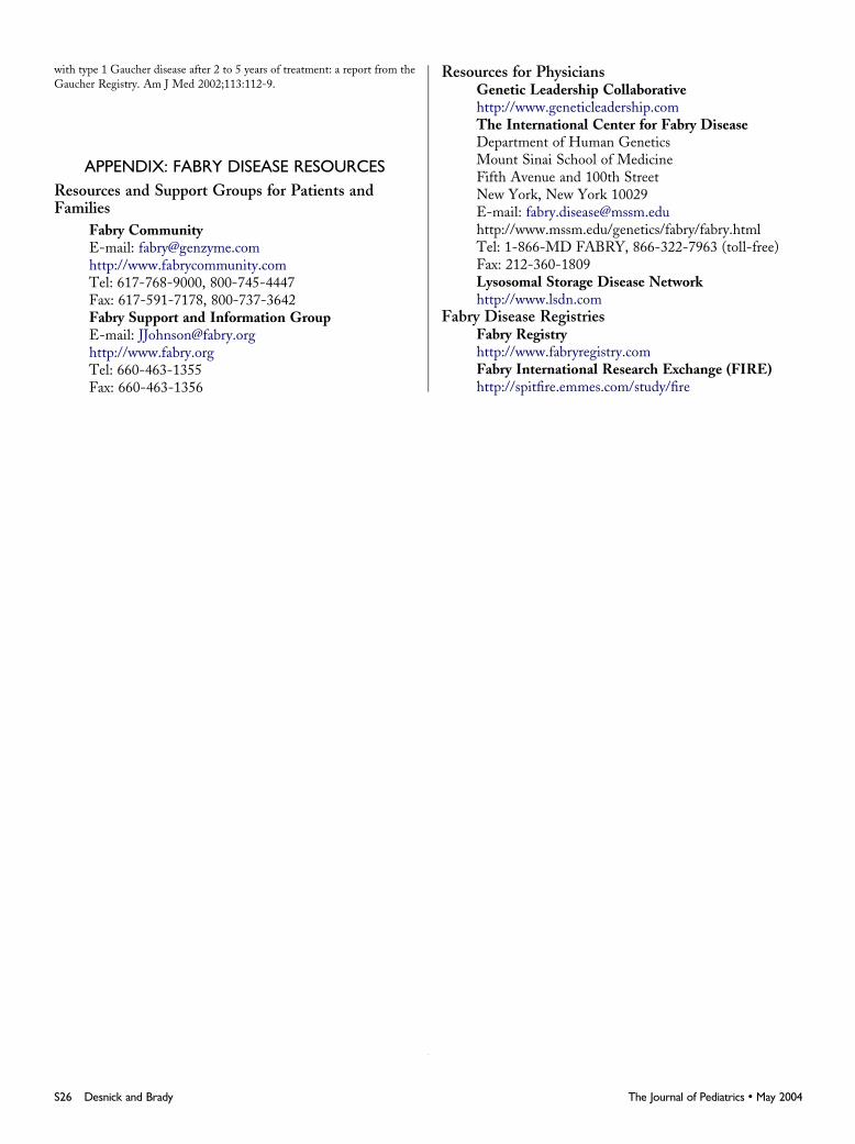
Fig 3. Whorled corneal opacity that does not affect vision. Thisopacity, seen only by slit-lamp microscopy, is found in virtually allmale patients with Fabry disease and 70% to 80% of female carriers.It is often more distinctive in females. Note the whorl-like raysemanating from a single vertex. Reprinted with permission.26
S21

Table I. Clinical manifestations of classic Fabry disease*
Age 4–16 yearsd Intermittent paresthesia and acroparesthesia consisting of chronic, burning, tingling pain in the hands and/or feet, usually beginning in earlychildhood. Can occur daily and can last minutes to days.
d Episodic Fabry crises of severe, incapacitating pain, lasting from days to weeks. Often precipitated by stress, illness, physical exertion, ortemperature changes and accompanied by fever and an elevated erythrocyte sedimentation rate. Very rare in carriers.
d Angiokeratomas that appear in adolescence and worsen in adulthood (Fig 2).d Whorled corneal opacity (Fig 2; also frequently observed in female carriers with or without disease manifestations).d Gastrointestinal problems, including diarrhea, abdominal discomfort, vomiting, nausea.d Hypohidrosis or anhidrosis.d Heat, cold, and/or exercise intolerance.d Mild proteinuria and urinary sediment containing globotriaosylceramide.
Late adolescence to adulthoodd Renal dysfunction that leads to uremia and hypertension and progresses to end-stage renal disease.d Cardiovascular dysfunction, including myocardial infarction, valvular abnormalities, arrhythmias, left ventricular hypertrophy.d Cerebrovascular complications, such as risk of early stroke, hemiplegia, hemianesthesia, transient ischemic attacks.d Pulmonary complications, such as airflow obstruction, dyspnea.
*Most patients with classic Fabry disease are male; however, female carriers can also have disease manifestations.
Fig 4. Inheritance of Fabry disease. Fabry disease is an X-linked recessive disorder. Statistically, 50% of the sons of a female carrier willhave the disorder, and 50% of her daughters will be carriers. None of the sons of an affected father will inherit the disease, but all of hisdaughters will be carriers. Carriers may also have clinical manifestations.
35% of female carriers also develop angiokeratomas, generallybeginning in adolescence. In female patients, the lesions areusually isolated, small macules on the breasts, groin, or flanks.
Ocular Abnormalities
Most boys with Fabry disease and >70% of carrier girlshave a characteristic corneal opacity, observed by slit-lampmicroscopy, that does not affect vision (Fig 4).
Skeletal and Growth Abnormalities
Children with Fabry disease do not have the skeletalabnormalities characteristic of other lysosomal disorders such
S22 Desnick and Brady
as Gaucher disease and several of the mucopolysaccharidoses(eg, Hurler [severe mucopolysaccharidosis type I], Hunter,Maroteaux-Lamy, Morquio diseases), but many children havesubtle evidence of musculoskeletal involvement. Retardedgrowth, delayed puberty, and sparse, fine facial and body hairare common.2 A characteristic facial dysmorphism is found inabout half of male patients and may be recognizable in earlyadolescence.3
Family History
A history of family members with similar symptoms;male relatives with early kidney disease, stroke, or cardiac
The Journal of Pediatrics � May 2004

Table II. Misdiagnoses of Fabry disease
Disease Misleading symptom
Acute appendicitis Severe right lower quadrant abdominal painCarditis Mitral value murmurChronic intermittent demyelinating polyneuropathy Pain, tingling in feet and handsGrowing pains Unexplained pain in extremitiesErythromelalgia Acute pain in the extremitiesLupus AngiokeratomasMultiple sclerosis Stroke-like findings on brain magnetic resonance imagesNeurosis Acute pain with no apparent causePetechiae AngiokeratomasRaynaud syndrome Pain and temperature sensitivity in the extremitiesRheumatic fever Pain accompanied by fever and an elevated erythrocyte sedimentation rateRheumatoid or juvenile arthritis Pain in the joints, elevated erythrocyte sedimentation rate
problems; or both is suggestive of Fabry disease and warrantsinvestigation. However, because the disease gene can be passedthrough the maternal line for one or several generations byasymptomatic or mildly affected female carriers, the absence ofa family history does not rule out the diagnosis. In addition, denovo mutations in the a-Gal A gene do occur, although rarely.
CLINICAL LABORATORY FINDINGSRoutine serum and urine chemistries, hematologic
indices, and pulmonary function tests typically are normal inaffected boys. Older boys with Fabry disease may have anelevated erythrocyte sedimentation rate, mild proteinuria, orisosthenuria. Urinary sediment examination reveals casts,erythrocytes, andcells containing theaccumulatedglycosphingolipid,which appears as Maltese crosses under polarization microscopy.
DIAGNOSISFabry disease can be diagnosed in male patients
by markedly deficient or absent a-Gal A activity in plasmaor peripheral leukocytes by using commercially available4-methylumbelliferyl-a-D-galactoside as substrate.24 Normalenzyme values vary depending on the enzyme source, substrateconcentrations, and assay variables.
In female patients, a very low a-Gal A level is alsodiagnostic of the carrier state for Fabry disease. However,normal or near-normal a-Gal A activity does not rule out thepossibility that a female is a carrier. Obligate heterozygotescan have normal a-Gal A activities because of randomX-chromosomal inactivation. Thus, all girls and women atrisk for carrying the disease gene should have their statusdetermined by molecular studies to detect the family’smutation.
Fabry disease can be diagnosed prenatally by demon-stration of an XY karyotype and deficient a-Gal A activity indirect or cultured chorionic villi or in cultured amniocytes.25 Ifthe family’s a-Gal A mutation is known, molecular studies canreplace or confirm the enzymatic diagnosis.
Fabry Disease in Childhood
GENETIC COUNSELINGGenetic counseling is essential after diagnosis, both to
provide patients and families with information about thenatural history of the disease and treatment options and toadvise parents of the likelihood that siblings, relatives, andfuture children will inherit the disease or carry the disease-causing gene (Fig 1). Parents should encourage other familymembers to have diagnostic testing and genetic counseling.
TREATMENTFabry disease was first identified a century ago, but until
now, no disease-specific treatment has been available. Patientshave been treated with supportive, nonspecific treatment forpain management, cardiac and cerebrovascular complications,and end-stage renal disease. These interventions may prolonglife, but their utility is limited because they do not address theunderlying cause of the disease—the deficient activity ofa-Gal A and the progressive accumulation of globotria-osylceramide.
Like all lysosomal storage disorders, Fabry disease is bestmanaged by a team of specialists headed by a physician withFabry expertise. Treatment should consist of both symptommanagement and enzyme replacement therapy. In addition,pediatricians should be sensitive to the effect of a chronic,progressive disease on children and their families. Genetic,family, and individual counseling are important resources tooffer Fabry families and patients.
How Children Should Be Followed
After the clinical diagnosis of Fabry disease is confirmedby enzyme assay or mutation analysis, all children shouldhave a detailed medical and family history taken. ABO bloodtype and secretor status should also be determined, becausethe presence of B blood group antigen (a glycosphingolipidcatabolized by a-Gal A) may be associated with a more severeprognosis. Pain; gastrointestinal complications; size, density,and distribution of angiokeratomas; hypohidrosis; eye findings;and all other signs and symptoms should be carefully
S23

Table III. Follow-up of children with Fabry disease
Evaluation at diagnosisd Detailed medical historyd Detailed family historyd Detailed physical examinationd Documentation of pain (frequency, character, intensity, and so forth), gastrointestinal symptoms, and angiokeratomasd Laboratory tests
d ABO blood type and secretor status (B blood group may be associated with a more severe prognosis)d Routine hematology, chemistries, and urinalysis
Routine follow-up (additional follow-up as indicated by symptoms or abnormal findings)d Annual detailed physical examination, documentation of level of pain, hypohidrosis, gastrointestinal symptoms, and angiokeratomasd Annual laboratory tests
d Routine hematology, chemistries, and urinalysisd For adolescents: urinary protein (including creatinine to albumin ratio), and creatinine clearance tests to monitor renal function
d Every other year for adolescents: echocardiography and electrocardiography to detect and monitor cardiac abnormalitiesd Early adulthood and/or before initiation of enzyme replacement therapy: baseline kidney, heart, and brain magnetic resonance imaging tomonitor disease progression and effect of therapy
documented at baseline and then at least annually (Table III).Monitoring and treating pain is especially important inchildren and may require additional follow-up. Annualevaluations should include a detailed physical examinationand routine hematology, chemistry, and urinalysis. In addition,because renal disease begins silently and patients with Fabrydisease as young as 16 years have developed renal failure,23
adolescent patients should have yearly serum creatinine,urinary protein (including a creatinine/albumin ratio), andcreatinine clearance tests to determine renal function. Simi-larly, adolescents should have an echocardiogram and electro-cardiogram at least every 2 years to detect and monitor cardiacabnormalities. In early adulthood or before initiation of enzymereplacement therapy, baseline magnetic resonance images ofkidney, heart, and brain are useful to document diseaseprogression and assess the effect of therapy.26
Girls who are Fabry carriers should have a completebaseline examination as described here by a physician withexpertise in Fabry disease. In female patients, disease mani-festations vary widely. Girls with no signs or symptoms ofdisease can be re-evaluated every 3 to 5 years. Symptomaticcarriers should be followed annually with tests focused on theirdisease manifestations.26
Symptom Management
Affected male patients should be encouraged to identifyand avoid stimuli that trigger pain crises, such as extreme heator cold, stress, or physical exertion. Patients with frequent andsevere pain can benefit from prophylaxis with diphenylhydan-toin,27 carbamazepine,28 or gabapentin.29 Narcotic analgesicsshould be avoided. Nonsteroidal antiinflammatory drugs aregenerally ineffective for pain relief. Pancrelipase or met-oclopramide can ameliorate gastrointestinal symptoms.21
Enzyme Replacement Therapy
Despite its often subtle presentation, the pathology inFabry disease begins at birth, or even before birth.30 Enzyme
S24 Desnick and Brady
replacement therapy supplies the patient with the biologicallydeficient protein and reverses metabolic and pathologicabnormalities.14-18
Clinical trials have demonstrated the safety and effec-tiveness of enzyme replacement therapy.14-17 Two differentpreparations of human a-Gal A—agalsidase alfa (Replagal;Transkaryotic Therapies, Cambridge, Mass) and agalsidasebeta (Fabrazyme; Genzyme Corp, Cambridge, Mass)—havebeen used at doses of 0.2 and 1 mg/kg, respectively.Biochemical comparison studies have shown that the a-GalA preparations are structurally and functionally very simi-lar31,32; therefore, the trial data can be taken together as a bodyof evidence supporting the use of enzyme replacement therapyin Fabry disease.14-18 In summary, human a-Gal A re-placement has been shown to decrease pain,16 to reverseabnormal cerebrovascular responses,18 and to deplete storagesignificantly of globotriaosylceramide in the plasma and thecapillary endothelium of the heart, kidney, and skin, majororgans of pathology in Fabry disease.17 In addition, globo-triaosylceramide was depleted in renal endothelial, mesangial,and interstitial cells and reduced in renal epithelial cells inresponse to human a-Gal A replacement.33
Therefore, experts recommend that enzyme replacementtherapy be initiated in all affected male patients with Fabrydisease as soon as clinical signs and symptoms (such as pain orisosthenuria) are observed.26 Carriers with substantial diseasemanifestations also should be treated with enzyme replacementtherapy. It is important to recognize that globotriaosylceramideaccumulation is progressive, and experience with enzymereplacement therapy in Gaucher disease and many otherinherited metabolic diseases has emphasized the importance ofearly intervention to prevent and avoid irreversible damage.
Both a-Gal A products were approved in Europe andelsewhere in 2001, and experience has been gained in morethan 500 patients, most of whom are age 16 years or older. Inthe United States, only agalsidase beta, which obtained accele-rated approval from the Food and Drug Administration inApril 2003, is available.
The Journal of Pediatrics � May 2004

Although results are not yet available, enzyme replace-ment therapy is now being evaluated in children with Fabrydisease in the United States and Europe. However, enzymereplacement therapy has been used safely by hundreds ofchildren with Gaucher disease,34 including children as youngas 1 year of age.
As with any rare disease, collecting and sharing ofinformation is essential. Disease registries provide an invalu-able repository of clinical information that can be used asa decision-making resource for clinicians to determine thenatural course of the disease, to assess the effectiveness ofvarious interventions, and to track individual patients regard-less of treatment choice or status. Therefore, we urge allphysicians treating patients with Fabry disease to enroll themin a disease registry.
CONCLUSIONSFabry disease has significant, although subtle, man-
ifestations in childhood. The benefits of early diagnosis areparticularly compelling with the advent of enzyme replace-ment therapy, which may prevent or even reverse life-threatening disease manifestations. Pediatricians should bealerted to the possible significance of unexplained pain,gastrointestinal disturbances, lack of or decrease in sweating,angiokeratomas, and corneal whorls or lens opacities, as well asunexplained left ventricular hypertrophy or mitral murmursand proteinuria, isosthenuria, or abnormal urinary sedimentanalysis in children and adolescents.
REFERENCES1. Brady RO, Gal AE, Bradley RM, Martensson E, Warshaw AL, Laster
L. Enzymatic defect in Fabry’s disease: ceramidetrihexosidase deficiency.
N Engl J Med 1967;276:1163-7.
2. Desnick RJ, Ioannou YA, Eng CM. Alpha-galactosidase A deficiency:
Fabry disease. In: Scriver CR, Beaudet AL, Sly WS, et al, eds. Metabolic and
molecular bases of inherited disease.NewYork:McGrawHill; 2001. p. 3733-74.
3. MacDermot KD, Holmes A, Miners AH. Anderson-Fabry disease:
clinical manifestations and impact of disease in a cohort of 98 hemizygous
males. J Med Genet 2001;38:750-60.
4. Colombi A, Kostyal A, Bracher R, Gloor F, Mazzi R, Tholen H.
Angiokeratoma corporis diffusum—Fabry’s disease. Helv Med Acta 1967;
34:67-83.
5. Meikle PJ, Hopwood JJ, Clague AE, CareyWF. Prevalence of lysosomal
storage disorders. JAMA 1999;281:249-54.
6. MacDermot KD, Holmes A, Miners AH. Anderson-Fabry disease:
clinical manifestations and impact of disease in a cohort of 60 obligate carrier
females. J Med Genet 2001;38:769-75.
7. Van Loo A, Vanholder R,Madsen K, PraetM, Kint J, De Paepe A, et al.
Novel frameshift mutation in a heterozygous woman with Fabry disease and
end-stage renal failure. Am J Nephrol 1996;16:352-7.
8. Puck JM, Willard HF. X inactivation in females with X-linked disease.
N Engl J Med 1998;338:325-8.
9. Desnick RJ, Simmons RL, Allen KY, Woods JE, Anderson CF,
Najarian JS, et al. Correction of enzymatic deficiencies by renal trans-
plantation: Fabry’s disease. Surgery 1972;72:203-11.
10. Nakao S, Takenaka T, Maeda M, Kodama C, Tanaka A, Tahara M,
et al. An atypical variant of Fabry’s disease in men with left ventricular
hypertrophy. N Engl J Med 1995;333:288-93.
11. von Scheidt W, Eng CM, Fitzmaurice TF, Erdmann E, Hubner G,
Olsen EG, et al. An atypical variant of Fabry’s disease with manifestations
confined to the myocardium. N Engl J Med 1991;324:395-9.
Fabry Disease in Childhood
12. Nakao S, Kodama C, Takenaka T, Tanaka A, Yasumoto Y, Yoshida A,
et al. Fabry disease: detection of undiagnosed hemodialysis patients and
identification of a ‘‘renal variant’’ phenotype. Kidney Int 2003;64:801-7.
13. Morgan SH, Crawfurd MA. Anderson-Fabry disease. BMJ 1988;
297:872-3.
14. Schiffmann R, Murray GJ, Treco D, Daniel P, Sellos-Moura M, Myers
M, et al. Infusion of alpha-galactosidase A reduces tissue globotriaosylcer-
amide storage in patients with Fabry disease. Proc Natl Acad Sci U S A
2000;97:365-70.
15. Eng CM, Banikazemi M, Gordon RE, Goldman M, Phelps R, Kim L,
et al. A phase 1/2 clinical trial of enzyme replacement in Fabry disease:
pharmacokinetic, substrate clearance, and safety studies. Am J Hum Genet
2001;68:711-22.
16. Schiffmann R, Kopp JB, Austin HA 3rd, Sabnis S, Moore DF, Weibel
T, et al. Enzyme replacement therapy in Fabry disease: a randomized
controlled trial. JAMA 2001;285:2743-9.
17. Eng CM, Guffon N, WilcoxWR, Germain DP, Lee P, Waldek S, et al.
Safety and efficacy of recombinant human alpha-galactosidase A—replace-
ment therapy in Fabry’s disease. N Engl J Med 2001;345:9-16.
18. Moore DF, Altarescu G, Herscovitch P, Schiffmann R. Enzyme
replacement reverses abnormal cerebrovascular responses in Fabry disease.
BMC Neurol 2002;2:4.
19. Brady RO, Schiffmann R. Clinical features of and recent advances in
therapy for Fabry disease. JAMA 2000;284:2771-5.
20. Filling-Katz MR, Merrick HF, Fink JK, Miles RB, Sokol J, Barton
NW. Carbamazepine in Fabry’s disease: effective analgesia with dose-
dependent exacerbation of autonomic dysfunction. Neurology 1989;39:
598-600.
21. Argoff CE, Barton NW, Brady RO, Ziessman HA. Gastrointestinal
symptoms and delayed gastric emptying in Fabry’s disease: response to
metoclopramide. Nucl Med Commun 1998;19:887-91.
22. Sheth KJ, Werlin SL, Freeman ME, Hodach AE. Gastrointestinal
structure and function in Fabry’s disease. Am J Gastroenterol 1981;76:246-51.
23. Nelis GF, Jacobs GJ. Anorexia, weight loss, and diarrhea as presenting
symptoms of angiokeratoma corporis diffusum (Fabry-Anderson’s disease).
Dig Dis Sci 1989;34:1798-800.
24. Desnick RJ, Allen KY, Desnick SJ, Raman MK, Bernlohr RW, Krivit
W. Fabry’s disease: enzymatic diagnosis of hemizygotes and heterozygotes:
alpha-galactosidase activities in plasma, serum, urine, and leukocytes. J Lab
Clin Med 1973;81:157-71.
25. Brady RO, Uhlendorf BW, Jacobson CB. Fabry’s disease: antenatal
detection. Science 1971;172:174-5.
26. Desnick RJ, Brady R, Barranger J, Collins AJ, Germain DP, Goldman
M, et al. Fabry disease, an under-recognized multisystemic disorder: expert
recommendations for diagnosis, management, and enzyme replacement
therapy. Ann Intern Med 2003;138:338-46.
27. Lockman LA, Hunninghake DB, Krivit W, Desnick RJ. Relief of pain
of Fabry’s disease by diphenylhydantoin. Neurology 1973;23:871-5.
28. Lenoir G, Rivron M, Gubler MC, Dufier JL, Tome FS, Guivarch M.
[Fabry’s disease: carbamazepine therapy in acrodyniform syndrome]. Arch Fr
Pediatr 1977;34:704-16.
29. Germain DP. [Fabry’s disease (alpha-galactosidase-A deficiency): recent
therapeutic innovations]. J Soc Biol 2002;196:183-90.
30. Elleder M, Poupetova H, Kozich V. [Fetal pathology in Fabry’s disease
and mucopolysaccharidosis type I]. Cesk Patol 1998;34:7-12.
31. Linthorst G, Blom G, Speijer D, Hollak C, Aerts J. Therapy for Fabry
disease: comparison of agalsidase alpha and beta enzyme preparations.
[abstract]. J Inher Metab Dis 2002;25(Suppl 1):115.
32. Lee K, Jin X, Zhang K, Copertino L, Andrews L, Baker-Malcolm J, et al.
Biochemical and pharmacological comparison of enzyme replacement thera-
pies for the glycolipid storage disorder Fabry disease. Glycobiology 2003;
13:305-13.
33. Thurberg BL, Rennke H, Colvin RB, Dikman S, Gordon RE, Collins
AB, et al. Globotriaosylceramide accumulation in the Fabry kidney is cleared
from multiple cell types after enzyme replacement therapy. Kidney Int 2002;
62:1933-46.
34. Weinreb NJ, Charrow J, Andersson HC, Kaplan P, Kolodny EH,
Mistry P, et al. Effectiveness of enzyme replacement therapy in 1028 patients
S25
with type 1 Gaucher disease after 2 to 5 years of treatment: a report from the
Gaucher Registry. Am J Med 2002;113:112-9.
APPENDIX: FABRY DISEASE RESOURCESResources and Support Groups for Patients andFamilies
Fabry CommunityE-mail: [email protected]://www.fabrycommunity.comTel: 617-768-9000, 800-745-4447Fax: 617-591-7178, 800-737-3642Fabry Support and Information GroupE-mail: [email protected]://www.fabry.orgTel: 660-463-1355Fax: 660-463-1356
S26 Desnick and Brady
Resources for PhysiciansGenetic Leadership Collaborativehttp://www.geneticleadership.comThe International Center for Fabry DiseaseDepartment of Human GeneticsMount Sinai School of MedicineFifth Avenue and 100th StreetNew York, New York 10029E-mail: [email protected]://www.mssm.edu/genetics/fabry/fabry.htmlTel: 1-866-MD FABRY, 866-322-7963 (toll-free)Fax: 212-360-1809Lysosomal Storage Disease Networkhttp://www.lsdn.com
Fabry Disease RegistriesFabry Registryhttp://www.fabryregistry.comFabry International Research Exchange (FIRE)http://spitfire.emmes.com/study/fire
The Journal of Pediatrics � May 2004


![Natural history of Fabry disease in females in the Fabry ... · Fabry disease is 1 in 117 000 male live births,[2] though estimates vary from 1 in 40000 to over 1 in 400 000. Fabry](https://img.dokumen.tips/doc/110x75/5f410e03f751a3285a719c0d/natural-history-of-fabry-disease-in-females-in-the-fabry-fabry-disease-is-1.jpg)