Embed Size (px)
Citation preview

EyeGate Pharmaceuticals, Inc.
Providing innovative products that enhance drug efficacy
and patient compliance to improve vision
Corporate Presentation

1
Forward Looking Statements
Some of the matters discussed in this presentation contain forward-looking statements that involve significant risks
and uncertainties, including statements relating to the prospects for the Company’s lead product EGP-437, for the
timing and outcome of the Company’s clinical trials, the potential approval to market EGP-437, and the Company’s
capital needs. Actual events could differ materially from those projected in this presentation and the Company
cautions investors not to rely on the forward-looking statements contained in, or made in connection with, the
presentation.
Among other things, the Company’s clinical trials may be delayed or may eventually be unsuccessful. The Company
may consume more cash than it currently anticipates and faster than projected. Competitive products may reduce or
eliminate the commercial opportunities of the Company’s product candidates. If the U.S. Food and Drug
Administration or foreign regulatory agencies determine that the Company’s product candidates do not meet safety or
efficacy endpoints in clinical evaluations, they will not receive regulatory approval and the Company will not be able to
market them. Operating expense and cash flow projections involve a high degree of uncertainty, including variances in
future spending rate due to changes in corporate priorities, the timing and outcomes of clinical trials, regulatory and
developments and the impact on expenditures and available capital from licensing and strategic collaboration
opportunities. If the Company is unable to raise additional capital when required or on acceptable terms, it may have
to significantly alter, delay, scale back or discontinue operations.
Additional risks and uncertainties relating to the Company and its business can be found in the “Risk Factors” section
of the Company’s Annual Report on Form 10-K filed with the SEC on February 23, 2017. The Company undertakes
no duty or obligation to update any forward-looking statements contained in this presentation as a result of new
information, future events or changes in the Company’s expectations, except as required by applicable law.

2
Company Overview
Ophthalmology company (NASDAQ: EYEG)
Platform 1: Crosslinked HA (eye drop formulation)
• Corneal Epithelial Defects:
• Positive results announced from pilot clinical trial
• FDA De Novo 510(k) filing by year-end 2017
• European CE Mark by year-end 2017
Platform 2: Proprietary delivery system (delivering EGP-437: corticosteroid)
• Cataract Surgery:
• Phase 2 trial to be initiated Q2 2017
• Supplemental NDA filing H2 2018
• Anterior Uveitis:
• Second Phase 3 underway
• NDA submission year-end 2017
Licensed to Valeant
Pharmaceuticals
(Bausch + Lomb)

Clinical Pipeline
3
Iontophoresis
Crosslinked HA
Two platforms in the
clinic with expected FDA
filings by year-end 2017
Crosslinked HA: OBG
Iontophoresis: EGP-437
Product Indication Stage Targeted Filing Dates
Large Corneal
Epithelial Defects
• Pilot Trial completed in patients having undergone
photorefractive keratectomy (“PRK”): Positive data
announced
• Next Trial: Initiation Q2 17
• 510(K) De Novo filing targeted for year-end 2017
• CE Mark targeted for year-end 2017
OBG
Crosslinked HA
EGP-437
Iontophoresis
Anterior Uveitis • 2nd Phase 3 Pivotal Trial: Enrolling • NDA filing targeted for year-end 2017
Cataract Surgery • Phase 1b/2a completed: Positive data announced
• Initiating Phase 2 Trial
• Believe only one Phase 3 trial required
• NDA supplemental filing targeted for H2 2018

4
CMHA-S Platform
Hyaluronic acid is a naturally occurring compound in the body
• ~15 grams of HA in an adult human body
• About 50% in the skin (promotes wound healing), also in the synovial fluid (natural lubricant)
• Rapidly degrades: one-third is naturally turned-over (degraded and synthesized) every day
Properties
• High-molecular weight HA is non-immunogenic
• HA binds up to 1,000 times its volume in water
• HA’s functions include: hydration, lubrication of joints, and providing a meshwork for cell migration
HA crosslinking prevents degradation and increases viscosity
• Crosslinking HA creates a 3D structure that stabilizes the molecule (resists degradation)
• Adheres longer to the ocular surface (90 minutes)
• Higher viscosity that thins with blinking and is non blurring
• Scaffolding matrix that protects the ocular surface

CMHA-S Platform
HA as a topical application is used for epithelial defects (derm wounds and dry
eye) and as an injection for protection of tissues during cataract surgery
Corneal epithelial defects can lead to ocular infections, inflammation, corneal
neovascularization, and vision loss if not treated promptly and healed rapidly
EyeGate has retained control of worldwide commercial rights to CMHA-S
First clinical product is eye drop formulation (liquid-gel)
The EyeGate Ocular Bandage Gel (OBG): a 0.75% concentration of crosslinked HA
• FDA 510(K) de novo filing targeted for year-end 2017
• European CE Mark targeted for year-end 2017, commercializing H1 2018
• HA has wound healing
properties: Supports cell
migration, required for repairing
epithelial defects
• In Europe/Japan HA is a first line
treatment for dry eye
Corneal compromise due to
eye surgeries such as cataract
surgeries, PRK and LASIK
Ocular trauma due to corneal
foreign bodies or chemical
burns
Corneal wounds due to
neurotrophic keratitis and
ulcers
Epithelial defects due to
contact lens wear, dry eye
and severe SPK
Epithelial Defects
5

CMHA-S Platform
EyeGate Ocular Bandage Gel (OBG) – proven efficacy and safety across several
animal studies (already commercialized in vet. space)
Efficacy of CMHA-S has been demonstrated in various animal pathology models:
• Post traumatic corneal stromal ulcers (dogs and cats)
• Corneal abrasion and alkali burn injuries (rabbits)
• Dry eye (rabbits and dogs)
1. EyeGate has human ophthalmic rights only. Visit http://www.bayerdvm.com/show.aspx/remend-cross-linking-video
Commercially available as a veterinary device
• Manufactured by SentrX Animal Care
• Sold in the U.S. and certain European countries by Bayer Animal Health
as Remend®
Corneal Repair1
• 5 years in dogs, cats and horses, with an excellent safety profile
6
Molly a 12 year old cat with a
non-healing corneal defect • Non-healing at 42 days (A)
• Ulcer healing after 12 days of using
0.75% CMHA-S (B)

Healing Corneal Abrasions and Alkali BurnsEfficacy Study: Rabbits1
CMHA-S treated cornea
exhibited “more normal”
epithelial and stromal
organization than control
group
7
1. Guanghui Yang, Ladan Espandar, Nick Mamalis and Glenn D. Prestwich, Veterinary Ophthalmology 2010
A. Fluorescein staining of corneal epithelial abrasions
B. Quantitative analysis at 24 hrs; 49 vs 83% complete
P < 0.01
Histology of alkali burn healingA. Control at Day 12 central wound with unhealed corneal epithelium
B. CMHA-S treated central epithelium and corneal stroma showing a better organization
than control
Wound closure rate of central corneal
epithelium faster in CMHA-S group
• Abrasion: Wound closure complete by 48
hours with CMHA-S
• Burns: Complete re-epithelization at Day 12 for
CMHA-S but not for control

Announced positive data evaluating ability of EyeGate OBG to accelerate
corneal surface re-epithelialization following bilateral photorefractive
keratectomy (PRK)
PRK is an efficacious alternative for patients seeking surgical correction of refractive errors
who are poor LASIK candidates
PRK surgery provides several advantages as indication to evaluate OBG’s ability
• Larger epithelial defects: All eyes randomized at time zero with same size defect
• Homogenous population: All eyes healthy (i.e. normal stem cell function) and will heal at ~ same rate
39 subjects randomized to one of 3 groups: both eyes received the same treatment
• Group 1: EyeGate Ocular Bandage Gel QID for 2 weeks after surgery
• Group 2: EyeGate Ocular Bandage Gel QID for 2 weeks after surgery in combination with a Bandage Contact Lens (BCL)
• Group 3: BCL and preservative-free artificial tears
8
EyeGate Ocular Bandage Gel (OBG)First Human Clinical Trial Completed

Excellent safety and tolerability
• No adverse events in OBG arm
• No corneal haze out to Day 28
~30% more patients healed by Day 3 with OBG than standard-of-care (BCL+AT)
Additionally, wound size was ~53% smaller as early as Day 1 (24 hrs post surgery) with OBG
Next Steps: File IDE Q1 17 followed by next trial Q2 17
9
EyeGate Ocular Bandage Gel (OBG)Data from First Human Clinical Trial
1. BCL = bandage contact lens and AT = artificial tears
# Subjects Closed Wound: Day 3 Surface Area (mm2)
per arm # % Day 1 Day 3
Arm 1: OBG 12 10 83.3% 18.5 0.02
Arm 2: OBG + BCL 14 9 64.3% 40.7 0.10
Arm 3: BCL + AT1 13 7 53.8% 39.5 0.37
Total Subjects Enrolled 39
OBG vs BCL: % better 54.8% 53.3% 94.4%

Meeting with FDA (Nov 2016) confirms device 510(k) de novo filing available for OBG
Device - Indication for Use (IFU): Acceleration of re-epithelialization of large corneal
epithelial defects in patients having undergone PRK
Broader IFU: Demonstrate benefit in additional clinical trial(s) based on size of defect and not
a specific underlying cause or indication
• Superiority claim against standard-of-care not necessary
10
EyeGate Ocular Bandage Gel (OBG)Device
CMHA-S
Promotes re-epithelization (wound healing)
Accelerates re-epithelization
Exhibits “more normal” epithelial and stromal organization and morphology
RESULT: POTENTIALLY FASTER RESTORATION OF VISION AND BETTER
VISUAL OUTCOMES

11
CMHA-S Solid or Film Formulations (2 Versions)Research Funded by Grants
Desired Properties of the Film:
• Easy to place, requiring no sutures or glue
• Allows for immediate stabilization of the eye following trauma
• No refrigeration or freezer required: room stable
• Prevents adhesions and scar formation between the globe and the conjunctiva
2. NSF SBIR Grant: Phase II Status
Films/Pellet: Topical sustained-release delivery vehicle placed in inferior fornix
• Release Profile: High-load product still releasing at 8 weeks (in vitro study ongoing)
• Retention Rate: Re-engineering design for longer retention on eye
• Delivery vehicle for short or long-term acute or chronic conditions including
• Antibiotic: bacterial conjunctivitis/keratitis
• Antihistamine: seasonal/perennial allergies
• Prostaglandins: glaucoma
1. DoD SBIR Phase II Grant: Ocular Surface Shield
A sterile, field-stable product easily applied to immediately
protect and promote healing of the ocular surface

Small electrical current (constant); current has same charge as active substance (drug)
Electrode creates repulsive electromotive forces (like charges repel)
Drug migrates toward return electrode, mobility a function of molecular weight and charge
Drug dose controlled by 2 variables: Current (mA) x application time (minutes)
Easy to use: ophthalmologist or optometrist in <5 minutes
More than 2,400 treatments performed in office setting
12
Iontophoresis Platform: A Non-Invasive Method
of Propelling Charged Active Compounds Into Ocular Tissues
EyeGate Applicator

13
EGP-437: A Potent Anti-inflammatory Agent(corticosteroid - dexamethasone phosphate)
Two indications licensed by Valeant: cataract surgery and anterior uveitis
Etiology assault based (cataract surgery) vs
primarily auto-immune (uveitis)
Inflammation of uveal tissue including iris
and/or ciliary body
Inflammation severity determined by number of
white blood cells in the anterior chamber of the
eye (Slit-lamp used)
Primary end-point is proportion of subjects with
zero cells in EGP-437 arm vs control arm
Cataract surgery incidence: ~4 million* annually in US
Uveitis incidence: ~26.6 to 102 per 100,000 annually in US
* Market Scope, 2015 Comprehensive Report on The Global IOL Market, June 2015

Standard of care for both indications: corticosteroid eye drops
• Example from first pivotal anterior uveitis trial: 2 EyeGate treatments vs. 154 eye
drop treatments
EGP-437: A Highly Differentiated ProductDramatically Reduces Patient Burden
14
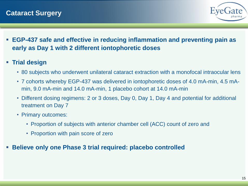
15
Cataract Surgery
EGP-437 safe and effective in reducing inflammation and preventing pain as
early as Day 1 with 2 different iontophoretic doses
Trial design
• 80 subjects who underwent unilateral cataract extraction with a monofocal intraocular lens
• 7 cohorts whereby EGP-437 was delivered in iontophoretic doses of 4.0 mA-min, 4.5 mA-
min, 9.0 mA-min and 14.0 mA-min, 1 placebo cohort at 14.0 mA-min
• Different dosing regimens: 2 or 3 doses, Day 0, Day 1, Day 4 and potential for additional
treatment on Day 7
• Primary outcomes:
• Proportion of subjects with anterior chamber cell (ACC) count of zero and
• Proportion with pain score of zero
Believe only one Phase 3 trial required: placebo controlled

16
Cataract SurgeryPositive results announced
EGP-437 safe and effective in reducing inflammation and preventing pain
• Cohorts receiving the 4.5 mA min and the 14 mA min doses of iontophoretic EGP-437
generated the most encouraging results
• Cell count (ACC) of zero in 20-30% of patients at day 7 and 70-80% of patients at day 28
• Percentage of patients in 4.5 and 14 mA-min doses with zero pain on day 1 was 70 and
90% respectively
• Phase 2b Trial initiation targeted for Q2 2017
*Durezol data from CDER Application Number 22-212: Medical Review for Durezol, studies ST-601A-002a and 002b. Durezol data shown is based on combined data from both studies. QID dose, ITT, LOCF.
EGP-437 data from 14mA-min dosed on Days 0, 1, and 4 (some subjects received additional dose at Day 7) and is ITT, LOCF.
**

Two 3 minute treatments of iontophoretically delivered EGP-437 (day 0 and day 7)
vs corticosteroid eye drops taken up to 8 times per day for 28 days
Primary end point: Percentage of subjects with ACC count of zero at Day 14
Safety: review of side effects, steroid induced increase in intraocular pressure
Anterior UveitisInitial Phase 3 Non-Inferiority Trial
17
Trial Design
193 Subjects randomized 2 arms - 1:1
• 2 EGP-437 iontophoresis treatments + placebo eye drops (N = 96)
• 2 placebo iontophoresis treatments + Pred Acet eye drops (N = 97)
Visit 1
Day 0
Visit 2
Day 7
Visit 3
Day 14
Visit 4
Day 28
Visit 5
Day 56
154 Pred Acet eye drop installations
154 Placebo eye drop installations
follow-up period
follow-up period
1st
Treatment
Iontophoresis
w/ EGP-437
or
w/Placebo
Primary endpoint
proportion of patients
w/ ACC count = 0
2nd
Treatment

18
1. ITT = Intent to Treat
2. Primary End Point (PEP): Total cell clearing (ACC) at Day 14
EGP-437 safe and effective in reducing inflammation vs positive control
• Successfully demonstrated similar response to standard of care (corticosteroid eye drops -
prednisolone acetate 1%)
• Lower incidence of increased intraocular pressure (IOP) with EGP-437 treatment
Confirmatory phase 3 trial ongoing: top-line data expected Q3 2017
Anterior UveitisResults

19
Valeant Pharmaceuticals – Bausch + Lomb (NYSE/TSX: VRX)
Exclusive license to manufacture, sell, distribute and commercialize throughout
the world for use in field of cataract surgery and uveitis
• Total upfront and milestone payments of $135 million
• Includes development milestones
• Royalties based on net sales: high single digits with upward adjustment based on
minimum sales for cataract surgery indication
EyeGate responsible for completion of the clinical development and FDA filing
for both indications
Valeant responsible for development outside U.S.
Valeant has right of last refusal for product outside of licensed fields
• For EGP-437 delivered with Iontophoretic EG II Delivery System
Licensing AgreementEG® II Delivery System + EGP-437

Trial confirms iontophoresis can non-invasively deliver
efficacious quantities to back of eye
ME: abnormal thickening of macula associated with
accumulation of excess fluid within the neurosensory retina
Efficacy: one-third of subjects responded
• Positive response from all subtypes (DME, RVO and CME)
Excellent Safety: no increase in IOP
Enrollment completed
• Under review for further development
• Value in preventing CME post cataract surgery
20
Macular EdemaResults Confirm non-Invasive Delivery to Retina
Number DME RVO CME
Phakic 9 6 3
Pseudophakic 9 4 3 2

21
Objective: Drug loaded contact lens with iontophoresis electronics
Two layer lens
Layer 1: sits on surface of eye – loaded with drug
Layer 2: sits on top of Layer 1 – incorporates iontophoresis electronics
Evolution of a PlatformAt Home Version
Visual center
ConductiveHaptic
ConductingPolymer

22
Summary
Ophthalmology company (NASDAQ: EYEG)
OBG 510(K) de novo filing targeted for year-end 2017: first and only eye drop
in the US with acceleration of re-epithelization claim
OBG CE Mark targeted for year-end 2017, commercial launch Q1 2018
EGP-437 NDA filing for Uveitis targeted for year-end 2017
EGP-437 supplementary NDA filing for ocular surgery H2 2018: effectively
controls post operative pain and inflammation without the need for drop
therapy