Embed Size (px)
Citation preview
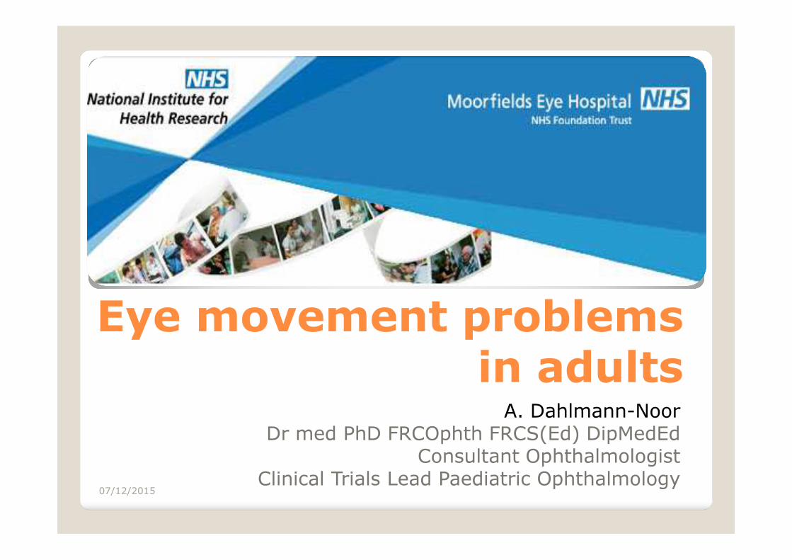
Eye movement problems
in adultsA. Dahlmann-Noor
Dr med PhD FRCOphth FRCS(Ed) DipMedEdConsultant Ophthalmologist
Clinical Trials Lead Paediatric Ophthalmology07/12/2015

OVERVIEW
� Horizontal misalignment
� (Cyclo)vertical misalignment
� Special forms of strabismus
� Pathological nystagmus and other ocular oscillations

HORIZONTAL MISALIGNMENT

HORIZONTAL MISALIGNMENT

Horizontal misalignment
� Comitant� 1. Esotropia� 2. Exotropia
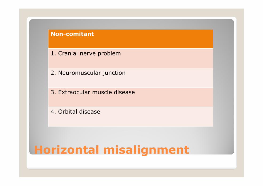
� Non-comitant� 1. Cranial nerve problem: causes/location� 2. Neuromuscular junction� 3. Extraocular muscle disease� 4. Orbital disease

Horizontal misalignment
� Comitant
Esotropia Exotropia
Infantile Infantile
Fully/partially accommodative Intermittent distance exotropia
Sensory Sensory
Sudden onset Sudden onset
Residual Residual
Consecutive Consecutive

Longstanding? Routine referral
What can we do?Assess, advise, operate
Horizontal misalignment
Non-comitant
1. Cranial nerve problem
2. Neuromuscular junction
3. Extraocular muscle disease
4. Orbital disease
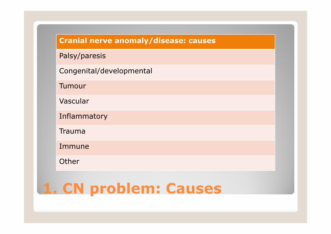
1. CN problem: Causes
Cranial nerve anomaly/disease: causes
Palsy/paresis
Congenital/developmental
Tumour
Vascular
Inflammatory
Trauma
Immune
Other


Sixth nerve palsy
Causes of sixth nerve palsy
Most common: vascular (diabetes,hypertension, atherosclerosis)
Trauma
Watch out for:
Raised intracranial pressure
Giant cell arteritis
Cavernous sinus mass
Brain stem tumour, aneurysm
Multiple sclerosis, sarcoidosis, vasculitis
Urgentreferral

What will we do?
Investigations for sixth nerve palsy
Check blood pressure, blood sugar
Check full blood count, inflammatory markers, renal function, cholesterol
Fundoscopy, Ishihara, neuro exam
Decide whether imaging is needed, and how urgently
Involve other hospital specialists if underlying condition such as MS identified
Urgentreferral

What will we do?
Management of sixth nerve palsy
Treatment for any underlying condition
Ophthalmic:
prisms
occlusion
After 6-12 months of stable measurements: surgery
Botulinum toxin
Urgentreferral

Sixth plus: horizontal gaze palsy
Horizontal gaze palsy
Limitation of abduction on one side(abducens nerve)
Limitation of adduction of the other eye(contralateral oculomotor nerve)
Cause: lesion affecting interneuronsfrom abducens nucleus to oculomotornucleus in the pons

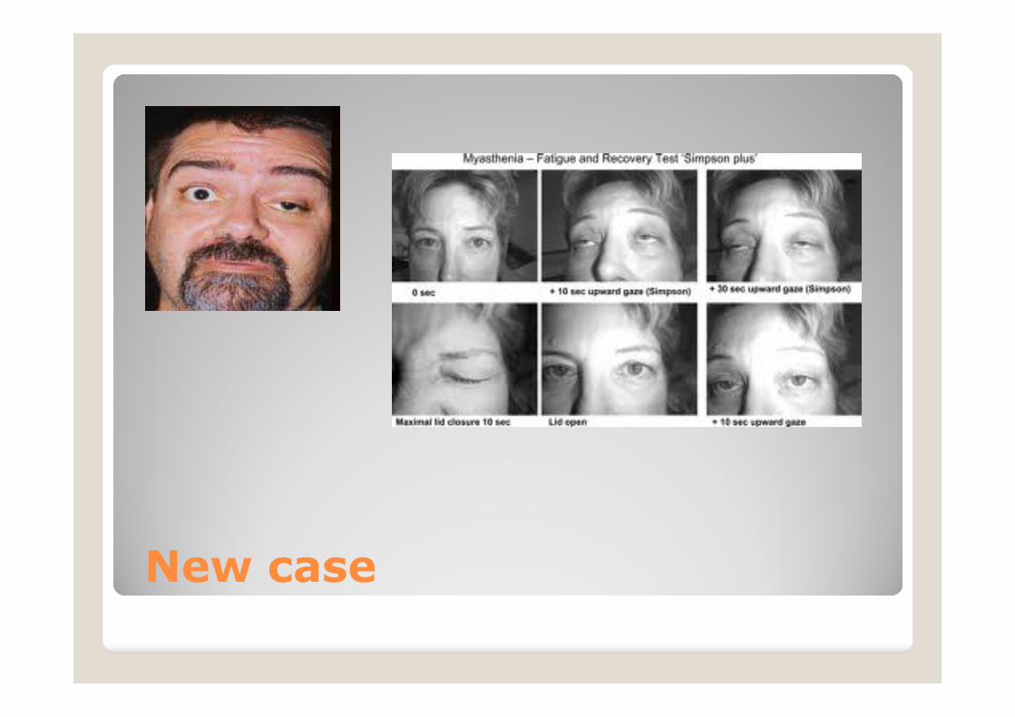
New case

Down and out
Ptosis
Dilated pupil (or not)
New case

Third nerve palsy
Urgentreferral
Causes of third nerve palsy
ANEURYSM (rare, posterior communicating artery)
Most common: vascular (diabetes,hypertension, atherosclerosis)
Trauma
Watch out for:
Giant cell arteritis
Cavernous sinus mass
Tumour
Multiple sclerosis, sarcoidosis, vasculitis

What will we do?
Investigations for third nerve palsy
Check blood pressure, blood sugar
Check full blood count, inflammatory markers, renal function, cholesterol
Fundoscopy, Ishihara, neuro exam
Decide whether imaging is needed, and how urgently
Involve other hospital specialists if underlying condition identified
Urgentreferral

What will we do?
Management for third nerve palsy
Treatment for any underlying condition
Ophthalmic:
prisms
Occlusion if diplopia (usually ptosis)
After 6-12 months of stable measurements: surgery
Botulinum toxin
Urgentreferral

New case
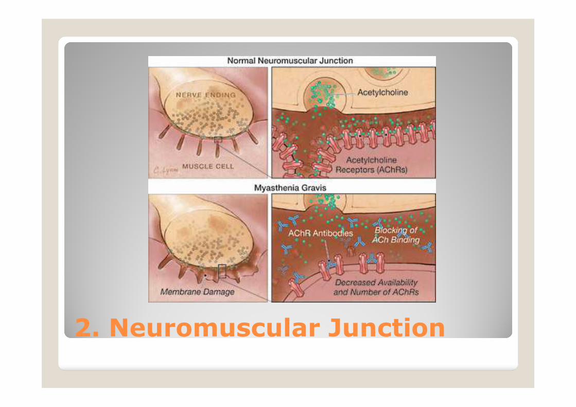
2. Neuromuscular Junction

2. Neuromuscular Junction
Neuromuscular junction: Myasthenia
Varying symptoms and signs
Fatiguability
Generalised symptoms of weakness, breathing problems, chewing/swallowing difficulties, change of voice
Tests: anti-acetylcholine receptor antibodies (negative in up to 50% of ocular only myasthenia), MuSK (muscle-specific kinase)
Chest MRI/CT: thymoma, lung tumour (Lambert Eaton)
Electromyography
Treatment: steroids, pyridostigmine, neostigmine


3. Extraocular muscle disease

3. Extraocular muscle disease
Extraocular muscle disease
Congenital/developmental
Tumour
Vascular
Inflammatory
Trauma
Immune
Other
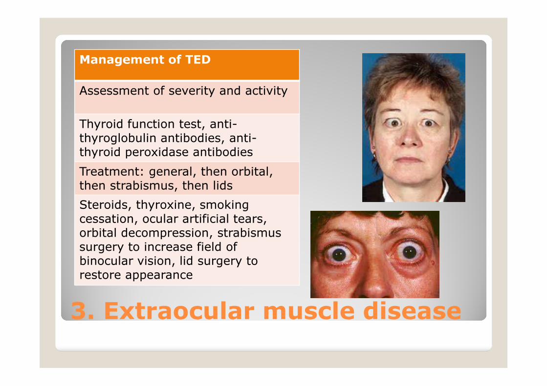
3. Extraocular muscle disease
Management of TED
Assessment of severity and activity
Thyroid function test, anti-thyroglobulin antibodies, anti-thyroid peroxidase antibodies
Treatment: general, then orbital,then strabismus, then lids
Steroids, thyroxine, smoking cessation, ocular artificial tears, orbital decompression, strabismus surgery to increase field of binocular vision, lid surgery to restore appearance

4. Orbital disease
Extraocular muscle disease
Congenital/developmental
Tumour
Vascular
Inflammatory
Trauma
Immune
Other


Cyclovertical misalignment
1. Apparent oblique muscle dysfunction
2. Paresis

Cyclovertical muscle dysfunction
� Inferior oblique overaction
� Very common, in particular in association with childhood strabismus
� Asymptomatic: others notice

New case
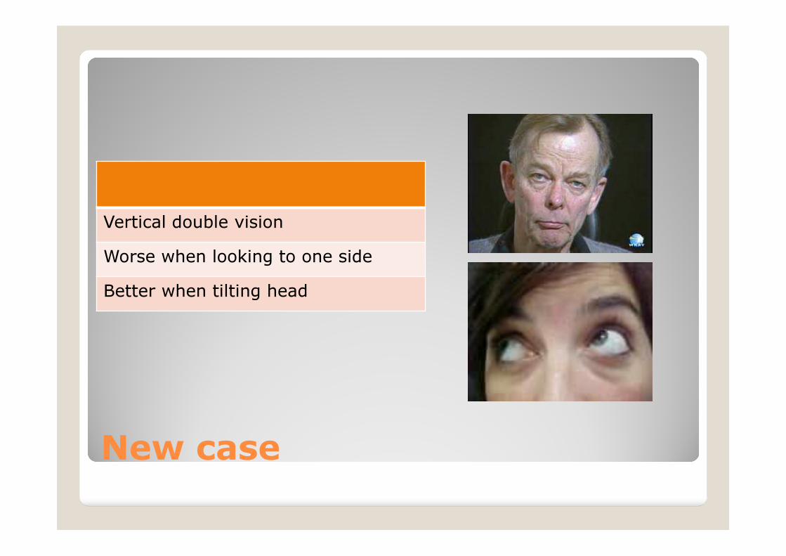
Vertical double vision
Worse when looking to one side
Better when tilting head
New case

Fourth nerve palsy
Usually longstanding and decompensating-> diplopia
If in doubt refer urgently
Causes of fourth nerve palsy
Most common: Congenital
Most common acquired: head injury
less common: vascular (diabetes,hypertension, atherosclerosis)
Watch out for:
Giant cell arteritis
Cavernous sinus mass
Tumour
Multiple sclerosis, sarcoidosis, vasculitis

What will we do?
Investigations for fourth nerve palsy
History: old photos head tilt?
Orthoptic assessment: vertical fusion range
Check blood pressure, blood sugar
Check full blood count, inflammatory markers, renal function, cholesterol
Fundoscopy, Ishihara, neuro exam
Decide whether imaging is needed, and how urgently
Involve other hospital specialists if underlying condition identified

What will we do?
Management for fourth nerve palsy
Treatment for any underlying condition
Ophthalmic:
prisms
occlusion
After 6-12 months of stable measurements: surgery


Special forms of strabismus
Restrictive/Mechanical Strabismus
Secondary to Muscular Disease
Associated with Orbital Bony Disease
Iatrogenic Cyclovertical Deviations (“Induced Adhesive Syndromes”)

Special forms of strabismus
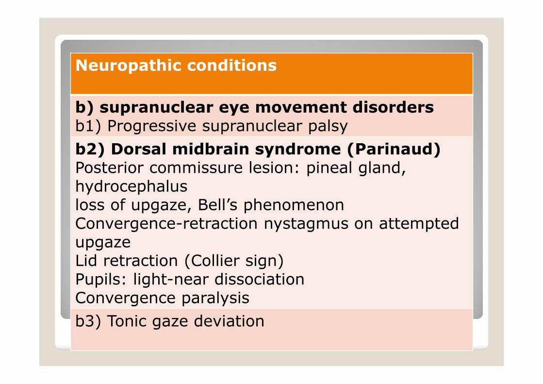
Neuropathic conditions
a) internuclear ophthalmoplegia
b) supranuclear eye movement disorders b1) Progressive supranuclear palsy b2) Dorsal midbrain syndrome (Parinaud) b3) Tonic gaze deviation
c) Skew deviation
d) Eye movement changes associated with Parkinson disease

Special forms of strabismus
Neuropathic conditions
a) internuclear ophthalmoplegia
Lesion in the medial longitudinal fasciculus
Usually multiple sclerosis or microvascular
Limitation of adduction plus contralateralnystagmus on abduction
Usually improve spontaneously
Diplopia: occlusion. Long-term: surgery

Special forms of strabismus
Neuropathic conditions
b) supranuclear eye movement disorders b1) Progressive supranuclear palsy Degenerative conditionBlurring of vision, photophobiaSlowing of saccades, then pursuitsSwallowing, speech, cognitive function, tone gait all progressively impaired
b2) Dorsal midbrain syndrome (Parinaud)
b3) Tonic gaze deviation
Special forms of strabismus
Neuropathic conditions
b) supranuclear eye movement disorders b1) Progressive supranuclear palsy
b2) Dorsal midbrain syndrome (Parinaud) Posterior commissure lesion: pineal gland, hydrocephalusloss of upgaze, Bell’s phenomenonConvergence-retraction nystagmus on attempted upgazeLid retraction (Collier sign)Pupils: light-near dissociationConvergence paralysis
b3) Tonic gaze deviation

Special forms of strabismus
Neuropathic conditions
b) supranuclear eye movement disorders b1) Progressive supranuclear palsy
b2) Dorsal midbrain syndrome (Parinaud)
b3) Tonic gaze deviation Lesion in frontal eye field (infarct)sustained horizontal conjugate deviation

Special forms of strabismus
Neuropathic conditions
c) Skew deviation Vertical misalignmentlesion of the vestibular input into nuclei of third, fourth, sometimes sixth nerve nucleusOne eye hypotropic/excyclotortedFellow eye hypertropic/incyclotorted

Special forms of strabismus
Neuropathic conditions
d) Eye movement changes associated with Parkinson disease Convergence deficitSometimes lid lag on downgazeHypometric saccadesImpaired smooth pursuitsSaccadic intrusions
Special forms of strabismus
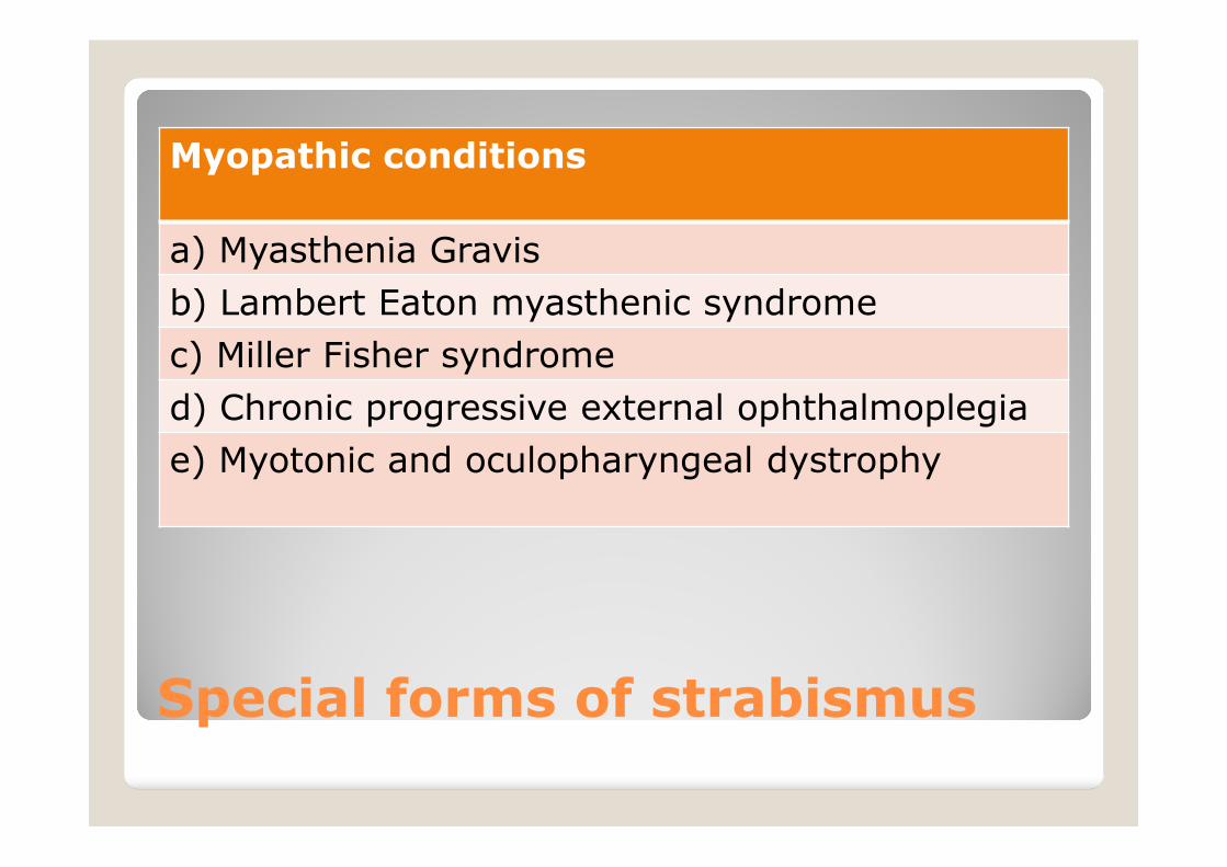
Myopathic conditions
a) Myasthenia Gravis
b) Lambert Eaton myasthenic syndrome
c) Miller Fisher syndrome
d) Chronic progressive external ophthalmoplegia
e) Myotonic and oculopharyngeal dystrophy

Special forms of strabismus
Myopathic conditions
a) Myasthenia Gravis
b) Lambert Eaton myasthenic syndrome
c) Miller Fisher syndrome Ophthalmoplegia, ataxia, areflexiaAuto-antibodies against ganglioside QG1b

Special forms of strabismus
Myopathic conditions
d) Chronic progressive external ophthalmoplegiaMutation in mitochondrial DNA: not enough ATP producedPtosis, progressive paresis of eye muscles,retinal salt-and-pepper appearance, cardiac conduction defect (Kearns-Sayre)

Special forms of strabismus
Myopathic conditions
e) Myotonic and oculopharyngeal dystrophy Expansion of unstable DNA trinucleotide repeats

Special forms of strabismus
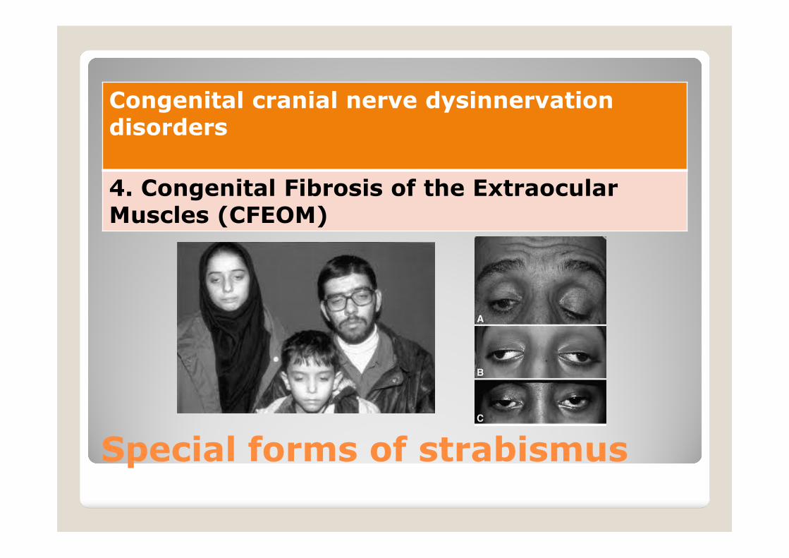
Congenital cranial nerve dysinnervationdisorders
1. Duane syndrome
2. Lower cranial nerve maldevelopment: Moebiussequence
3. Marcus Gunn Jaw-Eyelid Synkinesis Syndrome
4. Congenital Fibrosis of the Extraocular Muscles (CFEOM)
5. Restrictive Hypotropia in Adduction: Brown Syndrome

Special forms of strabismus
Congenital cranial nerve dysinnervation disorders
1. Duane syndrome
Missing development of abducensnucleus
Lateral rectus either not innervated, or innervated by branch from oculomotor nerve, usually inferior division
Result: deficit of abduction, sometimes deficit of adduction, narrowing of palpebral fissure on horizontal gaze; abnormal head posture

Special forms of strabismus
Congenital cranial nerve dysinnervation disorders
2. Lower cranial nerve maldevelopment: Moebius sequence
Sixth and seventh nucleus maldeveloped
Plus additional nucleiaffected (swallowing, speech etc)

Special forms of strabismus
Congenital cranial nerve dysinnervation disorders
3. Marcus Gunn Jaw-Eyelid Synkinesis Syndrome
Maldevelopment of lid innervationLevator palpebrae instead supplied by nerve providing input to muscles of masticationResult: eyelid lifts when chewing or sucking
Special forms of strabismus
Congenital cranial nerve dysinnervationdisorders
4. Congenital Fibrosis of the ExtraocularMuscles (CFEOM)

Special forms of strabismus
Congenital cranial nerve dysinnervationdisorders
5. Restrictive Hypotropia in Adduction: Brown Syndrome


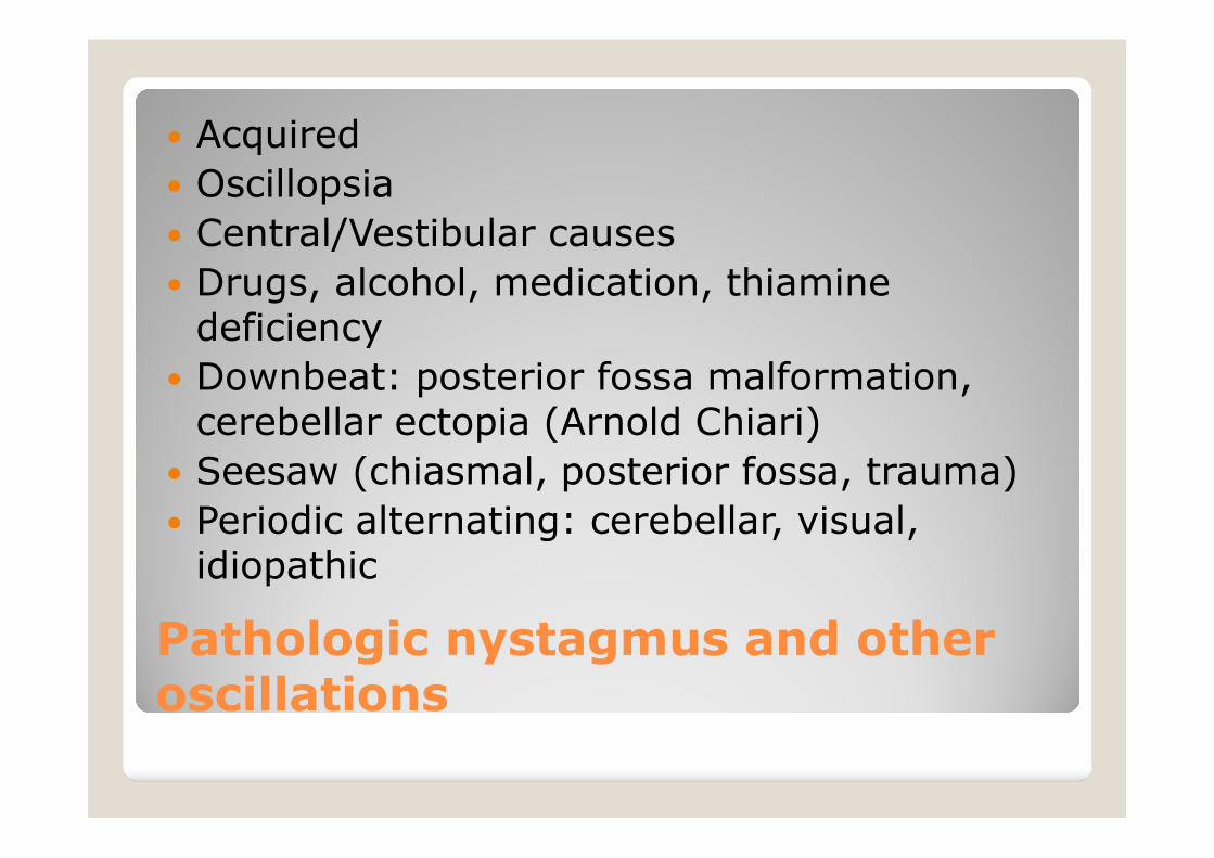
Pathologic nystagmus and other oscillations
� Acquired
� Oscillopsia
� Central/Vestibular causes
� Drugs, alcohol, medication, thiamine deficiency
� Downbeat: posterior fossa malformation, cerebellar ectopia (Arnold Chiari)
� Seesaw (chiasmal, posterior fossa, trauma)
� Periodic alternating: cerebellar, visual, idiopathic