Embed Size (px)
Citation preview

ORIGINAL RESEARCHpublished: 22 January 2016
doi: 10.3389/fnana.2016.00001
Extrinsic Sources of CholinergicInnervation of the Striatal Complex:A Whole-Brain Mapping AnalysisDaniel Dautan 1,2, Husniye Hacioglu Bay 1,3, J. Paul Bolam 1†, Todor V. Gerdjikov 2 andJuan Mena-Segovia 1,4*
1 MRC Anatomical Neuropharmacology Unit, Department of Pharmacology, University of Oxford, Oxford, UK, 2 Departmentof Neuroscience, Psychology and Behaviour, University of Leicester, Leicester, UK, 3 Department of Anatomy, School ofMedicine, Marmara University, Istanbul, Turkey, 4 Center for Molecular and Behavioral Neuroscience, Rutgers University,Newark, NJ, USA
Edited by:Jose L. Lanciego,
University of Navarra, Spain
Reviewed by:Pieter Voorn,
Vrije University, NetherlandsPhilip Winn,
University of Strathclyde, UK
*Correspondence:Juan Mena-Segovia
†Present Address:J. Paul Bolam,
MRC Brain Network Dynamics Unit,Department of Pharmacology,
University of Oxford, Oxford, UK
Received: 08 October 2015Accepted: 02 January 2016Published: 22 January 2016
Citation:Dautan D, Hacioglu Bay H, Bolam JP,
Gerdjikov TV and Mena-Segovia J(2016) Extrinsic Sources of
Cholinergic Innervation of the StriatalComplex: A Whole-Brain
Mapping Analysis.Front. Neuroanat. 10:1.
doi: 10.3389/fnana.2016.00001
Acetylcholine in the striatal complex plays an important role in normal behavior andis affected in a number of neurological disorders. Although early studies suggestedthat acetylcholine in the striatum (STR) is derived almost exclusively from cholinergicinterneurons (CIN), recent axonal mapping studies using conditional anterograde tracinghave revealed the existence of a prominent direct cholinergic pathway from thepedunculopontine and laterodorsal tegmental nuclei to the dorsal striatum and nucleusaccumbens. The identification of the importance of this pathway is essential for creatinga complete model of cholinergic modulation in the striatum, and it opens the questionas to whether other populations of cholinergic neurons may also contribute to suchmodulation. Here, using novel viral tracing technologies based on phenotype-specificfluorescent reporter expression in combination with retrograde tracing, we aimed todefine other sources of cholinergic innervation of the striatum. Systematic mapping of theprojections of all cholinergic structures in the brain (Ch1 to Ch8) by means of conditionaltracing of cholinergic axons, revealed that the only extrinsic source of cholinergicinnervation arises in the brainstem pedunculopontine and laterodorsal tegmental nuclei.Our results thus place the pedunculopontine and laterodorsal nuclei in a key andexclusive position to provide extrinsic cholinergic modulation of the activity of the striatalsystems.
Keywords: striatum, nucleus accumbens, cholinergic brainstem, cholinergic interneurons, acetylcholine
INTRODUCTION
The striatum (STR) plays a key role in learning, memory and motor control (Mink, 1996) and isimplicated in a variety of neurological disorders. It constitutes the main input nucleus of the basalganglia, receiving major afferents from the cortex, the thalamus and the dopaminergic midbrain(Bolam et al., 2000). Predominantly composed of GABAergic projection neurons, the striatumcontains a small proportion of cholinergic interneurons (CIN) that, despite their low number,have been proposed to provide the striatum with one of the highest concentrations of cholinergicmarkers in the brain (Macintosh, 1941; Hebb and Silver, 1961; Woolf et al., 1984; see review by Limet al., 2014).
Historically, the evidence suggested that CINs are the principal source of cholinergic markersin the striatum (Bennett et al., 2000; Wang et al., 2006; Ding et al., 2010; Goldberg et al., 2012;
Frontiers in Neuroanatomy | www.frontiersin.org 1 January 2016 | Volume 10 | Article 1
Dautan et al. Striatal Cholinergic Innervation
see review by Calabresi et al., 2000) but our recent analysisof brainstem cholinergic neurons [pedunculopontine nucleus(PPN) and laterodorsal tegmental nucleus (LDT)] usinganterograde conditional tracing, identified that these neuronstarget the striatum (Dautan et al., 2014; see also Saper andLoewy, 1982; Smith and Parent, 1986; Hallanger and Wainer,1988; Nakano et al., 1990). Similar to CINs, projectionsfrom the brainstem largely avoid striosomes, have extensiveramifications and give rise to both symmetric and asymmetricsynapses, although their proportions vary (Dautan et al., 2014).Notably, brainstem cholinergic projections exhibit a rostrocaudaltopographical organization, where the PPN (rostral brainstem,associated with motor circuits) predominantly targets thedorsolateral striatum, and the LDT (caudal brainstem, associatedwith limbic circuits) targets the dorsomedial striatum andnucleus accumbens. Thus, the extrinsic cholinergic innervationof the striatum follows a functional motor-to-limbic gradient, butthe extent to which this also involves other cholinergic structuresis not known, particularly in light of recently developedtechnologies that allow increased sensitivity to detect phenotype-specific labeling of axons.
Cholinergic innervation of the brain is widely distributed andpredominantly originates from eight anatomically segregatednuclei: medial septum (Ch1), the vertical limb of the diagonalband of Broca (Ch2), the horizontal limb of the diagonalband of Broca (Ch3), the nucleus basalis of Meynert (Ch4),the PPN (Ch5), the LDT (Ch6), the medial habenula (Ch7)and the parabigeminal nucleus (Ch8) (Mesulam et al.,1983a,b; Mufson et al., 1986; Mesulam and Geula, 1988;Mesulam, 1990). While there is some evidence of connectivitybetween neurons contained within these regions and thestriatum (e.g., Wall et al., 2013), it is uncertain whetherthese neurons are cholinergic and thus contribute to thecholinergic innervation of the striatum. Because of the potentialimplications of additional sources of acetylcholine in thestriatum, we investigated this possibility by targeting alleight brain cholinergic nuclei using a combined anterogradeand retrograde analysis of cholinergic neurons and createdmaps of their axonal distributions. Our results support theprevious findings of a direct cholinergic brainstem projectionto the striatum, and demonstrate that the PPN and LDTconstitute the only external source of acetylcholine to thestriatum.
MATERIALS AND METHODS
AnimalsAdult (250–350 g) male Long Evans (LE) wild types (n = 8) andChat::Cre+ (n = 19; Witten et al., 2011) rats were used for allexperiments. Rats were maintained on a 12 h light/dark cycle(light on 07:00 am) and had ad libitum access to water and food.All procedures were performed in accordance with the Society ofNeuroscience policy on the use of animals in neuroscience andthe Animals (Scientific Procedures) Act, 1986 (UK) under theauthority of a Project Licence approved by the Home Office andthe local ethical committee of the University of Oxford.
Stereotaxic InjectionsAll surgical procedures were performed during deep isofluraneanesthesia (2% in O2, IsoFlo, Schering-Plough). ChAT::Cre+
rats were injected in each hemisphere with adeno-associatedvirus serotype 2 carrying the fusion genes for the enhancedyellow fluorescent protein (AAV2-EF1a-DIO-eYFP) or mCherryprotein (AAV2-EF1a-DIO-mCherry; Gene Therapy CenterVirus Vector Core, University of North Carolina). Theviral vectors were injected in a volume of 300 nl forthe forebrain cholinergic structures (Ch1 to Ch4) to avoidspreading over contiguous cholinergic structures, whereasa volume of 500 nl was used for the other cholinergicnuclei (Ch5 to Ch8). The injection sites were randomizedfor hemisphere and fluorescent reporter. Viral injectionswere delivered in eight different locations corresponding tothe eight cholinergic groups described by Mesulam et al.(1983a,b) using the following stereotaxic coordinates (frombregma, in mm; DV ventral to the dura): Ch1 (medialseptum) AP +0.7, ML +0.2, DV −4.5; Ch2 (vertical limbof the diagonal band) AP +0.5, ML +0.4, DV −7.5; Ch3(horizontal limb of the diagonal band) AP +0.1, ML +1.6,DV −8.5; Ch4 (nucleus basalis of Meynert) AP +0.9, ML+2.5, DV −7.0; Ch5 (PPN) AP −7.3, ML +1.8, DV −7.2;Ch6 (LDT) AP −8.5, ML +1.0, DV −6.0; Ch7 (medialhabenula) AP−3.5, ML +0.3, DV−4.0; and Ch8 (parabigeminalnucleus) AP −4.5, ML +4.3, DV −5.5 (Paxinos and Watson,2007).
For the retrograde tracing studies, wild-type LE rats wereinjected bilaterally with cholera toxin b (Ctb, 2.5% in distilledwater, 400 nl over 10 min, Sigma-Aldrich) and Fluorogold (FG,2.0% in distilled water, 300 nl over 10 min, Fluorochrome)in dorsal striatum and nucleus accumbens. Injections sites,hemispheres and tracers were randomized across animals.Injections were delivered in the dorsal striatum and the nucleusaccumbens using the following stereotaxic coordinates (frombregma, in mm; DV ventral to the dura): dorsal striatumAP +0.5, ML +2.1, DV −4.5; nucleus accumbens AP +1.5,ML +1.8, DV−6.7.
For all experiments, injections were made using 1 µl syringes(Neuros 7001, Hamilton) at a rate of 50 nl/min and left to diffusefor 5 min before retraction of the syringe. Approximately 4 weeksfollowing AAV injections, or 10–15 days following tracersinjections, rats were humanely euthanized using a lethal doseof pentobarbital (>200 mg/kg, i.p.) and perfused transcardiallywith 0.05 M PBS, pH 7.4 (approximately 50 ml over 5 min),followed by 300 ml of 4% w/v paraformaldehyde in phosphatebuffer (0.1 M, pH 7.4) containing 0.1% glutaraldehyde (TAABLaboratories) over about 20 min. Brains were stored at 4◦C untilsectioning.
ImmunohistochemistryBrain blocks were formed using 2% agarose gel in PBS(Agarose, BIO-41025, Bioline). Coronal or parasagittal sectionswere cut at 50 µm thickness in PBS using a vibratingmicrotome (VT1000S, Leica). For each experiment, the sitesof injection were verified by fluorescent microscopy and onlythose with on-target injections were processed further. For
Frontiers in Neuroanatomy | www.frontiersin.org 2 January 2016 | Volume 10 | Article 1

Dautan et al. Striatal Cholinergic Innervation
the anterograde tracing study, one section every 300 µmof the entire brain was analyzed. Sections were incubatedovernight in a solution containing antibodies against greenfluorescent protein (GFP, which also detects eYFP; 1:1000dilution, raised in rabbit, A21311, Invitrogen), mCherry (1:500,raised in mouse, Millipore), choline acetyltransferase (ChAT;1:500, raised in goat, Millipore) and tyrosine hydroxylase(TH, in order to define striatal borders; 1:500, raised inchicken, Abcam). The antibodies were diluted in 1% normaldonkey serum (NDS) and 0.03% Triton X-100 in PBS. Afterseveral washes in PBS, the sections were incubated for aminimum of 4 h in secondary antibodies (all raised in donkey,Jackson Immunoresearch) conjugated to one of the followingfluorophores: Alexa Fluor 488 (1:1000), CY5 (1:1000), CY3(1:1000) or AMCA (1:500).
For the retrograde tracer injections, all sections that includedeach of the cholinergic nuclei (Ch1–Ch8) were collected.Following an overnight incubation with antibodies against ChAT(1:500) and Ctb (1:500, raised in mouse, Abcam), sections werewashed in PBS and then incubated in Cy3- or Cy5-conjugatedsecondary antibodies (1:1000 and 1:500, respectively; raisedin donkey, Jackson Immunoresearch). For FG detection, noadditional processing was required.
The processed sections were mounted on slides usingVectashield and then examined in a confocal microscope(LSM-510, Zeiss) at two magnifications (10×, 0.32 NAand 20×, 0.8 NA), using the corresponding filters (504nm for FG and YFP, 560 nm for Cy3 and mCherry, and650 nm for Cy5). Brightness and contrast of capturedimages were adjusted in Photoshop (Adobe Systems). AAV-injected sections were contoured and fully scanned on asingle Z-stack using StereoInvestigator (MicroBrightField;10×, 0.25 NA). For each scanned site, the top and bottomof the section were manually delimited based on thesection surface and single pictures were captured (2048× 1056 pixel resolution) at 10 µm below the surfaceof the section to ensure that the antibody completelypenetrated the section (frame size 860 × 660 µm).Scanning sites were selected using the StereoInvestigatorsystem incorporating a XYZ stage controller and a 25%overlap was selected to facilitate reconstruction. To avoidphotobleaching, an inter-acquisition interval of 50 ms wasapplied during scanning. Exposure time was automaticallyadapted for every site in order to keep the same intensitythreshold.
Analysis and Quantification of theDistribution of Cholinergic AxonsScans from AAV-injected brains were reconstructed off-linebased on the 2D serial section reconstruction module ofStereoInvestigator. Scans were then overlapped with outlinesof the rat brain atlas (Paxinos and Watson, 2007) with theaid of the TH and ChAT immunostaining. Injection siteswith GFP cell body expression less than 30% of the totalnumber of ChAT-positive neurons within the diffusion area, orwith positive soma located outside the borders of the nucleus
(for contiguous cholinergic structures), were excluded from theanalysis.
The density of AAV-positive fibers in each nucleus wasqualitatively assessed off-line. Representative levels for eachmain brain nuclei/region were scored using a predetermineddensity rating. The relative density was scored 4+ for manylabeled fibers covering approximatively more than 50% of theselected image surface (‘‘very dense’’). Nuclei were labeledas 3+ or 2+ when projections covered approximatively morethan 25% (‘‘dense’’) or less than 25% (‘‘moderate’’) ofthe surface, respectively. Structures were noted 1+ (‘‘few’’)when only few terminals were visible. All brains werescanned and only nuclei presenting similar scores in at leasttwo animals were considered. Fibers without terminals orarborization, defined as fibers en-passage, were excluded from themapping.
Analysis of the Distribution of RetrogradelyLabeled NeuronsBrains injected with retrograde tracers were sectioned andscanned as described above. Parasagittal and coronal sectionsthat included the striatum and all cholinergic nuclei wereexamined. Single optical sections containing cholinergic neuronswere obtained at high resolution (20×, 0.8 NA, step size 1 µm,2056 × 1056 pixels) for the fluorescent channels correspondingto the labeling of ChAT+, FG+ and Ctb+ neurons. Analysisof the distribution of retrogradely labeled neurons was carriedout using StereoInvestigator functions: the cholinergic nucleiborders were outlined based on ChAT-immunolabeling usingcontour tools, an overlay projection of the Z-stack was obtainedbased on average intensity tools, colocalization of ChAT, Ctband FG was quantified using the markers plugin. A minimum oftwo single optical sections were examined. Neurons within theborders of each cholinergic nucleus were classified as follows:(1) ChAT+/Ctb+; (2) ChAT+/FG+; (3) ChAT−/Ctb+; and(4) ChAT−/FG+. Data were confirmed in a minimum of fouranimals where Ctb and FG injections were alternated betweendorsal striatum and nucleus accumbens.
RESULTS
Conditional Labeling and Mappingof Cholinergic AxonsTransduction of cholinergic neurons in Ch1 to Ch8 areasin ChAT::Cre+ rats following the insertion of the reportertransgene produced strong and discrete eYFP or mCherrysignals in neurons immunopositive for ChAT (Figure 1).Reporter labeling was observed in cell bodies, dendrites andlocal axons within the sites of injection. No differences wereobserved in the labeling produced by eYFP or mCherry(data not shown), nor in the labeling specificity amongcholinergic structures. The labeling of cholinergic neurons withfluorescent reporters was confirmed by immunohistochemistryfor ChAT in the following structures: dorsolateral striatum(Figure 1A), nucleus accumbens (Figure 1B), medial septum(Ch1; Figure 1C), the vertical limb of the diagonal band
Frontiers in Neuroanatomy | www.frontiersin.org 3 January 2016 | Volume 10 | Article 1

Dautan et al. Striatal Cholinergic Innervation
FIGURE 1 | Transduction of cholinergic neurons in different brain regions. Coronal (Ch1 to Ch7) or sagittal (Ch8) sections showing the sites of cholinergicneuron transduction following injections of AAV2-DIO-EF1a-eYFP and AAV2-DIO-EF1a-mCherry into the striatum and the brain cholinergic nuclei (Ch1 to Ch8).Sections were immunolabeled for ChAT to outline cholinergic structures and verify the specificity of the reporter expression (eYFP, 92%; mCherry, 88%). eYFP ormCherry expression was observed in the dorsal striatum (A), nucleus accumbens (B), medial septum (Ch1; C), the vertical limb of the diagonal band of Broca(Ch2; D), the horizontal limb of the diagonal band of Broca (Ch3; E), the nucleus basalis of Meynert (Ch4; F), the pedunculopontine nucleus (Ch5; G), the laterodorsaltegmental nucleus (Ch6; H), the medial habenula (Ch7; I) and the parabigeminal nucleus (Ch8; J). All the injections were confined to their corresponding anatomicalborders, as defined by Paxinos and Watson (2007). Abbreviations: aca, anterior commissure; aq, aqueduct; cc, corpus callosum; GP, globus pallidus; hip,hippocampus: HDB, horizontal limb of the diagonal band of the nucleus of Broca; LDT, laterodorsal tegmental nucleus; lHb, lateral habenula; Mey, nucleus basalis ofMeynert; mHb, medial habenula; MITg, microcellular tegmental nucleus; ms, medial septum; ot, olfactory tubercle; PBN, parabigeminal nucleus; PPN,pedunculopontine nucleus; STR, striatum; V3, third ventricle; VDB, ventral limb of the diagonal band of the nucleus of Broca; VP, ventral pallidum. Scale bars: brainoutlines, 1000 µm; low magnification panels (left), 500 µm; high magnification panels (center and right), 50 µm.
of the nucleus of Broca (Ch2; Figure 1D), the horizontallimb of the diagonal band of the nucleus of Broca (Ch3;Figure 1E), the nucleus basalis of Meynert (Ch4; Figure 1F),the pedunculopontine nucleus (Ch5; Figure 1G), the laterodorsaltegmental nucleus (Ch6; Figure 1H), the medial habenula (Ch7;Figure 1I) and the parabigeminal nucleus (Ch8; Figure 1J).Injections in the striatum and nucleus accumbens producedlabeling of interneurons whose axons were contained withinthe striatal borders but extended ventrally or dorsally beyondthe site of injection. However, no overlap between the axonsfrom each region was detected, suggesting that the area ofinnervation of cholinergic axons is restricted within theirfunctional domains. Medial septum injections were targetedto its mediodorsal region to avoid overlap with the diagonalband of the nucleus of Broca, which resulted in bilateral
expression (Figure 1C). The vertical limb (Figure 1D) andthe horizontal limb (Figure 1E) are very ventral structuressurrounded by the ventral pallidum dorsally and the olfactorytubercle laterally; both show a high density of small cholinergicneurons. The nucleus of Meynert, situated ventral to theglobus pallidus (GP), contained loosely distributed, smallcholinergic neurons (Figure 1F), consistent with Mesulam et al.(1983a,b).
Medial septal (Ch1; Figure 2) cholinergic projections weremainly observed in the cingulate cortex (3+), the diagonal bandof the nucleus of Broca (3+), the lateral septum (3+), the ventralpallidum (3+), hippocampus (4+) and the reticular thalamicnucleus (2+) (Nyakas et al., 1987; Kalén and Wiklund, 1989;Senut et al., 1989). No labeled axons were observed in thestriatum or nucleus accumbens.
Frontiers in Neuroanatomy | www.frontiersin.org 4 January 2016 | Volume 10 | Article 1

Dautan et al. Striatal Cholinergic Innervation
FIGURE 2 | Mapping of cholinergic axons. Six representative coronal sections (anterior and posterior to bregma; columns) were selected to map the projectionsof the cholinergic neurons in the different cholinergic nuclei (Ch1 to Ch8; rows). For each cholinergic cell group, the rows depict schematic summaries where greenshaded areas indicate high density of fluorescently-labeled axons. Red squares indicate the site where the fluorescent images on the right were obtained.Abbreviations (if not defined previously): Accsh, nucleus accumbens shell; AccC, nucleus accumbens core; AID, agranular insular dorsal cortex, AIV, agranular insularventral cortex; BLA, basolateral amygdala; CA1, CA1 field of the hippocampus; CE, central amygdala (L, lateral; M, medial; C, capsular); Cg, cingulate cortex; Den,dorsal endopiriform nucleus; fmi, external capsule; GI, granular insular cortex; gcc, genu of the corpus callosum; GP, globus pallidus; IEn, intermediate endopiriformnucleus; InG, intermediate gray layer superior colliculus; IPN, interpeduncular nucleus (c, caudal; r, rostral); LDL, laterodorsal thalamic nucleus lateral part; LENt,lateral enthorinal cortex; Lms, lateral septum; LSD, lateral septum dorsal part; M, motor cortex; MD, mediodorsal thalamic nucleus (M, medial; L, lateral); MoDG,molecular layer dentate gyrus; Op, optic nerves layer superior colliculus; Pir, piriform cortex; PoDg, polymorph layer dentate gyrus; RSGa, retrospinal granular cortex;Rt, reticular thalamic nucleus; S, somatosensory cortex; SuG, superficial gray superior colliculus; VP, ventral pallidum; VPL, ventro-posterior thalamic nucleus lateralpart; VTA, ventral tegmental area. Scale bars: brain outlines, 1000 µm; fluorescent images, 200 µm.
The axons and terminals of cholinergic neurons locatedin the vertical limb of the diagonal band of the nucleusof Broca (Ch2; Figure 2) were found predominantly in themedial prefrontal cortex (3+), the cingulate cortex (4+), theorbital cortex (4+), the motor cortex (4+), the piriform cortex(4+), the lateral and medial septum (4+), the ventral pallidum(3+), the amygdala (4+), the rostral and caudal hippocampus(4+), zona incerta (3+), mediodorsal and reticular thalamic
nuclei (3+) (Kalén and Wiklund, 1989; Sarter and Bruno,2000; Henny and Jones, 2008). Injections in Ch2 did notproduce axon labeling in the striatum or the nucleus accumbens.However, a few projections were visible in the olfactorytubercle.
The cholinergic neurons of the horizontal limb of thediagonal band of the nucleus of Broca (Ch3; Figure 2) gaverise to similar projection patterns as Ch2 cholinergic neurons.
Frontiers in Neuroanatomy | www.frontiersin.org 5 January 2016 | Volume 10 | Article 1

Dautan et al. Striatal Cholinergic Innervation
eYFP-positive terminals were found mainly in the cingulatecortex (4+), medial prefrontal cortex (3+), motor cortex (3+),somatosensory cortex (3+), piriform cortex (4+), insular cortex(3+) and prelimbic cortex (3+). This population of neurons alsogave rise to projections to the ventral pallidum (4+), the amygdala(4+), hippocampus (4+), and reticular and mediodorsal thalamicnuclei (3+) (Záborsky et al., 1986; Kalén and Wiklund, 1989;Gaykema et al., 1990; Gritti et al., 1997; Henny and Jones, 2008).Analysis of the Ch3-injected rats never revealed positive axonswithin the striatum or nucleus accumbens. However, en-passagefibers within the olfactory tubercle were observed.
The labeling of cholinergic neurons in the nucleus basalisof Meynert (Ch4; Figure 2) revealed cholinergic projectionsprimarily to the orbital cortex (3+), the peduncular cortex (3+),the insular cortex (2+), the piriform cortex (3+), the ventralpallidum (4+) and the amygdala (4+) (Nagai et al., 1982; Woolfand Butcher, 1982; Pearson et al., 1983; Saper, 1984; Baskervilleet al., 1993; Schauz and Koch, 1999; Záborszky et al., 2015).No labeled axons were observed in the striatum, the nucleusaccumbens or the olfactory tubercle.
Injections in the PPN (Ch5; Figure 2) revealed weak axonallabeling in the anterior cingulate (1+), motor cortex (1+) andthe insular cortex (2+). Much stronger labeling was observed inthe ventral pallidum (3+), the medial and lateral septum (2+),the globus pallidus (2+), the amygdala (2+), the ventral anddorsal lateral thalamus (3+), the reticular thalamic nucleus (3+),the superior colliculus (3+), the dopaminergic ventral midbrainnuclei (3+), the LDT (3+) and the gigantocellular tegmentalfield (3+) (Mitani et al., 1988; Semba and Fibiger, 1992; Lavoieand Parent, 1994; Futami et al., 1995; Oakman et al., 1999;Mena-Segovia et al., 2004, 2008). Abundant labeled axons wereobserved in the dorsolateral striatum (3+), nucleus accumbens(3+) and olfactory tubercle (2+) (Dautan et al., 2014).
LDT-injected animals (Ch6; Figure 2) revealed labeled axonsand terminals in the ventral pallidum (3+), medial and lateralseptum (3+), globus pallidus (3+), amygdala (3+), reticularand medial thalamic nucleus (3+), inferior colliculus (2+),dorsal raphe (2+), gigantocellular tegmental field (3+) and themidbrain dopaminergic nuclei (3+) (Hallanger and Wainer,1988; Motts et al., 2008; Holmstrand and Sesack, 2011). YFP-positive axons were observed in the dorsomedial striatum (2+),nucleus accumbens (3+) and olfactory tubercle (4+) (Dautanet al., 2014).
Medial habenula-injected animals (Ch7; Figure 2) showeda strong and discrete descending pathway that followed thefasciculus retroflexus and terminated in the interpeduncularnucleus (3+) (Cuello et al., 1978; Ren et al., 2011; Kobayashiet al., 2013). Animals injected in the parabigeminal nucleus(Ch8; Figure 2) showed an ascending pathway that spreaddensely in the inferior (3+) and superior colliculi (4+) (Mufsonet al., 1986; Fitzpatrick et al., 1988). No rostrally projectingpathway was observed in animals injected either in the medialhabenula or the parabigeminal nucleus; detailed examinationof the striatum, accumbens and olfactory tubercle revealed nolabeling within their borders.
The descriptions above comprise the main areas ofinnervation for each of the cholinergic groups.
Retrograde TracingInjections into the striatum and nucleus accumbens(Figures 3A,C) revealed widespread cell body labeling(Figure 3E), but predominantly in the deep layers of themotor, somatosensory and limbic cortices (Figures 3B,D).Dorsal striatum injections also produced a strong signalin the thalamus, predominantly in the anterior thalamicnucleus (ATN), the central-median, the parafascicular and
FIGURE 3 | Distribution of neurons projecting to the dorsal striatum and nucleus accumbens. (A,C) Deposits of Ctb or FG were delivered into thedorsolateral striatum (STR) or nucleus accumbens (acc; this was alternated across animals), as shown in these examples from sagittal sections. (B) Representativeexamples of retrograde labeling in the motor cortex (M1 ctx) and substantia nigra pars compacta (SNc) following an injection in the dorsolateral striatum.(D) Representative examples of retrograde labeling in the prelimbic cortex (PrL ctx) and anterior thalamic nucleus (ATN) following an injection in the nucleusaccumbens. (E) Mapping of retrogradely labeled neurons across the brain at two representative sagittal levels (lateral to bregma: 0.4 and 1.55 mm; each dotrepresent a positive cell body; FG, green; Ctb, red). Scale bars: (A,C) 1000 µm; (B,D) 200 µm; (E) 5000 µm.
Frontiers in Neuroanatomy | www.frontiersin.org 6 January 2016 | Volume 10 | Article 1
Dautan et al. Striatal Cholinergic Innervation
the ventro-posterior nuclei. In contrast, nucleus accumbensinjections led to retrograde labeling mainly in the parafascicularand medial thalamic nuclei. Further labeling following dorsalstriatum injections was observed in the lateral substantianigra compacta, whereas injections delivered in the nucleusaccumbens produced labeling in the ventral tegmental areaand medial substantia nigra pars compacta. Other nuclei thatcontained retrogradely labeled neurons include the GP, theventral pallidum, dorsal raphe, ventral hypothalamus and locuscoeruleus.
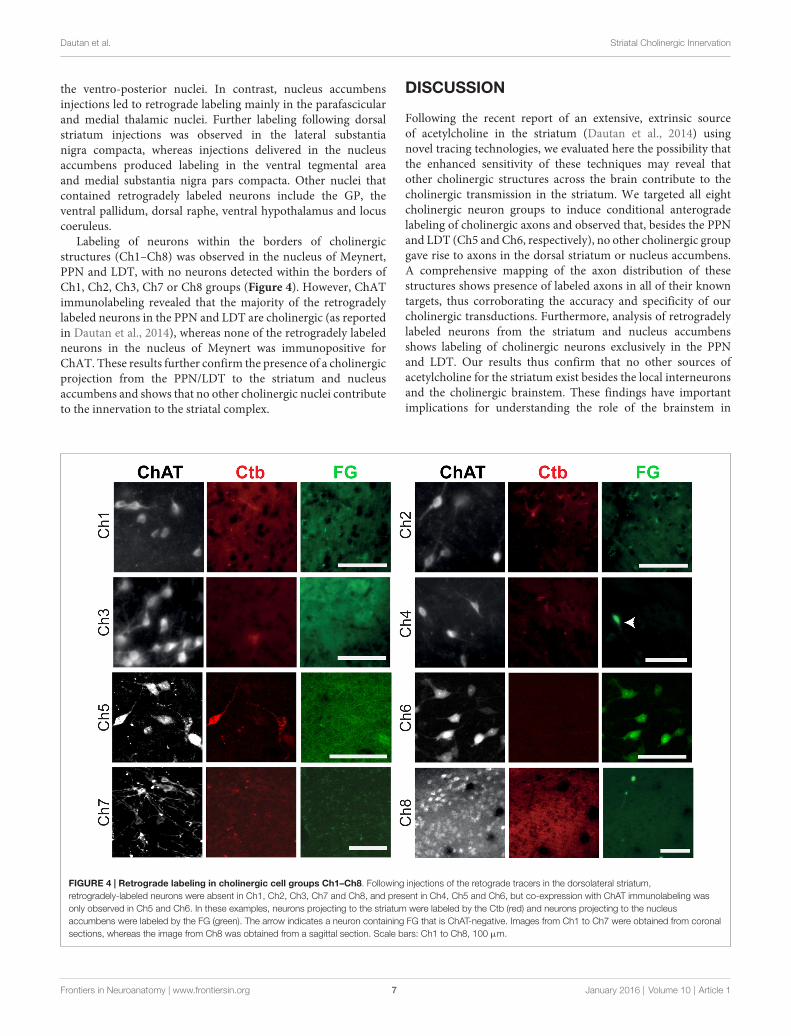
Labeling of neurons within the borders of cholinergicstructures (Ch1–Ch8) was observed in the nucleus of Meynert,PPN and LDT, with no neurons detected within the borders ofCh1, Ch2, Ch3, Ch7 or Ch8 groups (Figure 4). However, ChATimmunolabeling revealed that the majority of the retrogradelylabeled neurons in the PPN and LDT are cholinergic (as reportedin Dautan et al., 2014), whereas none of the retrogradely labeledneurons in the nucleus of Meynert was immunopositive forChAT. These results further confirm the presence of a cholinergicprojection from the PPN/LDT to the striatum and nucleusaccumbens and shows that no other cholinergic nuclei contributeto the innervation to the striatal complex.
DISCUSSION
Following the recent report of an extensive, extrinsic sourceof acetylcholine in the striatum (Dautan et al., 2014) usingnovel tracing technologies, we evaluated here the possibility thatthe enhanced sensitivity of these techniques may reveal thatother cholinergic structures across the brain contribute to thecholinergic transmission in the striatum. We targeted all eightcholinergic neuron groups to induce conditional anterogradelabeling of cholinergic axons and observed that, besides the PPNand LDT (Ch5 and Ch6, respectively), no other cholinergic groupgave rise to axons in the dorsal striatum or nucleus accumbens.A comprehensive mapping of the axon distribution of thesestructures shows presence of labeled axons in all of their knowntargets, thus corroborating the accuracy and specificity of ourcholinergic transductions. Furthermore, analysis of retrogradelylabeled neurons from the striatum and nucleus accumbensshows labeling of cholinergic neurons exclusively in the PPNand LDT. Our results thus confirm that no other sources ofacetylcholine for the striatum exist besides the local interneuronsand the cholinergic brainstem. These findings have importantimplications for understanding the role of the brainstem in
FIGURE 4 | Retrograde labeling in cholinergic cell groups Ch1–Ch8. Following injections of the retograde tracers in the dorsolateral striatum,retrogradely-labeled neurons were absent in Ch1, Ch2, Ch3, Ch7 and Ch8, and present in Ch4, Ch5 and Ch6, but co-expression with ChAT immunolabeling wasonly observed in Ch5 and Ch6. In these examples, neurons projecting to the striatum were labeled by the Ctb (red) and neurons projecting to the nucleusaccumbens were labeled by the FG (green). The arrow indicates a neuron containing FG that is ChAT-negative. Images from Ch1 to Ch7 were obtained from coronalsections, whereas the image from Ch8 was obtained from a sagittal section. Scale bars: Ch1 to Ch8, 100 µm.
Frontiers in Neuroanatomy | www.frontiersin.org 7 January 2016 | Volume 10 | Article 1

Dautan et al. Striatal Cholinergic Innervation
striatal modulation and enable us to define more clearly andin clear functional terms the role of cholinergic systems in themodulation of striatal/basal ganglia circuits.
Technical ConsiderationsThe use of a Cre recombinase rat line together with AAVinjections allowed us to target anatomically-restricted cholinergicgroups and map their projections. However, virus injectionsnecessary to reach a representative proportion of cholinergicneurons in each structure can potentially diffuse several hundredmicrons (Dautan et al., 2014), which becomes problematic forthose cholinergic cell groups that form a continuum (e.g., Ch1,Ch2, Ch3). To overcome this difficulty, preventing the spreadof the transduction over contiguous cholinergic groups andrestricting the labeling to the defined borders of each structure,the volume of the injections was adjusted for each structure basedon our preliminary assessments. Because our data is not used toevaluate the quantitative density of axons but it is rather basedon the qualitative expression, the variations on the virus injectionvolumes are unlikely to affect the conclusions of this study.
The expression of eYFP can give rise to a low signal tonoise ratio in thin axon shafts and small terminals, some ofwhich can be photobleached rapidly and thus become difficultto detect during on-line analysis. In order to circumvent thepossibility of false-negatives due to factors of this nature, weenhanced the YFP signal by immunostaining and performedthe analysis off-line, thus minimizing the exposure of the tissueto the fluorescent light. It is worth noting that YFP labelingproduces a complete labeling of axon terminals, as demonstratedpreviously by electron microscopy (Dautan et al., 2014), whichmay provide some advantage of sensitivity over conventionalanterograde tracers and may account for the detection ofpreviously unidentified targets. Additional validation of thedata from the ChAT::Cre+ rats was obtained by the use ofconventional retrograde tracers in wild-type rats. Our resultsshowed that no retrogradely-labeled cholinergic cell bodieswere observed in any of the cholinergic cell groups whoseaxons were absent from the striatal complex (i.e., Ch1–Ch4,Ch7 and Ch8), and in contrast, retrogradely-labeled cholinergiccell bodies were detected in the Ch5 (PPN) and Ch6 (LDT)regions, whose axons widely innervated the striatum and nucleusaccumbens.
Cholinergic Transmission in the StriatumThe effects of acetylcholine in the striatum are varied andcomplex (Sugita et al., 1991; Koós and Tepper, 2002; Goldberget al., 2012). Muscarinic and nicotinic receptors are presentat both the pre- and postsynaptic sites, and thus are able toregulate the activity of cortical, thalamic and dopaminergic
terminals, as well as the activity of striatal projection neurons anddifferent interneurons (Calabresi et al., 1998, 2000; Volpicelli-Daley et al., 2003; Bonsi et al., 2011; see review by Lim et al.,2014). The existence of an additional source of acetylcholine,provided by the brainstem, may be correlated to the diversity ofacetylcholine receptors. Dissecting these two cholinergic systemsthus becomes critical to fully understand the implications ofcholinergic signaling in the striatum. Furthermore, because thecholinergic brainstem provides collaterals to the thalamus andthe dopaminergic midbrain, two of the most important afferentsystems to the striatum, it is likely that their influence onstriatal circuits will be highly correlated with the thalamic andmidbrain inputs. Such connectivity thus puts the PPN/LDT as animportant station for striatal computations.
The lack of evidence from this advanced anatomicalexperimental approach of additional sources of acetylcholine tothe striatum emphasizes the key role of the cholinergic brainstemfor modulating striatal activity and basal ganglia function.Furthermore, because of the involvement of the PPN/LDT inneuropsychiatric disorders that affect predominantly the basalganglia, and whose pathophysiology is associated with abnormalcholinergic transmission, such as Parkinson’s disease (Hirschet al., 1987; Hall et al., 2014), Huntington’s disease (Picconiet al., 2006; Smith et al., 2006), progressive supranuclear palsy(Warren et al., 2005), and dystonia (Sciamanna et al., 2012),the evidence of a direct projection to the striatum opens newavenues for the interpretation of these abnormal processes andthe challenges they pose. Further work is necessary to define theroles of this pathway in the activity of the basal ganglia in heathand disease.
AUTHOR CONTRIBUTIONS
DD and JMS conceived the project; DD performed theexperiments; DD andHHB analyzed the data; DD, JPB, TVG andJMS wrote the article.
FUNDING
This work was supported by the Medical Research Council UK(MC-UU-12020/1 to JPB). DD was funded by a University ofLeicester PhD studentship. Access to data will be available onrequest.
ACKNOWLEDGMENTS
We thank K. Deisseroth and I. Witten for the donation ofChAT::Cre rats, and E. Norman, L. Conyers, and L. Black fortheir technical assistance.
REFERENCES
Baskerville, K. A., Chang, H. T., and Herron, P. (1993). Topography of cholinergicafferents from the nucleus basalis of Meynert to representational areas ofsensorimotor cortices in the rat. J. Comp. Neurol. 335, 552–562. doi: 10.1002/cne.903350407
Bennett, B. D., Callaway, J. C., and Wilson, C. J. (2000). Intrinsic membraneproperties underlying spontaneous firing in neostriatal cholinergicinterneurons. J. Neurosci. 20, 8493–8503.
Bolam, J. P., Booth, P. A. C., Hanley, J. J., and Bevan, M. D. (2000). Synapticorganisation of the basal ganglia. J. Anat. 96, 527–542. doi: 10.1046/j.1469-7580.2000.19640527.x
Frontiers in Neuroanatomy | www.frontiersin.org 8 January 2016 | Volume 10 | Article 1

Dautan et al. Striatal Cholinergic Innervation
Bonsi, P., Cuomo, D.,Martella, G.,Madeo, G., Schirinzi, T., Puglisi, F., et al. (2011).Centrality of striatal cholinergic transmission in basal ganglia function. Front.Neuroanat. 5:6. doi: 10.3389/fnana.2011.00006
Calabresi, P., Centonze, D., Gubellini, P., Pisani, A., and Bernardi, G. (1998).Endogeneous Ach enhances striatal NMDA-responses via M1-like muscarinicreceptors and PKC activation. Eur. J. Neurosci. 10, 2887–2895. doi: 10.1111/j.1460-9568.1998.00294.x
Calabresi, P., Centoze, D., Gubellini, P., Marfia, G. A., Pisani, A., Sancesario, G.,et al. (2000). Synaptic transmission in the striatum: from plasticityto neurodegeneration. Prog. Neurobiol. 61, 231–265. doi: 10.1016/s0301-0082(99)00030-1
Cuello, C. A., Emson, P. C., Paxinos, G., and Jessell, T. (1978). Substance Pcontaining and cholinergic projections from the habenula. Brain Res. 149,413–429. doi: 10.1016/0006-8993(78)90484-5
Dautan, D., Huerta-Ocampo, I., Witten, I. B., Deisseroth, K., Bolam, J. P.,Gerdjikov, T., et al. (2014). A major external source of cholinergic innervationof the striatum and nucleus accumbens originates in the brainstem. J. Neurosci.34, 4509–4518. doi: 10.1523/JNEUROSCI.5071-13.2014
Ding, J. B., Guzman, J. N., Peterson, J. D., Goldberg, J. A., and Surmeier, D. J.(2010). Thalamic gating of corticostriatal signaling by cholinergic interneurons.Neuron 67, 294–307. doi: 10.1016/j.neuron.2010.06.017
Fitzpatrick, D., Conley, M., Luppino, G., Matelli, M., and Diamond, I. T.(1988). Cholinergic projections from the midbrain reticular formation andthe parabigeminal nucleus to the lateral geniculate nucleus in the tree shrew.J. Comp. Neurol. 272, 43–67. doi: 10.1002/cne.902720105
Futami, T., Takakusaki, K., and Kitai, S. T. (1995). Glutamatergic and cholinergicinputs from the pedunculopontine tegmental nucleus to dopamine neuronsin the substantia nigra pars compacta. Neurosci. Res. 21, 331–342. doi: 10.1016/0168-0102(94)00869-h
Gaykema, R. P., Luiten, P. G., Nyakas, C., and Traber, J. (1990). Cortical projectionpatterns of the medial septum-diagonal band complex. J. Comp. Neurol. 293,103–124. doi: 10.1002/cne.902930109
Goldberg, A., Ding, J. B., and Surmeier, D. J. (2012). Muscarinic modulation ofstriatal function and circuitry. Handb. Exp. Pharmacol. 208, 223–241. doi: 10.1007/978-3-642-23274-9_10
Gritti, I., Mainville, L., Mancia, M., and Jones, B. E. (1997). GABAergic andother noncholinergic basal forebrain neurons, together with cholinergicneurons, project to the mesocortex and isocortex in the rat. J. Comp. Neurol.383, 163–177. doi: 10.1002/(sici)1096-9861(19970630)383:2<163::aid-cne4>3.3.co;2-t
Hall, H., Reyes, S., Landeck, N., Bye, C., Leanza, G., Double, K., et al. (2014).Hippocampal lewy pathology and cholinergic dysfunction are associatedwith dementia in Parkinson’s disease. Brain 137, 2493–2508. doi: 10.1093/brain/awu193
Hallanger, A. E., and Wainer, B. H. (1988). Ascending projections fromthe pedunculopontine tegmental nucleus and the adjacent mesopontinetegmentum in the rat. J. Comp. Neurol. 274, 483–515. doi: 10.1002/cne.902740403
Hebb, C. O., and Silver, A. (1961). Gradient of choline acetylase activity in nervefibers. Nature 189, 123–125. doi: 10.1038/189123a0
Henny, P., and Jones, B. E. (2008). Projections from basal forebrain to prefrontalcortex comprise cholinergic, GABAergic and glutamatergic inputs to pyramidalcells or interneurons. Eur. J. Neurosci. 27, 654–670. doi: 10.1111/j.1460-9568.2008.06029.x
Hirsch, E. C., Graybiel, A. M., Duyckaerts, C., and Javoy-Agid, F. (1987). Neuronalloss in the pedunculopontine tegmental nucleus in Parkinson disease and inprogressive supranuclear palsy. Proc. Natl. Acad. Sci. U S A 84, 5976–5980.doi: 10.1073/pnas.84.16.5976
Holmstrand, E. C., and Sesack, S. R. (2011). Projections from the ratpedunculopontine and laterodorsal tegmental nuclei to the anterior thalamusand ventral tegmental area arise from largely separate populations of neurons.Brain Struct. Funct. 216, 331–345. doi: 10.1007/s00429-011-0320-2
Kalén, P., and Wiklund, L. (1989). Projections from the medial septum anddiagonal band of Broca to the dorsal and central superior raphe nuclei: a non-cholinergic pathway. Exp. Brain Res. 75, 401–416. doi: 10.1007/bf00247947
Kobayashi, Y., Sano, Y., Vannoni, E., Goto, H., Suzuki, H., Oba, A., et al. (2013).Genetic dissection of medial habenula-interpeduncular nucleus pathwayfunction in mice. Front. Behav. Neurosci. 7:17. doi: 10.3389/fnbeh.2013.00017
Koós, T., and Tepper, J. M. (2002). Dual cholinergic control of fast-spikinginterneurons in the neostriatum. J. Neurosci. 22, 529–535.
Lavoie, B., and Parent, A. (1994). Pedunculopontine nucleus in the squirrelmonkey: projections to the basal ganglia as revealed by anterogradetract-tracing methods. J. Comp. Neurol. 344, 210–231. doi: 10.1002/cne.903440204
Lim, S. A., Kang, U. J., andMcGehee, D. S. (2014). Striatal cholinergic interneuronregulation and circuit effect. Front. Synaptic Neurosci. 6:22. doi: 10.3389/fnsyn.2014.00022
Macintosh, F. C. (1941). The distribution of acetylcholine in the peripheral andthe central nervous system. J. Physiol. Lond. 99, 436–442. doi: 10.1113/jphysiol.1941.sp003913
Mena-Segovia, J., Bolam, J. P., and Magill, P. J. (2004). Pedunculopontine nucleusand basal ganglia: distant relatives or part of the same family? Trends Neurosci.27, 585–588. doi: 10.1016/j.tins.2004.07.009
Mena-Segovia, J., Sims, H. M., Magill, P. J., and Bolam, J. P. (2008). Cholinergicbrainstem neurons modulate cortical gamma activity during slow oscillations.J. Physiol. Lond. 586, 2947–2960. doi: 10.1113/jphysiol.2008.153874
Mesulam, M. M. (1990). Human brain cholinergic pathways. Prog. Brain Res. 84,231–241. doi: 10.1016/s0079-6123(08)60908-5
Mesulam, M. M., and Geula, C. (1988). Nucleus basalis (Ch4) and corticalinnervation in the human brain: observations based on distribution ofacetylcholinesterase and choline acetyltransferase. J. Comp. Neurol. 275,216–240. doi: 10.1002/cne.902750205
Mesulam, M. M., Mufson, E. J., Levey, A. J., and Wainer, B. H. (1983a).Cholinergic innervation of cortex by the basal forebrain: cytochemistry andcortical connections of the septal area, diagonal band nuclei, nucleus basalis(substantia innominata) and hypothalamus in the rhesus monkey. J. Comp.Neurol. 214, 170–197. doi: 10.1002/cne.902140206
Mesulam, M. M., Mufson, E. J., Wainer, B. H., and Levey, A. I. (1983b).Central cholinergic pathways in the rat: an overview based on an alternativenomenclature (Ch1–Ch6). Neuroscience 10, 1185–1201. doi: 10.1016/0306-4522(83)90108-2
Mink, J. W. (1996). The basal ganglia: focused selection and inhibition ofcompeting motor programs. Prog. Neurobiol. 50, 381–425. doi: 10.1016/s0301-0082(96)00042-1
Mitani, A., Ito, K., Hallanger, A. E., Wainer, B. H., Kataoka, K., andMcCarley, R. W. (1988). Cholinergic projections from the laterodorsal andpedunculopontine tegmental nuclei to the pontine gigantocellular tegmentalfield in the cat. Brain Res. 451, 397–402. doi: 10.1016/0006-8993(88)90792-5
Motts, S. D., Slusarczyk, A. S., Sowick, C. S., and Schofield, B. R. (2008).Distribution of cholinergic cells in guinea pig brainstem. Neuroscience 154,186–195. doi: 10.1016/j.neuroscience.2007.12.017
Mufson, E. J., Martin, T. L., Mash, D. C., Wainer, B. H., and Mesulam, M. M.(1986). Cholinergic projections from the parabigeminal nucleus (Ch8) to thesuperior colliculus in themouse: a combined analysis of horseradish peroxidasetransport and choline acetyltransferase immunohistochemistry. Brain Res. 370,144–148. doi: 10.1016/0006-8993(86)91114-5
Nagai, T., Kimura, H., Maeda, T., McGeer, P. L., Peng, F., and McGeer, E. G.(1982). Cholinergic projections from the basal forebrain of rat to the amygdala.J. Neurosci. 2, 513–520.
Nakano, K., Hasegawa, Y., Tokushige, A., Nakagawa, S., Kayahara, T., andMizuno, N. (1990). Topographical projections from the thalamus, subthalamicnucleus and pedunculopontine tegmental nucleus to the striatum in theJapanese monkey, Macaca fuscata. Brain Res. 537, 54–68. doi: 10.1016/0006-8993(90)90339-d
Nyakas, C., Luiten, P. G. M., Spencer, D. G., and Traber, J. (1987). Detailedprojection patterns of septal and diagonal band efferents to the hippocampusin the rat with emphasis on innervation of CA1 and Dentate gyrus. Brain Res.Bull. 18, 533–545. doi: 10.1016/0361-9230(87)90117-1
Oakman, S. A., Faris, P. L., Cozzari, C., and Hartman, B. K. (1999).Characterization of the extent of pontomesencephalic cholinergic neurons’projections to the thalamus: comparison with projections to midbraindopaminergic groups. Neuroscience 94, 529–547. doi: 10.1016/s0306-4522(99)00307-3
Paxinos, G., and Watson, C. (2007). The Rat Brain in Stereotaxic Coordinates. 6thEdn. San Diego: Elsevier Academic Press
Frontiers in Neuroanatomy | www.frontiersin.org 9 January 2016 | Volume 10 | Article 1

Dautan et al. Striatal Cholinergic Innervation
Pearson, R. C. A., Gatter, K. C., Brodal, P., and Powell, T. P. S. (1983).The projection of the basal nucleus of Meynert upon the neocortexin the monkey. Brain Res. 259, 132–136. doi: 10.1016/0006-8993(83)91075-2
Picconi, B., Passino, E., Sgobio, C., Bonsi, P., Barone, I., Ghiglieri, V., et al. (2006).Plastic and behavioral abnormalities in experimental Huntington’s disease: acrucial role for cholinergic interneurons. Neurobiol. Dis. 22, 143–152. doi: 10.1016/j.nbd.2005.10.009
Ren, J., Qin, C., Hu, F., Tan, J., Qiu, L., Zhao, S., et al. (2011). Habenula‘‘cholinergic’’ neurons corelease glutamate and acetylcholine and activatepostsynaptic neurons via distinct transmission modes. Neuron 69, 445–452.doi: 10.1016/j.neuron.2010.12.038
Saper, C. B. (1984). Organization of cerebral cortical afferent systemsin the rat. J. Comp. Neurol. 222, 313–342. doi: 10.1002/cne.902220302
Saper, C. B., and Loewy, A. D. (1982). Projections of the pedunculopontinetegmental nucleus in the rat: evidence for additional extrapyramidalcircuitry. Brain Res. 252, 367–372. doi: 10.1016/0006-8993(82)90404-8
Sarter, M., and Bruno, J. P. (2000). Cortical cholinergic inputs mediating arousal,attentional processing and dreaming: differential afferent regulation of thebasal forebrain by telencephalic and brainstem afferents. Neuroscience 95,933–952. doi: 10.1016/s0306-4522(99)00487-x
Schauz, C., and Koch, M. (1999). Lesions of the nucleus basalis magnocellularisdo not impair prepulse inhibition and latent inhibition of fear-potentiatedstartle in the rat. Brain Res. 815, 98–105. doi: 10.1016/s0006-8993(98)01134-2
Sciamanna, G., Tassone, A., Mandolesi, G., Puglisi, F., Ponterio, G., Martella, G.,et al. (2012). Cholinergic dysfunction alters synaptic integration betweenthalamostriatal and corticostriatal inputs in DYT1 dystonia. J. Neurosci. 32,11991–12004. doi: 10.1523/JNEUROSCI.0041-12.2012
Semba, K., and Fibiger, H. C. (1992). Afferent connections of the laterodorsal andthe pedunculopontine tegmental nuclei in the rat: a retro- and antero-gradetransport and immunohistochemical study. J. Comp. Neurol. 323, 387–410.doi: 10.1002/cne.903230307
Senut, M. C., Menetry, D., and Lamour, Y. (1989). Cholinergic and peptidergicprojections from the medial septum and the nucleus of the diagonal band ofBroca to dorsal hippocampus, cingulate cortex and olfactory bulb: a combinedwheat-germ agglutinin horseradish peroxidase gold immunohistochemicalstudy. Neuroscience 30, 385–404. doi: 10.1016/0306-4522(89)90260-1
Smith, R., Chung, H., Rundquist, S., Maat-Schieman, M. L. C., Colgan, L.,Englund, E., et al. (2006). Cholinergic neuronal defect without cell lossin Huntington’s disease. Hum. Mol. Genet. 15, 3119–3131. doi: 10.1093/hmg/ddl252
Smith, Y., and Parent, A. (1986). Differential connections of caudate nucleusand putamen in squirrel monkey (Saimiri sciureus). Neuroscience 18, 347–371.doi: 10.1016/0306-4522(86)90159-4
Sugita, S., Uchimura, N., Jiang, Z. G., and North, R. A. (1991). Distinct muscarinicreceptors inhibit release of γ-aminobutyric acid and excitatory amino acidsin mammalian brain. Proc. Natl. Acad. Sci. U S A 88, 2608–2611. doi: 10.1073/pnas.88.6.2608
Volpicelli-Daley, L. A., Hrabovska, A., Duysen, E. G., Ferguson, S. M., Brakely,R. D., Lockridge, A., et al. (2003). Altered striatal function and muscariniccholinergic receptors in acetylcholinesterase knockout mice. Mol. Pharmacol.64, 1309–1316. doi: 10.1124/mol.64.6.1309
Wall, N. R., De La Parra, M., Callaway, E. M., and Kreitzer, A. C. (2013).Differential innervation of direct- and indirect-pathway striatal projectionneurons. Neuron 79, 347–360. doi: 10.1016/j.neuron.2013.05.014
Wang, Z., Kai, L., Day, M., Ronesi, J., Yin, H. H., Ding, J., et al. (2006).Dopaminergic control of corticostriatal longterm synaptic depression inmedium spiny neurons is mediated by cholinergic interneurons. Neuron 50,443–452. doi: 10.1016/j.neuron.2006.04.010
Warren, N. M., Piggott, M. A., Perry, E. K., and Burn, D. J. (2005). Cholinergic inprogressive supranuclear palsy. Brain 128, 239–249. doi: 10.1093/brain/awh391
Witten, I. B., Steinberg, E. E., Lee, S. Y., Davidson, T. J., Zalocusky, K. A.,Brodsky, M., et al. (2011). Recombinase-driver rat lines: tools, techniquesand optogenetic application to dopamine-mediated reinforcement. Neuron 72,721–733. doi: 10.1016/j.neuron.2011.10.028
Woolf, N. J., and Butcher, L. L. (1982). Cholinergic projections to the basolateralamygdala: a combined Evan’s blue and acetylcholinesterase analysis. Brain Res.Bull. 8, 751–763. doi: 10.1016/0361-9230(82)90102-2
Woolf, N. J., Eckenstein, F., and Butcher, L. L. (1984). Cholinergic systems in therat brain. Brain Res. Bull. 13, 751–784.
Záborsky, L., Carlsen, J., Brashear, H. R., and Heimer, L. (1986). Cholinergicand GABAergic afferents to the olfactory bulb with special emphasis on theprojections in the nucleus of the horizontal limb of the diagonal band. J. Comp.Neurol. 243, 488–509. doi: 10.1002/cne.902430405
Záborszky, L., Csordas, A., Mosca, K., Kim, J., Gielow, M. R., Vadasz, C., et al.(2015). Neurons in the basal forebrain project to the cortex in a complextopographic organization that reflects corticocortical connectivity patterns: anexperimental study based on retrograde tracing and 3D reconstruction. Cereb.Cortex 25, 118–137. doi: 10.1093/cercor/bht210
Conflict of Interest Statement: The authors declare that the research wasconducted in the absence of any commercial or financial relationships that couldbe construed as a potential conflict of interest.
Copyright © 2016 Dautan, Hacioglu Bay, Bolam, Gerdjikov and Mena-Segovia.This is an open-access article distributed under the terms of the Creative CommonsAttribution License (CC BY). The use, distribution and reproduction in other forumsis permitted, provided the original author(s) or licensor are credited and that theoriginal publication in this journal is cited, in accordance with accepted academicpractice. No use, distribution or reproduction is permitted which does not complywith these terms.
Frontiers in Neuroanatomy | www.frontiersin.org 10 January 2016 | Volume 10 | Article 1