Embed Size (px)
Citation preview
Extranuclear inheritance
Source of extranuclear genes
• Plants and animals both exhibit inheritance patterns non representative of nuclear inheritance such as – Variegated leaves in plants– Antibiotic resistance in chlamydomonas– Kappa toxins in the paramecium– Myoclonic eplilepsy and ragged red fiber disease in
humans
• All these problems are found to be independent of nuclear inheritance patterns (Mendelian ratios)
Types of ENH
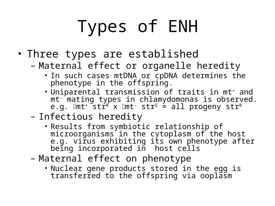
• Three types are established– Maternal effect or organelle heredity
• In such cases mtDNA or cpDNA determines the phenotype in the offspring.
• Uniparental transmission of traits in mt+ and mt- mating types in chlamydomonas is observed. e.g. mt+ strR x mt- strS = all progeny strR
– Infectious heredity• Results from symbiotic relationship of microorganisms in the
cytoplasm of the host e.g. virus exhibiting its own phenotype after being incorporated in host cells
– Maternal effect on phenotype• Nuclear gene products stored in the egg is transferred to the
offspring via ooplasm
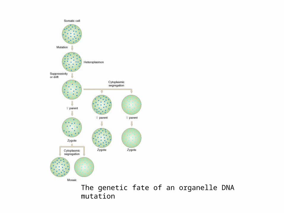
The genetic fate of an organelle DNA mutation
Organelle heredity
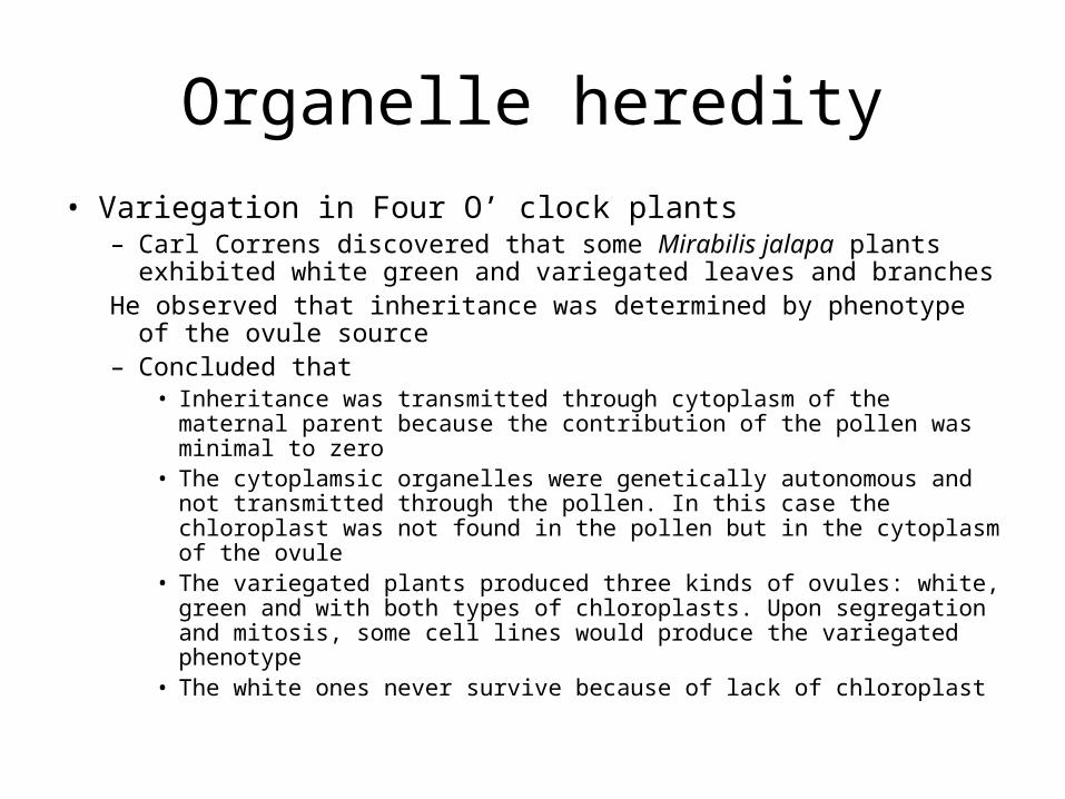
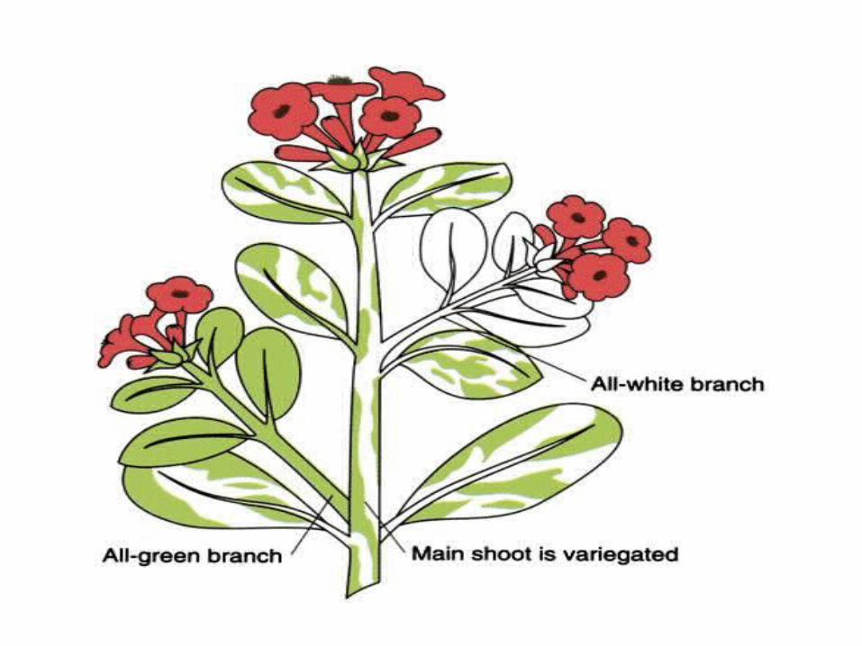
• Variegation in Four O’ clock plants– Carl Correns discovered that some Mirabilis jalapa plants
exhibited white green and variegated leaves and branchesHe observed that inheritance was determined by phenotype of the
ovule source – Concluded that
• Inheritance was transmitted through cytoplasm of the maternal parent because the contribution of the pollen was minimal to zero
• The cytoplamsic organelles were genetically autonomous and not transmitted through the pollen. In this case the chloroplast was not found in the pollen but in the cytoplasm of the ovule
• The variegated plants produced three kinds of ovules: white, green and with both types of chloroplasts. Upon segregation and mitosis, some cell lines would produce the variegated phenotype
• The white ones never survive because of lack of chloroplast
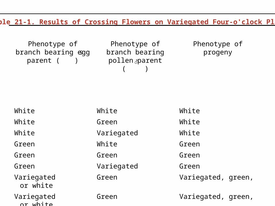
Table 21-1. Results of Crossing Flowers on Variegated Four-o'clock Plants
Phenotype of branch bearing egg parent ( )
Phenotype of branch bearing pollen parent
( )
Phenotype of progeny
White White White
White Green White
White Variegated White
Green White Green
Green Green Green
Green Variegated Green
Variegated or white Green Variegated, green,
Variegated or white Green Variegated, green,
Variegated or white Variegated Variegated, green,
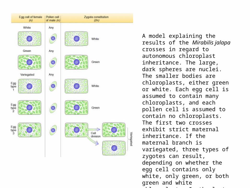
A model explaining the results of the Mirabilis jalapa crosses in regard to autonomous chloroplast inheritance. The large, dark spheres are nuclei. The smaller bodies are chloroplasts, either green or white. Each egg cell is assumed to contain many chloroplasts, and each pollen cell is assumed to contain no chloroplasts. The first two crosses exhibit strict maternal inheritance. If the maternal branch is variegated, three types of zygotes can result, depending on whether the egg cell contains only white, only green, or both green and white chloroplasts. In the last case, the resulting zygote can produce both green and white tissue, so a variegated plant results.
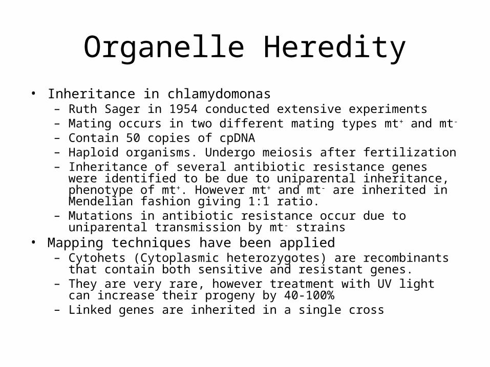
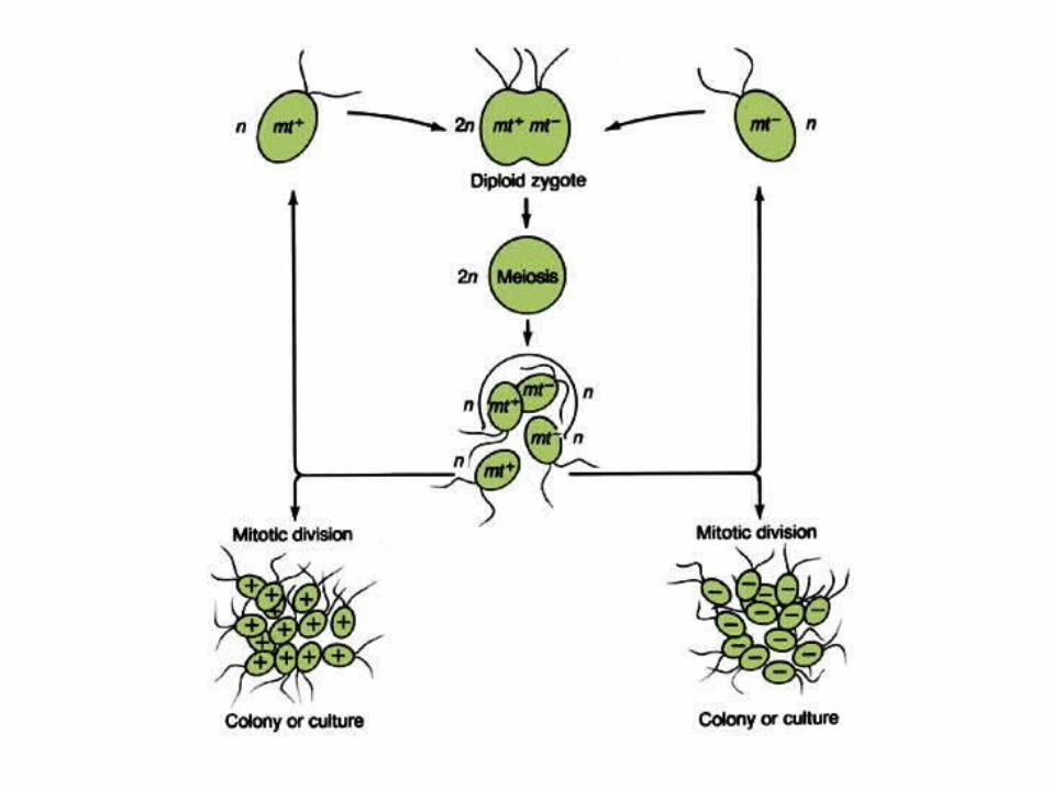
Organelle Heredity• Inheritance in chlamydomonas
– Ruth Sager in 1954 conducted extensive experiments– Mating occurs in two different mating types mt+ and mt- – Contain 50 copies of cpDNA– Haploid organisms. Undergo meiosis after fertilization– Inheritance of several antibiotic resistance genes were identified to be
due to uniparental inheritance, phenotype of mt+. However mt+ and mt- are inherited in Mendelian fashion giving 1:1 ratio.
– Mutations in antibiotic resistance occur due to uniparental transmission by mt- strains
• Mapping techniques have been applied – Cytohets (Cytoplasmic heterozygotes) are recombinants that contain
both sensitive and resistant genes. – They are very rare, however treatment with UV light can increase their
progeny by 40-100%– Linked genes are inherited in a single cross
Organelle heredity
• Poky Neurospora– Slow growing Neurospora inherits trait from maternal
plant– Contains abnormal quantities of cytochromes with cyt
a and b being minimal while cyt c in abundance (all three are found abundantly in wild types)
– Slow growth is attributed to these cytochromes in the mitochondria as they participate ATP synthesis. The mixing of poky cytoplasm somehow dilutes the normal concentration or produces some inactivating substance
– Heterokaryon test is performed to determine maternal effect
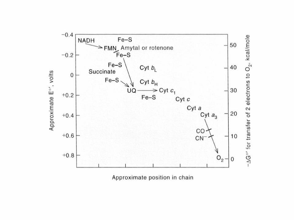
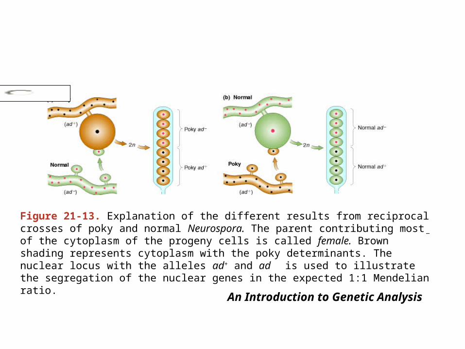
Figure 21-13. Explanation of the different results from reciprocal crosses of poky and normal Neurospora. The parent contributing most of the cytoplasm of the progeny cells is called female. Brown shading represents cytoplasm with the poky determinants. The nuclear locus with the alleles ad+ and ad
is used to illustrate the segregation of the nuclear genes in the expected 1:1 Mendelian ratio.
An Introduction to Genetic Analysis
BooksSearch.Books
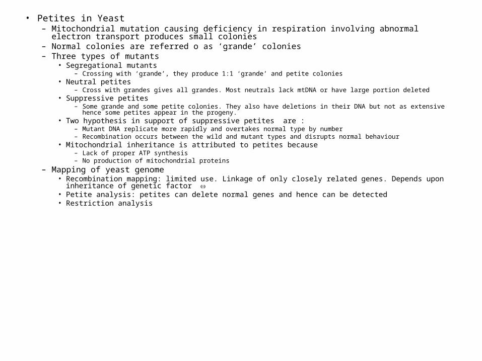
• Petites in Yeast– Mitochondrial mutation causing deficiency in respiration involving abnormal electron transport produces
small colonies– Normal colonies are referred o as ‘grande’ colonies– Three types of mutants
• Segregational mutants– Crossing with ‘grande’, they produce 1:1 ‘grande’ and petite colonies
• Neutral petites– Cross with grandes gives all grandes. Most neutrals lack mtDNA or have large portion deleted
• Suppressive petites– Some grande and some petite colonies. They also have deletions in their DNA but not as extensive hence some petites
appear in the progeny. • Two hypothesis in support of suppressive petites are :
– Mutant DNA replicate more rapidly and overtakes normal type by number– Recombination occurs between the wild and mutant types and disrupts normal behaviour
• Mitochondrial inheritance is attributed to petites because– Lack of proper ATP synthesis– No production of mitochondrial proteins
– Mapping of yeast genome• Recombination mapping: limited use. Linkage of only closely related genes. Depends upon inheritance of
genetic factor • Petite analysis: petites can delete normal genes and hence can be detected• Restriction analysis
Infectious heredity
• Kappa particles in paramecium– Certain strains of Paramecium aurelia produce toxin paramecin
that can kill sensistive strains– Hypothesized to be produced by temperate bacteriophages
infecting the paramecium– Transmitted via conjugation
• Infective particles in drosophila– Co2 sensitivity and sex ratio – Co2 sensitivity do not recover from anesthesia and are killed.
Sensitive mothers pass trait to all offsprings. Also injection of defective cytoplasm induces sensitivity in normal flies. Sensitivity due to presence of sigma virus specific to drosophila
– Sex ratio involves growth of predominantly drosophila bifasciata females at 21. Low percentage of males produced. Trait is passed from mother to daughter
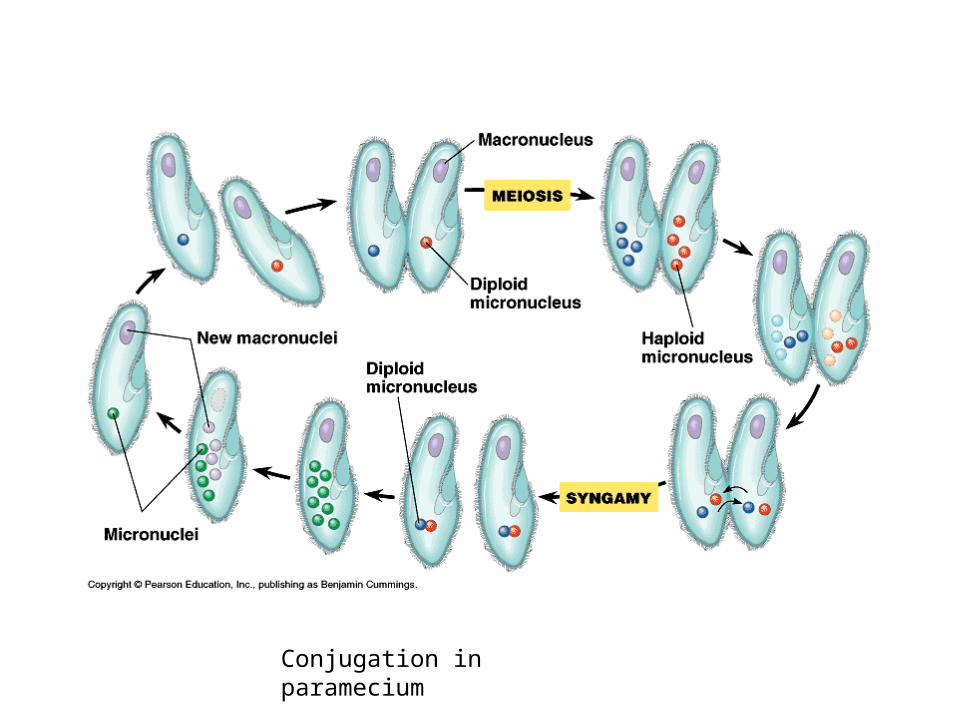
Conjugation in paramecium
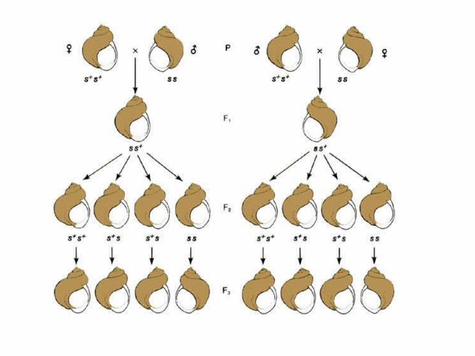
Maternal effect • Coiling in Limnaea peregra (snail)
– Alex Strutvant (linkage mapping fame) discovered that dextral (right handed) and sinistral (left handed) snail crosses always carried the coil of the maternal phenotype
dextral xsinistral=all progeny dextral
– Snails are hermaphrodites and can be cross fertilised as well as self fertilised
– Self fertilised F2 gives all dextral but F3 gives 3:1 dextral:sinistral– Maternal parents with DD and Dd genotype produce dextral coils– The coiling pattern is determined by the genotype of the parent
producing the egg regardless of its phenotype.• Orientation of the spindle in the first cleavage after fertilization determines
the direction of the coil• Spindle orientation is controlled by maternal genes acting on the developing
eggs• Genomic imprinting is also evidence of uniparental transmission
Maternal effect
• Ephestia pigmentation– The Mediterranean meal moth (Ephestia
kuehniella) has wild larvae with brown pigmentation and brown eyes because of a kynurenine that is a derivative of tryptophan.
– Mutation produces less pigment and red eyes. – A cross between heterozygous mother and
recessive father all larvae have brown eyes and pigment . However at least half of them loose the brown eyes and become red eyed
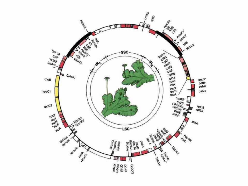
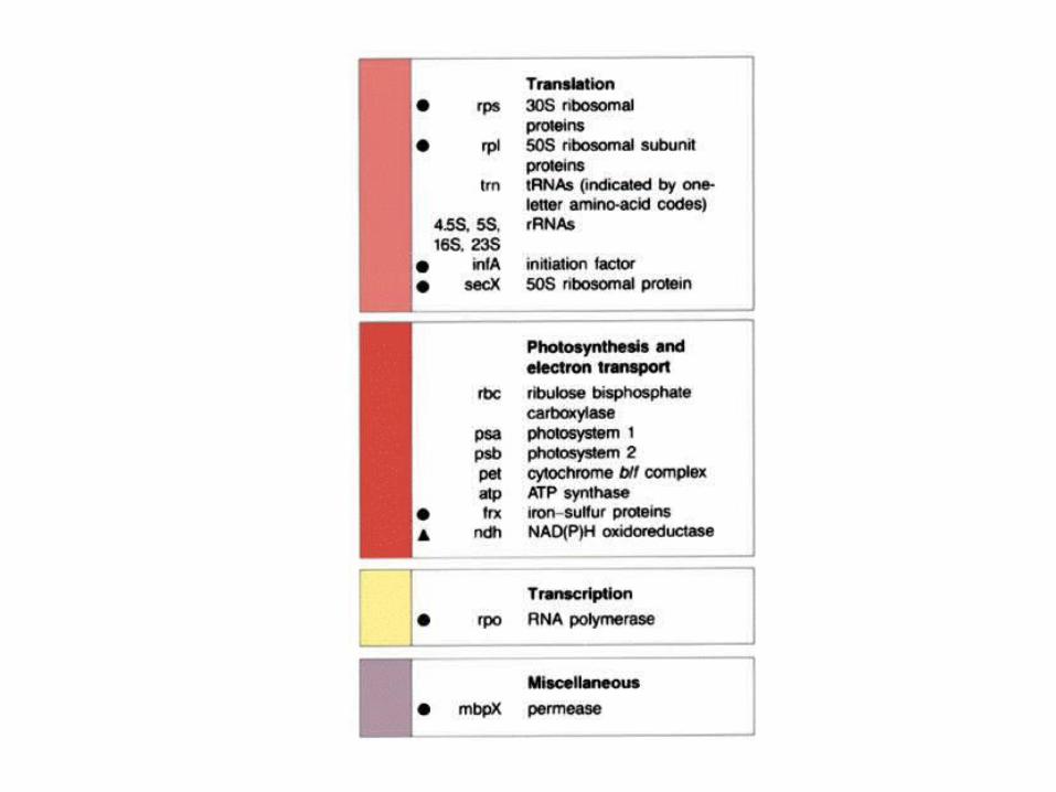
Molecular organisation of chloroplast genes
• Fairly uniform in size• 100-225kb in length• Circular, double stranded• Replicates semiconservatively• Shows variable base composition and bouyant
density from nuclear DNA• Contains introns and intergenic bases.
Duplications are also present • Carries genes for coding its own ribosomal and
protein production
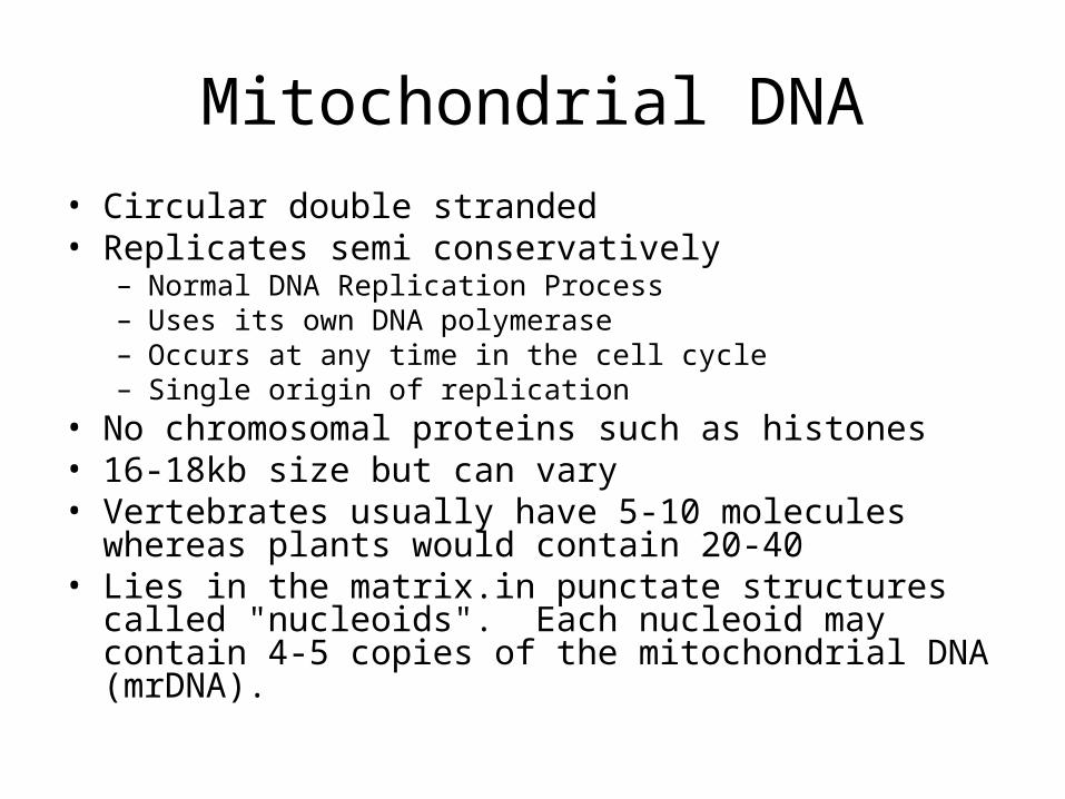
Mitochondrial DNA
• Circular double stranded• Replicates semi conservatively
– Normal DNA Replication Process– Uses its own DNA polymerase– Occurs at any time in the cell cycle– Single origin of replication
• No chromosomal proteins such as histones• 16-18kb size but can vary• Vertebrates usually have 5-10 molecules whereas plants
would contain 20-40• Lies in the matrix.in punctate structures called
"nucleoids". Each nucleoid may contain 4-5 copies of the mitochondrial DNA (mrDNA).
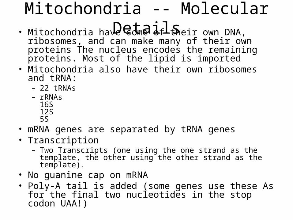
Mitochondria -- Molecular Details• Mitochondria have some of their own DNA, ribosomes,
and can make many of their own proteins The nucleus encodes the remaining proteins. Most of the lipid is imported
• Mitochondria also have their own ribosomes and tRNA: – 22 tRNAs – rRNAs
16S12S5S
• mRNA genes are separated by tRNA genes• Transcription
– Two Transcripts (one using the one strand as the template, the other using the other strand as the template).
• No guanine cap on mRNA• Poly-A tail is added (some genes use these As for the
final two nucleotides in the stop codon UAA!)
• Translation– mRNA is different from cytoplasmic mRNA, so
initiation is probably different– fMet tRNA (formylmethionyl) initiates
translation
• Some genes are translated in the Mitochondria, others are translated in the cytoplasm.
• Some coding dictionary changes
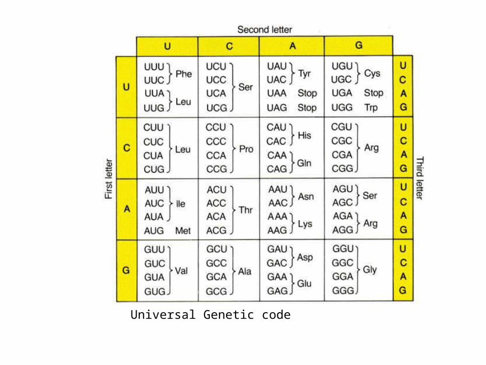
Universal Genetic code
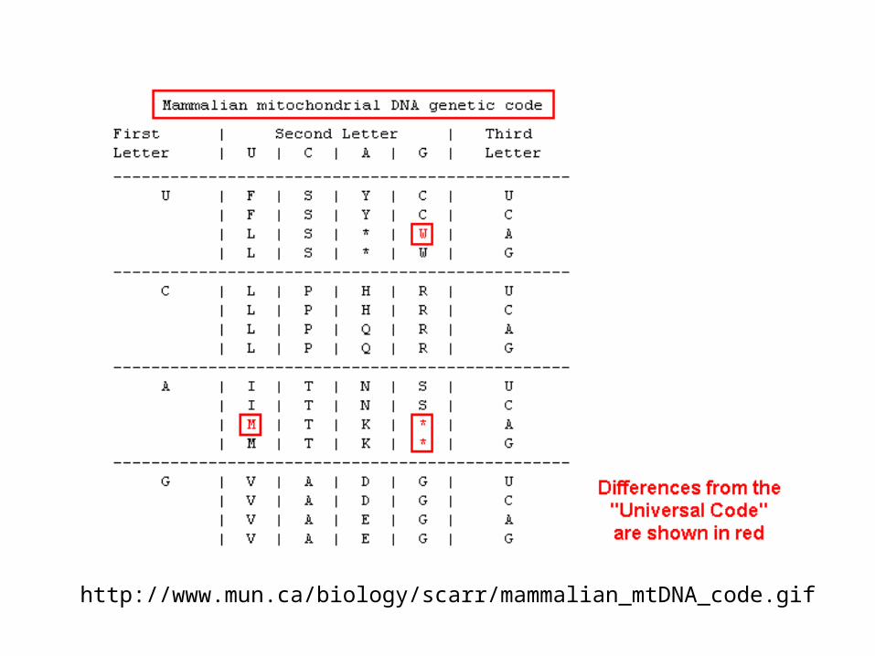
http://www.mun.ca/biology/scarr/mammalian_mtDNA_code.gif
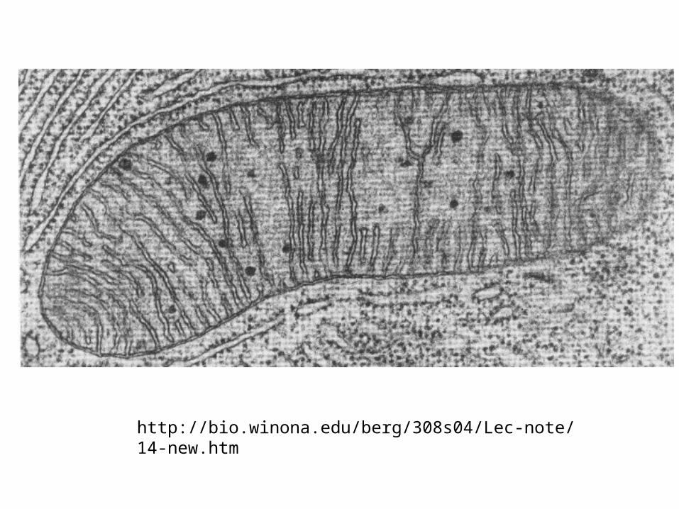
http://bio.winona.edu/berg/308s04/Lec-note/14-new.htm
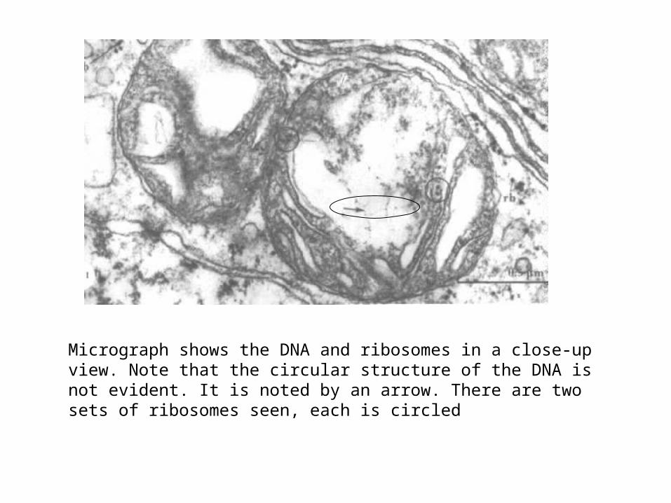
Micrograph shows the DNA and ribosomes in a close-up view. Note that the circular structure of the DNA is not evident. It is noted by an arrow. There are two sets of ribosomes seen, each is circled
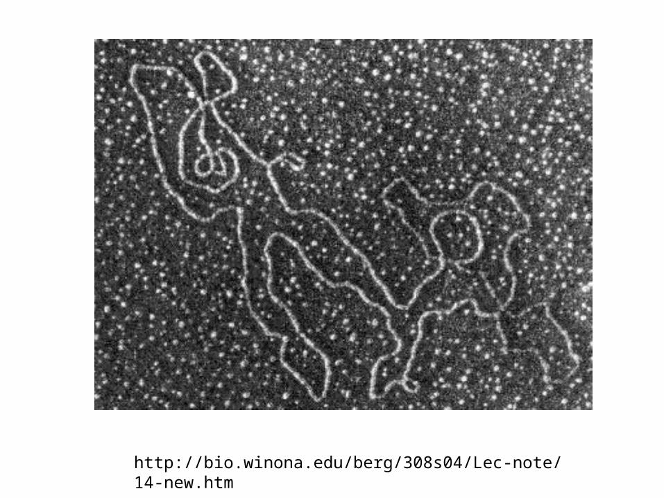
http://bio.winona.edu/berg/308s04/Lec-note/14-new.htm
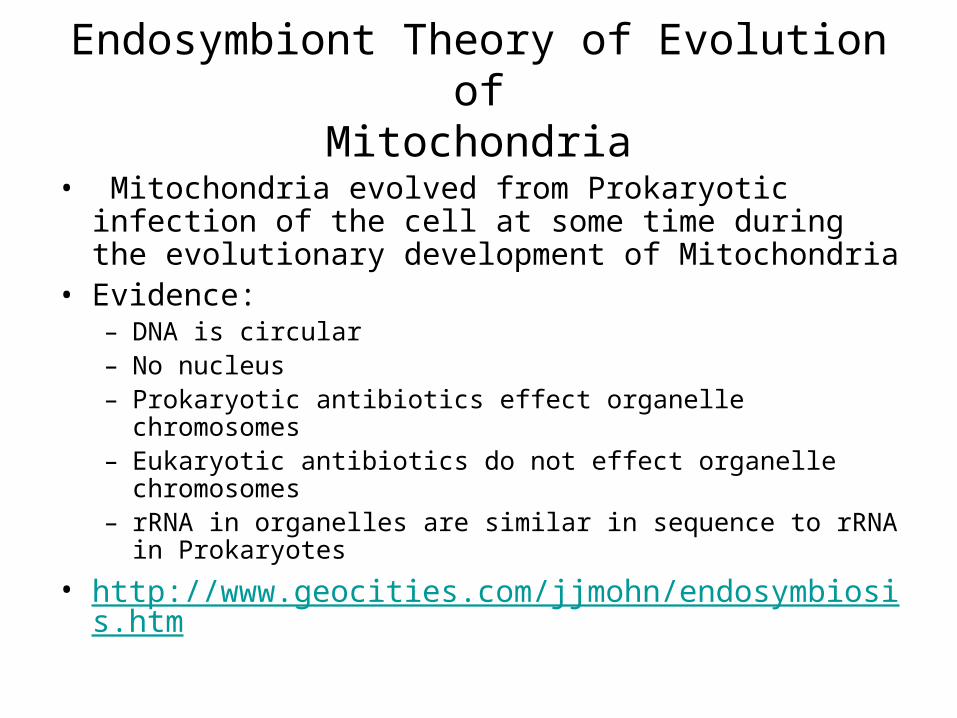
Endosymbiont Theory of Evolution ofMitochondria
• Mitochondria evolved from Prokaryotic infection of the cell at some time during the evolutionary development of Mitochondria
• Evidence:– DNA is circular– No nucleus– Prokaryotic antibiotics effect organelle chromosomes– Eukaryotic antibiotics do not effect organelle chromosomes– rRNA in organelles are similar in sequence to rRNA in
Prokaryotes
• http://www.geocities.com/jjmohn/endosymbiosis.htm
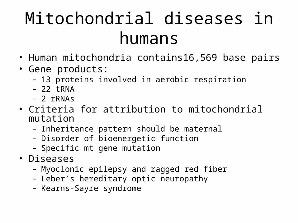
Mitochondrial diseases in humans
• Human mitochondria contains16,569 base pairs• Gene products:
– 13 proteins involved in aerobic respiration– 22 tRNA– 2 rRNAs
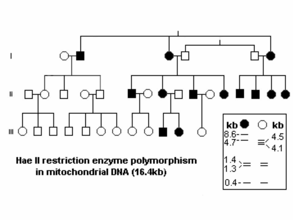
• Criteria for attribution to mitochondrial mutation– Inheritance pattern should be maternal– Disorder of bioenergetic function– Specific mt gene mutation
• Diseases– Myoclonic epilepsy and ragged red fiber – Leber’s hereditary optic neuropathy– Kearns-Sayre syndrome
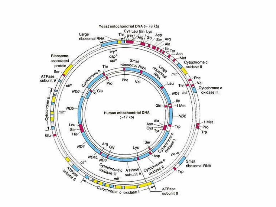
• Human mitochondrial DNA is 16,569 bp; encodes a number of mitochondrial proteins – Subunits 1, 2, and 3 of cytochrome oxidase – Subunits 6, 8,9 of the Fo ATPase – Apocytochrome b subunit of CoQH2-Cytochrome C reductase – Seven NADH-CoQ reductase subunits
• Since each cell contains hundreds of mitochondria and thousands of copies of the genome, the effects of the mutated mitochondria may be diluted out. As expected, those tissues or organs most likely to be affected would be the ones most dependent on oxidative phosphorylation (ATP production)
• In young persons it might not be picked up because even a person with 15% normal mitochondria might have enough to be healthy. However, aging patients may show a more severe disease phenotype
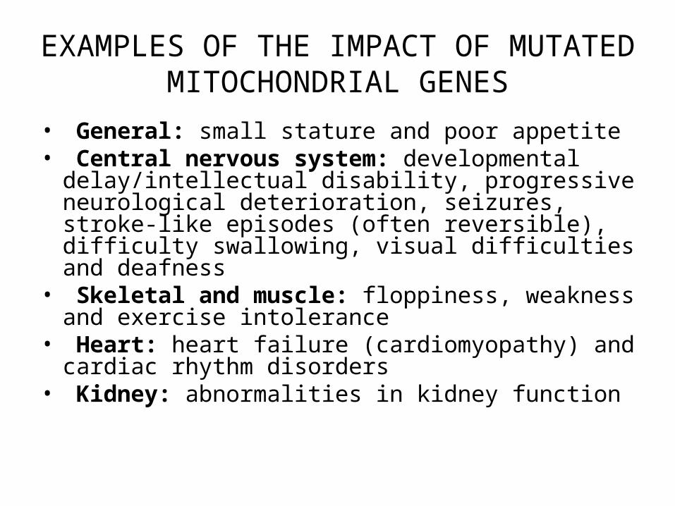
EXAMPLES OF THE IMPACT OF MUTATED MITOCHONDRIAL GENES
• General: small stature and poor appetite• Central nervous system: developmental
delay/intellectual disability, progressive neurological deterioration, seizures, stroke-like episodes (often reversible), difficulty swallowing, visual difficulties and deafness
• Skeletal and muscle: floppiness, weakness and exercise intolerance
• Heart: heart failure (cardiomyopathy) and cardiac rhythm disorders
• Kidney: abnormalities in kidney function
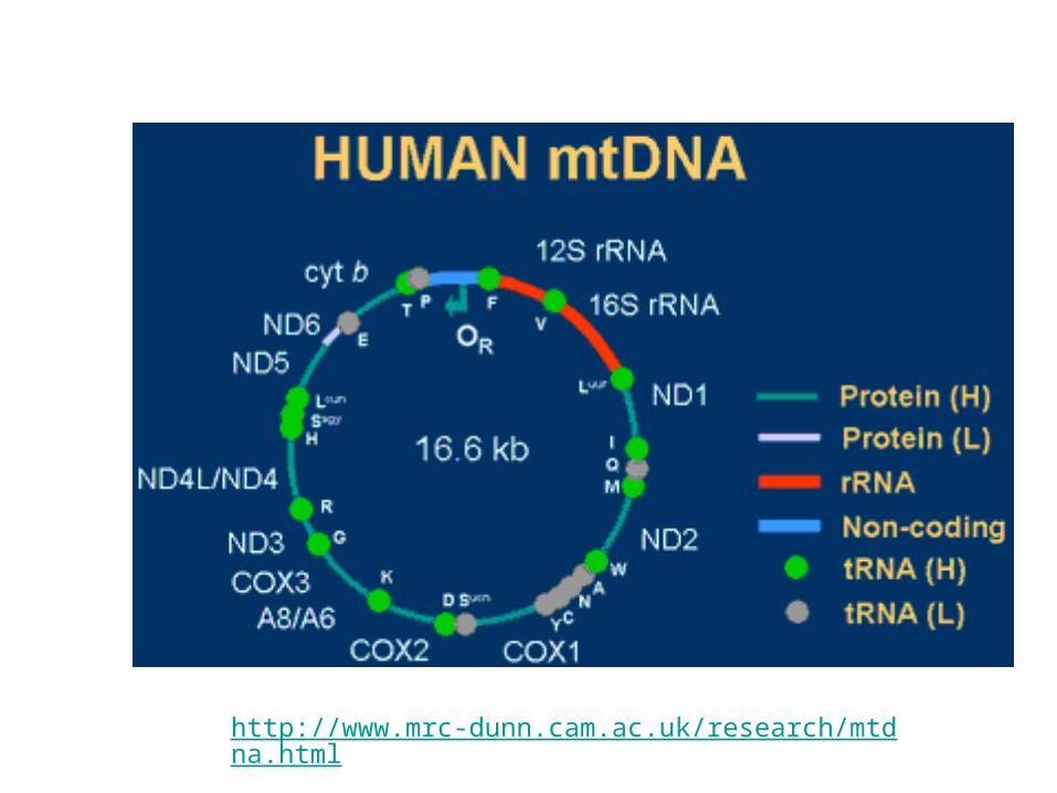
http://www.mrc-dunn.cam.ac.uk/research/mtdna.html
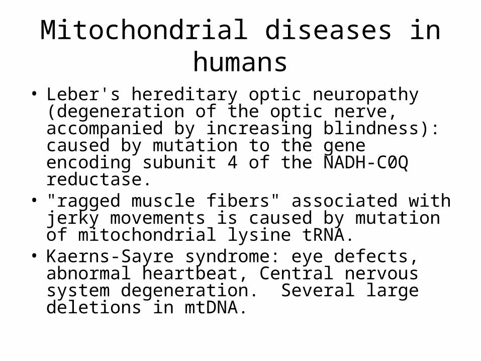
• Leber's hereditary optic neuropathy (degeneration of the optic nerve, accompanied by increasing blindness): caused by mutation to the gene encoding subunit 4 of the NADH-C0Q reductase.
• "ragged muscle fibers" associated with jerky movements is caused by mutation of mitochondrial lysine tRNA.
• Kaerns-Sayre syndrome: eye defects, abnormal heartbeat, Central nervous system degeneration. Several large deletions in mtDNA.
Mitochondrial diseases in humans
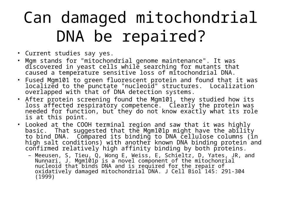
Can damaged mitochondrial DNA be repaired?
• Current studies say yes. • Mgm stands for "mitochondrial genome maintenance". It was discovered
in yeast cells while searching for mutants that caused a temperature sensitive loss of mitochondrial DNA.
• Fused Mgm101 to green fluorescent protein and found that it was localized to the punctate "nucleoid" structures. Localization overlapped with that of DNA detection systems.
• After protein screening found the Mgm101, they studied how its loss affected respiratory competence. Clearly the protein was needed for function, but they do not know exactly what its role is at this point.
• Looked at the COOH terminal region and saw that it was highly basic. That suggested that the Mgm101p might have the ability to bind DNA. Compared its binding to DNA cellulose columns (in high salt conditions) with another known DNA binding protein and confirmed relatively high affinity binding by both proteins. – Meeusen, S, Tieu, Q, Wong E, Weiss, E, Schieltz, D, Yates, JR, and Nunnari,
J. Mgm101p is a novel component of the mitochonrial nucleoid that binds DNA and is required for the repair of oxidatively damaged mitochondrial DNA. J Cell Biol 145: 291-304 (1999)


![Christopher paolini [inheritance cycle 04] - inheritance (pdf)](https://img.dokumen.tips/doc/110x75/554f25d2b4c905723a8b52b8/christopher-paolini-inheritance-cycle-04-inheritance-pdf.jpg)